Part 3 — SECTION VIII. THE SKIN AND THE SENSE-ORGANS. (continued 2)
George Haslam translation
d. The membranous labyrinth (Figs. 247, 248) has the following parts: the utricle and sinus superior, the recessus utriculi, the anterior semicircular canal and anterior ampulla, the external semicircular canal and external ampulla, the posterior semicircular canal and the posterior ampulla, the saccule, ductus endolymphaticus, and saccus endolymphaticus, the pars neglecta, the lagena cochleae, the pars basilaris cochleae, and the so-called tegmentum vasculosum.
In addition the following nerve-terminations can be distinguished: (1) The macula ac. recessus utriculi, (2) the three cristae acusticae ampullorum, (3) the macula ac. sacculi, (4) the macula ac. neglecta, (5) the papilla ac. lagenae cochleae, and (6) the papilla ac. basilaris cochleae.
The auditory nerve divides immediately beyond its origin from the medulla oblongata to form a ramus anterior and a ramus posterior, which course alongside each other for a short distance, the latter lying behind and above the former. The R. anterior runs forwards and outwards under the utricle, giving off the R. sacculi, which runs downwards and outwards; the R. rec. utriculi is then given off as a number of fibres, which run upwards and forwards; the main nerve then divides to form the R. ampullae anterioris and the R. ampullae externae, which course together for a short distance and then separate to reach their respective ampullae. The R. posterior runs backwards and outwards, gives off the R. lagenae, and then divides to form the R. neglectus, running upwards, the R. basilaris running downwards and backwards, and the R. ampullae posterioris which courses backwards and outwards.
(1) The utricle (utriculus) (Figs. 247 and 248 u) is irregularly cylindrical in form: commencing at the recessus utriculi it passes forwards and outwards; then backwards, inwards, and upwards, to terminate at the sinus posterior, where it is slightly contracted. At about its middle it is divided into an anterior and a posterior part by an incomplete, sickle-shaped partition, formed by the posterior semicircular canal opening obliquely into the utricle, and so causing a fold in the posterior wall: on the anterior wall there is no fold; the aperture left in the partition is the apertura utriculi.
The posterior part of the utricle receives the sinus superior, which is formed by the junction of the two vertical semicircular canals. The anterior part of the utricle receives the hinder dilated end of the external semicircular canal by an opening in its posterior wall, close to the apertura utriculi. In the lower wall or floor is the narrow opening leading into the saccule (canalis utriculo-saccularis): this opening is placed with its long axis parallel to the long axis of the utricle, with its broader end posterior, and the narrower end anterior.
(2) The recessus utriculi (Figs. 247 and 248 rec). The anterior end of the utricle widens and curves downwards and outwards to form the recessus utriculi; on its floor is a thin, kidney-shaped plate or otoliths, resting on the macula ac. recessus utriculi (mu), which receives the ramulus rec. utriculi; under it the ramulus amp. anterioris and the ramulus amp. externae run forwards and close together to reach their respective ampullae, which are close together at the antero-external wall of the recessus utriculi. The otolith is a glassy, homogeneous plate, with numerous vacuole-like spaces and striated borders; it covers the whole of the macula.
Fig. 247.
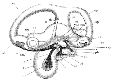
Fig. 248.
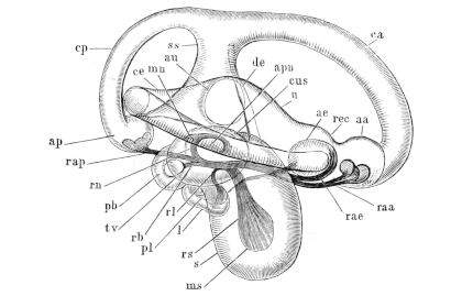
The right membranous labyrinth of Rana esculenta; after Retzius. Magnified 20 times. Fig. 247 seen from the inner side; Fig. 248 seen from the outer side.
aa Anterior ampulla. ae External ampulla. ap Posterior ampulla. apn Apertura partis neglectae. au Apertura utriculi. ca Anterior semicircular canal. ce External semicircular canal. cp Posterior semicircular canal. cus Canalis utriculo-saccularis. de Ductus endolymphaticus. l Lagena cochleae. mn Macula acustica neglecta. ms Macula acustica sacculi. mu Macula acustica recessus utriculi. pb Pars basilaris cochleae. pl Papilla ac. lagenae. ppb Papilla ac. basilaris. raa Ramulus ac. anterioris. rae Ramulus amp. externae. rap Ramulus amp. posterioris. rb Ramulus basilaris. rec Recessus utriculi. rl Ramulus lagenae. rn Ramulus neglectus. rs Ramulus sacculi. s Saccule. sp Sinus utriculi posterior. ss Sinus utriculi superior. tv Tegmentum vasculosum. u Utriculus.
(3) The anterior ampulla and semicircular canal (Figs. 247, 248, aa, ca). The anterior ampulla is a rounded, oval vesicle, with a depressed roof (Fig. 250 I); on the floor is a transverse low septum, which bears the crista acustica (cr) on its free border; seen from above the crista acustica (Fig. 249 II, aa, cr) has concave borders anteriorly and posteriorly; the ends are broad, rounded, and somewhat raised; and in the middle it presents a small elevation (Fig. 249 cr). On the crista acustica rests the cupula terminalis (Fig. 250 I, cu); this is arched above but of the same form as the crista acustica below, from which it is separated by an even slit-like space; the ends are not rounded but hollowed out. The substance of the cupula is very soft and has parallel striations, formed of fine fibres and running from above downwards; it separates very easily from the crista acustica. The anterior ampulla is directed forwards, outwards, and slightly upwards, to open into the anterior semicircular canal (canalis m. anterior) (ca), which curves first upwards and forwards, then backwards, inwards, and upwards, to open by means of a slightly dilated end into the sinus superior utriculi.
(4) The external ampulla and semicircular canal (Figs. 247, 248 ae, ce). The external ampulla lies immediately external to the anterior ampulla; it also is an oval vesicle, and corresponds with the anterior ampulla in size and shape, except that the roof is higher. The roof is directed backwards, the floor forwards; the septum transversum is low, placed vertically, and bears a triangular, slightly depressed crista acustica: the broader, rounded end of the crista is directed upwards, the apex downwards. The corresponding cupula terminalis is relatively high, and is of the same shape as the crista, and is striated. The external ampulla is continuous with the external semicircular canal (canalis m. externus) (ce), which courses outwards and backwards; then backwards, inwards, and slightly upwards, touches the roof of the posterior ampulla; then curves forwards and inwards to terminate by a slightly dilated end in the anterior part of the utriculus.
(5) The posterior ampulla and semicircular canal (Figs. 247, 248, and 249 ap, cp). The posterior ampulla commences at the posterior end of the sinus post. utriculi, and is directed outwards and backwards. It corresponds in all other points with the anterior ampulla. The posterior semicircular canal (ce) (canalis m. post.), into which the ampulla opens, curves upwards, inwards, and forwards, to open into the upper end of the sinus superior.
Fig. 249.
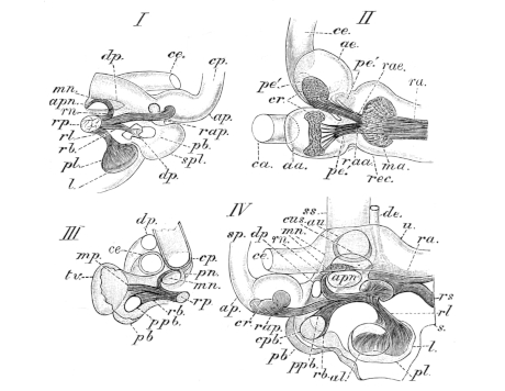
The membranous labyrinth of Rana esculenta; after Retzius.
I. Part of membranous labyrinth to show relations of the ductus and saccus endolymphaticus to the cochlear part; magnified.
II. The recessus utriculi and the external ampulla; magnified.
III. To show tegmentum vasculosum, pars basilaris, pars neglecta, etc.
IV. To show relations of the cochlear part to the pars neglecta.
aa. Ampulla anterior. ae. Ampulla externa. al. Apertura lagenae. ap. Posterior ampulla. apn Apertura partis neglectae. au. Apertura utriculi. ca. Canalis m. anterior. ce. External semicircular canal. cp. Posterior semicircular canal. cpb. Opening into pars basilaris. cr. Crista acustica. cus. Canalis utriculo-saccularis. de. Ductus endolymphaticus. dp. Ductus perilymphaticus. l. Lagena cochleae. ma. Macula ac. recessus utriculi. mn. Macula ac. neglecta. mp. Membrana basilaris. pb. Pars basilaris cochleae. pe′. Dark spot on either side of crest of ampulla. pl. Pars ac. lagenae. pn. Pars neglecta. ppb. Papilla ac. basilaris. ra. Ramus anterior. raa. Ramulus amp. anterioris. rae. Ramulus amp. externa. rap. Ramulus amp. posterioris. rb. Ramulus basilaris. rec. Recussus utriculi. rl. Ramulus lagenae. rn. Ramulus neglectus. rp. Ramus posterior. rs. Ramulus sacculi. s. Saccule. sp. Posterior semicircular canal. spl. Saccus perilymphaticus. ss. Sinus utriculi superior. tv. Tegmentum vasculosum. u. Utricle.
(6) The saccule (s), ductus endolymphaticus (de), and the saccus endolymphaticus (Figs. 247, 248, 249). The saccule (s) is an oval vesicle, placed under the anterior part of the utricle and directed outwards and downwards; below it is flattened in a direction from in front and within, outwards and backwards; above it is wider. In the inner and anterior surface is the rounded, oval macula acustica sacculi (Figs. 247 and 248 ms), to which is distributed the R. sacculi (rs) from above; a large otolith rests on the macula and occupies a large portion of the cavity, more especially the lower portion. The tubular ductus endolymphaticus (de) arises by a narrow oval opening placed in the upper and inner part of the wall, runs upwards and to the angle between the utricle and the sinus superior, continues in the same direction for a short space, and then curves inwards to pierce the apertura aquaeductus vestibuli, and so reach the cranial cavity. It then forms a large, thin-walled sac (saccus endolymphaticus), placed between the brain and cranium. The sac is very vascular and contains crystalline otoliths.
(7) The pars neglecta (Figs. 247, 248, 249) was described by Hasse as the ‘first part of the cochlea’ (‘Anfangstheil der Schnecke’), but according to Retzius it does not belong to the cochlea. It is placed above and in front of the pars basilaris cochleae, above and a little behind the lagena cochleae, therefore above the upper and posterior part of the saccule, and under the middle part of the utricle. It is really a prolongation of the saccule, with which it communicates by an elongated oval opening placed externally and immediately below the opening of the canalis utriculo-saccularis. It is an oval vesicle, with its roof intimately united with the lower wall of the utricle; anteriorly it is broad, posteriorly narrowed. The macula acustica neglecta is attached to the roof of the vesicle and consists of an anterior heart-shaped and a posterior semilunar portion united by a narrow connecting piece (Fig. 250 III and VII). The ramulus neglectus divides into two branches, which supply the two parts of the macula. On the macula acustica neglecta rests the membrana tectoria (Deiters); the membrane is S-shaped, with the anterior end narrow, the posterior broad (Fig. 250 VII); the borders of the membrane are pierced by numerous small round holes, the central part has smaller and fewer perforations, and is finely striated. From the middle of the posterior part of the upper surface a thicker portion projects downwards into the cavity of the pars neglecta; it has a narrow, deep notch for the nerve at about its middle; anteriorly it points towards a sickle-shaped piece, which is curved inwards. Canals run obliquely downwards and inwards from the apertures on the superior surface. The membrane is clear, homogeneous, partly finely striated, and corresponds with the membrana tectoria of the pars basilaris, etc.
(8) The lagena cochleae (Figs. 247, 248, and 249 l) is an oval swelling of the membranous labyrinth; it lies close to the sacculus with its broader end forwards, the narrower directed backwards. By a large rounded opening at the posterior end it communicates with the saccule by means of a rounded space common to the lagena cochleae and the cochlea. The papilla acustica lagenae (pl) is situated in the posterior wall of the lagena; it is elongated, oval, and supplied by the ram. lagenae. The papilla is covered by a plate-like otolith formed of numerous rounded particles.
Fig. 250.
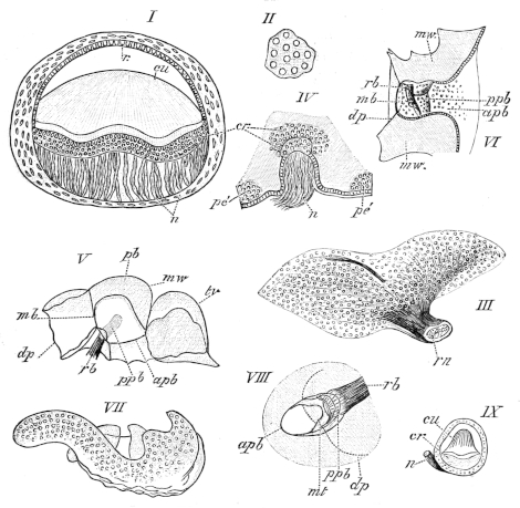
The membranous labyrinth of Rana esculenta; after Retzius.
I. Transverse section of the anterior ampulla. Magnified 50 times.
II. Piece from under surface of the cupula from the anterior ampulla. Vérick’s Syst., Obj. VIII. Oc. 3.
III. The macula acustica neglecta, seen from below. Vérick’s Syst., Obj. IV, Oc. 3.
IV. Part of wall of the anterior ampulla.
V. Part of the cochlea, the pars basilaris cut longitudinally. Vérick’s Syst., Obj. I, Oc. 3.
VI. Longitudinal section of the pars basilaris. Vérick’s Syst., Obj. I, Oc. 3.
VII. The macula ac. neglecta, seen from below. Vérick’s Syst., Obj. IV, Oc. 3.
VIII. The pars basilaris, seen from behind and the outer side. Vérick’s Syst., Obj. I, Oc. 3.
IX. Transverse section of the external ampulla. Magnified 175 times.
apb Oval opening into sacculo-cochlear space. cr Crista acustica. cu Cupula terminalis. dp Ductus perilymphaticus. mb Section of thinner wall of pars basilaris. mt Tectorial membrane. mw Thickened membranous wall. n Nerve-fibres. pb Pars basilaris. pe′ Area of coarsely granular cells. ppb Papilla acustica basilaris. r Epithelium on raphe. rb Ramulus basilaris. rn Ramulus neglectus. tv Tegmentum vasculosum.
(9) The pars basilaris cochleae (Figs. 247, 248, and 249 pb) is placed on the posterior thickened wall of the saccule and lies above and behind the lagena. It forms a small, oval, pocket-like protuberance, with the long axis directed from in front and above, backwards and outwards, its opening being directed forwards and outwards. The short ram. basilaris passes in from above to supply it. The walls of this dilatation are thick and stiff, with the exception of a small portion, the membrana basilaris (Hasse) (mb), which closes the opening into a small dilatation on the anterior inner wall. The ramulus basilaris (rb) divides into, at least, two branches, and passes close to the membrana basilaris (mb), where the elongated and oval papilla ac. basilaris (Fig. 250 ppb) is placed. The papilla is covered by a membrana tectoria (Fig. 250 mt), which is often found separated from the papilla, probably by the action of the reagents used. The form of this membrane is peculiar but will easily be understood from the figure (Fig. 250 mt). In structure it is similar to the corresponding structures found in other parts of the ear.
Fig. 251.
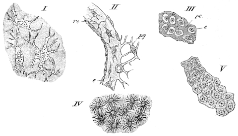
Preparations from the ear of Rana esculenta; after Retzius.
I. Part of the membranous wall seen from the surface. Vérick, Obj. VI, Oc. 3.
II. Transverse section of the membranous wall. Vérick, Obj. III, Oc. 3.
III. Epithelium from the neighbourhood of the macula ac. rec. utriculi. Vérick, Obj. III, Oc. 3. Oc. 3.
IV. Branched cells from the yellow spot on the floor of the anterior ampulla. Vérick, Obj. III,
V. Epithelium from the roof of the anterior ampulla. Vérick, Obj. III, Oc. 3.
re Epithelium of raphe. e Pavement epithelium. pe Protoplasmic cells. pg Perilymphatic tissue.
(10) The tegmentum vasculosum (Deiters) (Figs. 245, 248, and 250 tv) is an oval, shell-shaped dilatation of the membranous labyrinth; its long axis is directed from above and in front, downwards and backwards. The walls of the tegmentum are thin and intimately attached to the periosteum.
e. The minute structure of the membranous labyrinth (Figs. 251 252).
(1) The walls of the membranous labyrinth have the same general structure throughout: the walls are usually thicker at the nerve-terminations, in the ampullae, semicircular canals, pars neglecta, and especially the pars basilaris; the wall of the tegmentum tympani are the thinnest. The walls are transparent, homogeneous, refractive, and, at places, show a faint striation, which is, as a rule, not due to the presence of fibres; in parts of the recessus utriculi, and in the outer wall of the saccule, especially near the tegmentum vasculosum, more or less distinct fibres can be made out. Sections of the wall show spindle-shaped cells, with the processes usually arranged parallel to the surfaces; seen from the surface, the cells are seen to branch in all directions (Fig. 251 I, II). In the thinner parts of the walls the cells are few or altogether absent. The outer surface of the membranous labyrinth is uneven, in consequence of the attachment of the perilymphatic network. Blood-vessels are also attached to the outer surface, and pierce the wall, especially near the nerve-terminations.
The whole of the inner surface is lined with a layer of polygonal, tesselated epithelium-cells. The size and height of the epithelium varies in different parts. On the outer wall of the saccule the cells are large, but on the inner wall small; they are also large in the semicircular canals, except on a small raphe on the inner and outer side, where they are smaller but higher (Fig. 251 re); in the ampullae the cells are large, except on the roof. In the utricle and sinus superior they are also moderately large. In addition to the places mentioned, a smaller epithelium is found on the floors of the ampullae, in the recessus utriculi, and near all the nerve-terminations and on the sides of the ampullar septa. Surrounding the nerve-terminations of the macula rec. utriculi, macula sacculi, and papilla lagenae are found narrow, branched, yellowish cells (Fig. 251 pe) with spindle-shaped nuclei. Cells of a third kind, first described by Deiters, Hasse, and Kuhn, in the tegmentum vasculosum, and in the ampullae by Hasse and Kuhn, are also found in the utricle. They contain a yellowish pigment, and are collected into two sharply differentiated groups in each ampulla (Hasse has one placed before and one behind the septum on the floor). The cells are cylindrical, the upper parts striated, the lower narrower, and the bases again widened to a polygonal, more homogeneous plate, which is fixed to the wall. On the tegmentum vasculosum the corresponding cells are not so high.
(2) The nerve-terminations. The larger branches of the auditory nerve contain medullated fibres of various dimensions and bipolar, spindle-shaped ganglion-cells. The nerves pierce the walls obliquely or vertically, and retain their medullary sheaths until near their final distribution. On each of the nerve-terminations is found nerve-epithelium, which varies in height in different parts. In the crista acustica it measures 0.075 mm. in height in the middle part, 0.06 mm. at the sides; on the macula rec. utriculi 0.09 mm., on the macula sacculi 0.075 mm., on the papilla lagenae 0.06 mm., on the papilla part. basil. 0.045 mm., on the macula neglecta 0.075 mm. The epithelium is of two kinds, hair-cells and sustentacular cells.
Fig. 252.
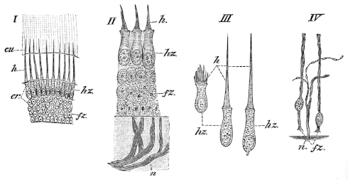
The nerve-terminations in the membranous labyrinth of Rana esculenta; after Retzius.
I. Vertical section through the crista acustica of the anterior ampulla. Vérick’s Syst., Obj. III, Oc. 3.
II. Vertical section through the macula ac. recessus utriculi. Vérick’s Syst., Obj. VIII, Oc. 3.
III. Three isolated hair-cells from the crista ac. of the anterior ampulla. Vérick’s Syst., Obj. VIII, Oc. 3.
IV. Two isolated sustentacular cells from the crista ac. of the anterior ampulla. Vérick’s Syst., Obj. VIII, Oc. 3.
cr Crista acustica.
cu Cupula terminalis.
fz Sustentacular cells.
h Hairs of hair-cells.
hz Hair-cells.
n Nerve-fibres.
α. The hair-cells (Fig. 252 hz) have, on the whole, elongated, flask-like forms, but are not all of the same length (0.024–0.04 mm.). The free ends of the cells are rounded, flattened, and yellowish, and each bears a stiff cilium, which is fixed by a broad base to the cell, and thins out towards its free end: the cilia vary in length; in the ampullae their greatest length is 0.13 mm., on the macula rec. utriculi 0.011 mm., and on the papilla lagenae 0.017 mm. The cells are granular, possess rounded oval nuclei, and are fixed by a fine, narrow process (Fig. 252 hz), though they usually seem to be rounded off without possessing a process.
β. The sustentacular cells. Under the hair-cells is a finely granular substance, possessing numerous rounded oval nuclei, which are placed in superimposed rows (Fig. 252 fz), the deepest row being placed close together and immediately on the membranous wall. After proper treatment and isolation these nuclei are seen to belong to narrow, elongated cells, which rest by a slightly widened base on the wall, and are continued upwards between the hair-cells to reach the surface of the epithelium, where their upper processes are again slightly widened.
γ. The nerve-fibres (Fig. 252 n) lose their medullary coats, ascend towards the epithelium, and frequently divide to form two unequal branches, which ascend to the level of the hair-cells, and curve so as to course horizontally as extremely fine varicose fibrillae; these frequently form a network, of which the exact method of termination has not been made out. In some cases a fine fibril may be traced to the base of a hair-cell, but a direct continuation of the one into the other has not yet been traced.
VI. THE EYE.
(Re-written by the translator.)
The organ of sight, the eyeball (bulbus oculi), together with its appendages (tutamina oculi), will be described in this chapter.
A. The Eye is flattened on the outer surface, more convex on the inner or deeper surface. Its principal axis is directed from behind, forwards and outwards.
The outer transparent portion of the eyeball is the cornea, which forms the outer boundary of the anterior chamber. The larger, white, opaque, and inner portion is the sclerotic coat, which, together with two deeper tunics, the choroid coat and the retina, enclose the posterior chamber of the eye. The pigmented ring placed behind the cornea is the iris, and the aperture it encloses the pupil. The lens is placed immediately behind the iris. On the inner side the optic nerve pierces the sclerotic to enter the eyeball.
a. The sclerotic coat (sclerotica s. sclera) forms about three-fourths of the surface of the eyeball; posteriorly it is pierced by the optic nerve at a point (porus opticus) nearer the temporal side than the nasal. The sclerotic coat consists of fibrous tissue externally, with a layer of hyaline cartilage internally (Helfreich). The fibrous layer is formed of bundles of parallel fibres, which cross each other, chiefly at right angles (Hoffmann). The cartilaginous layer ends just behind the line of insertion of the extrinsic muscle of the eye, and is thickest at the point of entrance of the optic nerve (Helfreich).
Fig. 253.
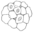
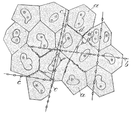
Endothelium from the inner surface of the sclerotic coat; after Hoffmann.
The sclerotic coat is rich in nerve-fibres, which form a close network; the fibres, however, do not unite but form the meshes of the network by simply crossing each at acute angles.
The deeper surface of the sclerotic coat is lined with a layer of large endothelial cells (Hoffmann), (Fig. 253), which form the outer wall of the capsule of Tenon.
b. The cornea and the anterior chamber. The cornea forms about one-fourth of the surface of the eyeball and is directly continuous with the sclerotic. In it five layers can be distinguished: a layer of stratified epithelium or conjunctiva, an anterior hyaline membrane, the true corneal substance, a posterior hyaline membrane, and a layer of endothelium.
(1) The corneal epithelium is a layer of stratified epithelium covering the superficial surface of the cornea. The superficial layer forms a beautiful mosaic of polygonal cells; the middle layers are polygonal in all sections, while the deepest layer is more or less columnar. Except in the most superficial layer, all the cells have serrated surfaces. Smaller cells possessing each two nuclei are also found between the columnar cells, and are evidently cells in process of division; according to Waldeyer, cell-proliferation may also take place in the middle layers.
The basal or deeper portions of the columnar cells possess a clear border, which reminds one of the hyaline border found on the free border of columnar epithelium in other parts. The cells are here so closely applied to one another that these borders have the appearance of a continuous, highly refracting membrane (Rollett); according to Henle, the border consists of a network of very fine processes from the cells above.
(2) The true corneal substance, and (3) the anterior hyaline membrane. The corneal substance consists of flat bundles of fibres arranged in laminae, with cement-substance and connective-tissue corpuscles interposed. The fibrils are extremely fine (0·0001 mm., Engelmann), and bound together into bundles by cement-substance. The bundles of the laminae are arranged at various angles, though many are placed at right angles to each other (Waldeyer).
Between the laminae are flattened spaces, which seen in section are spindle-shaped. By proper treatment they are seen to be irregular, branched spaces, which communicate by fine canals and form part of the Recklinghausen-canals or lymph-system. These spaces contain branched, connective-tissue corpuscles (Toynbee), and a colourless fluid.
The corpuscles (Fig. 254 e) do not fill the spaces which they occupy. They possess large nuclei, surrounded by granular protoplasm.
Fig. 254.
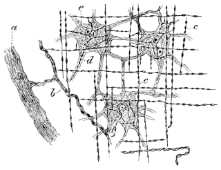
Preparation from cornea of Rana esculenta; after Klein, Hartnack’s Syst., Obj. VII, Oc. 3.
a Nerve of first order.
b Nerve of second order.
c Nerve of third order.
d Nerve of fourth order.
e Corneal corpuscles.
The canals by which these spaces communicate (‘Saftcanälchen’ of Recklinghausen) lie, in general, parallel to the surfaces of the cornea, and communicate by joining at acute angles or by short transverse branches. According to Lavdowsky, these canals have a distinct lining membrane.
The anterior hyaline87 layer (Bowman’s or Reichert’s lamella) is not so well seen in the frog as in some higher animals; it is simply a portion of the corneal substance, of somewhat denser structure than the rest, into which it passes by a gradual transition.
(4) The posterior hyaline membrane (Descemet’s membrane) is a highly elastic, very transparent layer, placed behind the true corneal substance; in the frog some few bundles of fibres belonging to the true corneal substance appear to pass into the posterior hyaline layer, although they cannot be traced further through its substance. The structure of the membrane is, in consequence of its transparency, unknown, though the above observation seems to point to a fibrillar origin.
(5) The corneal endothelium is a single layer of polygonal cells of 0·02 mm. diameter. The cells possess the power of altering their shape when stimulated (Klebs).
Fig. 255.
Preparation from cornea of Rana esculenta; after Klein. Hartnack’s Syst., Obj. X immers., Oc. 3.
a Endothelial cells.
b Nuclei of endothelial cells.
c Nerves of third order in the tissue of the cornea propria.
d Nerves of the fourth order.
(6) The nerves of the cornea are derived from the ramus ophthalmica trigemini; they pierce the sclerotic coat in front of the sclerotic cartilage and then course towards the cornea, at the margin of which they form a coarse network of medullated fibres. From this about thirty nerves pass towards the cornea, which they enter, and then very quickly lose the main part of their medullary sheaths. According to Wolff, a portion of the nerves retain their medullary sheaths, or in some cases appear to regain it after having lost it.
The nerves passing from the plexus (nerves of the first order, Klein) give off smaller branches, which for a short distance have a serpentine or rectilinear course. By a few anastomoses they form a loose plexus (nerves of the second order, Klein). After a longer or shorter course they give off numerous lateral fibres, or terminate in several such fibres arising at one point (nerves of the third order, Klein). These are distinguished by their size, varying only within small limits, and by the possession of more or less regularly placed varicosities; the clearer portions are longitudinally striated as though made up of fibrillae; they have a nearly rectilinear course, and, after a longer or shorter course, turn into a direction which is at right angles to the former one; lastly, they remain for long distances unbranched. These nerves are connected one with another by cross fibres running at right angles to them, and in this way a rectangular trellis-work is formed.
The fibrils (nerves of the fourth order) given off by these nerves form networks around the connective-tissue corpuscles, but no direct connection between nerve and corpuscle has been traced; they always appear to lie on that surface of the corneal corpuscle which is directed towards the superficial surface of the cornea (Klein). In the endothelium covering the membrane of Descemet these fibrils can be traced coursing along the margins of the cells (Fig. 255 d), and sometimes undergoing dichotomous division (Klein).
Almost all observers have described these fibrils as possessing varicosities; Hulke, and more recently Wolf, however, deny their presence. Lavdowsky traces nerve-fibrils to the nuclei of the connective-tissue corpuscles.
(7) The anterior chamber is the space between the cornea and the iris, and is filled with a watery fluid, the aqueous humour. At the circumference of the chamber are a number of spaces (spaces of Fontana), formed by interruptions in the tissue between the posterior surface of the cornea and the iris; the result is that bands or trabeculae (ligamentum pectinatum iridis) pass from the one structure to the other, and between these are the spaces of Fontana.
According to Angelucci these trabeculae are of three kinds: trabeculae passing from the cornea to the iris, formed of connective-tissue; trabeculae from the cornea to the ciliary processes, which contain elastic tissue; trabeculae from the interstitial connective-tissue of the ciliary muscle to the cornea, and formed almost entirely of elastic tissue.
At the junction of the cornea and sclerotic, and just in front of the spaces of Fontana, is a larger and similar space, which may be traced round the whole circumference of the cornea; this, the canal of Schlemm (Sinus circularis iridis), is held to be a venous plexus by some observers (Angelucci, and others), according to others it is a lymphatic space in connection with the anterior chamber (Schwalbe, and others). It is certain that the vessels can be very easily injected from the anterior chamber, although a direct communication has not yet been seen.
c. The choroid coat and the iris (tunica choroidea et iris, tunica vasculosa).
Fig. 256.
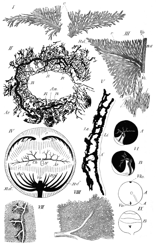
The vessels of the choroid and iris; after Hans Virchow.
I. The two roots of the V. bulbi superior. Magnified 10 times.
II. Vessels of the iris. Magnified 9 times.
III. Origin of the ventral vein.
IV. Schema of the choroid vessels; seen from the proximal pole.
V. Transverse section through the choroid at the equator.
VI. The origin of the choroid arteries from the ophthalmic artery. The greater part of the sclerotic has been removed. Left eye twice natural size. A. From the proximal pole. B. From the temporal side.
VII. Part of a choroidal artery attached to the choriocapillaris. Magnified 10 times.
VIII. A portion of the choriocapillaris, more highly magnified.
IX. The V. ophthalmica and V. bulbi superior on the sclerotic of the right eye. A. Seen from proximal pole. B. Seen from above. Twice natural size.
A Art. ophthalmica. A′ Art. choroidea. An R. nasalis of the ophthalmic artery. At R. temporalis of the ophthalmic artery. ch Area of membrana choriocapillaris. Le Outer pigmented layer of choroid. Li Inner pigmented layer of choroid. N Optic nerve. r Vasa recta. r′ Transitional part between choriocapillaris and ventral whorl. r″ Transitional part between choriocapillaris and upper whorl. R Branches of the circulus iridis major. Rd Distal root of vein of under surface of eye. Rd′ Distal root of nasal vein. Rd″ Proximal root of nasal vein. Rp′ Nasal root of ventral vein. Rp″ Temporal root of ventral vein. Vbs V. bulbi superior. Vh V. hyaloidea. Vo V. ophthalmica. Vp. Proximal root of vein of under surface of eye. Vs′ Nasal root of V. bulbi superior. Vs″ Temporal root of V. bulbi superior.
1. The choroid coat lines the deeper surface of the sclerotic coat, but is also prolonged under the cornea to form the iris. The choroid is firmly attached to the sclerotic in two positions, at the point of entrance of the optic nerve, and at the line of junction of the sclerotic and the cornea. Its external surface is closely applied to the deeper surface of the sclerotic, from which it is only separated by a very narrow serous cavity (supra-choroidal space), and to which it is attached by numerous vessels and nerves. The deep surface of the choroid is covered by the retina, to which it is closely attached, except at the ora serrata, the attachment being especially intimate at the processus ciliares.
The choroid coat consists of a fibrous layer containing corpuscles and traversed by a very rich vascular anastomosis. The corpuscles of this layer are deeply pigmented, in some cases to such an extent that the oval nucleus cannot be seen; the fibrous tissue is also pigmented, and has consequently a brownish tinge. That portion of the layer immediately below the sclerotic is termed the lamina fusca or suprachoroidea, the vessels on the deeper surface forming the membrana choriocapillaris. This again is lined on its deeper surface by a hyaline membrane.
α. The arteries (Fig. 256 VI; VII, VIII) supplying this coat are two branches of the arteria ophthalmica; these form a capillary network (Fig. 256 VII) resembling the corresponding structure found in mammals. The meshes have approximately the same size, while the capillaries themselves vary considerably in size. This network is, however, only complete on the nasal, temporal, and proximal part of the upper surfaces. Towards the corpus ciliare the meshes become wider and elongated; the capillaries then unite at acute angles parallel with the longitudinal axis of the eye. The network (choriocapillaris) exists in a simple layer within the two arteries which form it, and superficial to the veins (Virchow).
β. The veins of the choroid (Fig. 256 III, IV, IX) are (1) a vein which unites at the lowest point of the equator of the eye with the V. hyaloidea to form (2) the V. ophthalmica, two small branches of the V. bulbi superior, which unite outside the sclerotic, and (3) the vasa recta.
(1) The larger vein arises from the greater part of the under surface of the eye; it gives off branches to each side, which radiate to form a ‘whorl’ or star-shaped capillary anastomosis (Fig. 256 III), the two halves of which have no connection. A proximal and a distal root can be distinguished in the anastomosis; the distal lies towards the corpus ciliare, and occupies exactly one-fourth of the circumference of the choroid at its junction with the corpus ciliare.
(2) The two branches of the V. bulbi superior lie alongside the corpus ciliare on the upper surface, and each occupies one-fourth of the circumference; they form a similar though simpler figure (Fig. 256 I) to the foregoing, each forming one half.
(3) The vasa recta are numerous parallel vessels which arise in the iris, and coursing centrally empty themselves into the branches of the V. bulbi superior on the superior surface, and into the branches of the venous capillaries on the inferior surface.
2. The iris is covered anteriorly by a layer of endothelium, continuous with that covering the posterior surface of the cornea, and of similar character. The border of the pupil (margo pupillaris) is of a golden colour, outside this bright ring to its outer margin (margo ciliaris) the iris is black; the golden colour is due to the presence of cells containing a pale yellow pigment; the nuclei of these cells are round and granular; the cells themselves have rounded outlines (Hoffmann). The black portion of the iris contains more irregular, spindle-shaped cells, with round nuclei, which are hidden by a dense mass of pigment-granules (Iwanoff and Hoffmann).
The true substance of the iris consists of muscle, nerves, blood-vessels, and a connective-tissue stroma, but on the posterior surface is another layer of black, pigmented cells, and this is again covered with a hyaline membrane, in which, however, a fibrous structure may be made out (Koganeï).
The muscle-fibres are long, spindle-cells, which are abruptly swollen in the middle, where the nuclei are situated; the nucleus is oval, 0.009–0.0012 mm. in length, 0.0025 mm. broad, and occupies nearly the whole of the swollen part of the cell (Hoffmann, Grünhagen).
According to Koganeï the iris possesses a M. constrictor iridis (l. c. Berlin Sitzungsber.), but no M. dilatator iridis; in a former publication (l. c. Arch. mik. Anat.) he was unable to find any muscular fibre, and holds the muscle-fibres of Grünhagen to be connective-tissue elements.
The stroma consists of delicate connective-tissue fibrils, enclosing a very large number of pigmented, branched cells.
α. The arteries of the iris (Fig. 256 II) arise from an arch (see Vessels of Eye) formed by the A. ophthalmica in the corpus ciliare. It commences between the ventral and temporal surfaces by two branches: one courses along the temporal border, the other along the nasal, to meet each other on the nasal surface; the former courses through one-third, the latter embraces two-thirds of the circumference at the iris.
The temporal artery courses along the ciliary border during the first third of its course, it then gradually approaches the border of the pupil; the nasal artery runs at once towards the pupil. On the nasal border of the pupil they anastomose by their branches, and so form a circulus iridis major.
Except near their termination, no small vessels arise from this arterial circle; in Fig. 256 II, for example, only five larger branches are given off, three from the temporal side and two from the nasal. The five large branches run towards the circumferential border of the iris and break up into numerous vessels, which form a very irregular and open network. From this network arise the vasa recta already described.
d. The lens is almost spherical, and is composed of cellular elements enclosed in a capsule (capsula lentis).
The capsule is a homogeneous, transparent, structureless, and highly elastic membrane. The deeper surface of the anterior capsule is lined with a simple layer of regular nucleated six-sided epithelial cells.
Fig. 257.
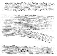
Fibres from the lens of the frog; after Hoffmann. Magnified 700 times.
The lens itself consists of long, flat fibres; seen from the surface these are broad, narrow edge-wise, and in section six-sided prisms. Those lying parallel to the anterior and posterior surfaces are broad and thicker, those towards the border are narrower. These cells are striated, both longitudinally and transversely (Arnold). The cells near the margin, however, have no transverse striation (Hoffmann). The cells of the central parts form a much closer and firmer structure than those at the periphery (Arnold). The peripheral cells are nucleated, and sometimes even possess two nuclei to one cell; the central cells have no nuclei (Arnold).
The cells are held together by a cement-substance and by their serrated surfaces; the serrations are the cause of the transverse striations. The fibres of the lens have a simple arrangement: commencing at the middle point or pole of one surface they pass over the equator to the opposite pole; consequently the long borders of adjacent cells are in juxtaposition, and their pointed extremities meet at points in the axis of the lens (Hoffmann).
Ritter has described short, nucleated cells in the centre of the lens; these are held by Babuchin to be cells which have been arrested in their development.
e. The retina is the innermost coat of the eye; in the recent state it is pale, soft, and smooth. The structures composing it are arranged in ten layers; from the deeper surface towards the choroid these are: the internal limiting membrane, the optic-fibre layer, the ganglion layer, the inner molecular layer, the inner nuclear layer, the outer molecular layer, the outer nuclear layer, the external limiting membrane, the layer of rods and cones, and the pigment layer.
These layers are held together by connective-tissue elements.
Fig. 258.
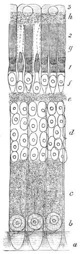
Vertical section through retina of frog; after Hoffmann. Magnified 500 times.
a Internal limiting membrane.
b Ganglion-cell layer.
c Internal molecular layer.
d Internal nuclear layer.
e External molecular layer.
f External nuclear layer.
g Layer of rods and cones.
h Pigmented epithelium layer.
1. Inner segments of rods and cones.
2. Outer segments of rods and cones.
3. Outer transparent segments of pigmented epithelium.
(1) The internal limiting membrane (Membrana limitans interna) will be described together with the connective-tissue elements (10).
(2) The optic-fibre layer is formed by the fibres of the optic nerve. The nerve-fibres in their course towards the eye are possessed of medullary sheaths, but on piercing the sclerotic these sheaths are lost. The fibres are now pale, non-medullated, and of very varying thickness. In the mass of fibres nothing can be seen except an extremely fine fibrillation and very fine varicosities; the latter, however, appear to be artificial productions (Hoffmann). This layer of fibres extends over the inner surface of the retina, and gradually thins from the point of entrance of the optic nerve to the limits of the retina.
(3) The ganglion-layer lies immediately without the nerve-fibre layer (Fig. 258 b). The ganglion-cells are small and usually pear-shaped. The cells possess large nuclei, round which is a thin layer of very granular protoplasm. The cells have inner and outer processes; the inner pass into the nerve-fibre layer, the outer into the inner molecular layer in more or less radiating directions. Manz claims to have traced a direct connection between the inner processes and the fibres of the nerve-fibre layer.
Each ganglion-cell, whatever its shape or size, has only one inner process, which is easily distinguished from the outer process by its being more glistening, by the possession of varicosities, and because this process never branches.
The outer processes are single (Schwalbe) or rarely double (Hoffmann), and have as a rule a direction at right angles to the inner processes. Each outer process is finely granular, which suggests rather a prolongation of the cell-substance than a true process. Frequently they are branched, sometimes forming two equal sized processes, which give off finer twigs; at other times they appear to pass through the whole of the inner molecular layer without undergoing division (Schwalbe). The processes do not inosculate (Santi Sirena).
(4) The inner molecular layer (Fig. 258 c) is 0·07–0·08 mm. thick (Hoffmann), and consists of a finely granular mass together with the outer processes of the ganglion-layer, and connective-tissue elements.
The granular matter consists of an extremely fine network or reticulum, through which numerous fine fibres course (Schultze, Kölliker, Manz, Heinemann, and others); according to Schultze the supposed molecules or granules of others (Henle, Merkel, and Retzius) are simply the fine meshes of this reticulum. The branched, outer processes of the ganglion-cells form a rich anastomosis in this layer.
(5) The inner nuclear layer (Fig. 258 d) contains parts of two kinds of cellular elements; these are radial nerve-fibres with large nuclei, and connective-tissue elements (see below, par. 10). The nerve-fibres are easily distinguished by their spindle-shaped varicosities; both cellular elements possess large oval nuclei. The bodies of the cells surrounding the nerve nuclei are almost filled by the nuclei, which have sharply-defined, rounded nucleoli. The fibres to which these cells are attached may be distinguished as inner and outer processes; the inner process is fine, irregularly varicose, and unbranched; the outer process is thicker, finely granular, and is not varicose (Schwalbe). At the margin of the outer molecular layer the outer processes divide, usually into two branches, and at an acute angle to each other, though sometimes at a right angle. The further course of these branches in the outer molecular layer is unknown.
(6) The outer molecular layer (Fig. 258 e) corresponds in general with the inner molecular layer as regards its structure; it is, however, much thinner.
(7 and 9) The outer nuclear layer and the layer of rods and cones (Figs. 258 f, g, 259). The rods and cones are intimately connected with the elements of the outer nuclear layer, hence the two layers are best described together.
The rods (bacilli) have two parts or limbs, an outer and an inner, which differ in structure, and in chemical and physical characters. The outer part is highly refractive, the inner more homogeneous and less refractive, the two parts being sharply differentiated from one another.
The outer part is also weakly doubly refracting, the inner has no trace of this property. The rods are 0·05–0·06 mm. in length, of which 0·035–0·04 mm. belongs to the inner limb. The outer end of the outer limb is more or less rounded; the whole has a longitudinal striation (Schultze), due to its being composed of rounded fibrils, about twenty-four to each rod (Hensen). The fibrils are sharply differentiated from each other and have a slightly spiral course; when seen in transverse section these outer limbs do not appear to be round (Schultze), although others hold them to be perfectly rounded (Hoffmann and others), and that the loss of the cylindrical form is due to the methods of treatment. According to Merkel the longitudinal striation is caused by a canalisation of the outer limb, which according to him encloses the processes of the pigmented epithelial layer; he is also of opinion that the spiral appearance is an artificial product. In the latter opinion he is probably wrong, as perfectly fresh rods examined in aqueous humour show the same spiral appearance (Hoffmann): against the canalisation view others observe that the longitudinal striation is most distinct near the inner limit of the outer limb, and that it is impossible to conceive that the processes of the pigment-cells should terminate with such extremely regular ends (Hoffmann).
Fig. 259.
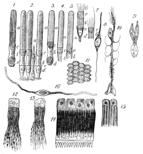
Various preparations from the eye of the frog: chiefly from the retina.
1. Rod from retina in aqueous humour, showing spiral striation.
2. Three rods and one cone after treatment with osmic acid.
3, 4, 5. Rods examined in recent state.
6, 7. Inner segments of two rods after treatment with osmic acid.
a Outer limb. b Inner limb. c Lenticular body. d Nucleus of outer nuclear layer. e External limiting membrane.
8. Nuclear body from inner nuclear layer.
9. Twin-cone.
10. Sustentacular fibre of retina.
11. Surface view of pigmented epithelium of retina.
12, 13. Isolated pigmented cells of retina.
14. Four pigmented cells, rods and cones, external limiting membrane, and part of outer nuclear layer attached.
15. Two pigmented cells; each showing three attached rods.
16. Muscle-fibre from the iris.
Figs. 1, 2, 3, 4, 5, 6, 7, 8, 9 are magnified 500 times. Fig. 10, 300 times. Fig. 16, 400 times. All are copied from Hoffmann’s figures.
Figs. 12, 13, 14, 15 are copied from Morano’s figures; Hartnack, Oc. II, Obj. 9.
In the central part of the inner end of the outer limb is seen a dark point when the structures are examined in transverse section (Ritter, Manz, Schiess, Schultze, and others). The cause of this is not clearly understood; some hold it to be a fibre (Ritter’s fibres), others hold it to be an artificial product (Hensen).
After treatment with certain reagents the outer limbs show a transverse striation, which is probably produced by the action of these reagents on the sheath of the outer limbs; that a sheath is present is proved by its possession of a different refractive index (Zenker, Schultze) to the rest of the outer limb, and this transverse striation is not seen until the whole organ has undergone considerable post-mortem changes (Hoffmann). Should this change be allowed to proceed a stage further, the outer limbs of the rods split transversely and form small discs from 0·0005–0·00055 mm. thick; this takes place in the outer limb only.
The inner segments of the rods (Figs. 258, 259) are short (0·020–0·022 mm.) and of the same thickness as the outer limbs. When perfectly fresh they appear homogeneous; very quickly changes commence, which are probably due to coagulation. A plano-convex figure (Fig. 259) is then seen at the outer portion of the segment (lens-shaped figure of Schultze); with staining reagents it gives the same reactions as the outer segment of the rods. The rest of this segment forms a short cylinder, which probably has no distinct sheath (Hoffmann, Merkel); some observers are inclined to think that a sheath exists (Landolt, Schwalbe).
The outer segments of the rods are of two chief sizes (Schwalbe). Those of the one kind are large; the second variety occurs less frequently, and the segments are shorter, measuring only 0·002 to 0·0025 mm. The inner segment is a long, thread-like process, except where it is swollen to enclose the lens-shaped body.
The rods are much more numerous than the cones, except at one small spot (macula lutea) on the posterior surface of the retina, where only cones are found (Krause).
The cones (coni) have each two segments like the rods (Figs. 258, 259). The outer segments are short (4–5 µ), they are slightly conical and terminate externally in a blunt point; they possess a longitudinal striation (Schultze), and very easily break up transversely into small discs, which, however, do not separate so completely as in the case of the rods, in consequence of the presence of a sheath continuous with a sheath on the inner segment.
The inner segments (Figs. 258, 259) have convex sides and measure 12–14 µ; like the corresponding parts of the rods they possess lens-shaped bodies at their junction with the outer segments, but the bodies differ in shape, being bi-convex or rather oval in form. The inner segments are enclosed in a delicate sheath continuous with that of the outer segments.
In some cases two cones are united to form a twin-cone; in such cases the one is always larger than the other (Fig. 259 9), and has several peculiarities which distinguish it from the smaller.
The smaller or secondary member of a twin-cone is longer, and possesses a lens-shaped body which is plano-convex. The larger or principal member of a twin-cone is shorter, has a plano-convex body, but also an oval, homogeneous, glistening body, which is directly attached to the plano-convex body. The shape of the two members is also different.
The outer nuclear layer (Fig. 258 f) is 14–16 µ thick; the nuclei lie in two layers. The nuclei belonging to rods and cones have the same characters, each nucleus being a large, oval, hyaline body, and enclosing a bright nucleolus. Each nucleus is surrounded by an extremely thin layer of finely granular matter. The inner processes of the nuclear bodies both of the rods and the cones extend to the outer molecular layer, are there dilated and serrated, where they become attached to the outer molecular layer (Schultze, Hoffmann). In some cases, however, the inner process of the nuclear bodies, belonging to the rods, forms only a short fine fibre.
In the case of twin-cones the corresponding parts in the outer nuclear layer possess two nuclei (Schultze).
(8) The pigment layer (Figs. 258 h, and 259 11, 12, 13, 14, 15) is not intimately attached to the rest of the retina. It consists of cylindrical cells in which two parts or segments are sharply differentiated; the external part, directed towards the choroid coat, is of pale, or colourless granular protoplasm, and occupies one-third of the length of the cell; this part encloses a large, round, nucleolated nucleus. This colourless segment of the cell also includes one or two bright yellow, fat globules (Morano). Seen from the surface the cells are hexagonal (Fig. 259 11). The remaining two-thirds of the cells consists of a brush formed of numerous fine pigmented processes; the ultimate terminations of the processes, which lie parallel to each other, are frequently unpigmented; each cell possesses thirty to forty such processes (Morano).
The processes extend between the rods and cones as far as the external limiting membrane (Figs. 258, 259), or sometimes a little further (Merkel, Morano, Hoffmann). The processes from one pigment-cell surround a number of rods and cones; according to Morano twelve to fifteen rods and cones may be encased or surrounded by the processes of a single cell.
The thickness of this layer varies from 60–70 µ; the nuclei of the cells have a diameter of 10–12 µ, the width of a single cell is from 20–25 µ (Hoffmann).
(10) The connective-tissue elements of the retina and the external and internal limiting membranes. The elements of the retina are supported by connective-tissue elements or sustentacular cells, which have a radial arrangement, and which form the two limiting membranes (Müller).
Each sustentacular cell (Fig. 259 10) has two segments, an inner and an outer, the boundary between these lying in the inner nuclear layer, and being marked by the presence of a large oval nucleus. The inner segment of each cell terminates internally in a wide ‘foot’ or base, or may form several such after having undergone division (Schultze): these bases together form a transparent, thin membrane, the internal limiting membrane (membrana limitans interna).
Within the ganglion-layer these cells possess peculiar appendages, which fit round the ganglion-cells and support them (Schwalbe).
The outer segments of the sustentacular cells extend into the outer molecular layer, and then break up into irregular processes which extend radially to the external limiting membrane, and which they probably form. The external limiting membrane (membrana limitans externa) is therefore a membrane corresponding to the internal limiting membrane, and formed by the flattened ends of the processes belonging to the sustentacular cells.
The sustentacular cells have a distinct, resistant cell-wall (Schwalbe); the cell-contents are a finely granular protoplasm, and a large oval, nucleated nucleus placed in the inner nuclear layer.
f. The ciliary processes have the same structure as the rest of the choroid coat: the vessels form more or less longitudinal meshes and are more irregular than in the rest of the choroid.
g. The posterior chamber and vitreous body (Fig. 260). The vitreous humour occupies the greater portion of the cavity of the eyeball, i.e. the posterior chamber. The humour consists of a mass of cells enclosed in a transparent hyaloid membrane (membrana hyaloidea), which is in contact with the internal limiting membrane of the retina.
The cells forming this structure are small, flattened, transparent, and nucleated (Iwanoff and Virchow); according to the former observer the cells have contractile powers.
The hyaloid membrane is described as structureless by Schwalbe, as fibrous by Pappenheim, Bowman, and Fuikbeiner.
The vessels of the vitreous body (Fig. 260) are as follows. The A. hyaloidea arises at the lowest point of the corpus ciliare; it almost immediately divides into two branches, which form a ring at a distance of about 0·5 mm. from the lens and lying on the surface of the vitreous body (Fig. 260 I, II). One, R. nasalis, passes to the nasal side and courses through one-fourth of the circle; the other, R. temporalis, courses through three-fourths of the circle. The branches are all given off proximally and at right angles to the circle (Fig. 260 I, II). From the R. nasalis only one branch arises, from the R. temporalis seven, the first of which corresponds in point of origin with the branch from the R. nasalis. The branches on the nasal and temporal surfaces of the vitreous body are the shortest.
Fig. 260.
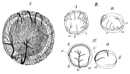
The vessels of the vitreous body; after Hans Virchow.
I. Vessels of the vitreous body; seen from the deeper pole and slightly from above. Magnified 6 times.
II. Arteries of the vitreous body of the right eye.
A Seen from the proximal pole. B Seen from the nasal side.
III. Veins of the vitreous body of the left eye.
A Seen from the proximal pole. B Seen from the nasal side.
a Nasal vein. b Temporal vein. c Branch (constant) of temporal vein. n R. nasalis of the ophthalmic artery. t R. temporalis of the ophthalmic artery. t′ Termination of the R. temporalis. V Ventral vein.
These branches form a capillary network (Fig. 260 I) with elongated meshes, formed by the capillaries anastomosing at acute angles. The capillary network is more dense towards the middle of the proximal surface than in other parts.
The veins arising from this network are three in number (Fig. 260 III); two of these accompany the arteries from their origin, and form a somewhat similar circle around the lens, while the third passes backwards along the ventral surface of the vitreous body to the papilla nervi optici. The nasal vein, however, takes a more proximal course than the corresponding artery, the branches of which it crosses; consequently the venous ring is not so perfect as the arterial. The nasal vein is larger and the temporal vein smaller than the corresponding arteries.
The ventral vein is formed near the papilla nervi optici by the union of two smaller branches. The capillary system of these vessels has the usual structure of capillaries, the cells being united by cement-substance (Zimmermann).
The blood-vessels of the vitreous body are accompanied by lymphatics; according to Iwanoff they completely enclose the capillaries: Zimmerman contradicts this view, as he has been unable to find lymphatics on that side of the capillaries directed towards the vitreous body.
B. Appendages of the eye.
The appendages of the eye are the eye-muscles (see pp. 55–59), the eyelids, the Harderian gland, and the lachrymal duct.
a. The eyelids are two in number, an upper and a lower. The upper eyelid is intimately attached to the eyeball and follows the movements of that organ.
The lower eyelid (membrana nictitans) is much larger than the upper and has the same functions as the lower eyelid of higher vertebrates. It forms a transparent covering for the eyeball, and is raised by a special muscle (see p. 58); functionally it takes the place of both eyelids of higher vertebrates.
The lower eyelid is a prolongation of the skin, but has only a few pigment-cells, except at its free margin, and no serous glands. Mucous glands are found in two or three rows, closely applied to one another, on the superficial surface of the lid; on the deeper surface they are wanting. The stroma of the lid, like the cutis, is of connective-tissue.
Nerve-fibres can be traced in all directions through the substance of the lid, forming a wide-meshed plexus. Around each gland the plexus becomes finer and by numerous branchings much closer; from the plexus twigs are given off, which divide to form a number of fibrils traceable into the epithelial cells of the glands (Openchowski).
The vessels of the lower eyelid have been investigated by Stricker, (l. c.); according to him they possess some interesting peculiarities. Many of these capillaries course within a lymphatic vessel, in some places the capillary being contracted by a projection from its inner wall; where this is found the accompanying lymphatic is correspondingly dilated. Such points are especially met with where the capillaries branch; in many cases the capillary was contracted to such an extent that the blood-corpuscles were unable to pass the obstruction. Stricker further observed in the living tissue that such constrictions could take place in a part which a short time previously had been comparatively wide and dilated; further, that many of the nerves were enclosed in similar lymphatics. Langer, however (l. c.), describes the vessels as being accompanied by an irregular network of small lymphatic vessels.
Fig. 261.
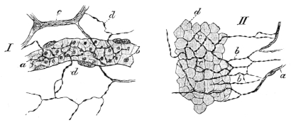
Preparations from the nictitating membrane of Rana esculenta to show distribution of nerves.
I. Preparation of the nictitating membrane to show nervous supply to a capillary vessel; after Klein. Hartnack, Oc. III, Obj. 8.
a Capillary vessel. b Blood-corpuscles. c and d Non-medullated nerve-fibres.
II. To show distribution of nerves in the epithelium; after Klein. Hartnack, Oc. IV, Obj. 8.
a Subepithelial nerve-fibres. b c Fine fibrils between the deepest epithelial cells. d Deepest epithelial cells.

The distribution of the nerves in the lower eyelid has been described by Klein. In the epithelium they form a network resembling that found in the cornea (Fig. 261 II); along the blood-vessels the fine fibrils form a perivascular network, which supplies fine twigs to the walls of the vessels (Fig. 261 I). He distinguishes three kinds of pigmented cells.
b. The Harderian gland is situated at the inner angle of the eye, and is pear-shaped in form. It consists of a number of racemose glands held together by connective-tissue, the whole being enclosed in a relatively thick and strong capsule of connective-tissue.
The alveoli have a diameter of 0·040–0·060 mm.: they possess a lining of epithelium and a lumen which varies considerably in size. The epithelial layer is bounded externally by a membrana propria. The cells are placed eccentrically, they are cylindrical, and composed of finely granular protoplasm; each cell contains a pale, rounded nucleus. The ducts of the alveoli are lined with a single layer of cylindrical epithelium, the cells of which are usually shorter and narrower than those of the epithelium of the alveoli; the ducts open into a single main tube, lined with similar epithelium but strengthened externally by a layer of connective-tissue. The glands secrete a fluid which moistens the free surface of the eye.
The Harderian glands are surrounded by a rich capillary anastomosis which completely invests the alveoli.
c. The lachrymal duct opens behind and below into the nasal cavity (see p. 389), anteriorly it can be traced forwards, as a small tube imbedded in connective-tissue and lying immediately beneath the skin, to the outer angle of the eye, where it opens by numerous tubules.
The lachrymal duct is lined with ciliated columnar epithelium.