HISTORY OF DEVELOPMENT AND SUPPORTING EVIDENCE (continued 5)
Reading edition
A comparison of these drawings shows that as digestion proceeds the antrum appears gradually to elongate and acquire a greater capacity, and that the constrictions make deeper indentations in it. But when the fundus has lost most of its contents, the longitudinal and circular fibres of the antrum contract to make it again shorter and smaller. Its change of form, however, compared with the rest of the stomach, is slight.
The first region to decrease markedly in size is the preantral part of the pyloric portion. The peristaltic undulations, caused by the circular fibres, start at the beginning of this portion, and gradually, by their rhythmic recurrence, press some of the contents into the antrum. As the process continues, the smooth muscle fibres with their remarkable tonicity contract closely about the food that remains, so that the middle region comes to have the shape of a tube (Figs. 3 and 4—1.30 P.M. to 2.30 P.M.), with the rounded fundus at one end and the active antrum at the other. Along the tube very shallow constrictions may be seen following one another to the pylorus.
At this juncture the longitudinal fibres which cover the fundus like radiating fingers, and the circular and oblique fibres reaching in all directions about this spherical region, begin to contract. Thus the contents of the fundus are squeezed into the tubular portion. This process, accompanied by a slight shortening of the tube, goes on until the shadow cast by the fundus is almost wholly obliterated (Fig. 5—5.30 P.M.).
The waves of constriction moving along the tubular portion press the food onward as fast as they receive it from the contracting fundus, and when the fundus is at last emptied they sweep the contents of the tube into the antrum (Fig. 5—5.00 P.M. to 6.00 P.M.). Here the operation is continued by the deeper constrictions till, finally (in this instance at 6.12 P.M.), with the exception of a slight trace of food in the fundus, nothing is to be seen in the stomach at all.
The food in the fundus may possibly be slightly affected by the to-and-fro movements of the diaphragm in respiration. With normal breathing the upper border of the cardiac portion swings through about one centimetre; with dyspnœa, or deep breathing, through one and a half or two centimetres. Since the lower border does not move so much, the contents are gently pressed, and then released from pressure, at each respiration. The pyloric portion is moved very little by the diaphragm, the oscillation being less than a half centimetre.
Moritz has pointed out the value of an organ like the stomach for holding the bulk of the food and serving it out a little at a time, so that the intestines may not become congested during their digestive and absorptive processes. All of the advantages supposed to be thus secured to the intestines may be claimed also for the stomach itself. For the preceding description indicates, and experiments to be described later prove, that the stomach is composed of two physiologically distinct portions: the busy antrum, over which during digestion constriction-waves are running in continuous rhythm; and the cardiac part, which is an active reservoir, pressing out its contents a little at a time as the antral mechanism is ready to receive them.
The Movements of the Stomach in Vomiting
The appearance of the stomach during vomiting has been studied particularly by Openchowski. He says that when an emetic is given there follows a quivering of the stomach-wall, which, beginning near the pylorus, shows itself later in the antral and middle regions of the stomach. The quivering afterwards passes into a contraction, most strongly marked in the antral part, since the peristaltic waves running down to the antrum from above are continually growing deeper. At the same time the fundus expands spherically. The increased contraction in the pyloric part drives the contents towards the more dilated portion, and thence they are forced into the œsophagus by abdominal pressure.
The same phenomena occur when a cat is given apomorphine hypodermatically. First the upper circular muscles relax and become so flaccid that the slightest movement of the abdomen changes the form of the fundus. Then there are apparently irregular twitchings of the fundus wall. Soon a deep constriction starts about three centimetres below the cardia and, growing in strength, moves towards the pylorus. When it reaches the transverse band the constriction tightens and holds fast, while a wave of contraction sweeps over the antrum. Another similar constriction follows. In the interval the transverse band relaxes slightly, but tightens again when the second wave reaches it. Perhaps a dozen such waves pass; then a firm contraction at the beginning of the antrum completely divides the gastric cavity into two parts. This same division of the stomach into two parts at the transverse band is to be seen when mustard is given. Now, although the waves are still running over the antrum, the whole preantral part of the stomach is fully relaxed. A flattening of the diaphragm and a quick jerk of the abdominal muscles, accompanied by the opening of the cardia, now force the contents of the fundus into the œsophagus. As the spasmodic contractions of the abdominal muscles are repeated, the gastric wall again tightens around the contained food. Antiperistalsis I have seen only once; then, while the cat was retching, a constriction started at the pylorus and ran back, over the antrum, completely obliterating the antral cavity.
It will be recalled that the principal difference between the movements of the stomach and their effects as described by Beaumont, and Hofmeister and Schütz on the one hand, and Rossbach, Roux and Balthazard, and myself on the other hand, is that the former observed constrictions completely dividing the stomach at the transverse band, and the antrum then squeezing its contents into the intestines; whereas the latter have seen the constrictions moving forward as narrowing rings, but not separating the gastric cavity into two parts.
With the exception of peristalsis in the antrum, the gastric movements at the beginning of emesis are almost exactly the same as those Beaumont, and Hofmeister and Schütz, declare to be the normal contractions of the stomach. Their observations were made, however, when the organ was subjected to unnatural stimulation. In the excised stomach, observed by Hofmeister and Schütz, not only were all nervous connections severed, but likewise all flow of blood to the organ was entirely stopped, and the cutting off of the blood supply is regarded as one of the most powerful predisposing causes of peristaltic action. The thermometer-tube used by Beaumont was an irritant to the stomach, as he himself admits. “If the bulb of the thermometer,” he writes, “be suffered to be drawn down to the pyloric extremity, and retained there for a short time, or if the experiments be repeated too frequently, it causes severe distress and a sensation like cramp, or spasm, which ceases on withdrawing the tube, but leaves a sense of soreness and tenderness at the pit of the stomach.” Moritz also noticed that a rubber sound introduced into the human stomach proved to be a source of irritation. It seems reasonable to suppose, therefore, that these observers did not see the normal movements, but the actions resulting from abnormal irritation.
The Effect of the Movements of the Stomach on the Food
In my first observations on the active stomach a bulging of the stomach-wall was to be seen in front of the passing waves. But as food did not immediately appear in the intestine, and as, after the pylorus relaxed, the gastric contents did not diminish rapidly enough to allow the supposition that all of the food squeezed forward by the waves was immediately forced through the pylorus, it was assumed that a part, at least, of the food under pressure was forced back towards the cardia through the constriction-ring. This inference was stated in the preliminary notice of my work. Roux and Balthazard also observed the passage of the undulations over the pyloric part, but state merely that the function of the constrictions is the propulsion of food into the intestine, without mentioning what must be regarded as a very important function; namely, the mixing effect of the waves.
Most writers have agreed that the result of the active and passive movements of the stomach is to force the contents hither and thither, thus mixing them and the gastric juice together. Two observers, Beaumont and Brinton, have attempted to explain the manner of the mixing. Beaumont, after noting how the thermometer-tube, used by him to indicate the gastric motions, was affected, describes the circulation of the food as follows: “The bolus as it enters the cardia turns to the left, passes the aperture, descends into the splenic extremity, and follows the great curvature towards the pyloric end. It then returns, in the course of the small curvature, makes its appearance again at the aperture, in its descent into the great curvature, to perform similar revolutions.” Brinton bases his theory of the circulation of the food on an analogy between the movement of a constriction over the stomach and the passage of a septum with a central perforation along the interior of a cylinder full of liquid. The result in both cases, he declares, must be a peripheral current of advance, and a central current of return. Thus in the stomach there would be peripheral currents from the cardia along the walls of the stomach to the pylorus, where they would unite and run as an axial current back to the cardia.
Certain a priori objections may be urged against each of these conclusions. In the first place Beaumont’s observations were made on a subject having a gastric fistula, and the adhesions between the stomach and the abdominal wall would prevent the fundus from acting quite normally in relation to its contents. Beaumont’s conclusions, furthermore, are based on the movements of a thermometer-tube introduced through the fistula, and on the recognition of particles of food which he had seen before as they passed the fistulous opening: the first method, as has been shown, made the conditions in the stomach more abnormal than they were previously; the second gave uncertain knowledge of the course of the food when out of the observer’s sight. Brinton’s hypothesis states the probable movements of fluid contents acted on by a passing constriction. But it may be objected that the conditions assumed by him do not exist in all parts of the stomach. For not only is there no peristalsis visible in the fundus, but with the usual food the fundus contents are not liquid. Moreover, the constrictions at the beginning of the pyloric portion are very slight and move slowly. The food in front of them is, accordingly, not under much greater pressure than the food behind them. The axial current which might result, therefore, could not be strong enough to go far into the cardiac portion.
It is easily possible to test experimentally the validity of these two theories by watching the action of pieces of food which throw a black shadow in a dimly outlined stomach. For this purpose little paste pellets of bismuth subnitrate, with starch enough to keep the form, were given with the customary meal. These pellets, it was found, did not break up in the stomach during the gastric digestion of soft bread. Several times I have been fortunate in getting two of the little balls in the axis of the stomach and about a centimetre apart. As the constriction-wave approached them, both moved forward, but not so rapidly as the wave. Now when the constriction overtook the first ball, the ball moved backward through the constricted ring, in the direction of least resistance. The wave then overtook the second ball, and it also passed backward to join its fellow. At the approach of the next wave they were both pushed forward once more, only to be again forced backward, one at a time, through the narrow orifice. As the waves recurred in their persistent rhythm, the balls were seen to be making progress—an oscillating progress—towards the pylorus; for they went forward each time a little farther than they retreated. This to-and-fro movement of the pellets was much more marked in the antrum, where the waves were deep, than in the middle region. On different occasions from nine to twelve minutes have elapsed while the balls were passing from where the waves first affected them to the pylorus; which means that on the way they were moved back and forth by more than a half hundred constrictions.
If the pylorus does not relax, it is evident that a wave approaching it pushes the food into a blind, elastic pouch, the only exit from which is through the advancing constricted ring. The constrictions are deeper near the end of the antrum, and the rings are small; consequently, the food is squirted back through them with considerable violence. As has been noted, the pylorus opens less frequently for a while after a solid piece of food comes to it. In such a case the slow driving waves squeeze the hard morsel and the soft food about it up to the sphincter, only to have the whole mass shoot back, sometimes half-way along the antrum. Over and over again the process is repeated till the sphincter at last opens and allows the more fluid parts to pass. Hofmeister and Schütz, and Moritz have disclaimed any selective action of the pylorus, and declare that solids are driven from the pylorus to the fundus by antiperistalsis. The action of the pylorus which I have seen, however, is more like that described by the earlier investigators; for during digestion there was no antiperistalsis, and the sphincter, separating the fluids from the solids, caused the solids to remain and undergo a tireless rubbing. Frequently, when several of these balls have been given at the same time, they have all been seen in the antrum after the stomach was otherwise empty. Here they remain to be softened in time by the juices, or to be forced through the pylorus later, for solids do pass into the intestine. Thus when the teeth neglect their work the stomach attempts to perform their function; the relative inefficiency of the gastric method of grinding and its interference with the normal gastric activities point an obvious hygienic moral.
During the process of digestion the food in the cardiac portion gives no sign of currents. Balls which lie in the fundus immediately after the food is ingested keep their relative positions until the cardiac portion begins to contract, and then move very slowly towards the antrum. Moreover, the food in the fundus of a cat has the same mushy appearance when examined after gastric peristalsis had been active for an hour and a half that it had when ingested. The contents of the antrum, on the other hand, look quite different and have the consistency of thick soup. The inactivity of the food in the fundus can also be proved by feeding first five grams of bread and bismuth, then five grams without bismuth, and finally five grams again with bismuth in it. The stomach contents are thus arranged in two dark layers along the curvatures, with a light layer between. Tracings made on tissue paper show that ten minutes after peristalsis commenced the stratification had entirely disappeared in the pyloric part, but that an hour and twenty minutes thereafter the layers were still clearly visible in the cardiac region.
The value of the circulation of the food, as described by Beaumont and Brinton, lay in the supposition that the contents of the stomach were thus brought near to the secreting gastric wall, and that the gastric juice could thus more readily exert its action. Although my observations do not support their theories of mixing currents running throughout the stomach, they still show that the pyloric portion is an admirable device for bringing all of the food under the influence of the glandular secretions of that region. For, when a constriction occurs, the secreting surface enclosed by the ring is brought close around the food lying within the ring in the axis of the stomach. As this constriction passes on, fresh areas of glandular tissue are continuously pressed in around the narrow orifice. And also, as the constriction passes on, a thin stream of gastric contents is continuously forced back through the orifice, and thus past the mouths of the glands. The result of this ingenious mechanism is that every part of the secreting surface of the pyloric portion is brought near to every bit of food, before the latter leaves the stomach, a half hundred times or more, as evidence by the moving ball.
Salivary Digestion in the Stomach
The absence of movement in the fundus would seem to give the food during its stay there little opportunity to become mixed with the gastric juices, and thus to undergo peptic digestion. The truth of this supposition can easily be proved experimentally by feeding a slightly alkaline meal, and later testing the chemical reaction of the contents of various parts of the stomach. A cat which had been without food for fifteen hours was given eighteen grams of mushy bread made slightly alkaline with sodium carbonate. One hour and a half after the cat had finished eating she was killed, and the stomach laid bare by opening the abdomen. A very small hole was then made through the wall in the fundus region, and another similar hole was made into the antrum. By means of a glass pipette food was extracted first from the periphery of the fundus; this food was slightly acid. The cleaned pipette was then introduced two and a half centimetres into the fundus contents, and the food thus extracted gave the original alkaline reaction. Specimens of the liquid contents of the antral and middle regions, taken from various depths, were all strongly acid. A dog killed an hour and three-quarters after eating showed similar differences between the reactions of the food in the fundus and the food in the pyloric portion. So, as a matter of fact, the food does not become acid at a uniform rate in all parts of the stomach, as would be the case if Beaumont’s and Brinton’s theories of mixing currents were true. Moreover, if the facts accorded with their notions, the saliva, which ceases to act in the presence of more than 0.003 per cent free hydrochloric acid, and is destroyed when the percentage of acid proteids is large, would manifestly have its service as a ferment limited to the relatively short time during which the stomach contents, in the process of thorough mixing, were reaching that degree of acidity. There is, however, no movement of food in the fundus, and the alkaline food received from the œsophagus remains alkaline in this region for a considerable period. The nutriment, therefore, if well chewed and thus mixed with saliva, can undergo salivary digestion in the fundus for a considerable period without interference by the acid gastric juice.[32]
From all these observations the conclusion must be that the fundus acts as a reservoir for the food, in which the digestion of sugars and starches may take place; and that the pyloric portion with its simple but marvellous peristaltic mechanism, by a single process, triturates the food, brings it near to the active glands, stirs it thoroughly with their secretions, and expels the products into the intestines.
The Inhibition of Stomach Movements during Emotion
Early in the research a marked unlikeness was noticed in the action of the stomachs of male and female cats. The peristalsis seen with only a few exceptions in female cats failed to appear in most of the males, although both had received exactly the same treatment. Along with this difference was a very striking difference in behaviour when bound to the holder; the females would lie quiet, mewing occasionally, but purring as soon as they were gently stroked. The males, on the contrary, would fly into a violent rage, struggle to be loose from their fastenings, bite at everything near their heads, cry loudly, and resist all attempts to quiet them. On account of this difference only female cats were used for some time, and the significance at first attributed to the action of the males was almost forgotten when the following incident recalled it and suggested that the excitement caused the suspension of the stomach movements. On October 23, 1897, a male cat was fed at 12.00, but was not placed on the holder till ninety minutes later. The waves were passing at the rate of six a minute. The cat fell into a rage and the waves suddenly stopped.
A few days later an observation on a female with kittens explained the absence of gastric movements in the males. While the peristaltic undulations were coursing regularly over the cat’s stomach, she suddenly changed from her peaceful sleepiness, began to breathe quickly, and struggled to get loose. As soon as the change took place, the movements in the stomach entirely disappeared; the pyloric portion relaxed and presented a smooth, rounded outline. I continued observing, and stroked the cat reassuringly. In a moment she became quiet and began to purr. As soon as this happened the movements commenced again in the stomach; first a few constrictions were visible near the end of the antrum, then a few near the sharp bend in the lesser curvature, and finally the waves were running normally from their habitual starting-place. By holding the cat’s mouth closed between the thumb and last three fingers, and covering her nostrils with the index finger, she could be kept from breathing. At the first sign of discomfort the fingers were removed. This experiment was repeated a great many times on different cats, and invariably the evidence of distress was accompanied by a total suspension of the motor activities of the stomach and a relaxation of the antral fibres.
No amount of kneading or compression of the abdomen with the fingers, short of making the cat angry, would cause the waves to stop; so that the cat’s movements, in themselves, were not the source of the inhibition. And since expressions of strong feeling on the part of the animal always accompanied cessation of the constriction-waves, the inhibition was probably due to nervous influence. It has long been common knowledge that violent emotions interfere with the digestive process, but that the gastric motor activities should manifest such extreme sensitiveness to nervous conditions is surprising.
Summary
1. By mixing a harmless powder, subnitrate of bismuth, with the food, the movements of the stomach can be seen by means of the Röntgen rays.
2. The stomach consists of two physiologically distinct parts: the pyloric part and the fundus. Over the pyloric part, while food is present, constriction-waves are seen continually coursing towards the pylorus; the fundus is an active reservoir for the food and squeezes out its contents gradually into the pyloric part.
3. The stomach is emptied by the formation, between the fundus and the antrum, of a tube along which constrictions pass. The contents of the fundus are pressed into the tube and the tube and antrum slowly cleared of food by the waves of constriction.
4. The food in the pyloric portion is first pushed forward by the running wave, and then by pressure of the stomach-wall is returned through the ring of constriction; thus the food is thoroughly mixed with gastric juice, and is forced by an oscillating progress to the pylorus.
5. The food in the fundus is not moved by peristalsis, and consequently it is not mixed with the gastric juice; salivary digestion can therefore be carried on in this region for a considerable period without being stopped by the acid gastric juice.
6. The pylorus does not open at the approach of every wave, but only at irregular intervals. The arrival of a hard morsel causes the sphincter to open less frequently than normally, thus materially interfering with the passage of the already liquefied food.
7. Solid food remains in the antrum to be rubbed by the constrictions until triturated, or to be softened by the gastric juice, or later it may be forced into the intestine in the solid state.
8. The constriction-waves have, therefore, three functions: the mixing, trituration, and expulsion of the food.
9. At the beginning of vomiting, the gastric cavity is separated into two parts by a constriction at the entrance to the antrum; the cardiac portion is relaxed, and the spasmodic contractions of the abdominal muscles force the food through the opened cardia into the œsophagus.
10. The stomach movements are inhibited whenever the cat shows signs of anxiety, rage, or distress.
THE MOVEMENTS OF THE INTESTINES STUDIED BY MEANS OF THE RÖNTGEN RAYS[33] By W. B. Cannon From the Laboratory of Physiology in the Harvard Medical School Extracts from American Journal of Physiology, 1902
Introduction
The investigation of intestinal movements has been beset by the same difficulties that characterised the investigation of the gastric mechanism. Pathological subjects or animals subjected to the disturbing action of drugs and anæsthetics and of serious operations have been the only sources of our knowledge. A considerable difference of opinion as to the nature of the normal movements in the intestines has resulted from observations made under these necessary abnormal conditions. The slowly advancing peristaltic wave and the Pendelbewegung, or swaying movement, described by Ludwig, have been regarded as true physiological processes. Concerning antiperistalsis and the swiftly running vermicular contraction, observers are not so nearly in agreement. The activity of the large intestine has been described as simply peristalsis of a slower rate than that seen in the small intestine.
The best known of the intestinal movements is the normal peristaltic wave. This wave is slow, having a rate of about two centimetres per minute, is regular, and by most observers is said to move always in one direction. The progress of the contraction, as suggested by Nothnagel’s experiments, and as clearly stated by Mall and by Bayliss and Starling, is dependent upon a local reflex. According to Mall, when an object stimulates the mucosa there occurs above the point of stimulation a constriction which forces the object downward; and as it moves downward new regions immediately above the mass are by this reflex brought into constriction, and thus the wave and its stimulus advance together. “At the same time,” Mall states, “a sucking force, due to active dilatation below the body, may have a tendency to drag it down.” In what manner an active dilatation of the intestinal wall may occur so as to produce a “sucking force” he does not make wholly clear. Bayliss and Starling, in describing normal peristalsis in the intestine, state that the contractions above the bolus increase until there is a strong tonic constriction. This passes the bolus onward, and as it advances the ring of constriction follows it. While the bolus is passing down, the gut above it is traversed by waves running as far as the constricted ring. These observers state the law of intestinal peristalsis thus: “Local stimulation of the gut produces excitation above and inhibition below the excited spot.”
The pendulum movements are characterised by a gentle swaying motion of the coils, and are accompanied by rhythmical contractions of the intestinal wall. They continue with undiminished force after paralysis of the local nervous mechanism by nicotine or cocaine; they have been called, therefore, myogenic or myodromic contractions. Observers have described them variously as shortenings and narrowings of the gut, rhythmically repeated at nearly the same intestinal circumference; as alternating to-and-fro movements of the long axis without changes in the lumen; as local or extensive periodic contractions and relaxations mainly of the circular musculature; and as waves involving both muscular coats of the intestine, and travelling normally from above downward at a rapid rate (2 to 5 cm. per second). They have been seen in the dog, and in the rabbit and cat. In the cat Bayliss and Starling noticed that when the lumen of the gut was distended by a rubber balloon, there appeared rhythmical contractions, which nearly always were most marked at about the middle of the balloon; i. e., the region of greatest tension. This form of constriction, which, as my observation shows, is an indication of the manner in which the rhythmical contraction acts in the cat’s intestine, Bayliss and Starling seem to have regarded with slight attention, since it did not accord with the law of peristalsis.
The swift vermicular wave may pass the whole length of the intestine in about a minute. It is often seen just after death, as well as in pathological states such as intestinal anæmia or hyperæmia, and when the bowel contains gases and organic acids from decomposing food. Starling is inclined to regard this type of intestinal activity as an exaggeration of the rhythmic type; Mall, on the other hand, places it in a class by itself, and declares that its service is to rid the intestine rapidly of irritating substances. Nothnagel, who designates this form of movement with the term Rollbewegung, thinks it is transitional between a physiological and a pathological activity.
The existence of antiperistalsis has been so much questioned that it will be considered in a special section of this paper, where my observations may be conveniently introduced.
The common understanding of the manner in which food passes through the intestinal canal is that the chyme ejected from the stomach is pressed downward by a peristalsis, which passes slowly over a part or all of the small intestine. The peristaltic waves of the colon are supposed to constitute an independent group, similar to those of the small intestine, but weaker and slower. Movements of the food other than the uninterrupted advance have been mentioned by some observers. Starling states that the effect of the pendulum movement is to mix the contents of the intestine and bring them into intimate contact with the mucous membrane. Grützner writes that he has been brought “by strange and peculiar observations” to believe that the fluid contents of the small intestine move irregularly forward, then forward and back, then perhaps remain quiet for some time, only to pass backward for a long distance, and finally to move forward steadily to the end. In this manner the food is mixed and brought into contact with the absorbing walls. The to-and-fro shiftings of the food Grützner ascribed to advancing and retrograde contractions of the intestinal musculature, and he argued that even circular constrictions must force the liquid contents away in both directions. To support his contention, Grützner introduced mercury into the intestine and observed it with the Röntgen rays. After noting a backward and forward movement of the mercury he dismissed the method, saying, “Many a flash must come from the Röntgen tube before the normal movement of the intestinal contents is made entirely clear by this method.”
The following account is a summary of many repeated observations on different animals, and is a contribution to a clear understanding of the normal movements of the intestines and their contents.
The Movements of the Small Intestine
When the food has been distributed through the intestine so as to present the appearance shown in Figure 1, a noticeable feature in most or all of the loops is the total absence of movement. If the animal remains quiet, however, only a few moments elapse before peculiar motions appear in one or another of the loops, or perhaps in several, and last for some time. These motions consist in a sudden division of one of the long, narrow masses of food into many little segments of nearly equal size; then these segments are again suddenly divided and the neighbouring halves unite to make new segments, and so on, in a manner to be more fully described. I have called this process the rhythmic segmentation of the intestinal contents. Further observation reveals peristalsis here and there, and under certain circumstances the typical swaying movements may be seen. All these phenomena are now to be considered in detail.
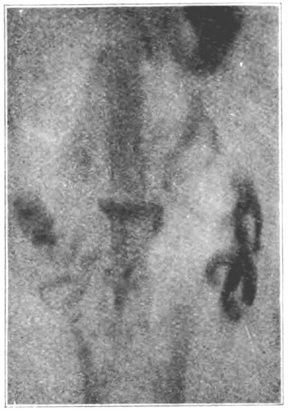
Figure 1.—Appearance of food in the intestines 5¾ hours after eating. This and other radiographs reduced two-thirds.
Rhythmic segmentation of the intestinal contents.—This is by far the most common and the most interesting mechanical process to be seen in the small intestine. The nature of the process may best be understood by referring to the diagram in Figure 2. A string-like mass of food is seen lying quietly in one of the intestinal loops (line 1, Fig. 2). Suddenly an undefined activity appears in the mass, and a moment later constrictions at regular intervals along its length cut it into little ovoid pieces. The solid string is thus quickly transformed, by a simultaneous sectioning, into a series of uniform segments. A moment later each of these segments is divided into two particles, and immediately after the division neighbouring particles (as a and b, line 2, Fig. 2) rush together, often with the rapidity of flying shuttles, and merge to form new segments (as c, line 3, Fig. 2). The next moment these new segments are divided, and neighbouring particles unite to make a third series, and so on. At the time of the second segmentation (line 3, Fig. 2) the end particles are left small. Observation shows that these small pieces are not redivided. The end piece at A simply varies in size with each division; at one moment it is left small, at the next moment it is full size from the addition of a part of the nearest segment, and a moment later is the small bit left after another division. The end piece at B (probably the rear of the mass) shoots away when the end mass is divided, and is swept back at each reunion to make the large end mass again, only to be shot away and swept onward with each recurrence of the constrictions. Thus the process of repeated segmentation continues, with the little particles flitting towards each other and the larger segments shifting to and fro, commonly for more than half an hour without cessation. From the beginning to the end of a period of segmentation the food is seen to have changed its position in the abdomen to only a slight extent; whether this change is a passing of the food along the loop, or a movement of the loop itself, it is impossible to tell from the shadows on the screen. The change of position, however, is much less conspicuous than the lively division and redivision which the mass suffers so many times from the busy, shifting constrictions.
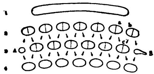
Figure 2.—Diagram representing the process of rhythmic segmentation. Lines 1, 2, 3, 4 indicate the sequence of appearances in the loop. The dotted lines mark the regions of division. The arrows show the relation of the particles to the segments they subsequently form.
From this typical form of rhythmic segmentation there are several variations. Sometimes, and especially when the mass of food is thick, the constrictions do not make complete divisions and are so far apart that the intermediate portions are relatively large. Moreover, the constrictions do not take place in the middle of each portion, but near one end; thus each portion is constricted, not into halves, but into thirds. If a little pointer is placed at the middle of a segment, when the segments are completely divided into halves, in a few seconds the pointer will be in the middle of the clear space between two segments; but in a few seconds more the first phase will return and the pointer will again be indicating a segment,—two operations intervene between similar phases. When, however, the portions are constricted into thirds, the indicator shows it, since three operations intervene between similar phases. The manner of these changes is made clearer by reference to the diagram in Figure 3. That each portion is constricted into three pieces is proved also by watching the gradual reduction of the portion at the left end of line 1 through lines 2 and 3, and also in the gradual formation of a full-sized portion at the right end of lines 2, 3, and 4. When food undergoing this process is watched, it appears to be affected by a series of constrictions, each of which starts at one end of the mass and marches through to the other end, leaving its impress at short intervals along the length. The progression of the dotted lines from right to left in a, b, c, and d, etc., Fig. 3, gives a notion of these advancing constrictions.
Another variation of the segmentation is shown in Figure 4. In this type there are evidently divisions and subdivisions, i.e., one more operation between the appearance and the reappearance of the same phase than is present in the simple division of the small segments in a long string of food (Fig. 2). This form of segmentation is fairly typical for the constrictions seen in food advancing through the intestine. Sometimes the divisions occur in the middle of a long string of food and leave the ends wholly unaffected.
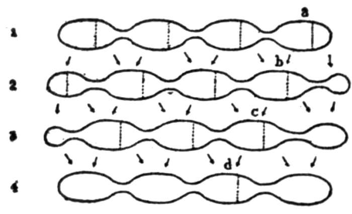
Figure 3.—Diagram showing the relations of the portions when they are constricted into three pieces. The dotted lines indicate regions of constriction; the arrows indicate the relationship of the pieces to the portions they subsequently form.
A remarkable feature in the segmentation of the food is the rapidity with which the changes take place. The simplest way of estimating the rate of division is to count, not the number of times the partition of the food recurs in the same place, but the number of different sets of segments observed in a given period. Thus in Figure 4 the appearances of lines 1, 2, 3, 4, etc., would be counted, and not merely lines 1, 4, etc. Repeated observations on different animals have shown that the most common rate of division in long, thin chains of food varies between twenty-eight and thirty times in a minute; i. e., there is a change from one set of segments to another set every two seconds, and a return of the same phase every four seconds. In some cases the rate is as low as twenty-three times per minute. The larger masses seem to be associated with a slower segmentation; the operations indicated in Figure 3, for example, occurred from eighteen to twenty-one times in a minute, so that the same phase reappeared only once in eight or nine seconds. The segmentation frequently continues for more than half an hour; in one instance it was seen to persist with only three short periods of inactivity for two hours and twenty-two minutes. At the rate of thirty segmentations per minute it is clear that a slender string of food may commonly undergo division into small particles more than a thousand times while scarcely changing its position in the intestine.
I have seen once, in a cat only lightly etherised, the exterior of an intestine which was dividing the food as above described. An hour and a half after a meal of salmon the anæsthetic was given, the abdomen opened, and the flaps raised so as to form walls. Warm salt solution was then poured into the abdominal cavity, and the floating coils left covered with the transparent omentum. The gastric peristaltic waves were running regularly; on the intestine there were visible at various places during the period of observation regions of constriction which had the appearance shown in Figure 3, except that the rings were relatively nearer together. New rings of constriction took place on the same side of all the bulging parts at the margin of the constricted portion (cf. dotted lines, Fig. 3). As new rings occurred the old relaxed, but apparently with tardiness, for the contents gurgled as if forced through the narrowed lumen. The constrictions recurred irregularly and at much longer intervals than in the normal animal. The contracted rings were pale and bloodless.
The effect of the process of rhythmic segmentation proves it an admirable mechanism. The food over and over again is brought into closest contact with the intestinal walls by the swift kneading movement of the muscles. Thereby not only is the undigested food intimately mixed with the digestive juices, but the digested food is thoroughly exposed to the organs of absorption. Mall has shown that contraction of the intestinal wall has the effect of pumping the blood from the submucous venous plexus into the radicles of the superior mesenteric vein, and thus materially aids the intestinal circulation. Moreover, lacteals loaded with fat will in a few moments become empty unless the intestine is slit lengthwise, so that the muscles cannot exert compression. The rhythmic constrictions, therefore, both propel the blood in the portal circulation and act like a heart in promoting the flow of lymph in the lacteals. This single movement with its several results is an excellent example of bodily economy; the repeated constrictions, as already shown, thoroughly churn the food and digestive fluids together, and also plunge the absorbing mucosa into the very midst of the food masses: but not only are the processes of digestion and absorption favoured by these movements; they also, by compression of the veins and lacteals of the intestinal wall, serve to deport through blood and lymph channels the digested and absorbed material.
Peristalsis.[34]—The phenomena of peristalsis and segmentation are usually combined in some manner while the food passes through the small intestine. Peristalsis is observed normally in two forms: as a slow advancing of the food for a short distance in a coil, and as a rapid movement sweeping the food without pause through several turns of the gut. The latter form is frequently seen when the food is carried on from the duodenum; and it may readily be produced in other parts of the small intestine by giving an enema of soapsuds.
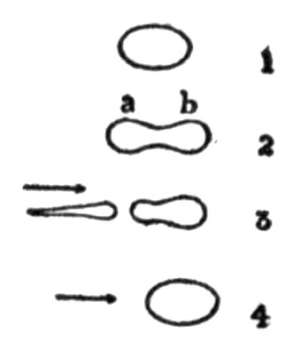
Figure 4.—Diagram showing combined peristalsis and segmentation.
When a mass of food has been subjected for some time to the segmenting activity of the intestine, the separate segments, instead of being again divided, may suddenly begin to move slowly along the loop in which they lie. That this movement is not a swinging of the coil as a whole, but a peristaltic advance of separate rings of its circular musculature, is made probable by the fact that the succeeding segments follow along the same path their predecessors have taken. The advance of the little pieces may continue for seven or eight centimetres, when finally the front piece stops or meets other food. Then all the succeeding pieces are swept one by one into the accumulating mass, which at last lies stretched along the intestine, a solid string manifesting no sign of commotion.
Another form of slow peristalsis is frequently observed when the food is pushed forward, not in small divisions, but as a large lump. The relatively long string of food is first crowded into an ovoid form as the forward movement begins, and as it is collecting thus, it seems at the last to be suddenly formed into a more rounded ball, as if the mass were pulled or pushed together at the two ends. The next moment it is indented in the middle by a circular constriction (as shown in Fig. 4, line 2), which spreads it in both directions along the loop. The trailing portion (a) is next cut in two, and the severed part sometimes flies back over its course about a centimetre. Now the whole mass is swept together again and slightly forward as shown in line 4, Fig. 4, and the segmenting process is repeated. At stage 3, Fig. 4, a constriction sometimes appears around the middle of the advanced portion (b). Thus, with many halts and interruptions, the food slowly advances.
A slight variation of the movement just described is observed when the amount of food is greater and extends farther along the intestine. Under such circumstances, as the mass moves forward, constrictions appear just in front of the rear end, which separate it from the main body, and cause it to shoot backward sometimes through the distance of a centimetre. The main body meanwhile is not disturbed. No sooner has the rear section been shot away than it is swept forward again into union with the rest of the food, and the whole mass then advances until another interfering constriction repeats the process.
Rhythmic segmentation and the pendulum movement.—There is little doubt that the segmentation of the food which I have seen is due to an activity of the intestinal musculature similar to that which causes the so-called pendulum movement. This activity, as already noted, is rhythmic, and, although accounts differ, analytical methods prove that it involves both the longitudinal and the circular layers of muscle. Observations of the effect of the rhythmic contractions upon the food show that the action of the circular fibres is most prominent. It is probable, however, that the longitudinal fibres also play an important part in the process of segmentation. Examination of Figure 2 makes clear that in line 2 the regions of constriction appear between the regions of constriction in line 3; before c can be formed, therefore, the constriction between a and b must relax. Contraction of the longitudinal fibres between two segments would help to enlarge the constricted lumen of the gut. It seems probable that, as the constrictions on either side of c occur, the longitudinal fibres between them contract; almost simultaneously the constriction between a and b relaxes, and the two particles are thus brought swiftly together. A similar process naturally would take place for each of the shifting segments. Thus the function of the longitudinal muscles would be to contract between new rings of constriction and thereby aid in relaxing the former ring between them. During my one observation of the segmenting process, as seen on the surface of the intestine, I could not be sure that the distance between neighbouring segments was shortened as the constriction relaxed; that activity of the longitudinal fibres is present, however, is indicated by observations of Raiser on the intestines of the rabbit and the cat. Raiser observed the outer surface of the coils, and describes the normal movement as an alternate contraction and relaxation of single divisions of the longitudinal fibres; he notes that these short divisions shift. But whether they shift in alternation with the shifting circular constrictions, as seems probable, is an interesting point not yet determined.
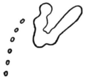
Figure 5.—Tracing showing segmentation of chyme in the duodenum. This and other tracings reduced two-thirds.
Bayliss and Starling state that the swaying pendulum movements are essentially due to peristaltic waves recurring in the same place and running rapidly downward. This form of the movements I have seen only once. At this time about 90 c.c. of soapy water had been injected. This procedure has the effect of exaggerating in every particular the movements of the small intestine. In this instance a broad constriction appeared about the middle of a long string of food and persisted there while it spread down the gut. As the contraction spread, the gut swayed slowly to and fro before it. Then there was a relaxation, followed by a recurrence of the constriction in the same place, a spreading of the contraction, and a swinging of the loop just as before. This phenomenon was repeated again and again, till finally the string was divided and the forward piece pushed through a tortuous course to the colon.
The course of the food in the small intestine.—Chyme is not forced from the stomach by every wave that passes over the antrum, but only at intervals. When the pylorus relaxes, the food, moved towards the pylorus under considerable pressure, is squirted along the duodenum for two centimetres or more. Careful watching of this food shows that usually it lies for some time in the curve of the duodenum until additions have been made to it from the stomach, and a long, thin string of food is formed. While it is resting in this place it is exposed to the outpouring of the bile and pancreatic juices. All at once the string becomes segmented (see Fig. 5) and the process of rhythmic segmentation continues several minutes, thoroughly mixing the intestinal digestive juices with the chyme. In this region the alternate positions of the segments are sometimes far apart, and the to-and-fro movements of the particles may be a relatively extensive and very energetic swinging. Finally the little segments unite into a single mass, or form in groups, and begin to move forward. The peristalsis here, as already mentioned, is much more rapid than the normal peristalsis elsewhere in the small intestine. The masses, once started, go flying along, turning curves, whisking hither and thither in the loops, moving swiftly and continuously forward. After passing on in this rapid manner for some distance the food is collected in thicker and longer strings, resembling the strings seen characteristically in the other loops. Towards the end of digestion the small masses shot out from the stomach, after a few segmentations, may move on in the rapid course without being accumulated in a larger mass until the swift movement ceases.
During the first stages of digestion in the cat’s small intestine the food usually lies chiefly on the right side of the abdomen; during the last stages the loops on the left side contain the greater amount of food. In these loops the food remains sometimes for an hour or more with no sign of movement. All at once a mass begins to show irregular depressions and elevations along its length, and then suddenly it is divided, at first partially, later completely, into many little equal parts, and these repeatedly undergo division and reunion, division and reunion, over and over again, in the manner described above as rhythmic segmentation. After a varying length of time the activity wanes and the little segments are carried forward individually and later brought together, or join and move on as a single body, or they may reunite and lie quietly for some time without further change. Thus by a combined process of kneading and peristaltic advance the food is brought to the ileocæcal valve to enter the large intestine. Records from ten different animals show that salmon does not appear in the small intestine until an hour or an hour and a half after the food is eaten. Inasmuch as five or six hours elapse after eating before this food begins to be seen in the colon, it is evident that the chyme takes four to five hours to pass the length of the small intestine. It is interesting to note that the operations are considerably shortened if the meal has consisted of bread and milk.
The Competence of the Ileocæcal Valve
The ileocæcal valve in the cat is situated three or four centimetres from the blind end of the cæcum. Its position is usually marked in shadows of the food in the colon by a slight indentation, towards which masses about to enter the colon are ordinarily directed from a point somewhat distant in the small intestine (see Fig. 6).
Regarding the competence of the ileocæcal valve many observations have been made. Grützner has reviewed the evidence bearing on the question and concludes that the valve is not competent, least of all for liquids. He declares that as soon as liquids or thin fluid masses appear in the upper part of the colon they pass in many instances into the small intestine the moment that the pressure on the colon side rises slightly. If the colon contains a solid or a thick, mushy mass, the passage towards the small intestine is scarcely possible, because every increase of pressure in the large intestine must force the two lips of the valve together and close it.
The importance of the competence of the ileocæcal valve under normal conditions cannot be appreciated until the function of the first part of the colon is considered. In order that this part of the intestinal mechanism may perform its service, the competence of the valve for the food which enters the colon from the ileum should be perfect. As a matter of fact, such is the case. Not only does the activity of the colon prove this statement, but the failure of every attempt to drive the food in the colon back through the valve into the ileum confirms the proof. Again and again I have tried, by manipulation through the abdominal wall, to press the normal contents of the colon downward with sufficient force to cause them to return to the small intestine, but without success. The valve held perfectly.
The Movements of the Large Intestine
When the large intestine is full, palpation through the abdominal wall demonstrates that the material in the lower descending colon and in the sigmoid flexure is usually composed of hard, incompressible lumps, while that in the ascending and transverse colon and the cæcum is soft, permitting the walls of the gut to be easily pushed together. The condition of the contents in these two regions seems to indicate a rough division of the large intestine into two parts, and the mechanical activities of these two parts verify the differentiation. In the descending colon the material is very slowly advanced by rings of tonic constrictions (see Fig. 7); in the ascending and transverse colon and in the cæcum by far the most common movement is an antiperistalsis.
Antiperistalsis in the colon.—The colon of cats which have been without food for a day usually contains enough gas to make the position of the gut distinguishable with the fluorescent screen (see Fig. 1). The first food to enter the colon from the small intestine is carried by antiperistaltic waves into the cæcum (Fig. 1), and all new food as it enters is also affected by these waves. Thus the contents of the colon, instead of being driven immediately toward the rectum by slow peristalsis, as is the general opinion, are first repeatedly pushed toward the cæcum by an antiperistaltic action.
These antiperistaltic waves follow one after another like the peristaltic waves of the stomach (see Figs. 5, 6, and 10). They begin either on the more advanced portion of the food in the colon (when only a small amount is present), or at the nearest tonic constriction, which is usually at the turn between the transverse and descending colon (Figs. 7 and 8.) The waves rarely run continuously for a long time. When the colon is full, it is usually quiet. The first sign of activity is an irregular undulation of the walls, then very faint constrictions passing along the gut towards the cæcum. These constrictions may first appear only on the ascending colon. As they continue coursing over the intestine they become deeper and deeper, until there is a marked bulging between successive constrictions. When the waves have thus become more prominent, they are seen to start near the end of the transverse colon and pass without interruption to the end of the cæcum. After these deepest waves have been running for a few minutes the indentations grow gradually less marked, until at last they are so faint as to be hardly discernible. The final waves are sometimes to be observed only at the end of the transverse colon.
Such a period of antiperistalsis lasts from two to eight minutes, with an average duration of four or five minutes. The periods recur at varying lengths of time; in one instance a period began at 1.38 P.M. and was repeated at 2.06, 2.34, 2.55, 3.15, and at 3.36, when the observation ceased; in another instance a period began at 2.43 P.M., and was repeated at 2.57 and at intervals of from ten to fifteen minutes thereafter while the animal was being watched. The waves have nearly the same rate of recurrence as those in the stomach; about five and a half waves pass a given point in a minute, i. e., eleven waves in two minutes. This rate has proved fairly constant in different cats and at different stages in the process of digestion; in one case, however, the waves passed at the rate of nine in two minutes.
The stimulating effect of rectal injections on the movements of the small intestine has already been noted. Enemata have also pronounced stimulating action on the antiperistalsis of the colon. Usually the almost immediate result of a rectal injection of warm water is the appearance of deep antiperistaltic waves, which often continue running for a long period. In one case, after an injection of 50 c.c. of warm water, the waves followed one another with monotonous regularity during an observation lasting an hour and twenty minutes. The manner in which this antiperistaltic mechanism affects nutrient enemata introduced into the bowel will be discussed in the section devoted to the question of antiperistalsis.
These constrictions passing backward over the colon do not force the normal contents back through the valve into the small intestine again. I have seen hundreds of such constrictions, and only twice have there been exceptions to this rule,—once under normal conditions, when a small mass slipped back into the ileum, and at another time when a large amount of water had been introduced into the colon. The importance of the competence of the ileocæcal valve is now apparent; indeed, antiperistalsis in the colon gives new meaning and value to the location of a valve at the opening of the ileum. For, inasmuch as the valve is normally competent, the constrictions repeatedly coursing towards it force the food before them into a blind sac. The effect on the food must be the same as the effect seen in the stomach when the pylorus remains closed before the advancing waves. The food is pressed forward by the approach of each constriction; but since it cannot go onward in the blind sac, and is, moreover, subjected to increasing pressure as the constriction comes nearer, it is forced into the only way of escape, i. e., away from the cæcum through the advancing constricted ring. About twenty-five waves affect every particle of food in the colon in this manner during each normal period of antiperistalsis. The result must be again a thorough mixing of the contents and a bringing of these contents into close contact with the absorbing wall—a process which has already been variously repeated many times in the stomach and in the small intestine.
Two other movements have been observed in the ascending colon, but they are rare appearances. The first of these was a serial sectioning of the contents noticed in an animal given castor oil with the food. A constriction separated a small segment in the cæcum; another constriction then cut off a segment just above the first, and with the disappearance of the first constriction the two separated segments united. A third segmentation took place above the second, and the changes occurred again. Thus the whole mass was sectioned from one end to the other; and no sooner was that finished than the process began again and was repeated several times. A slight modification of this movement was observed in a colon containing very little food. The mass was pressed and partially segmented in the manner characteristic of the small intestine, and was thus again and again spread along the ascending colon, and each time swept back into a rounded form by antiperistalsis. The second of the two movements mentioned above consisted in a gentle kneading of the contents. This was caused by broad constrictions appearing, relaxing, appearing, relaxing, over and over again, in the same place. When several of these regions were active at the same time, they gave the food in the colon the appearance of a restless undulatory mass. Once a constriction occurred and remained permanently in one place, while the bulging parts on either side of it pulsated alternately, at the rate of about eighteen times in a minute, with the regularity of the heart-beat. Although these phenomena are somewhat striking, they are not usual, and are in no way so important as the antiperistalsis.
The changes when food enters the colon.—The passage of food through the ileocæcal valve seems to stimulate the colon to activity. As food is nearing the ileocæcal valve the large intestine is usually quiet and relaxed (Fig. 6, 4.00), though occasionally indefinite movements are to be observed; and sometimes just before the food reaches the end of the ileum the circular fibres of the colon in the region of the valve contract strongly, so that a deep indentation is present there. The indentation may persist several minutes; it disappears as the muscles relax just previous to the entrance of the food. The food is moved slowly along the ileum and is pushed through the valve into the colon. The moment it has entered a strong contraction takes place all along the cæcum and the beginning of the ascending colon, pressing some of the food onward, and a moment later deep antiperistaltic waves (Fig. 6, 4.03) sweep down from the transverse colon and continue running until the cæcum is again normally full, i. e., for two or three minutes.
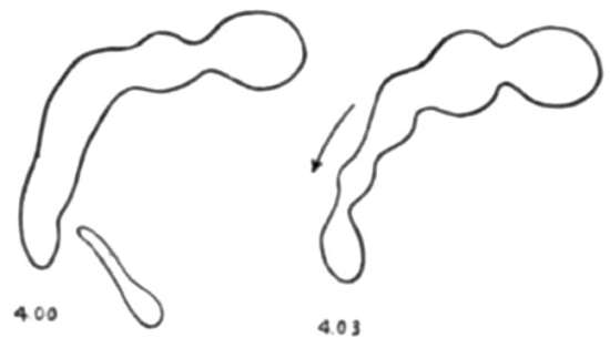
Figure 6.—Tracings showing changes when food enters the colon and also the first tonic constriction. 4.00, the colon relaxed as food approaches in the ileum. 4.03, the colon contracted and traversed by antiperistaltic waves after the food has entered.
The appearance of tonic constrictions.—It has already been noted that as the food accumulates in the ascending colon it is at first confined to this region by antiperistaltic waves. With further accessions, however, the contents naturally must be pressed more and more into the transverse and descending colon. In the early stages of this accumulation, while the food lies chiefly in the ascending colon, the only activity of the muscular walls is the antiperistalsis. As the contents extend along the intestine a deep constriction appears near the advancing end and nearly separates a globular mass from the main body of the food (Fig. 6). The contents of the large intestine progress farther and farther from the cæcum; meanwhile new tonic constrictions appear which separate the contents into a series of globular masses. And as the number of these divisions increases they take a position farther from the cæcum, so that they are present chiefly in the descending colon (Fig. 7). Raiser has recorded a similar appearance in the terminal portion of the rabbit’s colon, in which deep circular constrictions separate the scybalous masses. He maintains that these masses are pushed onward by the constrictions. Comparing tracings made at rather long intervals (forty-five minutes), I found that the rings disappear from the transverse colon, and then are present with the waste material in the descending colon. Thus in the cat also these rings, which seem with short observation to be remaining in one position, are in reality moving slowly away from the cæcum, pushing the hardening contents before them. The contents at this stage are no longer fluid, and consequently they must offer considerable resistance to a force pushing them through the colon. It is an advantage to have this pultaceous substance propelled in divisions rather than in a uniformly cylindrical mass, since the fibres along the length of the mass are thereby rendered effective. Such are the functions of the persistent rings; they form the waste matter into globular masses at the end of the transverse colon and slowly push these masses onward.
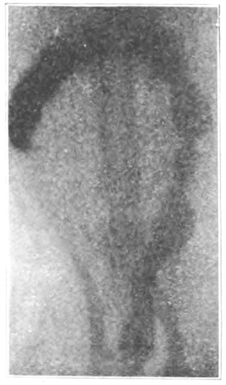
Figure 7.—Radiograph showing the region of tonic constrictions (descending colon) and the region of antiperistalsis (transverse and ascending colon).
In the transverse colon, which is free from the slowly moving rings, the antiperistaltic waves have full sway. In the region of the tonic rings an infrequent or even a slowly periodic relaxation and contraction are often to be observed. These changes seem to take place in all the rings at about the same time. Once I saw antiperistaltic waves running over the uppermost of four segments, but since the rings on either side of the segment held tightly, the waves had merely the effect of churning the material of the segment and did not move it onward. Inasmuch as the material in these segments at first is soft, so that the segments are easily compressible, while the fæcal masses which are the final result are relatively hard and dry, it follows that even within the confines of these persistent rings some absorption is taking place.
Defecation
The process of clearing the colon is a process of repeated reduction of the amount of material present. Figure 8 (3.11) is a radiograph showing the food in the colon at 3.11 P.M. About 3.25, with a slow, sweeping movement, the gut swung around so that the ascending colon was lying in the position of the last half of the transverse colon, and the transverse colon had taken the position of the descending part (Fig. 8, 3.25). At the same time the tonic constrictions disappeared and were replaced by a strong, broad contraction of the circular muscle, tapering the contents off on either side in two cones. The region of strongest contraction was apparently drawn downward with the rest of the gut by a shortening of the descending colon. As the intestine swung around, more material was forced into the rectum, and when the swinging of the intestine stopped, the constriction which divided the lumen passed slowly downward, and with the aid of the muscles surrounding the abdominal cavity, pushed the separated mass out of the canal.[35] After the terminal mass had thus been pushed out, the colon with the remainder of its contents returned to nearly its former position (Fig. 8, 3.46). About two hours afterward this remnant had been spread throughout the length of the large intestine by means of the slowly moving rings. Figure 7 is a radiograph of the same colon pictured in Figure 8; the radiograph was taken at 11.50 A.M., and at 12.15 P.M. the material in the lower descending colon was forced out in the manner above described. Within three hours the remaining portion had been spread into the evacuated region, as shown in Figure 8, 3.11. The manner in which the material is spread from the region of the antiperistaltic waves into the region of the slowly advancing rings presents a problem. During normal living new food constantly arriving in the colon must force the old contents forward just as the later parts of a meal force forward the earlier parts; there is no doubt, however, that most of the contents of the cæcum and the ascending colon may be passed onward even during starvation. The emptying of these regions, according to my observations, is never complete; for after considerable time has elapsed and the large intestine is cleared and dilated with gas, some substance is still to be detected in the cæcum and clinging to the walls of the ascending colon. The only activities manifested here are the antiperistaltic waves and the strong tonic contraction of the whole circular musculature shown in Figure 6. It is clear that the latter activity would serve to press into the transverse colon a considerable portion of the contents of the ascending colon, and the remnant seen clinging to the walls would be the part not thus pressed forward.
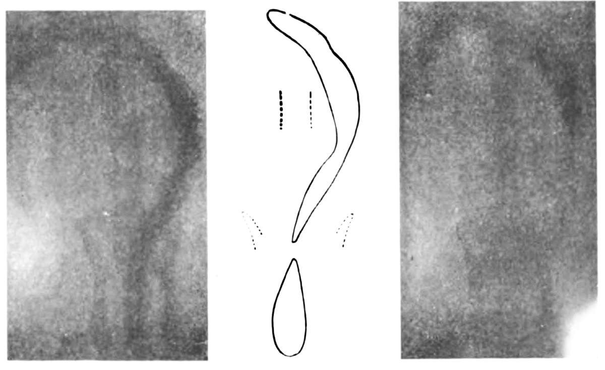
Figure 8.—Two radiographs and a tracing showing the changes taking place in defecation. 3.11, material in the colon. 3.25, colon carried downward and terminal mass separated. 3.46, after defecation, when the colon returns to former position. Defecation occurred at 3.27.
Twice I have seen appearances which might account for the emptying of the first portion of the large intestine in a more thorough manner than that above described. At one time, without apparent stimulation, strong tonic contraction occurred along the entire length of the ascending colon, which forced the contents almost wholly into the transverse portion. This action seemed merely an exaggerated form of that observable after food passes the ileocæcal valve (see Fig. 6). At another time, after a mass of food had passed through the ileocæcal valve, after the ascending colon had contracted generally and the antiperistaltic waves had coursed over it in the usual manner, a deep constriction appeared at the valve and ran upward without relaxation nearly the length of the ascending colon, pushing the contents before it. For an instant the wave paused; then the constriction relaxed and the food returned towards the cæcum. These observations indicate that either a general contraction of the wall of the large intestine or a true peristalsis may be effective in pressing waste matter from the region where antiperistalsis is the usual activity into the region where the slowly advancing rings may carry it on to evacuation (see Fig. 7).
The Question of Antiperistalsis
In 1894 Grützner published an observation and made an assumption about which there has since been much controversy. He maintained that when normal salt solution, holding in suspension hair, powdered charcoal, or starch grains, is injected into the rectum, it is carried upward into the small intestine and may even enter the stomach. These experiments have been repeated by several observers. Some have confirmed Grützner’s results; others have failed, after using most careful methods, to find any evidence of the passage of the injected material back to the stomach, and they have declared that the apparent success was due to carelessly allowing the food of the animal to become contaminated with the test materials, so that these were introduced into the stomach by way of the mouth. That antiperistalsis does not occur in the small intestine seems to be proved by Mall’s experiment of reversing a portion, sewing it in place, and then finding that the food does not pass the reversed region, but collects at the upper end. Sabbatani and Fasola reversed stretches of small intestine of varying length, and found that the reversed portions allowed fluids to pass, but that the persistence of the physiological direction of movement caused an accumulation of undigested food in the region of the upper suture. However a portion of the intestine lay in relation to the rest, it always manifested the normal peristalsis. Many other observers working directly on the intestine confirm this testimony and state that the progress of the constriction-rings is always downward, and that antiperistalsis is not physiological. In 1898, however, Grützner took his stand again in favour of a backward movement in the intestines, and in a somewhat metaphysical manner argued that peristalsis and antiperistalsis belong to each other just as relaxation of muscle is related to contraction. He assumed that as the contents are advanced by slow peristalsis, so are they returned by a similar movement in the opposite direction, and he mentions several pathological cases (fistula of intestine) to substantiate the assumption.
By means of the X-rays it is possible to see just what takes place when a fluid is injected into the rectum. For the purpose of determining how nutrient enemata are received and acted upon in the intestines, I have introduced thin, fluid masses in large and small amounts, and thick, mushy masses in large and small amounts, in different animals. The enemata consisted of 100 c.c. of milk, one egg, ten to fifteen grams of bismuth subnitrate, and two grams of starch to hold the bismuth powder in suspension. To make the thick enema all these were stirred together and boiled to a soft mush; to make the thin enema all the parts were boiled together except the egg, which was added after the boiled portion was cooled. The small amount injected was 25 c.c.; the large amount almost 90 c.c., about the capacity of the large intestine when removed from the body. The animals were given first a cleansing injection, and after this was effective the nutrient material was introduced. In order to make sure of the observation, a control radiograph was first taken to show no bismuth food present, and other radiographs taken at varying intervals after the injection to record the course the food was following.
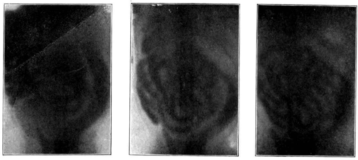
Figure 9.—- Radiographs showing that after a large nutrient enema (about 90 c.c.) has been given the food is forced more and more from the large into the small intestine. The enema was introduced at about 1.40 P.M. At 3.00 segmentation was occurring in many loops.
These experiments show that when small amounts of nutrient fluid are introduced they lie first in the descending colon. In every instance antiperistaltic waves are set going by the injection, and the material is thereby carried to the cæcum. When large amounts are injected they stop for a moment in the region between the transverse and descending colon, as if a constriction existed there. Then a considerable amount of the fluid passes the point, and antiperistaltic waves carry it to the cæcum. In any case the repeated passing of the waves seems to have the effect of promoting absorption, for in the region where these waves continue running, the shadows become gradually more dim, and finally the bismuth appears to be only on the intestinal walls; in other regions, e. g. in the descending colon, the shadows retain their original intensity. Small injections have never in my experience been forced even in part into the small intestine; but with the larger amounts, whether fluid or mushy, the radiographs show many coils of the small intestine containing the bismuth food.
The passage of the injected material beyond the ileocæcal valve is probably due entirely to antiperistalsis in the colon,—a factor unknown to both Grützner and his opponents. The valve, which is thoroughly competent for food coming normally from the small intestine into the large, is curiously incompetent for a substance, even of the consistency of thick cream, introduced in large amount by rectum. When the valve first permits the food to enter the ileum, the fluid pours through and appears suddenly as a winding mass occupying several loops of the intestine (Fig. 9, 1.50, about ten minutes after the injection). The mass is continuous from the valve to the other end; antiperistalsis is therefore not visible in the small intestine under the circumstances of this experiment. The antiperistaltic waves of the colon, however, continue running; the transverse and ascending colon are thus almost emptied, and the small intestine more and more filled with food (Fig. 9, 2.15 and 3.00). After a short time the typical segmenting movements can be seen in the loops, busily separating the food into small masses, and over and over again dividing and redividing them.
I have never seen food material pass back from the colon so far as the stomach; but once, about ten minutes after an injection of 100 c.c. of warm water, the cat retched and vomited a clear fluid resembling mixed water and mucus. In the fluid were two intestinal worms still alive.
The importance of the mechanism by which nutrient enemata are passed backward in the intestine is evident. In the colon the nutrient material is worked over by the antiperistaltic waves, intimately mixed with whatever digestive juices may be present, and exposed to the organs of absorption in that region. If the enemata are large, the digestive and absorptive processes are by no means confined to the colon, but may take place along extensive surfaces of the small intestine. I have repeatedly seen rhythmic segmentation active throughout many loops of the small intestine, thus exposing the injected food to the same mixing and absorbing processes as affect the nutriment which has come through the stomach in a normal manner.
The Effect of Emotions and Sleep
Observations on the stomach of the cat showed that the peristalsis is inhibited whenever the animal manifests signs of anxiety, rage, or distress. Since the extrinsic innervation of a large part of the intestinal tract is the same as that of the stomach, it is of interest to note the effect of emotional states on the movements of the intestines. Esselmont, in a study of the dog’s intestine, noted constantly after signs of emotion a marked increase of activity lasting for only a few moments. Fubini also observed that fear occasioned more rapid peristalsis. There is no doubt that many emotional states are a strong stimulus to peristalsis, but it is equally true that other emotional states inhibit peristalsis. In the cat the same conditions which stop the movements of the stomach stop also the movements of the intestines.
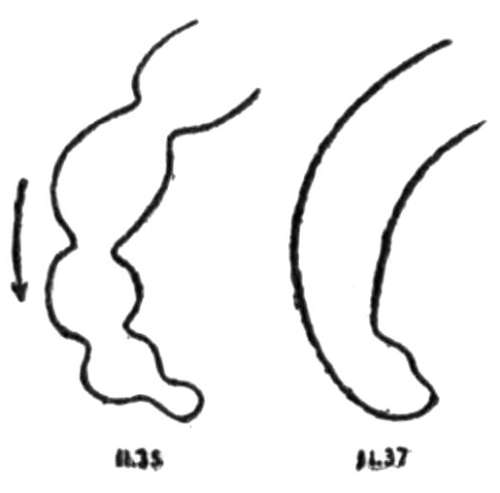
Figure 10.—Tracings showing the effect of excitement on antiperistalsis in the colon.
The female cats used in these observations ordinarily lie quietly on the holder and make no demonstration. Sometimes, however, with only a little premonitory restlessness, the cat suddenly flies into a rage, lashing her tail from side to side, pulling and jerking with every limb, and biting at everything near her head. During such excitement, and for some moments after the animal becomes pacified again, the movements, both of the large and small intestine, entirely cease. Such violence of excitement is not necessary to cause the movements to stop; a cat which was restless and continually whining while confined to the holder showed no signs of intestinal movements during any period of observation (one period lasted more than an hour), although the changes in the distribution of the food observable from one period to the next proved that movements were going on during the quiet intermissions. In another cat, uneasy and fretful for fifty minutes, no activity was seen; then she became quiet for several minutes, and peristalsis of the small intestine appeared.
When the segmentation process in the small intestine is stopped by excitement the segments unite and the series of parts returns to the form of a solid string. The change occurring in the large intestine when the antiperistalsis is inhibited by excitement is shown in Figure 10. The tonic constrictions in the descending colon are apparently not affected by emotional states, for they do not seem to relax in the excitement which causes the movements to cease.
By holding the mouth and nostrils closed, or by pressing between the rami of the jaw, the breathing may be stopped. As soon as the cat shows distress from lack of breath every form of intestinal movement stops.
The statement is sometimes made in text-books of physiology that the gastric and intestinal mechanisms cease to act during sleep. It is worthy of note that nearly all the animals curled up and slept during the time between observations; nevertheless, the progress of the food through the intestines continued. The statement is also made that at night, even without sleep, the intestines are almost entirely at rest; that this is their normal time for repose. I have seen both large and small intestines actively at work, however, from half past nine until half past ten o’clock at night.
Summary
1. Bismuth subnitrate, 10 to 33 per cent, mixed with the food renders the movement of the intestinal contents, and thereby the movements of the intestinal walls, visible on the fluorescent screen.
2. The activity most commonly seen in the small intestine is the simultaneous division of the food in a coil into small segments, and a rhythmic repetition of the segmentation each time applied to the new segments formed from parts of those just divided. In the cat this rhythmic segmentation may proceed at the rate of thirty divisions per minute. The effects of the constrictions causing the segmentation are the mixing of the food and the digestive juices, the bringing of the digested food into contact with the absorbing mechanisms, and the emptying of the venous and lymphatic radicles of their contents by compression of the intestinal wall.
3. Peristalsis is usually combined with segmentation. As the food is advancing, interfering constrictions often separate the rear end of the mass from the main body. The separation is momentary, however; the rear end is swept into union with the main body again, and the whole mass is pushed onward until another constriction repeats the changes.
4. The ileocæcal valve is thoroughly competent for food entering the colon from the ileum.
5. The usual movement of the transverse and ascending colon and the cæcum is an antiperistalsis. This recurs in periods about every fifteen minutes, and each period lasts commonly about five minutes; the waves recur during a period at the rate usually of eleven waves in two minutes. This antiperistalsis gives new significance to the ileocæcal valve; for the food, now in a closed sac, is thoroughly churned and mixed by the constrictions running towards the cæcum, and again exposed to absorbing walls without any interference with the processes in the small intestine.
6. As soon as new food enters the large intestine a strong general contraction takes place along the cæcum and ascending colon, forcing some of the food onward; a moment later antiperistaltic waves begin to pass.
7. With the accumulation of material in the transverse colon, deep tonic constrictions appear one after another and carry the material into the descending colon, leaving the transverse and ascending portions free for the antiperistaltic waves.
8. In emptying the large intestine the material in the lower descending colon is first carried out by combined peristalsis and pressure of abdominal muscles; the remainder of the material is then spread into the evacuated region, and this region is again cleared; the second remainder may be similarly affected. In normal life the new food arriving in the colon must force forward the old contents of the ascending and transverse colon.
9. The observations have revealed no evidence of antiperistalsis in the small intestine, but since the ileocæcal valve will allow nutrient material under pressure to pass backward, the antiperistalsis of the large intestine may force into the small intestine a considerable portion of a large nutrient enema. Segmentation in the small intestine affects such an enema precisely as it affects food which has passed normally through the stomach.
10. Signs of emotion, such as fear, distress, or rage, are accompanied by a total cessation of the movements of both large and small intestines. The movements continue in the cat both during sleep and at night.
THE BATTLE CREEK LABORATORIES THE MAMMOTH SANITARIUM AND THE LARGE ADOPTED FAMILY OF DR. AND MRS. J. H. KELLOGG
[A report of one experiment has been selected from Modern Medicine relative to the work of the laboratories connected with the Battle Creek Sanitarium because it relates to the effect of cooking and mastication upon food in illustration of the statement of Dr. Campbell pertaining to these aids to digestion. Much more evidence could be had from the Sanitarium reports, but sufficient has already been given herewith from various authoritative sources to justify our claims of the great importance of mouth-treatment in human nutrition.
It may be said here, however, that the trial of thorough mouth-work as an aid to digestion, which has been in progress at the Sanitarium for more than a year, and which has finally been accepted and prescribed as the first requirement of the treatment of patients, is of the utmost significance. This is, by far, the largest sanitarium in the world, having some hundreds of physicians, nurses, and other attachés, and treating many thousands of patients annually. The “cure” is based upon natural methods of recuperation, and while all of the staff, both medical and surgical, are fully equipped diplomatists, and whereas the organisation has a legally and professionally accepted medical school of its own, so-called medicines are rarely used, and never except as antidotes to specific poisons. Nature is assisted by scientific means to do the curing, and now that an economic nutrition to relieve the exhausted system of the patient from all possible strain through ample mouth-treatment of food, as intended by the anatomical, dental, and chemical plan on which man is constructed, has been tried and accepted as a fundamental principle of the institution, it gives a practical indorsement of the claims set forth in “Glutton or Epicure,” and in this present book, and declares that they are of greatest importance in securing health and efficiency.
The Battle Creek Sanitarium is a philanthropic and humanitarian institution operating under a perpetual charter which compels the use of all the profits gained to foster the spread of the humanitarian work. More than sixty branches of the parent institution have been established in or near large cities in different parts of the world, under the title of The American Medical Missionary Association, and each of these branches conducts a life-saving business on Good Samaritan principles. The organisation started its medical missionary work some thirty-seven years ago, with almost no capital and only one patient, in a small two-storey frame house, in the then small village of Battle Creek, Michigan. The incorporators were religious enthusiasts who believed that Christianity should be expressed in works as much as in faith, in curing the sick and healing the wounded, and thus preparing the unfortunate for the reception of moral and spiritual inspiration.
The best evidence that this scheme of procedure to attain the ultimate end was a good one is shown by the success of the institution in its growth from such small beginning to the immense proportions of the present time, with one of its buildings nearly a thousand feet in length and five storeys in height and numerous other buildings radiating from the main one and scattered about it in a finely wooded park. Fire came and destroyed the old building and all its contents, but yet it was soon rebuilt, and the concern goes on growing and growing, because the foundation principle of the institution is the beautiful Golden Rule, and the method of treatment employed is taken from the open book of Nature.
While the organisation was primarily based upon a special religious creedal enthusiasm, it has become so broadly altruistic as to suggest a return to original Christianity as defined in the Sermon on the Mount. In such Christian expression honest agnostics, born Buddhists, and the tolerant of all the different Christian creeds may join and say amen!
One of the splendid results of an economic nutrition, attained by following the natural requirements and impulses, is the curing of many diseases, among them several forms of constipation. The writer has a genuine admiration for the spirit that is the motive power of the Battle Creek Sanitarium and firm belief in the Christianity demonstrated in the work, especially in the private experiment of Dr. and Mrs. Kellogg, with their family of adopted waifs. Twenty-four children of unfortunate parents, waifs so unfortunate in their attractability as to be hopelessly neglected, have been gathered under this sheltering roof and are showing their mettle and gratitude by splendid behaviour and brilliant accomplishment in a manner that any proud parent might approve. To miss any opportunity to express gratitude to Dr. and Mrs. Kellogg for giving us such a splendid example of the true meaning of practical Christianity would be showing symptoms of the worst form of constipation; viz., constipation of appreciation and affection.—Horace Fletcher.]
EXPERIMENTAL INVESTIGATION OF THE INFLUENCE OF MASTICATION AND COOKING OF FOOD, ETC., IN THE LABORATORIES OF THE BATTLE CREEK, MICHIGAN, SANITARIUM, UNDER THE DIRECTION OF DR. J. H. KELLOGG
From Modern Medicine
The table clearly shows the effect of cooking and the effect of mastication upon the salivary digestion of food. Column 1 shows the results obtained after an ordinary test meal consisting of 1½ ounces of water biscuit to 8 ounces of water; column 2, 1½ ounces of water biscuit ground fine, mixed with water and swallowed without chewing; column 3, test meal consisting of 1½ ounces of raw wheat flour and 8 ounces of water; column 4, test meal consisting of 1½ ounces of unground pearled wheat with 8 ounces of water.
Water biscuit, well chewed. Water biscuit, not chewed. Raw flour. Raw wheat.
1 2 3 4
Total acidity (A) 0.142 0.140 0.204 0.136
Calculated acidity (A´) 0.156 0.132 0.186 0.128
Total chlorine (T) 0.296 0.284 0.332 0.272
Free HCl (H) 0.050 0.028 0.056 0.052
Combined chlorine (C) 0.106 0.104 0.130 0.076
Fixed chlorides (F) 0.114 0.152 0.146 0.144
Maltose (M) 1.088 0.272 0.000 0.000
Dextrine and soluble starch (D) 0.812 0.548 0.300 0.448
COEFFICIENTS
Digestion of albumin (a) 0.82 0.97 1.00 1.00
Digestion of starch (b) 0.71 0.42 0.00 0.00
Salivary activity (c) 1.17 1.11 1.14 1.37
Fermentation (x) 5.00 11.00 6.00 6.00
Chlorine liberation (m) 0.80 0.70 0.85 0.71
Several points of interest are to be noted in the above table, the first and most conspicuous of which is the fact that the saliva did not act at all upon the raw flour and raw wheat, as shown by the total absence of maltose in the cases represented in columns 3 and 4. The small amount of dextrine and soluble starch shown was, perhaps, already present in the raw grain, but this point I have not investigated. It is clear, however, that no sugar was produced when raw starch was taken, whereas the amount of sugar produced after the ordinary test meal was more than 1 gram in each 100 c.c. of stomach fluid; in other words, the stomach fluid contained more than one per cent of sugar without taking into account the amount which had been absorbed.
The figures for maltose in column 2 represent a test meal in which little or no saliva was mixed with the test meal, the food being swallowed without chewing, indicating very slight action of the saliva, the amount of maltose found in the stomach fluid being but a trifle more than one-fourth the amount obtained after an ordinary test meal. The amount of soluble starch and dextrine was less than half the normal amount in the case of the raw flour, and but little more in the case of the raw wheat.
Another point of interest is the increased amount of lactic acid found in the test meal taken without chewing, represented in column 2. The coefficient of fermentation which represents the number of milligrams of lactic acid (as expressed in terms of HCl) found in 100 c.c. of stomach fluid was more than double that found after the same kind of test breakfast properly masticated, represented in column 1. The results of this experiment distinctly associate acid fermentation with imperfect mastication and imperfect salivary digestion.
Another fact noted in a comparative study of the results of the analysis of over 5000 stomach fluids, which very strongly confirms this idea, is that starch conversion is usually complete in cases of apepsia, while lactic acid is conspicuous by its absence. In nearly all cases of apepsia which I have encountered, numbering about forty cases in all, the most delicate tests for lactic acid have failed to show its presence except in the most minute quantities; in most cases it was entirely absent.
There are a number of other points of interest in the above table in addition to those which relate particularly to starch digestion. One of the most noteworthy of these is the fact that the digestion of albumen was not unfavourably influenced by the neglect to masticate the food, the coefficient of digestion, in fact, being raised from .82 to .97. This coefficient is a qualitative and not a quantitative index. The higher coefficient indicates a more perfect elaboration of proteids and a close approach to an absolutely perfect proteid digestion.
Another fact of perhaps even greater interest has relation to the digestion of albumen when the wheat was eaten raw, in the form of either flour or wheat. The coefficient of proteid digestion in both cases, as shown in columns 3 and 4, was 1.00, indicating perfect elaboration of the albuminoids. From this it appears that raw gluten, or the proteids of wheat, is digested more perfectly when taken in a raw state than when cooked, the very opposite of which we have seen to be true of starch. The digestion of raw starch may take place in the intestines, by the action of the pancreatic juice, but cannot take place in the stomach, for the reason that the saliva has not the power to penetrate the cellulose envelope of the starch granule, and hence cannot digest raw starch.
This fact coincides in a most interesting manner with the biological fact that man is by nature a frugivorous animal. In the process of ripening, the starch of fruits undergoes a hydration similar to that which takes place in cooking and in pancreatic digestion, whereby the insoluble starch is converted into soluble starch, dextrine, and sugar. This explains, also, why well-ripened fruit may be eaten raw with impunity, while unripe fruit and farinaceous food of all sorts require cooking. In his diet, man, like his nearest relative, the monkey, being naturally a frugivorous animal, may eat fruits in the state in which Nature has provided them; but when he introduces other natural products into his bill of fare, he must adopt artificial means for securing the preparation for digestion which Nature makes in the ripening process of fruits.
The coefficient of chlorine liberation (m) is very nearly uniform, indicating that the mastication of food and the cooking of food have little influence upon this digestive function.
The coefficient of salivary activity (c) was determined independently for each test breakfast. Its practical uniformity indicates that there was no essential change in the character or quality of the saliva to account for the differences shown by the totals in relation to the stomach digestion of starch.
DR. EDWARD HOOKER DEWEY AND THE “NO BREAKFAST PLAN”
The “No Breakfast Plan,” evolved from the long experimental experience of Dr. Dewey, to secure much needed rest for the stomach and intestines, is described in a book bearing that title which can be had direct from the author by addressing him at his home, Meadville, Penn., U. S. A.
“No Breakfast” is, evidently, a misnomer, but means, in the present application, an appetite earned after arising from sleep. The writer, for instance, often begins work so early in the morning that by the time the ordinary breakfast is ready he has already done a fair day’s work.
The writer has no reported details of the work of Dr. Dewey to add to this volume. In “Glutton or Epicure” full appreciation of this Esculapian Luther is expressed and extracts of his writings are reprinted. In fighting for more than forty years for the principle of less abuse of the tired body of man, Dr. Dewey has rendered a service that some time will be reckoned very great; and while there is no scientific report of the good doctor’s work to call for introductory comment, it would be equally unhealthy to miss an opportunity to express gratitude for what he has done for us all.
PROFESSOR JAFFA AND THE FRUITARIANS
Professor Jaffa, too, of the University of California, has been doing most valuable service in testing the usefulness of fruits and nuts as human foods. He generously furnished the author with elaborate tables of his results, covering several years of observation, showing low nitrogen possibilities similar to those demonstrated by the writer and his colleagues at Cambridge and Yale. These have since been published, and relating to special kinds of foods, as they do, suggest a wide range of choice among the fruits of earth; but the collected evidence of this book shows that human nutrition is best served when the appetite, being kept at normal, is allowed to make selection from the whole range of nutritious products furnished by good Mother Nature.
DR. H. P. ARMSBY
In the Oct. 16th, 1903, number of Science, also, is an interesting article by Dr. H. P. Armsby on the heat values and muscular energy values of different food elements and their isodynamic replacement of each other under various conditions.—Horace Fletcher.]