Chapter II · CRANIOLOGY
Frederic Taber Cooper translation
Having finished the study of general biological questions and of the body considered in its entirety, we may now pass on to analyse its separate parts, treating in connection with each of such parts the social and pedagogic questions which may pertain to it.
The parts of the body which we shall take under consideration are: the head, the thorax, the pelvis and the limbs.
The Head.—When we pass from the body as a whole to a more particularised study of the separate parts, it is proper to begin with the head because it is the most important part of the whole body. The older anthropology, and biological and criminal anthropology as well were very largely built up from a study of the head; a study so vast and important that it has come to constitute a separate branch of science: craniology.
The fact is that the characteristics manifested by the cranium are chiefly in the nature of mutations rather than variations, and consequently the anthropological data relating to the cranium correspond more directly to the characteristics of the species, or in the case of man, to the characteristics of race. Hence they are of special interest to the general study of anthropology. But when these imitative characteristics, which are naturally constant and have a purely biological origin, undergo alterations, they are to be explained, not as variations, but as pathological deviations; and for this reason criminal anthropology has drawn a very large part of its means of diagnosis of anomalies and of degeneration from malformations of the cranium.
Furthermore, the cranium together with the vertebral column represents not only the characteristics of species, but also those of the genus; in fact, it corresponds to the cerebro-spinal axis, which is the least variable part of the body throughout the whole series of vertebrates; just as, on the contrary, the limbs represent the most variable part. Indeed, if we study separately the cranio-vertebral system and the limbs, through the whole series of vertebrates, we shall discover gradual alterations in the former, and sudden wide alterations in the latter. The cerebro-spinal axis (and hence the cranio-vertebral system) shows from species to species certain progressive differences that suggest the idea of a gradual sequence of modifications (from the amphioxus to man) to which we could apply the principle, Natura non facit saltus: while the limbs on the contrary, even though they preserve certain obvious analogies to the fundamental anatomic formation of the skeleton, undergo profound modifications—being reduced in certain reptiles to mere rudimentary organs, developing into the wing of the bird, the flying membrane of the bat, and the hand of man.
Since it is not only a characteristic of species and race, but of genus as well, the cranium constitutes one of the most constant anatomical features. For the same reason it is less subject to variations due to environment, and from this point of view offers slight interest to pedagogic anthropology. But since the cranium contains the organ on which the psychic manifestations depend, we have a deep interest in knowing its human characteristics, its phases of development, and its normal limits.
Head and Cranium
The term Head is applied to the living man; the Cranium, from which this branch of science takes its name, is the skeleton of the head. The cranium is composed of two parts, which may be virtually separated, in the lateral projection, by a straight line passing through the external orbital apophysis and extending to the auricular foramen, thus separating the facial from the cerebral portion of the cranium. Hence the cranium is the skeleton of the head in its entirety, and is divisible into the cerebral cranium and the facial cranium.
The Cranium.—The cranium is a complex union of a number of flat, curved bones united together by means of certain very complicated arborescent sutures, and forming a hollow osseous cavity of rounded form. I will briefly indicate the bones which form its external contour. On the anterior part is the frontal bone, terminated by the suture which unites it to the two parietal bones: the coronal suture; while the two parietal bones are joined together by the median or sagittal suture, which forms a sort of T with the other suture.
On the posterior side is the occipital bone, which is also joined to the two parietal bones, by means of the occipital or lambdoidal suture. Below the two parietal bones, in a lateral direction, are the two temporal bones; and between the temporal and parietal bones are situated the great wings of the sphenoid. The main body of the sphenoid is at the base of the cranium. Besides these there is another, internal bone, the ethmoid.
The Face.—The skeleton of the face is composed of fourteen bones; some of these are external and lend themselves to measurement; others which are internal and hidden contribute to the completion of the delicate scaffolding of this most important portion of the skeleton. The principal bones of the face are: the two zygomatic bones (articulating with the temporal, frontal and maxillary bones); the two nasal bones (articulating with the frontal and with the ascending branch of the maxillary, and uniting above to form the bridge of the nose; this is a bone of great importance in anthropology, because it determines the naso-frontal angle and the formation of the nose); the two upper maxillary bones, or upper jaw (articulating together in front to form the subnasal region; laterally with the zygomatic bones; above with the nasal bones; internally with each other, to form the palate, and posteriorly with the palatine bones); the mandible or lower jaw (a single bone, and the only movable bone in the cranium), articulating with the temporal bones by means of a condyle, and the separate parts of which are distinguished as the body of the mandible and the ascendant branches, which are united to the cranium.
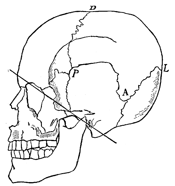
Fig. 39.—Note the line of division between the cerebral and facial cranium; in addition to this the sutures are shown which divide the frontal, parietal, occipital and temporal bones. PD. Coronal Suture; DL. Sagittal Suture; AL. Lambdoidal Suture.
The bones of lesser importance, which are interior and hidden are: the two lacrymal bones (situated at the inner angle of the orbitary cavity), the vomer or osseous septum of the nose; the two bones in the nose which lie on each side of the vomer and are known as the turbinated bones (concha nasalis); and the two palate bones (which form the backward continuation of the palatine vault constituted by the maxillary bones).
Human Cranium and Animal Cranium.—The dividing line between the cerebral and facial cranium is of great importance in anthropology, because the relative proportions between these two parts of the cranium form a human characteristic, contrasting widely with the animal characteristics; and they offer a simple criterion for determining the higher or lower type of the human cranium. (Compare in this connection Fig. 40, skulls of the higher mammals and of man.)
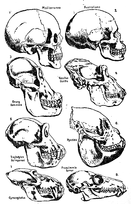
Fig. 40.
The illustration represents a number of different animal skulls; and at the top are two human skulls, the one of an Australian and the other of a European. It will be seen that the proportions between the facial and cerebral portions are very different; in the animals, even in the higher orders such as the primates (orang-utan, gorilla, etc.), the facial and masticatory parts predominate over the cerebral.
One might even say that the skeleton gives us at a glance the characteristic psychological difference; the animal eats, man thinks; that is, the animal is destined only to vegetate, to feed itself; man is an entirely different species; he has a very different task before him; he is the creative being, who, through thought and labour, is destined to subjugate and transform the world.
There are still other characteristic differences between the animal and the human skull. The cerebral cranium of the ape is not only smaller but it is furnished with strong bony ridges, to serve as points of attachment for powerful muscles intended to protect the cranial cavity. The human skull is completely devoid of such ridges; it is perfectly smooth, with delicate contours; it might be described as "frail and naked"; for the word nakedness precisely expresses the absence of those defences with which the cranium of the anthropoid ape is so abundantly provided. Accordingly, the human cranium is undefended by soft tissues; and even the bony walls themselves are far from thick. If we take a transverse section of the bones of the cranium, we find that they are formed of two very thin layers of bone united by a porous, osseous substance; the external layer is in direct contact with the muscles of the scalp, and the internal layer with the brain. These two layers differ widely in their degree of elasticity: the external layer is so elastic that if it receives a bruising blow (provided this is not so heavy as to surpass its limits of elasticity) it will yield even to the point of touching the inner layer and then spring back to its original position without leaving any perceptible trace of the blow received (this is especially true in the case of infants),[36] while the inner layer is so unelastic as to appear almost as brittle as glass: so much so, for example, that the indirect shock of the same contusion may cause it to splinter into fragments, which may either penetrate the substance of the brain, or produce hemorrhages, or inflammatory reactions in the meninges—and sometimes may constitute the sole cause of epilepsy, and various forms of inflammation of the brain (even resulting in idiocy), and sometimes of meningitis and death.
Contusions on the heads of children, and in general blows resulting from falls or other causes, must be taken into serious consideration, in the history of the individual, even though they have left no profound traces externally.
This human characteristic of nakedness, of the absence of powerful bodily defences, is not limited to the head alone, but is diffused over the entire morphological organism. Man, considered as an animal, is weak; he is born naked and he remains naked, and destitute of those natural defences which explain the endurance and the survival of other species; neither the fur nor the plumage of mammals and of birds nor the bony shields of reptiles and scales of fishes serve as defences for this vertebrate, who has raised himself to the highest eminence in the zoological scale; neither the muscular strength and powerful teeth of the felines, nor the talons of the birds of prey have been his arms of conquest.
Nevertheless, man who has conquered the earth and overcome all his powerful biological enemies, owes his survival, equally with all other living creatures, to his victory over other animals and over his environment. Wherein lies the special strength of this little, feeble being, who has become the lord of the earth? It lies in his brain. The arms of this conqueror are wholly psychic. It is his intelligence which has prevailed over the might of other animals and enabled him to acquire the means of adapting himself to his environment, or else of adapting his environment to himself. His intelligence, which sufficed him as a weapon with which to achieve victory in the struggle for existence, is also the means which still permits him to continue on the road toward self-perfectionment.
The morphological importance attached by anthropologists to the cerebral cranium depends precisely upon this: that it is the envelope of the brain. If we examine the interior of the human cerebral cranium, we find that it has adapted its bony contours so faithfully to those of the soft tissues that it bears the imprint of the various parts of the brain (cerebrum, cerebellum), the convolutions, and even the blood-vessels of the meninges. Accordingly, a study of the cerebral cranium amounts to an indirect study of the brain itself.
Characteristics of the Human Cranium.—The characteristics of the human cranium are all associated with the great development of the volume of the brain. Let us assume that we have an elastic vessel, representing in form an animal cranium, open at the base through an orifice corresponding to the occipital foramen. If we inflate this vessel, it will not only begin to enlarge at the expense Of its folds (ridges), and to stretch and distend its walls (thinness and fragility of the cranial bones); but furthermore it will undergo a change in form, acquiring a more pronounced rotundity and pushing upward in its anterior part above the face. This part, rising erect above the face, and determined by the volume of the brain, is the forehead. Animals do not have an erect forehead; their orbits continue backward in an almost horizontal line, giving them an extremely receding brow. Corresponding to this preponderance of the cerebral portion, the facial portion retires below the brow, the mandibles do not extend beyond the anterior axis of the brain, and are so far diminished in volume that they assume, as compared with animals, a new function; in short, the mouth is no longer merely the organ of mastication, but also the organ of speech; its animal part has been spiritualised.
The Evolution of the Forehead.—Inferior Skull Caps; the Skull of the Pithecanthropus; the Skull of the Neanderthal Man. The forehead is so distinctly a human characteristic that mankind has not needed the help of anthropology in order to realise its importance—and as a sign of superiority, nobility or sovereignty, has placed upon the forehead the crown of laurel, or the crown of nobility or kingship.
Has the forehead always been a human characteristic, or have we acquired it little by little? Such a problem is associated with the evolution of the brain. There are in existence certain remains of the skeletons of primitive men, which show them to have possessed a cerebral cranium inferior in volume to that now attained by the human species; and in these remains the forehead is also profoundly different from that of to-day, in that it is much lower and slants backward, while the supraorbital arches are very prominent. Such is the evidence of the "cranial caps," discovered in the early geological strata.
In the tertiary strata of the island of Java, which in that remote epoch of the earth's history must, together with Sumatra, have formed part of the continent of Asia, which is considered as the "laboratory of races," a skull was found by Dubois which raised the problem whether it should be classed as that of an ape superior to those now existing, or of a primitive man. Prior to this discovery, it had been maintained that man did not make his appearance until the quaternary period. This supposed primitive man was called by his discoverer the Pithecanthropus, pithecanthropus erectus.
Remains that are unquestionably human occur in the quaternary period, in which however skeletons are very rare, as compared with relics of human labour or social life, relics which are found scattered everywhere throughout Asia and Europe as well (chipped flints). The various remains of skeletons show us skulls much inferior to those of modern man, but superior to that of the pithecanthropus. In treatises of general anthropology reproductions are given of human crania known as the Spy or Neanderthal type, belonging to the epoch when the gigantic mammoth still roamed the earth. The forehead is very low and receding and the orbital arches are enormously developed; while the cerebral capacity calculated from the cranial dimensions is inferior to that of modern man.
Consequently, as the brain increases in volume in the course of the revolution of the race, the cranium not only shows a corresponding volumetric increase, but at the same time alters its form, thus producing the forehead which little by little rises from a receding to an erect position, and becomes high where it was formerly low, while at the same time the prominent orbital arches disappear. Accordingly, we may consider the forehead as the skeletal index of the cerebral volume, and hence of the relative anthropological and intellectual superiority.
In addition to its above-mentioned value, it also furnishes us with a biological principle of much importance: the relation between the volume and form of the cranium.
While the volume has a significance that is relative to the mass of the body, the significance of the form is absolute.
Let us examine these two skulls: normal human skulls of our own epoch; one of the Celtic race (Fig. 46) and the other Sardinian (Fig. 43); that of the Celtic race is much larger and rounder; that of the Sardinian is very much smaller and more elongated.
If we were considering only the volume, we might say that it was simply a case of a microcephalic and a macrocephalic: two terms (microcephaly and macrocephaly) that fall within the province of pathology. On the contrary, these two skulls are normal, but they belonged to individuals characterized by differences of race; the one (small skull) having a low stature; the other (large skull) having a tall stature.
The volume of the head therefore bears a relation to that of the body; the volume has a relative significance. But the form in both of them reveals a state of normality; the two skulls have a high and erect forehead, and exhibit in their whole contour a fine and regular development. Therefore the form has an absolute significance. It even proves to us the normality of the volume, a fact which could not be determined by the volume alone.
Another mechanical correspondence between volume and form is disclosed when we compare the skull of a new-born child with that of an adult. The skull of the new-born child is much smaller in volume; but the form shows the relatively enormous volumetric development of the brain; in fact the skull is protuberant and the forehead bulges forward above the face (front bombé), while corresponding to this index of cerebral development is the enormous preponderance of the cerebral cranium over the facial cranium, which is so small as to be almost reduced to a simple rudiment.
Hence the form by itself alone reveals the infantile character of the cerebral volume, which, in relation to the bulk of the body is of far greater dimensions than in the adult. In fact, if a child simply increased in volume and its growth was not the sum total of a morphological evolution, the adult man would become a monster; his macrocephaly would be so exaggerated that his neck could not sustain the weight of the head (If the relations between the proportions in infancy were maintained through life the adult man would have a head with a perimeter of 130 centimetres, = 4 ft. 3 in.).
Aside from its mechanical relations to the volume, the form has characteristics dependent upon biological factors, such as the sex and the race. The female cranium in fact has a straighter forehead than the male and the orbital arches are absolutely wanting, while the entire surface of the cranium is smoother and more rounded.
Similarly, the different races exhibit forms determined by biological factors and not by mechanical causes—for instance, the degree of dolichocephaly (elongated cranium) and of brachycephaly (short cranium).
Hence the form is life's manifestation not only of the characteristics proper to the species, but also of the mechanical adaptations demanded by the material composing the body.
It may be said that the volume and the form of the cranium are dependent upon two different biological potentialities: the volume is mainly determined by the cerebral mass; the form, on the contrary, is mainly determined by the bony structure—no matter how completely form and volume coincide in their reciprocal mechanical relations.
That is, the attainment of a given volume of head depends upon the development of the brain; the bone follows this development passively, is the index of it, the skeletal representation of it, but never the determining factor.
At one time it was thought, on the contrary, that a precocious ossification of the cranial cavity would arrest the development of the brain; microcephaly was believed to be caused by a precocious closing of the sutures of the cranial bones; and there was a certain period when the surgical treatment of microcephaly consisted in the removal of a portion of the cranial bone, in order to allow the brain to develop freely.
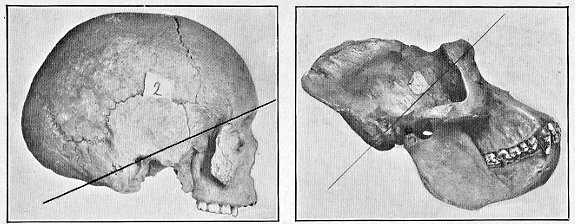
Fig. 41. Fig. 42. Dividing line in human skull, as compared with that of gorilla.
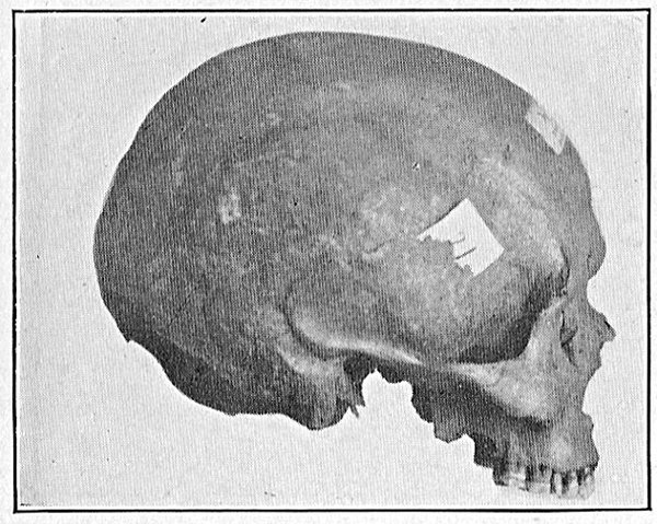
Fig. 43.—Rounded ellipsoidal cranium.
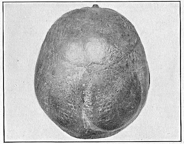
Fig. 44.—Brachycephalic cranium (vertical norm)
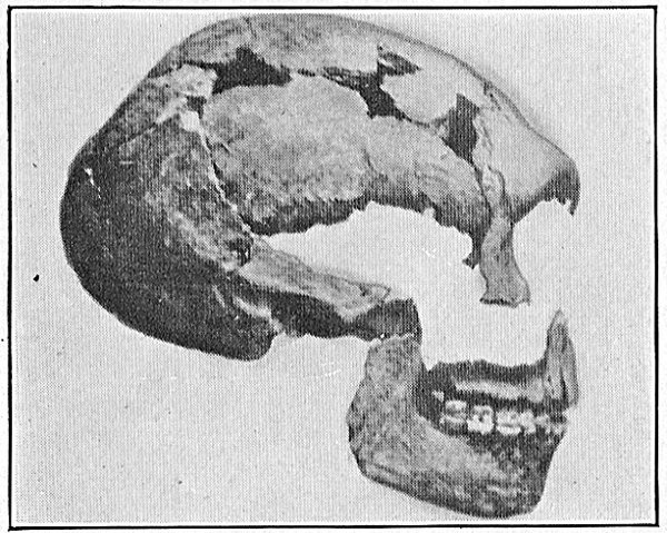
Fig. 45.—Remains of spy cranium.
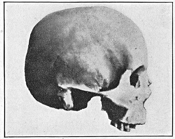
Fig. 46.—Brachycephalic cranium.
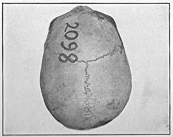
Fig. 47.—Egyptian cranium, 21st dynasty, ovoid type.
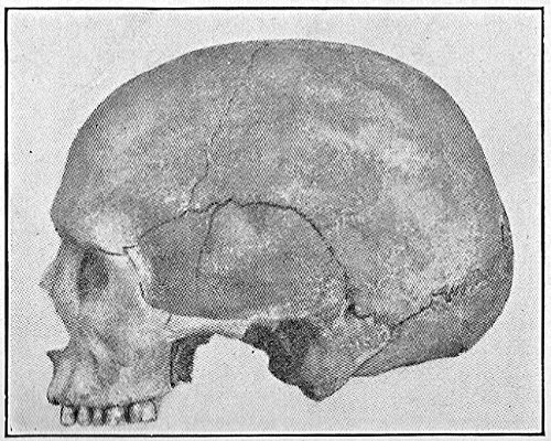
Fig. 48.—Dolichocephalic cranium, from lateral norm.
But the failure of such attempts afforded additional proof of the fact that the volumetric development of the cranium depends upon the brain alone.
If a precocious or abnormal suture occurs in the cranial bones, there does not follow an arrest of development, but simply a malformation; which is precisely in proportion to the potentiality of the brain, which grows less where the suture has been formed, and in compensation grows more than normally where the conditions of the bones permit of cerebral expansion; and a deformity results. Microcephaly on the contrary shows inferiority of form (smallness, receding forehead, etc.), but not malformation.
Anomaly of form, therefore, results only from anomaly of skeletal development, and is frequently found in conjunction with a normal development of the brain.
Consequently malformations of the cranium do not have the grave significance of biological inferiority or of degeneration that they were at one time believed to have; but frequently they must be considered in connection with pathological conditions resulting for the most part in delayed development in the embryo or in early infancy, producing a thickening of the bone, or a partial suturation of the points, or parts, or of the entire suture (punctiform synostosis, partial or total); sometimes the sutures remain unaltered, and the deformation must be attributed to various disturbances connected with the nutrition of the skeleton in the course of intrauterine evolution (hereditary syphilis, denutrition of the mother during pregnancy, etc.). In short, a cranium that is abnormal in form is an indication of pathological occurrences or of physiological errors that have resulted in altering the normal growth of the individual.
There are many anomalies in the form of the cranium, but here we will cite only the two principal ones, because they are the most frequent and most likely to be encountered in individuals whose growth has been retarded (from lack of nutrition) and consequently constitute signs of physiological inferiority often associated with social caste. These two forms are: scaphocephaly and plagiocephaly.
The scaphocephalic cranium (Figs. 51, 52), is characterised by being very narrow and flattened laterally; while the forehead and the occiput project in front and behind, the two parietal bones meet above almost in an angle, so that, if it were turned upside down, the vault of the cranium would have the appearance of the hull of a ship.
The plagiocephalic cranium is a cranium which is unsymmetrical in respect to its longitudinal axis; that is, it is not equally developed on the right and on the left.
As a matter of fact, our bilateral symmetry is an ideal standard rather than an absolutely attainable reality; we are all of us a little larger on one side and a little smaller on the other, but to so slight a degree as to escape superficial observation, so that in general we have apparently a bilateral symmetry—that is, we appear to be symmetrical according to the testimony of our senses; but a more delicate examination proves that this is not true. Plagiocephaly therefore represents an exaggerated case of a normal fact. Plagiocephaly may be simple or compound; it is simple when the asymmetry is partial; namely, when it is confined to the anterior or posterior portion; it is compound when it is total; and in such case we find a complete diagonal correspondence: for instance, if the right nodule in the frontal region is more prominent, the left nodule is more prominent in the left occipital region, or vice versa. In general it may be said that the various forms of plagiocephaly are produced by asymmetry of the nodules or of the flattened surfaces of the cranium. Even in the case of microcephaly and of macrocephaly, which are substantially anomalies of volume, we find corresponding characteristic abnormalities of form. The microcephalic cranium is of inferior type, suggesting that of the ape—in other words, it is a cranium which has mechanically adapted itself to a brain of inferior volume: the macrocephalic cranium, especially if the abnormality is due to rickets or to hydrocephaly, calls to mind the infantile type of cranium; it has the characteristic bulging forehead, while mechanical adaptation frequently renders it very round (pathological brachycephaly). We will take up this question again when we come to speak in particular of malformations and to describe the technical methods of cranioscopy. What more particularly concerns us now is a consideration of the normal form of the cranium and its morphological evolution.
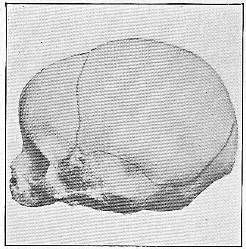
Fig. 49.—Cranium of new-born child (lateral norm).
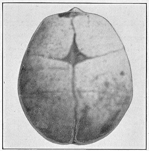
Fig. 50.—Cranium of new-born child (vertical norm).
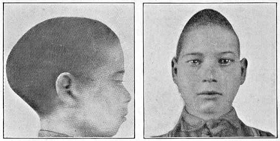
Fig. 51. Fig. 52.
Scaphocephalic cranium.
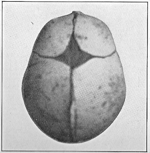
Fig. 53.—Cranium of new-born child seen from above, showing polyhedric contour due to nodules of ossification; fontanelle of the bregma; and suture dividing the two frontal bones.
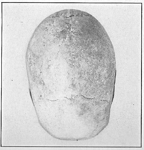
Fig. 54.—Ellipsoides (classified by Sergi).
The Morphological Evolution of the Cranium through the Different Periods of Life. Embryogeny. Order of Appearance of the Points of Ossification and of Synostosis of the Sutures.—In its successive transitions through the different periods of life, the cranium not only acquires successively greater volume, but it assumes forms corresponding to the different grades of morphological evolution. We may group its transformation under five different periods: 1. from conception until birth (embryonic evolution); 2. from birth until the end of the third year (infantile evolution); 3. from three years old until twenty (youthful evolution); 4. from twenty to forty (adult age); 5. from forty to the end of life (involution).
First Period.—In the earliest stages of intrauterine life the cranium consists of a membranous skin, enclosing the primitive cells of nerve tissue constituting the brain; it has a cartilaginous basal part, destined later to form the base of the skull (basioccipital and basisphenoid bones). But all the rest (the vault or cap of the cranium) remains in a membranous state, so that at this period the head of the embryo has not yet acquired a definite form.
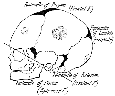
Fig. 55.—Cranium of new-born child. Showing nodules and fontanelles.
In the second month of intrauterine life the phenomena of ossification have already begun to take place; that is, a fine network has formed, spreading over almost the entire surface, which proceeds to fill up its interstices with calcareous salts. This process, however, is more rapid and more intense at certain points (points of ossification), from which it cannot properly be said that the ossification radiates, but rather that at these points the general process is intensified and concentrated. There are five principal points of ossification: two frontal, two parietal and one occipital, which appear clearly defined and projecting like nodules, imparting to the cranium, when seen from above, a pentagonal form, which is the normal form of the infant cranium.
Second Period.—At birth the cranium has not yet completed the process of ossification, nor are the normal number of bones that will eventually compose the adult cranium, as yet definitely determined. Therefore the cranium of the new-born child has three distinct characteristics:
It is not yet uniformly rounded, but polyhedral because of the noticeable prominence of the five primitive nodules or centres of ossification (2 frontal, 2 parietal, 1 occipital, Figs. 53, 55).
Since the process of ossification of the bones is not yet completed, certain membranous portions or cranial fontanelles still remain, which are especially wide at the points where several bones meet. The principal fontanelle is that of the bregma (at the juncture of the two frontal with the two parietal bones, quadrangular). Next comes that of the lambda, which is much smaller (juncture of the two parietal bones with the occipital, triangular), and lastly the fontanelles of the asterion and the pterion, on opposite sides of the temporal bones, the former being situated behind and the latter in front.
Since the process of ossification is incomplete, the fusion of bony portions into entire bones, such as they are destined to be when complete development is reached, has not yet been accomplished; that is to say, certain bones of the cranium are still divided into several portions. For example, the frontal bone in the new-born child is composed of two bones, separated by a longitudinal suture that is destined to disappear, and the occipital bone is composed of four parts, namely, the base, the squama and the two condyles (basioccipital, exoccipital and superoccipital bones).
During the first period of three years, while the brain is increasing notably and rapidly in volume, the cranium undergoes various and interesting transformations. The pentagonal form of the cranium tends steadily to become rounder, because the primitive nodules are diminishing, or even disappear, although in this regard many individual varieties result; and the processes of ossification reach their completion. This is the most important period of growth, during which the individual development of the perfect cranial form may be attained, provided the rhythm of growth between the brain and its envelope remains harmonious; or again, certain deformations may be definitely established, owing to the intervention of some pathological condition or a disturbance of nutrition, altering either the internal volume or the normal process of ossification of the bony covering.
The first closing of the fontanelles takes place, in our race, in those of the asterion (posterior to the temporal bones), and next in those of the pterion; and it sometimes happens, as an anomaly of growth that leaves no external trace in the living man, that a little bone is formed, duplicating the shape of the fontanelle itself; such little bones, very common in abnormal crania, are called Wormian bones. They may occur in connection with any of the fontanelles, but especially with that of the bregma.
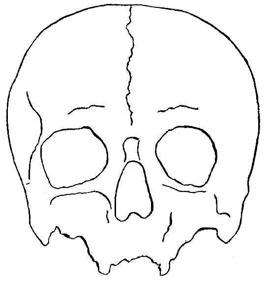
Fig. 56.—Cranium of adult with abnormal medio-frontal suture.
The fontanelle of the lambda generally closes during the first year; and the last of all the fontanelles to close is the largest, which is situated toward the front of the head, at the bregma, and is well known, even by the common people, and can easily be felt upon a child's head; it generally closes toward the end of the second year; and its characteristics may furnish valuable indications of abnormality or insufficiency of the child's development. For example, if it diminishes and disappears ahead of time, this may constitute the first symptom of microcephaly, or at all events, of submicrocephaly (i.e., a case of microcephaly that is not very pronounced). On the contrary, when this fontanelle remains dilated and delays its normal closing, this is a sign of organic weakness and debilitating disease (cachexia, rickets, myxedema). Furthermore, the fontanelle in question may alter its characteristic appearance in certain forms of sickness. In the case of hydrocephaly it becomes distended, while in enteritis, on the contrary, in which the organism parts with a large proportion of liquid, it becomes depressed.
The sutures also undergo notable changes during this period of life. The first to become effaced is the metopic or medio-frontal suture, which is destined to close and form a single bone; by the end of the first year it is obliterated throughout the middle third of its length, and thereafter the process of suturation spreads upward and downward until it is completed at the end of the second year (Welcker, Haeckel, Humphry). Sometimes, however, this suture is not obliterated until very late, and there are anomalous cases where it has remained throughout life, giving the forehead a characteristic form (pronounced frontal nodules and a slight palpable furrow along the medial line of the forehead).
During this same time a fusion has also taken place between the occipital squama and the two lateral or condyloid portions; but the resultant whole still remains separated from the corpus or base of the occipital bone, which will not become welded into one solid piece with the rest before the age of seven years.
At the age of three, the ossification of the cranial vault has been completed. In place of being depressed and protuberant, as it was at birth, the cranium has grown upward and forward in the frontal region, assuming an almost definitive form; the volume of the cranium has at the same time undergone an exceedingly rapid growth, attaining proportions very near to those of an adult.
From the age of three onward the head grows slowly, and its transformations are much slighter and fewer. The cranial capacity which at birth is 415 cubic centimetres, becomes at the age of three, 1,200, at the age of fifteen, 1,393, and in the adult, 1,400 cu. cm. respectively. Accordingly we might say that at the age of three a sort of repose has been established in the growth both of the the brain and of the cranium; this is the age at which an awakening begins in the child of that intelligence which is to put him in touch with the external world, and it is also the age at which he may begin his education in school.
Third Period.—There follows a slow and parallel growth of both brain and cranium. The ossification of the cranium itself reaches completion. At the age of seven the occipital is definitely solidified into a single bone and between the years of fifteen and twenty the body of the sphenoid also becomes welded to the occiput. This process of synostosis begins from the interior of the cranium, and only subsequently manifests itself externally. Consequently, the basilar suture closes at the time when the last large molars, the so-called "wisdom teeth," appear. After this period, the base of the cranium can no longer undergo any sort of growth, and in the case of uneducated persons the complete development of the cranium is definitely accomplished.
Fourth Period.—But in the case of cultured persons, those who form the class of brain-workers, the brain continues to grow, although extremely slowly, up to the age of thirty-five or even forty, thanks to the sutures which still remain completely intact and which still make an expansion of the bony envelope possible.
After this comes the beginning of the
Fifth Period.—The period of involution, during which the synostosis (closing) of all the cranial sutures will successively occur, until in advanced old age the cranium becomes composed of a single bone, just as in the embryo it was formed of a single membrane.
The synostoses which occurred in the early periods had an evolutive significance and were associated with the growth of the body and the intelligence. These later synostoses, on the contrary, have an involutive significance and are associated with the physiological decay of the organism and at the same time with that of the psychic activities.
The first point at which synostosis takes place is in the region of the obelion, that is, near the middle of the suture which, unites the two parietal bones; shortly afterward, the fronto-parietal sutures begin to unite along the pterion. At the age of forty-five, the obeliac synostosis has progressed as far as the lambda, and that of the fronto-parietal suture to the bregma; and at fifty the ossification is very nearly accomplished, at least on the right-hand side (according to Broca's series of crania). At seventy the squama of the temporal bone unites with the parietal, and at eighty the entire cranium has become a single bone.
These processes are subject to no small number of individual variations; there have been cases of persons who, although very old, still preserved many of their cranial sutures intact and their psychic activities remained correspondingly alert (men of genius). Conversely, the closing of the sutures sometimes begins as early as the thirty-fifth year. A diagnosis of age, as determined by the skeleton, is consequently only approximate.
During the periods of growth the cranium may exhibit transitory anomalies; it is very common to encounter in the heads of children of the lower social classes, who are consequently subject to denutrition, malformations which represent various degrees and forms of plagiocephaly, and which subsequently disappear completely, as the development of the cranium advances. Anomalies of form must therefore be judged differently in the case of the child than in that of the adult.
It may even happen that the five primitive nodules persist for a long time and even remain as a definitive form of the adult cranium constituting, according to Sergi, a distinct variety, the pentagonal cranium. But this is quite rare. From the frequency with which this form is to be observed in schools attended by children of the poorer classes, it is better to regard it as due to a delay in morphological evolution, which will probably disappear later on.
Normal Forms of the Cranium
We are indebted to Sergi for an exact knowledge of the normal forms of the cranium. Such forms are racial characteristics and are invariable, as Sergi has succeeded in proving by a comparison of the most ancient forms of the cranium with recent forms. Accordingly this authority takes the cranial formation as the basis for his classification of races. We have no direct interest, so far as concerns the special scope of our own science, in the value of this theory of classification—a theory, by the way, already divined, although very imperfectly and under a different form, by French and German anthropologists. Sergi's studies of cranial forms interest us solely as a diagnostic test of normality as compared with abnormality. For it is due to these researches that certain forms that used to be considered pathological, have come to be recognised as normal.
The normal forms of the cranium may be grouped, according to Sergi, under nine primary varieties, each of which includes sub-varieties.
These nine varieties are named as follows:
I. Ellipsoid; II. Ovoid; III. Pentagonoid; IV. Rhomboid; V. Beloid; VI. Cuboid; VII. Sphenoid; VIII. Spheroid; IX. Platycephalic.
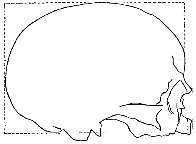
Fig. 57.—Ellipsoides depressus cranium.
I. Ellipsoid (Fig. 58).—This form is recognised by inspecting the cranium according to the vertical norm (see in the chapter on Technique the method of cranioscopy).
The cranial contour recalls an ellipse in which no trace of the nodules remains, and in which the occiput is not in the least flattened; while the anterior half of the cranium closely corresponds to the posterior half.
The sub-varieties are differentiated by their greater breadth and length, by the form and protrusion of the occiput, and also by the height of the cranium measured vertically.
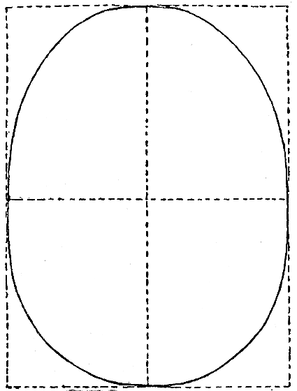
Fig. 58.—Ellipsoid cranium.
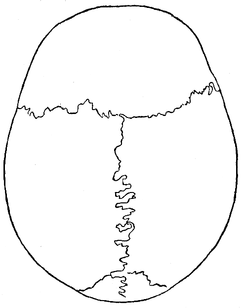
Fig. 59.—Ovoid cranium.
Accordingly, the sub-varieties have a binominal nomenclature indicating, in addition to the fundamental characteristic (variety) the qualitative characteristic of the sub-variety (e.g., ellipsoids depressus; compare Fig. 57, showing a cranium seen laterally).
II. Ovoid.—This form of cranium, seen from above, is that of an ovoid, with the broader portion corresponding to the parietal bones, at the point where the characteristic embryonal nodules are situated. The protrusions of the parietal bones are apparent (swellings) but not angular (nodules). The occiput protrudes and is broad (Fig. 59).
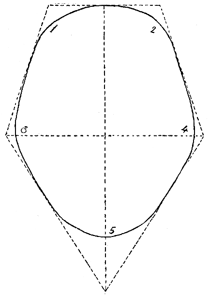
Fig. 60.—Pentagonoid cranium.
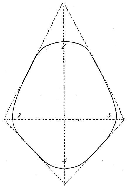
Fig. 61.—Rhomboid cranium.
III. Pentagonoid.—In this form, persistent traces of the five primitive embryonal nodules are still plainly visible, giving the contour of the cranium, when seen vertically, the appearance of a pentagon. The protuberances, however, are quite smooth and not pointed, as in the embryonal cranium.
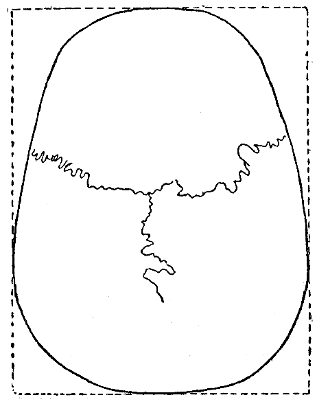
Fig. 62.—Beloid cranium.
IV. Rhomboid.—This form is similar to the pentagonoid, excepting that the parietal breadth is much more notable in proportion to the forehead, which is much narrowed and has lost its nodules.
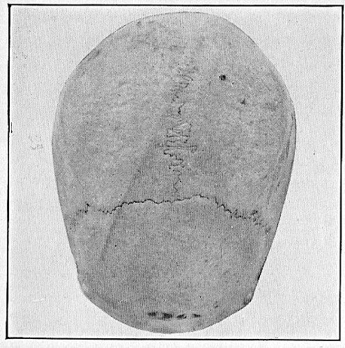
Fig. 63.—Ovoids (classified by Sergi).
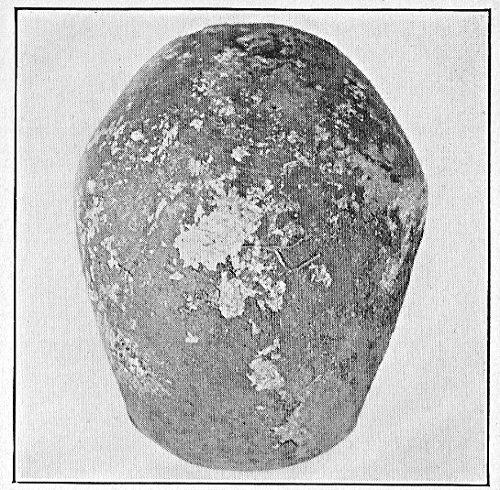
Fig. 64.—Pentagonoides acutus (Sergi's collection).
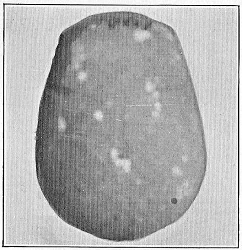
Fig. 65.—Beloides lybicus (classified by Sergi).
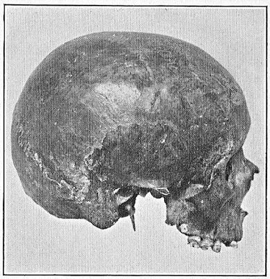
Fig. 66.—Platycephalus orbicularis (classified by Sergi).
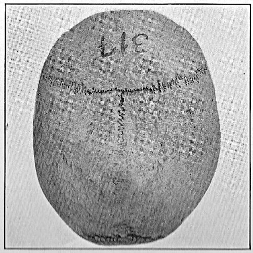
Fig. 67.—Platycephalus ovoidalis (classified by Sergi).
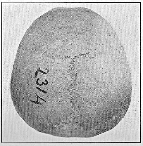
Fig. 68.—Spheroidal cranium, vertical norm (Sergi's collection).
V. Beloid.—The beloid, or arrow-head cranium is like the ovoid with the occiput more flattened, so that the widest portion is further back than in the ovoid; toward the front it becomes narrower, constituting altogether an admirably shaped type of head.
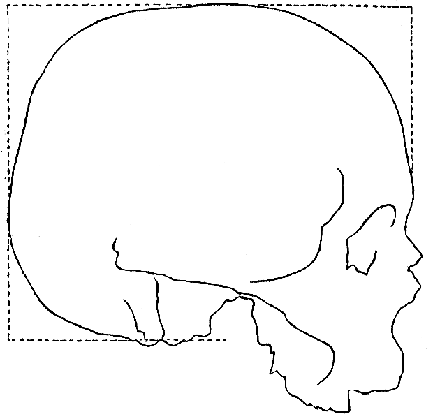
Fig. 69.—Cuboid cranium.
VI. Cuboid.—This form is most clearly perceived when the cranium is seen either sidewise or from the rear. Not only the face, but the lateral and occipital walls as well are flattened; so also is the forehead, which in general is quite vertical.
VII. Sphenoid (cuneiform).—The broadening between the two parietal bones is usually far back and very evident, while the cranium narrows toward the front. The occiput is flattened.
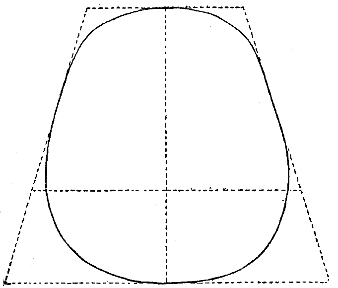
Fig. 70.—Sphenoid cranium.
VIII. Spheroid.—Seen vertically, it presents the appearance of a very broad ellipse; all the curves tend to become spherical. The forehead, however, is not notably vertical.
IX. Platycephalic.—The fundamental characteristic of this type of cranium is that it is flattened on top, or rather, since such flattening cannot be absolute, the arch of its vault is a segment of a circle of very large diameter (Sergi), with the result that this cranium has the appearance of being very low vertically and very broad laterally. When seen vertically it may present a wide variety of contours, ellipsoid, ovoid, pentagonoid, etc., but its distinguishing characteristic remains that of the flattened vault.
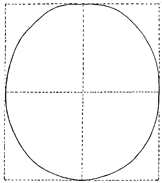
Fig. 71.—Spheroid cranium.
Sub-varieties.—Sphenoids trapezoids, or trapezoid cranium. Observed from the vertical norm, this form appears as a variety of the sphenoid; and when seen laterally it is characterised by the lines of its contour forming a trapezium. Starting from the vertex of the cranium one line slants toward the forehead and another toward the occiput, which is very massive. In the figure given below, the quadrangle drawn in solid lines serves to indicate the correct position of the cranium, while the trapezium formed of dotted lines gives us its characteristic form.
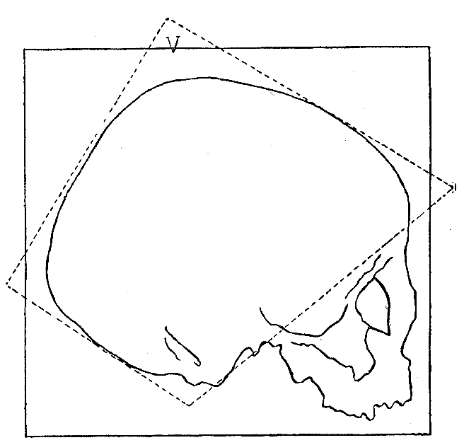
Fig. 72.—Trapezoid cranium.
Among the forms described by Sergi, are several which were formerly held to be abnormal, such, for instance, as the platycephalic cranium and the pentagonoid. Similarly, when the surfaces of the cranium showed a tendency toward flatness, or when there were cranial protuberances, even though these were destined to disappear, they were regarded as malformations. Before this high authority offered us his guidance, there were certain forms, frequently encountered, that it was difficult to define, for example, the trapezoid cranium, which often presents a notable vertico-occipital flattening, with the vertex notably higher than the forehead.
There are also certain forms of cranium having the frontal region more restricted than the parietal region, or slanting down from a much elevated vertex, which have been proved to be normal forms; while still another error previously made was that of trying to judge the forehead on the criterion of a single model, deviations from which were much too readily relegated to the category of abnormalities. The most regular and beautiful forms, and the ones that are commonest in our racial stocks are the ellipsoid, ovoid and sphenoid. In my work on the women of Latium, precisely one of the points that I noted was the frequent occurrence of certain sub-varieties of the ellipsoid and the sphenoid.
In order to recognise the forms of the cranium, a certain training is necessary which each one must acquire for himself. Observations of the cranium will make it easier to judge of the form in relation to the head, at least, when the latter is not too much hidden by the hair, as often happens in the case of young children.
A knowledge of the normal forms of the cranium will also guide us in our judgment of many abnormal forms, which very often present the appearance of exaggerations of normal types.
Thus, for example, the acrocephalic cranium (much raised in the parieto-lambdoideal region and sloping forward toward the brow, while the occipito-lambdoideal region is flattened) recalls the trapezoid; and the clinocephalic cranium (in which the coronal suture forms a slight girdle-like indenture and divides the contour of the cranium, when observed along the vertical norm, in two curves, a lesser anterior and a greater posterior curve, resembling a figure of 8) recalls certain varieties of ovoid cranium described by Sergi. This brings us to a principle that is very interesting to establish, namely, that frequently anomalies represent exaggerations of the racial or family type.
The Cephalic Index
Retzius was the first to take the cranium under consideration as a basis for a classification of the human races; and he attempted to determine a concept of its form by means of a numerical formula expressing the relation between the length and width of the cranium (cephalic index). Thus he distinguished the races into brachycephalics, or those having a short head; and dolichocephalics, or those having a long head. Following Retzius, who may be regarded as the founder of craniology, Broca adopted, completed and expanded this method, deriving from the cranium, or rather from the particular character given by the cephalic index, a key, as it were, suited to unlocking the intricate mysteries of hybridism among the human races. Consequently the cephalic index was not confined, as regards its importance, within the same limits as all the other indexes, but was raised by the French school, warmly seconded by Italian anthropologists, to the dignity of a fundamental determinant of the ethnic type, as definitely as, for example, the vertebral column serves as basis for a classification including all species of vertebrates.
The Germans refused to accept the cephalic index as determining the classification of races; but while seeking to prove themselves independent of it, they continued to regard the form of the cranium as a basis of classification (Rütimeyer, von Höller, and to-day Virchow), but without ever having identified, as Sergi has now done, existing forms as normal types of race.
The cephalic index is obtained by the well-known formula expressing the relation between the maximum transverse diameter of the skull (see "Technique") and the maximum longitudinal diameter reduced to 100, and is expressed as follows: Ci = 100×d/D (the cephalic index is equal to a hundred times the lesser diameter divided by the greater; in the present case the lesser diameter is the transverse).
This proportion between linear measurements cannot properly sum up the form of the cranium. We can, for example, conceive of a microcephalic cranium having a normal cephalic index, since the relation between the two maximum diameters necessary for deducing the index, does not tell us, for example, either the dimension of the cranium or the form of the forehead.
If, for instance, we should imagine a photograph of a cranium enlarged a hundred diameters, the reciprocal relations between the length and the width would still remain unchanged.
In order to demonstrate that the cephalic index does not determine the form of the cranium, Sergi makes use of a number of different geometric figures, such as a triangle, an ellipse, a trapezoid inscribed within equal rectangles, and which consequently have an equal base and equal altitude, that is, the same proportion between length and width.
It follows that skulls corresponding more or less closely in shape, trapezoidal, trigonocephalic, ellipsoidal, plagiocephalic, and hence both normal and abnormal, can be expressed by a cephalic index having the same identical figures.
But, although the cephalic index is far from being descriptive in regard to the form of the cranium, it constitutes an anthropological datum that has two advantages: 1. It depends upon measurements and is therefore accessible to those who, not being anthropologists, lack the trained eye that can distinguish with careful accuracy the true forms of the cranium in their manifold variety. Furthermore, since the measurement of maximum diameters is sure and easy and may be obtained with exactness, regardless of the thickness of the hair, it may be applied in anthropological research to all subjects. 2. The cephalic index, even if it does not give us the form, does give us a fact which has a bearing upon the form, namely, whether the cranium is long or short; in other words, it substantially represents the most real and evident difference between the different types of cranium. And since the cranium has a visibly spheroid form, that is, with smooth and rounding surfaces, and constantly adheres to this generic delineation, the fact of being longer or shorter introduces a definite differentiation into the general and accepted form, and gives a very simple and concise indication of it, that conveys the idea more clearly than a description would.
Granting the practicality of this line of research, the cephalic index may also be accepted as an index of form, so long as there is no intention of going deeply into minute differentiations for systematic purposes. Professor Sergi himself, author of the system that forms the basis of the study of cranial forms, urged me to exclude from a practical course in pedagogic anthropology the classification of forms, limiting the concept of form to that included in the cephalic index.
The cephalic index has the additional advantage of having been extensively studied and consequently of having an abundance of mean averages for comparison that are of great practical use. Furthermore, the idea it gives regarding the cranium by means of one simple figure serves to convey certain fundamental principles with great clearness.
In dealing with figures that determine an anthropological datum of such high importance, it is necessary to define its limits and its nomenclature.
Various authors have introduced their own personal classification of the cephalic index, and no small confusion in nomenclature has resulted; so much so that a need was felt of establishing a uniformity of numerical limits and of the relative terminology, in other words, of simplifying the scientific language.
Accordingly, a congress was held at Frankfort in 1885, at which the following nomenclature was established by international agreement:
CEPHALIC INDEX.—Nomenclature established at Frankfort
Dolichocephalia = 75 and below
Mesaticephalia = from 75.1 to 79.9
Brachycephalia = from 80 to 85
Hyperbrachycephalia = 85.1 and above.
Previous to this, the most widely varied classifications were in use, and the leading authorities had all introduced into the literature of the subject their own personal classifications. Here are some of the more important:
Broca: Dolichocephalics = 75 and below
Subdolichocephalics = from 75 to 80
Subbrachycephalics = from 80 to 83.3
Brachycephalics = 83.3 and above.
Ranke: Dolichocephalics = 74.9
Mesaticephalics = from 75 to 79.9
Brachycephalics = 80 and above.
Kollman: Dolichocephalics = 73.9 and below
Mesaticephalics = from 74 to 79.9
Brachycephalics = from 80 to 86.9
Hyperbrachycephalics = 87 and above.
Retzius and Davis: Dolichocephalia = 79 and below
Brachycephalia = 80 and above.
Topinard: Dolichocephalics 64 and below = Ultradolichocephalics.
65 True dolichocephalics.
66
67
68
69
70 Subdolichocephalics.
71
72
73
74
Mesaticephalics 75 True mestaicephalics
76
77 (Mean average.)
78 Submesaticephalics
79
Brachycephalics 80 Subbrachycephalics.
81
82
83
84
85 True brachycephalics.
86
87
88
89
90 and above = Ultrabrachycephalics.
It remains to determine the extreme limits of oscillation of the index, both in relation to the normal mean and in relation to the fluctuations of this important ethnic datum in a given population.
Topinard, as we have seen, gives as his mean figures for the extreme normal limits among the human races 64 and 90.
Deniker gives, as his mean averages for the human races, the following figures: For dolichocephaly, 69.4 (natives of the Caroline Islands; Australia); For brachycephaly, 88.7 (the Ayssori of the Transcaucasus; Asia).[37] But we know that a mean is obtained from figures either greater or smaller than the mean itself, so that the limits of individual variation must exceed that of the given figures.
Accordingly the oscillation of the normal cephalic indices may be given as ranging from 70 to 90.
In regard to abnormalities (extreme human limits of the cephalic index) the authorities give 58 for dolichocephaly (scaphocephaly) and 100 for brachycephaly (in which case the cranium is round and known as trochocephalic; it is met with among the insane).
Between oscillations of such extremely wide range in the normal cephalic index, the number chosen as a medial figure to serve the purpose of dividing the dolichocephalics from the brachycephalics is that of 80, which is included within the division of brachycephaly. In spite of the nomenclature established at Frankfort, there is a distinct scholastic advantage, because of the greater simplicity of memorising and fixing the idea, in reverting to the nomenclature of Retzius, who classes as brachycephalics all crania from 80 upward, and as dolichocephalics all those below 80. It is certainly strange to class all crania from 80 to 90 without distinction as brachycephalics, and then to alter the name and call a cranium with an index of 79.9 a dolichocephalic. It has been found that there is always a slight difference between the index taken from measurements of the cranium and that obtained from measurements of the head. According to Broca, it is necessary to subtract two units from the cephalic index taken from a living person, in order to obtain that of the cranium; thus, for example, if the cephalic index (taken from life) is 80, the cranial index (taken from the skeleton) would be 78. Such differences are due to the disposition of the soft tissues. Consequently, even according to the simple subdivision of Retzius, a person who was brachycephalic during life, would become dolichocephalic after he was dead.
But this is what always happens in biology, whenever we try to establish definite limits. Life undergoes an insensible transition through successive limits and forms, and this fact constitutes the grave difficulties and the apparent confusion of biological systems. In determining degrees of difference, it is necessary to have recourse constantly to special methods, which teach us to recognise general properties and to use them as a basis in dividing living creatures into separate groups (see in the section on Method, "Mean measurements and formation of series in relation to individual variations").
Hence, for mnemonic purposes, we need remember only the single number, 80.
But if we wish to adopt the nomenclature of Frankfort, it is necessary to keep in mind two figures denoting limits, 75 (inclusive) for dolichocephaly, and 80 (inclusive) for brachycephaly.
75 dolichocephalics
mesaticephalics
80 brachycephalics
85 ultra
These constitute, as it were, two centres, beyond which, on this side and on that, we may picture to ourselves the individual variations drawn up in martial line. In this case, the space between 75 and 80, in other words, the limits of mesaticephaly, may be interpreted as due to oscillations between dolicho- and brachycephaly according to the laws of variability, which is analogous to what takes place in the case of oscillations in the opposite direction (70-75 dolichocephaly; 80-85 brachycephaly). From this point of view, these two numbers, 75 and 80, constitute median centres of two different types.
But according to Broca and his school—and this view is accepted by many anthropologists—mesaticephaly should be regarded as constituting a fusion of the two other types, the brachy- and dolichocephalic, whence it follows that mesaticephalics would be hybrids. Other authorities, on the contrary, exaggerating the conception of the fixity of the cephalic index in a given race, admit the existence of mesaticephalic races.
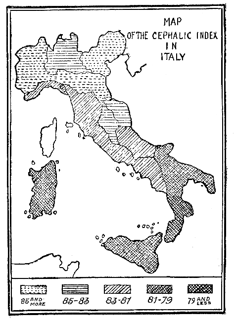
Fig. 73. Map of the Cephalic Index in Italy.
But it has been observed that the greater number of mesaticephalics are to be found in regions where dolichocephaly prevails; in certain districts of Africa, as for example, in Somaliland, not a single brachycephalic exists, yet none the less the mesaticephalics are numerous. Accordingly, mesaticephaly may be classed with dolichocephaly and regarded as one of its variations, while it seems to be independent of brachycephaly. Therefore the nomenclature of Retzius may for many good reasons be chosen and adopted in our schools. In conclusion, we shall regard the brachycephalics and dolichocephalics as the two fundamental types; and shall adopt the figure 80, included among the brachycephalics, as the limit of separation. The different grades of dolicho- or brachycephaly are to be determined by mean averages, and the oscillations due to individual variations, by series.
Hence it is important to determine the mean average and the oscillation of the cephalic index for the different races; and this is of interest to us as educators, in order to establish the limits of normality.
The practical method of studying the cephalic index is according to geographical distribution.
Here are a few general data of the cephalic index relative to its distribution:
The most dolichocephalic of all peoples are found in Melanesia, Australia, India and Africa. In the Fiji Islands the mean cephalic index is 67; in the Caroline Archipelago it is 69; in various regions of India, 71; that of the Hottentots, 74; of the Bantus, 73. Belonging to the dolichocephalics or mesaticephalics are the populations of the extreme south of Europe (Mediterranean race) and at the extreme north (English, Scotch). On the contrary, the races of western Europe and of central Asia are brachycephalic (Celts, Mongols). The most brachycephalic of all these peoples are met with in the Transcaucasus; their mean average is 88.7. There also exists a notable brachycephalic type in France (Savoyards, 86.9; inhabitants of the upper Loire, 87.4); also in Dalmatia, 80, while the Lapps of Scandinavia are also ultrabrachycephalic, 87.4.
On very general lines, it may be said that the dolichocephalics are the Eurafrican races (including the Mediterranean race, with which the first civilisations are associated: Egyptian, Greek and Roman) who migrated from the Mediterranean basin into Europe; and the brachycephalics are the Eurasian races, who on the contrary migrated from continental Asia across western Europe (the Aryans).
As far as regards Italy, its population is by no means evenly constituted. The median index given by Livi for Italy, deduced from observation of more than 29,000 subjects is 80; in regard to regional distribution, the results are shown in the following table:
Piedmont 85.9
Emilia 85.2
Venctia 85.0
Lombardy 84.4
Umbria 84.1
Marches 84.0
Liguria 82.3
Tuscany 82.3
Campania 82.1
Abruzzo and Molise 81.9
Latium 81.0
Basilicata 80.8
Apulia 79.8
Sicily 79.6
Calabria 78.4
Sardinia 77.5
Let us remember that if the cephalic index were measured directly from the cranium, the result would be one or two units less, hence the mean average of the cranial index would be about 78.
The accompanying map represents still more clearly the geographical distribution. The results show that in Piedmont, in Emilia, and in Northern Italy in general the inhabitants are more brachycephalic; while in the south and more especially in the island possessions we find the more dolichocephalic part of the population. The highest degree of dolichocephaly is found in Sardinia.
But if, instead of the cartographic summary herewith reproduced, we could examine the exhaustive one with which Livi has illustrated his great work on Anthropometry, we should discover that the distribution does not follow the great regional lines; but that as a matter of fact certain human groups exist, isolated like little islands, which have a cephalic index in marked contrast to that of the remaining population of the same region.
Thus, for example, at Lucca, in the midst of a brachycephalic population, there is a pronouncedly dolichocephalic group; and in the midst of the dolichocephalic population of Abruzzo and the neighbouring provinces, there exists at Chieti a strongly brachycephalic group. Besides these and similar groups contrasting with the regional type, there exist a multiplicity of differences, from one successive boundary line to another, so that the limits of the cephalic index may be determined with great minuteness in the various regions.
Livi's large charts lend themselves with great clearness to this sort of analytical study, which would be found to be very profitable to teachers.
It is also quite instructive to compare the different charts representing various anthropological data of ethnical importance; such, for example, as that of the distribution of stature and that of the distribution of pigmentation. These data are regarded by anthropologists as attributes of race. Well, in these three charts it is evident at the first glance that there is a notable resemblance in distribution, so much so than an eye untrained to observation would be likely to confuse them. The cephalic index, the stature, the colour of the skin are consequently of almost uniform distribution. Corresponding to the most pronounced brachycephaly, we have the tallest stature and the fairest complexion; corresponding to the most pronounced dolichocephaly, we find instead the lowest stature and the most brunette types. Such an accumulative coincidence, in certain communities, of characteristics, in contrast to those that are found combined in certain other communities, reveal the existence in Italy of two different races. One of these races seems to have descended from over the Alps; the other, to have landed on the shores of the Mediterranean. The first belong to the Eurasians; the second to the Eurafricans.
In my work upon the population of Latium, the mean cephalic index obtained by me is 78. The distribution according to the localities studied affords the mean averages noted in the following table, in which I have also recorded the maximums and minimums, and the percentage of brachycephalic and dolichocephalic individuals who contributed to the given means:
CEPHALIC INDEX AMONG THE PEOPLE OF LATIUM (According To Montessori)
Provinces Mean cephalic index Minimum Maximum Dolichocephalics, per cent. Brachycephalics, per cent.
Rome 78 73 89 63 37
Castelli Romani 76 70 79 100 —
Tivoli 80 76 87 59 41
Velletri 79.5 75 86 50 50
Frosinone 80.7 75 87 43 57
Civitavecchia 78.5 78 80 65 35
Bracciano 77 75 80 65 35
Orte 83.6 75 90 11 89
Acquapendente 79.4 76 81 60 40
The results show a preponderance of brachycephalics or of dolichocephalics in the places where the mean cephalic index is respectively highest for brachycephaly (Orte) or for dolichocephaly (Castelli Romani). Furthermore, the extreme maximum and minimum figures are found to be included in these groups (90 at Orte and 70 at Castelli).
It should be noted that at Castelli Romani the mean average is mesaticephalic (76), notwithstanding the absence of brachycephalics; this average is based on figures showing an extremely pronounced dolichocephaly (ranging to 70!). The groups at Castelli and at Orte also showed characteristics in respect to stature (see page (111)); at Orte the mean stature is 1.61 m., with a maximum of 1.70 m. (very tall statures for women), and at Castelli the mean stature is 1.47 m., with a minimum of 1.42 m. (low statures).
Similarly, in regard to pigmentation, I found at Orte a prevalence of blonds, and at Castelli of brunettes. Hence the conclusion may be drawn that at Castelli and at Orte there exist groups of human beings who are of almost pure race, in the midst of a population in which racial types have become attenuated or hidden; but in centres like these we still find persistent testimony as to the ethnic factors that combined to form the people of Latium: the one, a blond, tall, brachycephalic race; the other, dark, small, and dolichocephalic.
The Cephalic Index at Different Ages of Life.—Another quality that renders the cephalic index of great importance is that it remains constant in the course of growth, since the two maximum diameters, the antero-posterior and the transverse, increase at very nearly the same rate, excepting during the earliest years, at which time the length of the cranium increases slightly more than the width. According to some authorities it is in the second year, according to others it is in the fourth or seventh, that the cephalic index becomes constant (Binet, Deniker, Pearson, Fawcette, Ammon, Johannson, and Westermarck).
The following table is one that I have drawn up on the basis of Quétélet's figures:
CEPHALIC INDEX
Age Males Females Age Males Females
At birth 83 83 11 years 80 79
1 year 80 80 12 years 80 79
2 years 80 80 13 years 80 79
3 years 80 80 14 years 80 79
4 years 79 79 15 years 80 79
5 years 79 79 16 years 80 79
6 years 79 79 17 years 80 79
7 years 79 79 18 years 80 79
8 years 79 79 19 years 80 79
9 years 80 79 20 years 80 79
10 years 80 79 —- — —
Since it has been observed that the cranium in the course of its growth may assume forms, amounting even to apparent malformations (due chiefly to "bumps," either symmetrical or asymmetrical), which disappear during the evolution of the individual, the cephalic index, for the very reason that it does not represent a faithful description of the form, gives us precious aid in judging the cranium of the child, because it accurately determines the proportions between length and breadth which are destined to persist even in the adult, and hence serve to give, even in infancy, a sure indication of the ethnic type to which the child belongs.
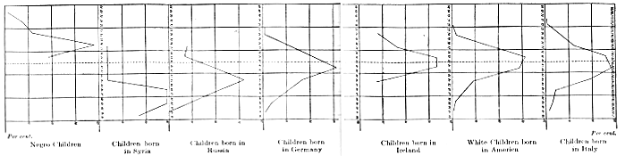
Per cent.
Negro Children
Children born in Syria
Children born in Russia
Children born in Germany
Children born in Ireland
White Children born in America
Children born in Italy
Fig. 74.
We owe to Dr. Ales Hrdlicka the extremely important graphic chart, which I will proceed to summarise, of the cephalic indices of children of various races: the central dotted line corresponds to the index 80: consequently the brachycephalics are indicated on the right, and the dolichocephalics on the left (Fig. 74).
In the case of Italy, the graphic line extends between the two extreme figures of 70 and 90, which are precisely the extreme limits that we have already noted for individual adults, in the case of the women of Latium: moreover, the curve is perceptibly symmetrical, although the brachycephalics are in the majority; a fact already established by Livi's mean averages. One might say that this curve was a graphic representation of Livi's two-colour method in his map of the cephalic index: one-half of Italy is brachycephalic and the other half is dolichocephalic; but since brachycephaly prevails in the northern half, a wider extent of territory is occupied by brachycephalics.
In America, where emigration brings every variety of humanity, the curve is even more symmetrical, and rests on a broader basis, representing widely separated extremes. Ireland also shows a very perceptible symmetry, the population being a mixture of Celts (brachycephalics) and of Scotch (northern blond dolichocephalics).
In Germany there is a prevalence of brachycephalics; we are here approaching the eastern regions from which the Eurasian race came through emigration. Here the Slavs and Celts (brachycephalics who immigrated into Europe at various epochs) are intermingled with a notable percentage of dolichocephalics (Teutons).
But in Russia, a region still further east, and similarly in Syria, we find an almost pure race: the curves lie wholly within the field of brachycephaly.
On the contrary, the dark-skinned children given in the last chart, and belonging to African races and tribes of American Indians, are all of them dolichocephalic.
According to Binet and other writers, the cephalic index and the cranial volume are the two anthropological data on which the criterion of normality of children's heads must be based.
When we observe a child's head which is apparently malformed, we cannot call it abnormal; it is not abnormal unless it has a volume notably too small (submicrocephaly, microcephaly) or too large (rickets, hydrocephaly); and a cephalic index exceeding the normal limits, in other words, exaggerated (scaphocephaly, trochocephaly, pathological brachycephaly occurring in hydrocephalics).
The Volume of the Cranium
The volume of the cranium owes its importance, as we have already seen, to the fact that the cranium represents the envelope of the brain, and is consequently normally determined, as regards its dimensions, by the cerebral volume. Accordingly, in normal cases, when we speak of the cranial volume, we are speaking by implication of the cerebral volume; and all anthropological questions regarding the volumetric development of the cranium in reality have reference to the brain.
In abnormal cases, on the contrary, it may happen that the bony covering is not a skeletal index of the brain; in fact, pathological cases may occur analogous to those we have already observed in discussing the etiology of cranial malformations, in which the flat bones of the cranial vault undergo a notable thickening, so that as a result the greater volume of the cranium is due to the increased quantity of bony substance, and not of brain tissue, and is very heavy, so that it readily droops over upon the shoulder: pachycephalic cranium.
Another cause for lack of correspondence between the cerebral and the cranial volume may be the abnormal production of cerebro-spinal fluid within the brain: hydrocephalic cranium.
The Development of the Brain.—In the earliest period of embryonal life, the brain consists of a single vesicle, the continuation of which forms the spinal marrow: later on, this vesicle divides into three superimposed vesicles which represent respectively the embryonal beginnings of the anterior, middle and posterior brain; continuing their development, the anterior and posterior brains each divide in turn into two other vesicles, so that there result in all five primitive vesicles of the brain, superimposed one upon another (see Fig. 75); the anterior vesicle which is destined to grow enormously, dividing into two parts, right and left, with a longitudinal division, will constitute the cerebral hemispheres; the second vesicle will constitute the optic thalami; the third vesicle, the corpora quadrigemina; the fourth vesicle, the cerebellum, and the fifth vesicle, the medulla oblongata.
When complete development is attained, the cerebral hemispheres completely cover the other parts of the brain, besides which they themselves are covered over with a multiplicity of folds constituting the convolutions. If we take a cross-section of the hemispheres, we find that they consist of an outer layer of gray matter formed of nerve cells, and of a central mass of white matter, formed of fibres.
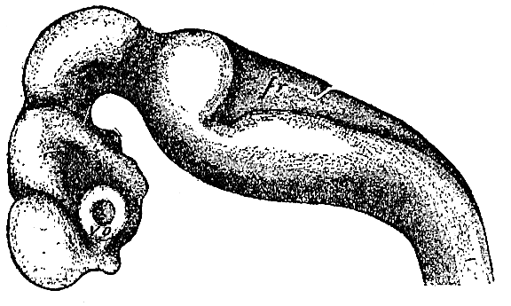
Fig. 75. Brain of a Human Embryo after the Fourth Week.
The study of the convolutions is quite important from the anthropological standpoint, because their number is not identical in the different branches of the human race, and also because they differ both in number and in arrangement from the convolutions in the brain of the anthropoid apes. But however interesting they may be, considered as differentiating characteristics, we cannot linger over a study of this kind, which has a purely theoretic importance, and for the present cannot be applied in any practical and direct way to our problems of pedagogic anthropology. It will be sufficient to note rapidly that at the present time the study of the convolutions has received a new impulse through the labours of certain distinguished investigators, among whom we must once more include Dr. Sergio Sergi. Instead of studying the surface convolutions, Dr. Sergi studies the internal folds which are disclosed by separating the lips of the cerebral fissures; and from these he draws deductions which to a large extent correct those made by previous scientists, in regard to the eventual ancestry of the different species, the marks of biological superiority or inferiority, the differences in the brain due to sex, etc.
The surface fissures which divide the cerebral hemispheres into convolutions are shown in the two accompanying figures (Figs. 76 and 77), the first of which shows the outer side of the hemispheres, and the second the inner side.
Of chief importance to us is the arrangement of convolutions and furrows on the outer surface of the hemispheres.
The points to be noted are the following: the two great fissures, Rolando's, running longitudinally, and Silvius's running transversely, which, together with the perpendicular fissure, divide the hemisphere into four lobes: the frontal lobe and the parietal lobe, situated respectively in front and behind Rolando's fissure; the temporal lobe, situated below Silvius's fissure, and lastly, the occipital lobe at the posterior apex of the hemisphere.
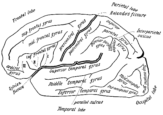
Fig. 76.—Cerebral hemisphere; external face.
In the third frontal convolution are situated Broca's centres, which are believed to be the seat of articulate speech; while along Rolando's fissure, in the ascendant convolutions, is the locality designated by physiologists as the motor centres.
The occipital lobe is the location of the zone of sight; and the temporal lobe, that of hearing.
It is important for us to observe the volume of the brain, and therefore that of the head, in relation to the rest of the body; it is enormous in the embryo; and even at birth and during childhood the head is quite voluminous as compared with the body, as appears from the diagram in Fig. 16, in which a new-born child and an adult man are reduced to the same scale, each retaining his relative bodily proportions. In Fig. 22 a new-born child is shown in two positions: from the front and from behind; the head is very large and the cranial nodules are plainly visible. Figs. 80 and 81 represent the same child at the age of six months and a year and a half; in the first picture the head is still very large as compared with the body, and the forehead protrudes (infantile forehead); in the second, the proportion between head and body has already altered.
A knowledge of the laws governing the growth of the brain is of particular importance in relation to pedagogic anthropology.
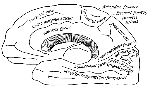
Fig. 77.—Cerebral hemisphere, internal face.
Within the last few years anthropologists have established certain principles that are well worthy of notice:
The child's head is normal when its volume and cephalic index come within the limits of normality (even if the shape appears abnormal: Simon, Binet, etc.).
When the volume of the head is too small it frequently indicates psychic deficiency; when it is too large, even up to the age of twenty years, it indicates a predisposition to precocious mortality (see below).
Very frequently when the size of the head is larger than normal and is not due to pathological causes (rickets, hydrocephaly, etc.), it is associated with an excessive development of the brain, and also with an intellectual precocity. A high percentage of this type die before reaching the age of twenty years; and this fact confirms the popular belief that children who are too intelligent or too good cannot live long.
This indication alone ought to be sufficient to prove the pedagogic importance of the cerebral volume.
The researches made by various authors in regard to the growth of the brain are not rigorously in accord as to the limits of volume: but they do agree as to the rhythm of growth.
Welcker gives the following figures:
WEIGHT OF THE BRAIN IN GRAMS (According to Welcker)
Age Males Females
At birth 400 360
Two months 540 510
One year 900 850
Three years 1,080 1,010
Ten years 1,360 1,250
Accordingly, the weight of the brain is doubled before the end of the first year; according to Massini it is very nearly doubled at the end of the first six months:
MASSINI'S FIGURES AS TO THE WEIGHT OF THE BRAIN
Age Total weight Increase
At birth 352 68 279
First month 420 211
From first to third month 631
From third to sixth month 675 44 63
From sixth month to 1 year 694 19
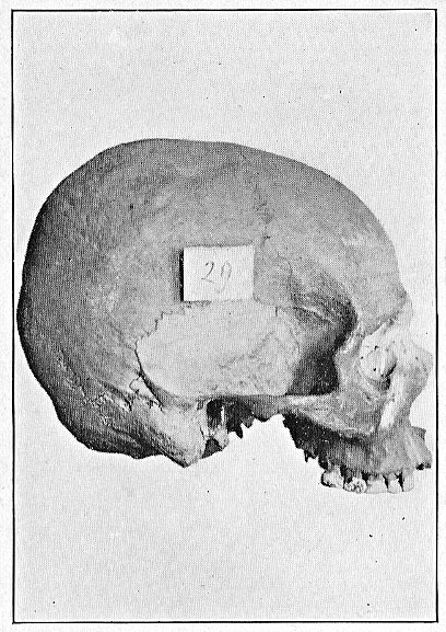
Fig. 78.—Spheroidal cranium lateral norm (Sergi's collection).
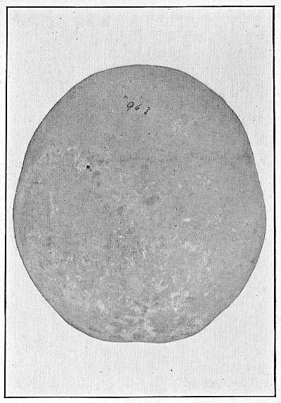
Fig. 79.—Spheroids typicus (from Sergi's collection).
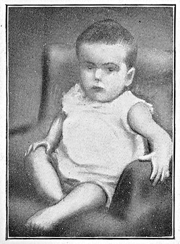
Fig. 80.—A child six months old.
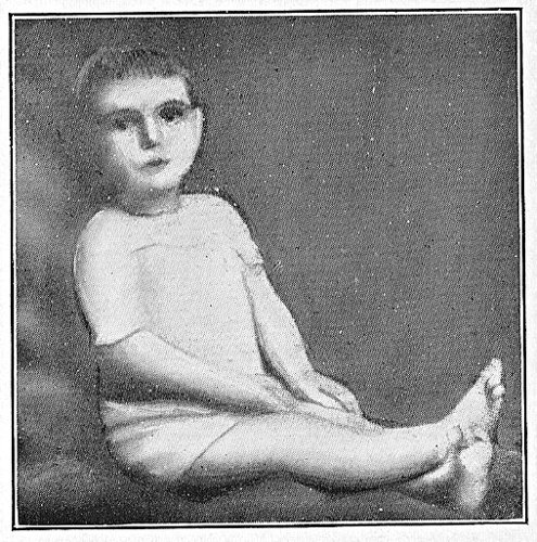
Fig. 81.—The same child a year and a half old.
It follows from these figures that by the end of the sixth month the weight of the brain is already very nearly doubled; but the maximum growth takes place between the ages of one month and three, after which it shows a notable diminution of rate.
But while the weight of the whole body is increased threefold by the end of the first year, that of the brain is very far from being tripled, since the rate of growth is still further diminished during the second six months; in fact even according to Welcker the weight at the end of the first year has little more than doubled.
Accordingly the rhythm of cerebral growth is not identical with that of the increase in weight of the body taken as a whole.
According to Massini, the relation between the cerebral weight and the weight of the body, at the various successive ages, is as follows:
RELATION BETWEEN WEIGHT OF BRAIN AND TOTAL WEIGHT (According to Massini)
Age Brain Body Age Brain Body
At birth 1 8 2 years 1 15
First month 1 9 3 years 1 14
From first to third month 1 9
to sixth month 1 10
one year 1 12 25 years 1 40
In other words, the body grows more rapidly than the brain, and consequently, than the head: a fact which results in the different proportions already noted between head and body.
The rhythm of brain growth considered by itself has been set forth in a most noteworthy and accurate fashion by Boyd, based on the study of about two thousand cases; from the figures given by Boyd, I have calculated the amount of increase from period to period, as well as from year to year, the whole result being set forth in the following table:
RHYTHM OF GROWTH OF BRAIN (Males: According to Boyd)
Age Weight in grams Difference for each period Difference for each year Relative epoch Proportion to maximum reduced to 100
At birth 331 — — — 24.2
From birth to 3 months 493 +162 — — 36.0
From 3 to 6 months 603 +110 — — 44.1
From 6 months to 1 year 777 +174 +446 1st year 56.8
From 1 to 2 years 942 +165 +165 2d year 69.0
From 2 to 4 years 1,097 +155 +77 2d- 4th 80.4
From 4 to 7 years 1,140 +43 +14 4th- 7th 83.4
From 7 to 14 years 1,302 +162 +23 7th-14th 95.3
From 14 to 20 years 1,374 +72 +12 14th-20th 100.5
From 20 to 30 years 1,357 — — — 99.3
From 30 to 40 years 1,366 +9 +0.9 30th-40th 99.3
From 40 to 50 years 1,352 -14 -1.4 40th-50th 98.9
From 50 to 60 years 1,343 -9 -0.9 50th-60th 98.3
From 60 to 70 years 1,315 -28 -2.8 60th-70th 96.9
From 70 to 80 years 1,289 -26 -2.6 70th-80th 95.3
From 80 to 90 years 1,284 -5 -0.5 80th-90th 94.2
In the above table, the first column of figures gives the mean average weight of the brain, obtained by direct observation of individual subjects; while from all the others the rhythm of cerebral growth and involution throughout the successive periods of life may be computed.
We see that the maximum growth takes place in the first years of life, the intensity is greater in the first year than in the second, and greater in the first three months than in those that follow. Already at the end of the first year the brain has surpassed one-half of the maximum weight which the individual is destined to attain in adult life (last column: proportions computed on scale of 100). A notable rate of increase continues up to the age of four, after which it moderates, but receives a new impulse at about the fourteenth year (period of puberty); hence it appears that at this important epoch of life the brain not only shares the general rapid growth of the body, but that by the end of the fourteenth year the brain has already practically completed its development; in fact, assuming that 100 represents its complete development, the weight of the brain is already 95.3; and at thirty it will be only 99.3.
By studying the above table we can obtain a clear analysis of these phenomena.
For women, Boyd gives the following figures:
THE GROWTH OF THE BRAIN IN WOMEN (Figures Given by Boyd)
Age Weight Proportion to the maximum reduced to 100
At birth 283 22.8
Three months 452 36.5
From 3 to 6 months 560 45.2
From 6 months to 1 year 728 58.8
From 1 to 2 years 844 68.1
From 2 to 4 years 991 80.8
From 4 to 7 years 1,136 91.7
From 7 to 14 years 1,155 93.3
From 14 to 20 years 1,244 100.4
From 20 to 30 years 1,238 100.0
From 30 to 40 years 1,218 98.3
From 40 to 50 years 1,213 97.9
From 50 to 60 years 1,221 98.2
From 60 to 70 years 1,207 97.4
From 70 to 80 years 1,167 94.2
From 80 to 90 years 1,125 90.8
The rhythm of growth of the female brain is analogous to that of the male, except for the more precocious attainment of the maximum weight, which corresponds to the more precocious evolution of the female organism.
It should be noted that in the tables above cited the maximum is actually given as occurring at the age of twenty; and that after this period the weight diminishes again, subsequently increasing up to an age that varies according to the sex. But this maximum at the age of twenty must be considered as one of the false results of mean averages; and it must be explained on the ground that after the twentieth year the death rate has eliminated a series of individuals whose heads were abnormally large, and that a majority of the survivors were those whose heads had developed within normal limits.
This fact is further confirmed by Wagner's figures, cited by Broca:
MEAN WEIGHT OF THE BRAIN (According TO Wagner)
Age Men Women
Under 10 years 985 1,033
From 11 to 20 years 1,465 1,285
From 21 to 30 years 1,341 1,249
From 31 to 40 years 1,410 1,262
From 41 to 50 years 1,391 1,261
From 51 to 60 years 1,341 1,236
Above 60 years 1,326 1,203
Here again we have a false maximum at twenty, which nature subsequently corrects through mortality.
From such knowledge we obtain certain important rules of hygiene.
The normal brain which exceeds the common limits of volume is not, in an absolute sense, incompatible with life. We need only to call to mind certain men of genius who had the brains of a giant.
Accordingly a brain which exceeds the limits demands of the individual who possesses it that he shall live according to certain special rules of hygiene. Children and young people who are too intelligent, too good, in other words, children of the elite class demand a special treatment, just as much as any other class of beings that pass beyond the bounds of average normality. Parents and teachers ought to be enlightened in regard to these scientific principles; the growth of individuals who are exceptional in regard to their intelligence and their emotions, should be supervised as though it were something precious and fragile. Such individuals are destined to be more subject than others to infective maladies, which frequently prove fatal, developing symptoms of meningitis and cerebral affections. Consequently a hygienic life, psychic repose, an avoidance of emotional excitement, moderate physical exercise in farm or garden, a prolonged stay in the open country, might be the salvation of children of this type, who often are over-praised and over-stimulated by friends and relatives, and consequently subjected to continual excitement and surménage to a degree destructive to their health.
Extreme Individual Variations of the Volume of the Brain.—In regard to individual variations, the authorities give various figures, from which the following have been selected as most noteworthy for their accuracy of research:
NORMAL EXTREMES OF INDIVIDUAL VARIATIONS IN THE VOLUME OF THE BRAIN
Authors Age: from 20 to 60 years From 60 to 90
Maximum Minimum Maximum Minimum
Calori 1,542 1,024 1,485 1,080
Bischoff 1,678 1,069 1,665 1,080
Without distinction of age:
Broca Maximum 1,830 Minimum 1,049
These figures refer to individuals belonging to European races.
Comparison with the Brains of Apes.—The brain of the great anthropoid apes (Chimpanzee, Orang-utan, Gorilla), whose total weight of body is comparable to that of man, weighs on an average 360 grams, and the greatest weight which it can attain is 420 gr.
Specific Gravity of the Human Brain.—In normal individuals, the average specific gravity is 1.03; in insane persons it is slightly higher: 1.04.
The Relation between the Weight of the Brain and the Cranial Capacity: Figures given by Lebon:
Weight of the brain in grams Cranial capacity in cubic centimetres
1,450 1,650
1,350 1,550
1,250 1,450
1,150 1,350
Figures given by Manouvrier:
Weight of the brain in grams Cranial capacity in cubic centimetres
1,700 1,949
1,450 1,663
1,250 1,432
1,000 1,147
Increase in the Volume of the Brain.—Studies regarding the growth of the head, although not yet complete, have gone sufficiently far to give us some useful ideas. In regard to the volume in a general sense, the cranium in its growth obeys the cerebral rhythm.
We shall speak in the section on Technique of the methods of measuring the head: at present it will suffice to point out that the measurements may be made directly upon the cranium, and the cranial capacity calculated directly from the head: and that the maximum linear measurements are sufficient to indicate the volume—such measurements being the three maximum diameters, longitudinal, transverse, and vertical, and the maximum circumference. Even the forehead, as an index of the general volume of the brain, is of interest in researches relating to the volumetric growth of the head.
Regarding the growth of the several cranial dimensions, the most accurate and complete knowledge is furnished by Binet's researches among the school-children of Paris (1902).
This author has made special investigations into the rhythm of growth of the cranium and of the face, with special reference to the period of puberty. The following are the mean averages obtained by him, relative to the three diameters corresponding to the three maximum dimensions of the head:
MEAN AVERAGES OF CEPHALIC MEASUREMENTS TAKEN UPON CHILDREN OF DIFFERENT AGES (Binet: From the schools of Paris)
Measurement Kindergartens Lower primary schools Upper primary schools Normal schools
4 years 5 years 8 years 10 years 12 years 14 years 14 years 16 years 18 years
Antero-post. diameter 169.5 173.9 174.7 177.1 181.5 181.5 185.3 188.3 190.4
Transverse diameter 140.6 141.7 145 145.7 147.9 150.1 155.5 152.3 156.7
Vertical diameter 118.8 121.6 122 122.8 127.6 129.7 128.1 131.4 130.8
It is evident that these figures contain inaccuracies, especially in regard to the vertical diameter (where the subsequent two-year period gives a smaller measurement than the preceding) due to the fact that the averages were obtained from an insufficient number of subjects or from subjects differing too widely in intelligence (from schools of different grades). For this reason Binet summarises the differences in growth, that is, the increase in relation to the diameters, under broad groups (six year groups, from four to ten years, and from ten to sixteen), in order to determine whether puberty exerts a sensible influence upon the cranial growth. The result is contained in the following table:
INCREASE OF THE THREE MAXIMUM DIAMETERS OF THE HEAD IN MILLIMETRES FROM FOUR TO EIGHTEEN YEARS OF AGE
Age in years: from — to — 4-6; 6-8; 8-10 10-12; 12-14; 14-16 16-18
Antero-posterior diameter 5.6; 0.8; 2.4 4.4; 1.8; 5 2.1
8.8 11.2
Transverse diameter 1.1; 3.3; 0.7 2.2; 3.9; 0.5 4.4
5.1 6.6
Vertical diameter 2.8; 0.4; 0.8 4.8; 2.3; 2.5 0.6
4.0 9.6
From which it appears that there exists, in regard to the head, a puberal acceleration of growth.
These conclusions of Binet are indirectly confirmed by the researches of Vitale Vitali regarding the development of the forehead in school-children; since it is well known that the forehead represents the index of the general growth of the cerebral cranium.
Vitale Vitali based his observations upon school-children and students between the ages of ten and twenty. He not only measured the width of the forehead (frontal diameter; see Technique), but also measured its height, obtaining the percentage of its relation to the width (frontal index).
These are his figures:
FRONTAL INDEX AND DIAMETER ACCORDING TO AGE
(Vitale Vitali: Researches Among Scholars and Students From 10 To 20 Years Old)
Age Frontal index Frontal diameter Amount of increase
11 years 73.05 107.5 —
12 years 74.11 112.0 4.5
13 years 74.14 112.5 0.5
14 years 74.80 114.4 1.9
15 years 75.67 116.8 2.4
16 years 77.24 120.1 3.3
17 years 77.02 120.6 0.5
18 years 77.36 121.5 0.9
19 years 77.60 122.8 1.3
20 years 77.15 122.1 0.7
Accordingly, between the years of fourteen and sixteen there is a puberal acceleration of growth, accompanied by an elevation of the forehead (high frontal index).
Vitali gives, as extreme limits of the frontal index, 68 and 83.
But in order to give a better illustration of the author's figures, his own words may be quoted: "It appears from our observations that the forehead begins to develop in notable proportions during the fourteenth year, and that the development of the frontal region as compared with the parietal region continues to augment up to the sixteenth year; after this it still increases, but only by a few millimetres, until the end of the sixteenth year. The cephalic development is completed between the sixteenth and eighteenth years. This observed fact is of great importance in relation to the development of the intellect."
The most complete figures at the present time on the growth of the brain, are those of Quétélet, which follow its development from birth until the fortieth year. They are summarised in the following table:
INCREASE IN THE CIRCUMFERENCE OF THE BRAIN AND IN ITS THREE MAXIMUM DIAMETERS (According to Quétélet)
Age Circumference in millimetres Maximum diameters
Antero-post. Transverse Vertical
Men Women Men Women Men Women Men Women
At birth 335 335 120 120 100 100 80 80
1 year 440 439 158 157 127 126 105 105
2 years 471 469 168 167 135 134 113 113
3 years 486 483 171 170 137 136 117 115
4 years 496 493 174 173 138 137 119 116
5 years 503 500 176 175 139 138 120 117
6 years 508 505 178 177 140 139 121 117
7 years 513 509 179 178 142 140 122 118
8 years 519 512 180 179 143 141 123 118
9 years 523 515 181 180 144 141 124 119
10 years 527 517 182 180 145 142 125 119
11 years 531 518 183 181 146 142 126 120
12 years 535 519 184 181 147 143 127 121
13 years 539 520 185 182 147 143 128 122
14 years 543 521 186 182 148 144 129 123
15 years 547 523 186 183 149 144 130 124
16 years 551 525 187 183 150 145 130 125
17 years 555 528 188 184 151 145 130 125
18 years 561 531 189 184 152 146 131 126
19 years 563 533 190 185 153 146 131 126
20 years 564 535 191 185 153 147 131 126
25 years 564 537 191 186 153 147 131 127
30 years 564 538 191 186 153 147 131 127
40 years 564 538 191 186 153 147 131 127
It appears from the foregoing table that after the twenty-fifth year the growth of the cranium practically ceases in all directions. In regard to the rhythm of growth, the problem is rendered clearer by the following table, which gives the annual increase:
ANNUAL INCREASE IN THE MAXIMUM CRANIAL MEASUREMENTS IN MALES (From Figures Given by Quétélet)
Age Circumference Antero-post. diameter Transverse diameter Vertical diameter
1 105 38 27 25
2 31 10 8 8
3 15 3 2 4
4 10 3 1 2
5 7 2 2 1
6 5 2 1 1
7 5 1 1 1
8 6 1 1 1
9 4 1 1 1
10 4 1 1 1
11 4 1 1 1
12 4 1 1 1
13 4 1 1 1
14 4 1 1 1
15 4 1 1 1
16 4 1 1 1
17 4 1 1 1
18 4 1 1 1
19 4 1 1 1
20 1 1 1 1
It appears from the above table that the total growth of the cranium takes place to a notable extent during the early years of life; as regards the diameters, the longitudinal diameter grows faster during the first few months than the transverse; but after the first year, the two maximum diameters which determine the cephalic index increase in very nearly the same proportion (constancy of the cephalic index throughout life). The vertical diameter on the contrary undergoes a relatively much greater increase than the two others, since, although much shorter than the transverse, it nevertheless overtakes and surpasses it in its absolute annual increase.
This corresponds to the fact that the first two diameters are indexes of growth relative to the base of the cranium, while the vertical diameter is the index of expansion of the cranial vault, which more directly follows the growth of the brain and elevates the forehead as it pushes upward.
Quétélet's figures, however, fail to show in the rhythm of growth that puberal acceleration which has been observed to take place in the growth of the brain. This contradicts the researches of Vitali and also those of Binet.
Similar studies have been made a number of times during the last few years, especially in America, but with English tables of measurement, and with little uniformity in the results obtained by the different investigators.
Among the most recent and most complete figures should be cited those of Bonnifay[38] in which however the measurement of the vertical diameter is lacking, or in other words the third element needed, in conjunction with the dimensions of length and breadth, to give the volumetric factors.
CRANIAL MEASUREMENTS AT DIFFERENT AGES (According to Bonnifay)
Age from — to — Absolute figures Amount of Increase
Circumference Antero-posterior diameter Transverse diameter Circumference Antero-posterior diameter Transverse diameter
Birth to 15 days 343.9 116.3 93.4 — — —
15 days to 2 months 368.7 126.3 99.1 24.8 10.0 5.7
3 months to 4 months 388.8 132.7 106.0 20.1 6.4 6.9
6 months to 1 year 429.8 145.4 118.2 41.0 12.7 12.2
1 year to 2 years 459.7 154.3 129.3 29.9 8.9 11.1
2 years to 3 years 473.5 161.9 133.3 13.8 7.6 4.0
3 years to 4 years 487.4 166.2 136.3 13.9 4.3 3.0
4 years to 5 years 495.7 169.9 138.3 8.3 3.7 2.0
5 years to 6 years 497.8 171.9 140.4 2.1 2.0 2.1
6 years to 7 years 504.4 172.8 141.1 6.6 0.9 0.7
7 years to 8 years 511.6 175.2 143.7 7.2 2.4 2.6
8 years to 9 years 514.1 176.1 144.3 2.5 0.9 0.6
9 years to 10 years 514.7 176.4 144.2 0.6 0.3 0.9
10 years to 11 years 519.8 177.1 146.6 5.1 0.7 2.3
11 years to 12 years 521.1 177.5 145.7 1.3 0.4 0.1
12 years to 13 years 529.7 180.1 147.8 8.6 2.6 1.2
13 years to 14 years 533.1 178.1 148.5 3.4 — 0.7
14 years to 17 years 548.8 182.4 152.2 15.7 2.3 3.7
22 years to 27 years 549.1 186.6 153.2 0.3 4.2 1.0
Among the linear measurements of the cranium, the one which serves to give the most exact index of volume is the maximum circumference.
This index, nevertheless, is not a perfect one, in the same sense that the stature, for instance, is a perfect index in respect to the body, because in the case of the cranium another element enters in: the form. The cranial circumference of an extremely brachycephalic cranium (almost circular) may contain a larger surface (and consequently include a larger volume), than a maximum circumference of the same identical measure, which belongs to an extremely dolichocephalic cranium (approaching the shape of an elongated ellipse). This may be easily understood if we imagine a loop of thread laid out in the form of a circle: if we pull it from two opposite sides, the enclosed area diminishes until it finally disappears as the two halves of the thread close together, while the length of the thread itself remains unaltered.