Topographic Anatomy.
Reading edition
A close study of the lesions of the rectum and their manifestations and some experience in discussion of these subjects have convinced me of the necessity of methods of greater accuracy than those generally employed in designating the precise situation of a pathologic feature in this organ. Mensural methods of designating the situation of strictures in the rectum are of no surgical value.
The manner of the application of the details in a given method of treatment for a disease situated in the fixed or anal portion of the rectum should differ essentially from that employed in the application of the same principles of treatment to a similar disease situated in the movable abdominal rectum; and the application of the details of a given kind of treatment should differ, too, according to the situation of the disease at one point or another in the circumference of the rectum. The prognosis as well as the treatment of rectal disease is determined not only by the pathologic character of the lesion, but also by its anatomic situation. “Two inches up,” or “one and a half inches from the anus,”[15] if one were positively sure of the precise location referred to as the anus, would point to very different anatomic parts, whether the subject were male or female, and whether young or old, and whether thin or stout.
The rectum, the terminal 8 or 10 inches (20.32 or 25.40 cm.) approximately of the intestine, presents an upper abdominal and essentially movable portion, which is about three-fourths the length of the entire rectum, and a lower coccygeoischioperineal and essentially fixed anal portion, which is about one-fourth the length of the rectum. The upper half of the movable rectum is, in the majority of adult persons, completely invested with peritoneum, which sometimes provides this portion with a mesentery. The lower half of the movable rectum begins at a point about the upper border of the second sacral bone, and is not completely invested with peritoneum; the peritoneum is reflected from the sides of the rectum toward the lateral masses of the sacrum, so that the posterior wall of this part is not covered by peritoneum. The movable rectum begins opposite the sacroiliac synchondrosis. If a subject lies in the dorsal posture, and if the abdomen be normal, the upper end of the rectum will lie opposite the left sacroiliac synchondrosis. If the patient is inverted to the knee-chest posture or its equivalent, and if the abdomen is normal, the upper end of the rectum, if distended, is usually found nearer the right sacroiliac synchondrosis. The movable abdominal rectum terminates at the levator ani muscle.
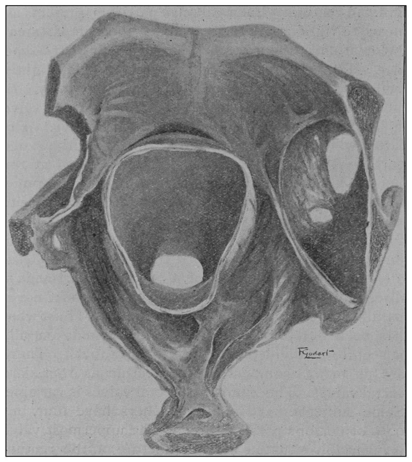
Fig. 19.
—Drawing of specimen No. 281 in the Anatomical Museum of the Royal College of Surgeons, London. The dissection was made by Mr. William Pearson. The drawing shows in the lower part of the field the coccyx, in the upper part of the field the pubes.
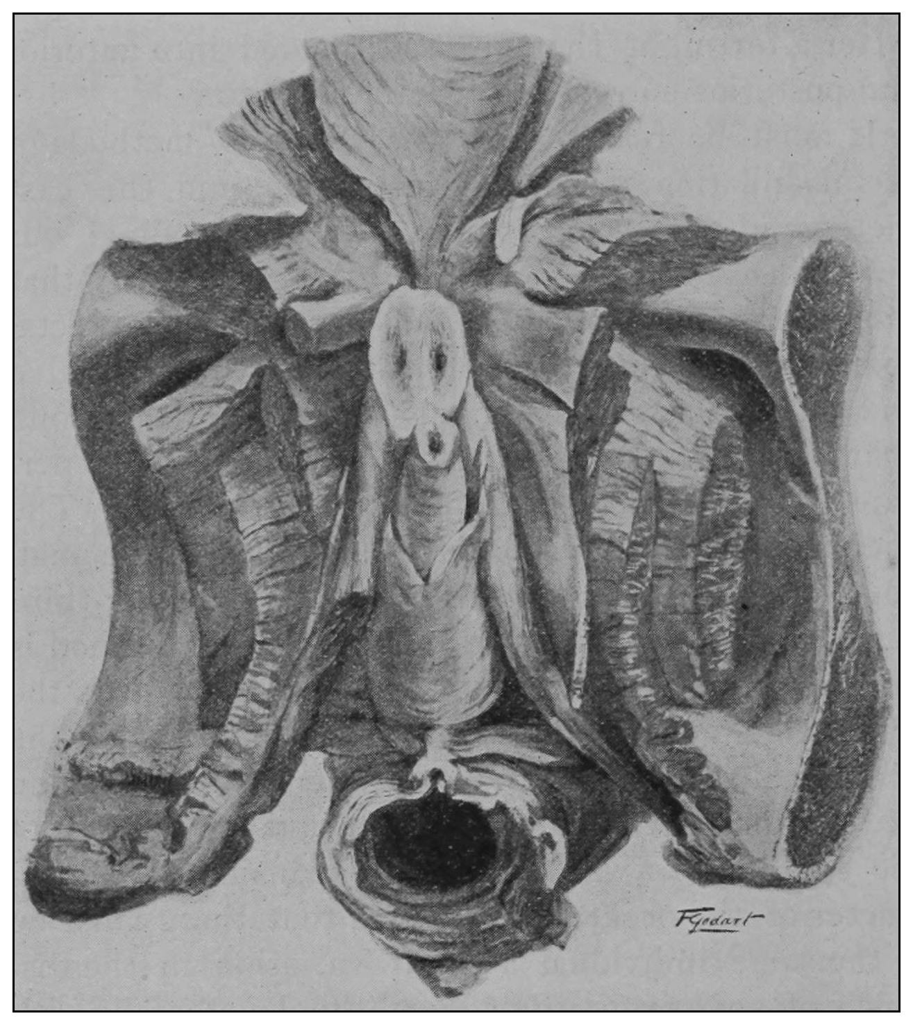
Fig. 20.
—Drawing of specimen No. 284 in the Anatomical Museum of the Royal College of Surgeons, London. The dissection was made by Mr. William Pearson. The drawing shows in the lower part of the field behind the rectum the coccyx, in the upper part of the field the pubes.
The fixed anal rectum begins at the levator ani and coccygeus muscles. The levator ani has its origin at the sides of the bodies of the pubic bones, the coccygeus at the spines of the ischii, and the levator ani has additional origin from the fascia and bony parts on a line between these two points. The fibers of these muscles are directed downward and inward to the fixed anal rectum; many fibers are blended into the contiguous structures of the pelvic floor. The coccygeolevator muscles may be compared to an opened slat-fan, the apex having its place at the anus, and its long border representing the line of origin of the muscles, Fig. 19. The ental sphincter ani muscle, situated a few lines below the levator ani, is made up of an aggregation of the fibers of the circular non-striated muscular intestinal coat, Fig. 20. The ectal sphincter ani striated muscle is situated immediately beneath the external skin. It serves the present occasion to describe the ectal sphincter of the male as a loop of muscle thrown about the terminal end of the rectum and hitched to the terminal bone of the coccyx, and in the female as a longer loop of muscle twisted upon itself so as to make a tandem-loop, which, in the form of the figure eight (8), is thrown about the vagina and terminal end of the rectum.
In the passive subject the finger discovers the ectal sphincter as a broad, relaxed band of muscle situated beneath the external skin and surrounding the infra-anal depression. In action the ectal sphincter is contracted and is retracted from beneath the external skin to a point beneath the mixed mucocutaneous integument. In such a state of ectal sphincter contraction the fixed portion of the rectum is lengthened, or in other words, the pelvic floor is deepened one-half inch (1.27 cm.), approximately. The finger progressing, it engages the tonicly contracted ental sphincter muscle which the delicate touch discovers as vibratory in its grasp of the finger, presenting to the touch the sense of a sharp rigid ring, and again as a flat band snugly applied about the finger. A few lines above the ental sphincter the finger discovers the upper limits of the fixed or anal portion of the rectum surrounded by the somewhat V- or U-shaped borders of the levator ani muscle; the anterior quadrant is not encircled by the levator ani. The fixed portion of the rectum presents, then, to the touch, three landmarks: the usually relaxed ectal sphincter, the usually contracted ental sphincter, and the levator ani muscles. The normal levator ani muscle can not by the contraction of its fibers close the upper end of the fixed rectum. It is possible, however, for the patient to contract his levator upon the finger of the examiner to quite an appreciable degree, a fact which led Mr. Harrison Cripps to state that the levator ani can close the upper end of the fixed rectum. If a tubular speculum of seven-eighths of an inch (2.22 cm.) diameter be introduced through the fixed rectum and then withdrawn through the levator ani to a point above the ental sphincter and the patient be directed to contract the muscles of the pelvic floor, it may be observed that the axis of the fixed rectum is directed more forward at its upper end, and that the depression between the levatores ani is partially encroached upon by the levator fibers but is in no sense effaced; hence, we must recognize that the lower fixed portion of the rectum presents a canal of an hour-glass form, expanded above as a concavity between the borders of the levatores ani, narrowed at its middle part by the contracted ental sphincter, and expanded below where it is surrounded by the relaxed ectal sphincter muscle.
In the same subject the length of the fixed anal rectum is variable with a state of activity or passivity, and in a state of activity there are variations in its length of at least one inch (2.54 cm.) between a contracted, uplifted pelvic floor and that of a depressed floor with anal eversion; both of which conditions may rapidly follow one upon the other while the examiner’s finger is engaged in diagnosis. Again, variations in depth of the fixed anal rectum are quite noticeably regulated by the size of the finger introduced. The thumb may find a fixed anal rectum of two inches (5.08 cm.) in depth, while the little finger discovers it but a little more than an inch (2.54 cm.). Because of the bony confines to the tissues of the ischiorectal space, displacement of its structures to open the anus must occur in the vertical direction, the larger the finger the greater the displacement upward.
Passing the finger beyond the borders of the levator ani, the distal phalanx enters the movable or abdominal rectum, where it may be hooked over the pelvic floor. In some instances, if the finger be directed backward and crowded with a boring maneuver through the loose folds of the movable rectum, and provided the folded knuckles displace upward the pelvic floor, the finger may be made to engage the lowermost of the rectal valves, which will contract about the finger with a rhythmic action and mislead the uninformed, inexperienced and undiscriminating explorer to think that he is but now encountering the ental sphincter muscle, or, perhaps, that he has discovered a stricture—the phantom stricture? In the passive rectum, this valve is usually about three inches (7.62 cm.) above the lower border of the ental sphincter muscle. The great range of mobility of the pelvic diaphragm permits a finger of two inches (5.08 cm.) length to be hooked over a valve which under some circumstances may be an inch and a half (3.81 cm.) beyond its reach.
The pelvic floor in the infant is often less than one-half inch (1.27 cm.) in depth. The depth of the pelvic floor in the adult, from the lower border of the relaxed ectal sphincter ani muscle to the levator ani muscle, is extremely variable. In the aged male, because of senile enlargement of the prostate, the fixed rectum may seem to be three inches (7.62 cm.) in depth. In the aged female, because of senile atrophy of the generative organs and contiguous structures, the pelvic floor may be much less than an inch (2.54 cm.) in depth. In the adipose and in emaciated subjects because of the character of the tissues occupying the ischiorectal space, there are the greatest variations in the depth of the pelvic floor. Hence, it is obvious that the palpable muscular landmarks of the fixed rectum are situated at variable positions in the different sexes, and that the length of the fixed rectum is changed in the same person at different periods of life and in differing conditions of flesh.
The visible topographic features of the fixed rectum are several, and under the influence of disease may become somewhat changed in appearance and situation. In the male they are readily discoverable by putting the subject in the dorsal posture, separating the nates with the hands and placing the thumbs on bits of dry cotton to prevent their slipping just exterior to the anus. Simultaneously to the patient’s bearing-down impulse the thumbs may be used to evert the anal mucous membrane. The anterior and posterior borders of the anus may in a measure be everted by a somewhat similar manipulation. In the female the visible landmarks of this region may be inspected by entering the finger into the vagina and placing the thumb over the perineum; the former is to push the rectum down while the thumb draws the external skin forward over the perineum. The posterior segment may be exposed by pushing the post anal skin toward the coccyx; the lateral segments may be exposed with the finger and thumb of the left hand while the right hand keeps up the initial pressure. Marked pigmentation of the anal skin is observable in a circumscribed area about the anus; beneath this area of darkened skin, intimately attached to the skin, lies the surgically unappreciated corrugator cutis ani. Within the borders of the everted anus the complexion fades to a light gray, within which zone is sometimes noticeable the distinctly lighter zone known as Hilton’s white line. Above, is noticeable an undulating zone of deeply red mucous membrane the lower border of which has been given the name of linea dentata (Stroud). Between the linea dentata and the white line is a zone occupied by several pyramidal elevations about half an inch (1.27 cm.) in length, to which has been given the names: columns of Morganni and pecten of Stroud. Their bases are of a somewhat purple reddish color. Toward their apices they may pale and terminate in projecting white eminences. The bases of these pyramids, which, I believe, may without impropriety be called anal pilasters, are extremely vascular and their structure partakes somewhat of the character of erectile tissue. The apex contains a nerve end-bulb. Under the influence of disease the color of these bodies is changed. Their antemortem and postmortem appearance is quite different. There are usually present in each anus from four to eight of these bodies. Between these projections and at their lower borders, sometimes, there is discoverable a thin fold of membrane. The saccule which it, together with the anal wall and pilasters on either side, forms, has been known as the saccule Horneri, pocket of Physic, and anal pocket, sometimes inaptly called rectal pocket. Its cavity is about the size of a split pea. The three typic visible topographic features of the fixed rectum, then, are the white line of Hilton, the pecten of Stroud, or anal pilasters, and the linea dentata.
The mucocutaneous membrane of which these visible landmarks are a part, rests upon a quantity of loose connective tissue, which permits of a great range of mobility of these features independent of movement of the structures constituting the palpable landmarks of the fixed or anal rectum.
Fig. 21.
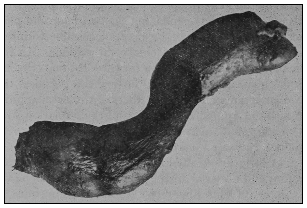
Fig. 22.
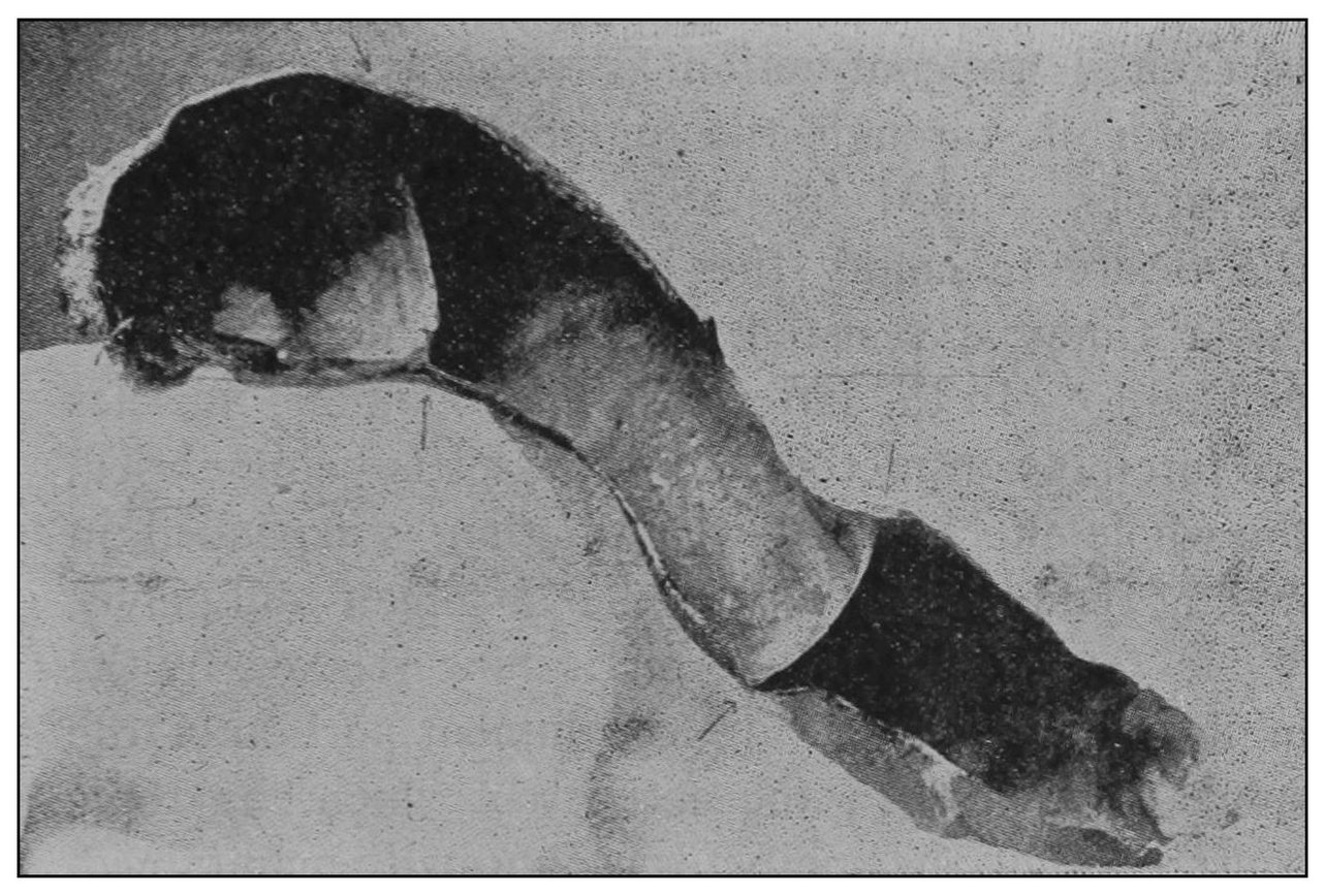
Fig. 23.
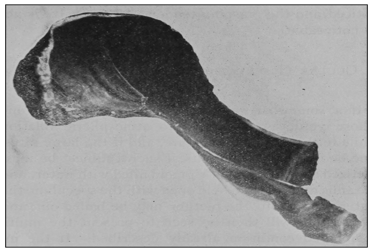
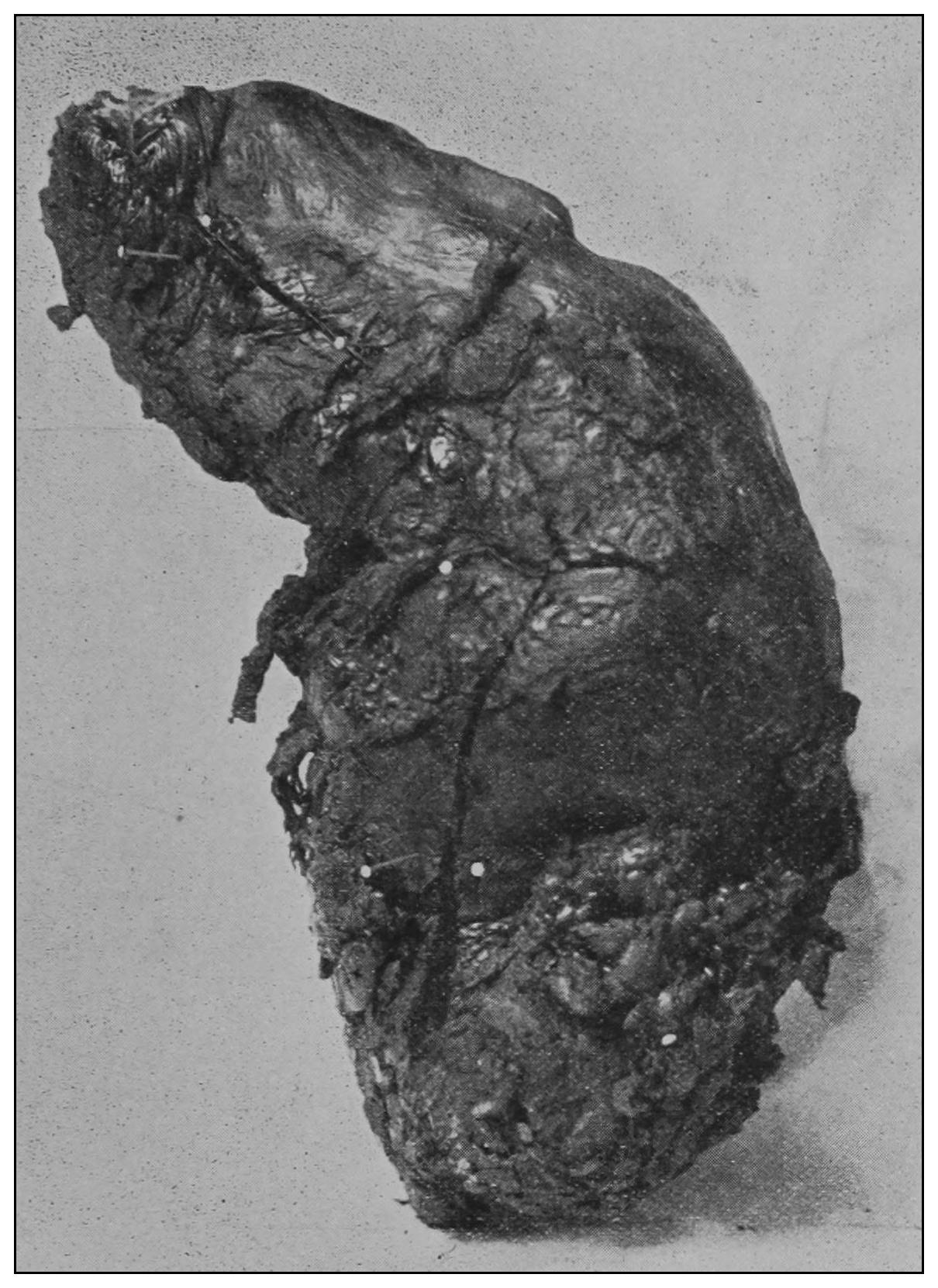
Fig. 21.—From a photograph of an external view of the paraffin cast-filled rectum. The rough surface shows the place of attachment of the peritoneum.
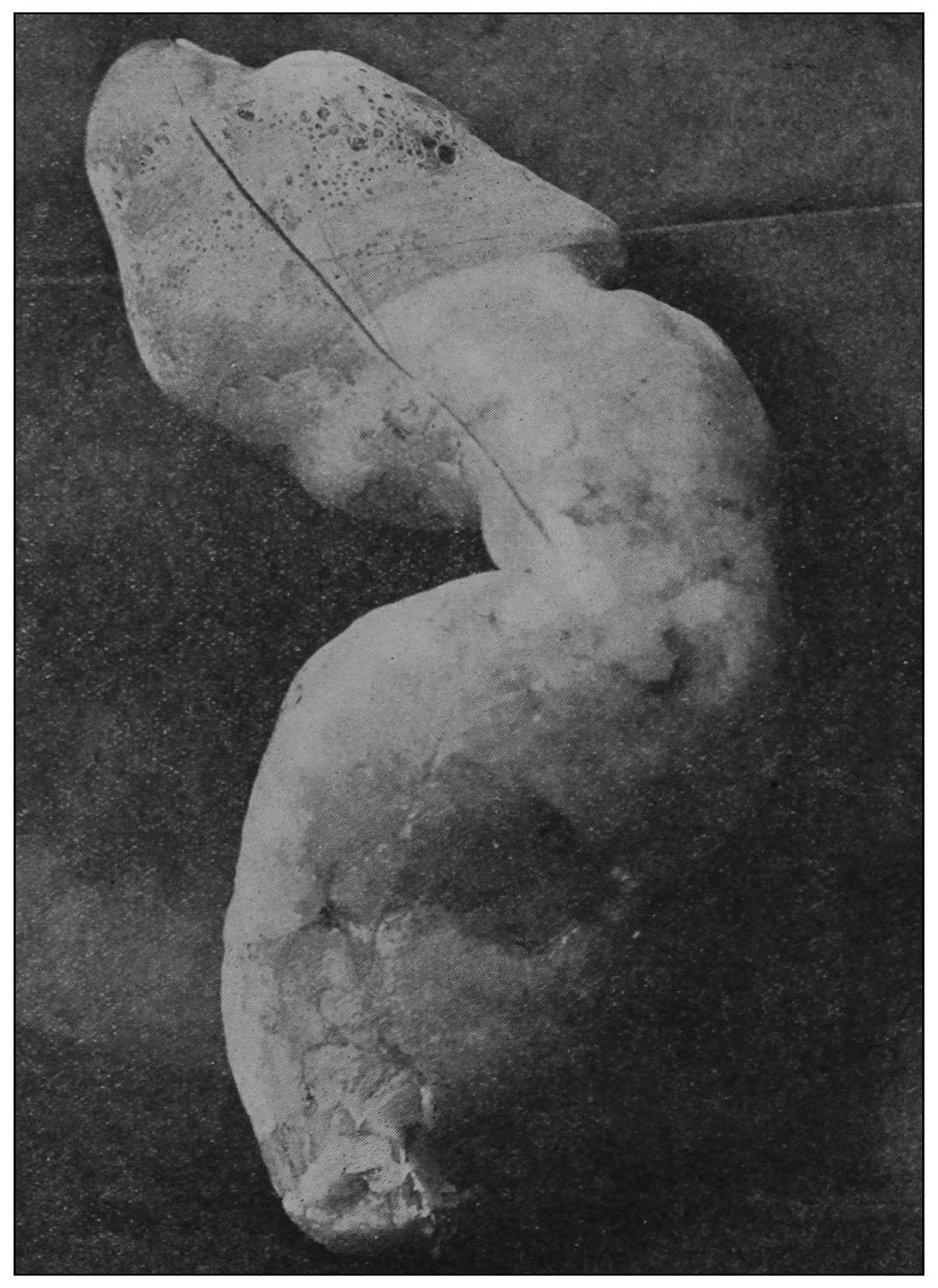
Fig. 22.—Interior view of the left half of the rectum of an adult. Filled with melted paraffin, the subject being in the proctocolonoscopic posture. The rectum is distended only to the degree of normal atmospheric inflation. When the wax was hardened the rectum was removed, varnished, dried and cut open longitudinally. The picture is a reproduction of a photograph of the interior of the left half of the rectum. The anus is at the picture’s left. The valves are respectively two and one-half and seven inches (6.35 and 17.78 cm.) from the anus.
Fig. 23.—Knee-chest posture. Left lateral half-interior view. Upper rectum and sigmoid out of focus.
Fig. 24.—Posterior view of a specimen carefully dissected to show the muscular supply to the valve-bases.
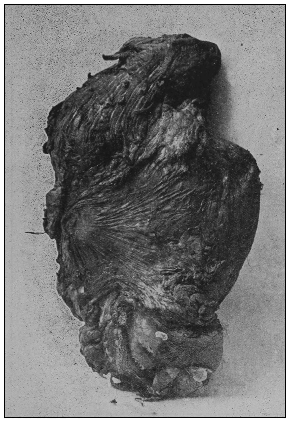
Fig. 25.—Posterior half occupied by its cast.
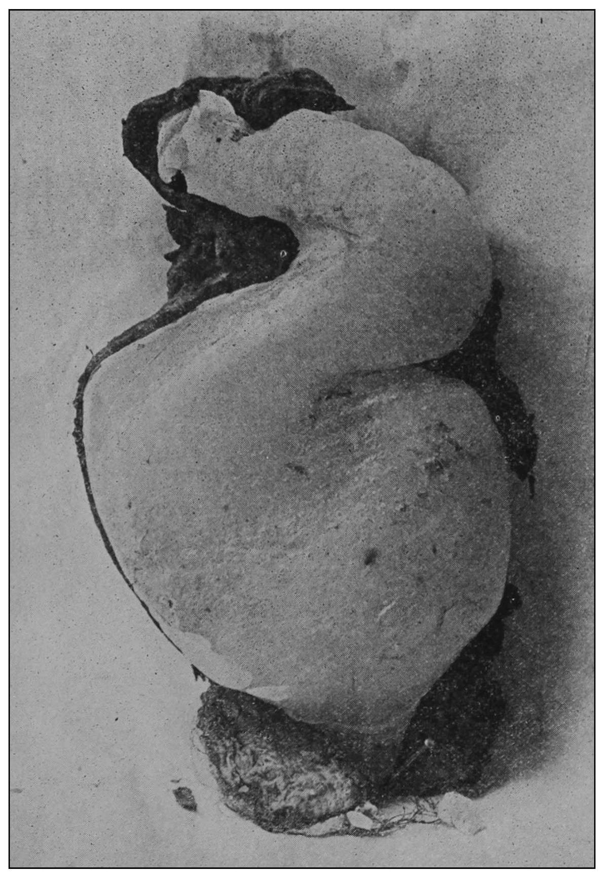
Fig. 26.—Anterior half of specimen shown in Fig. 25.
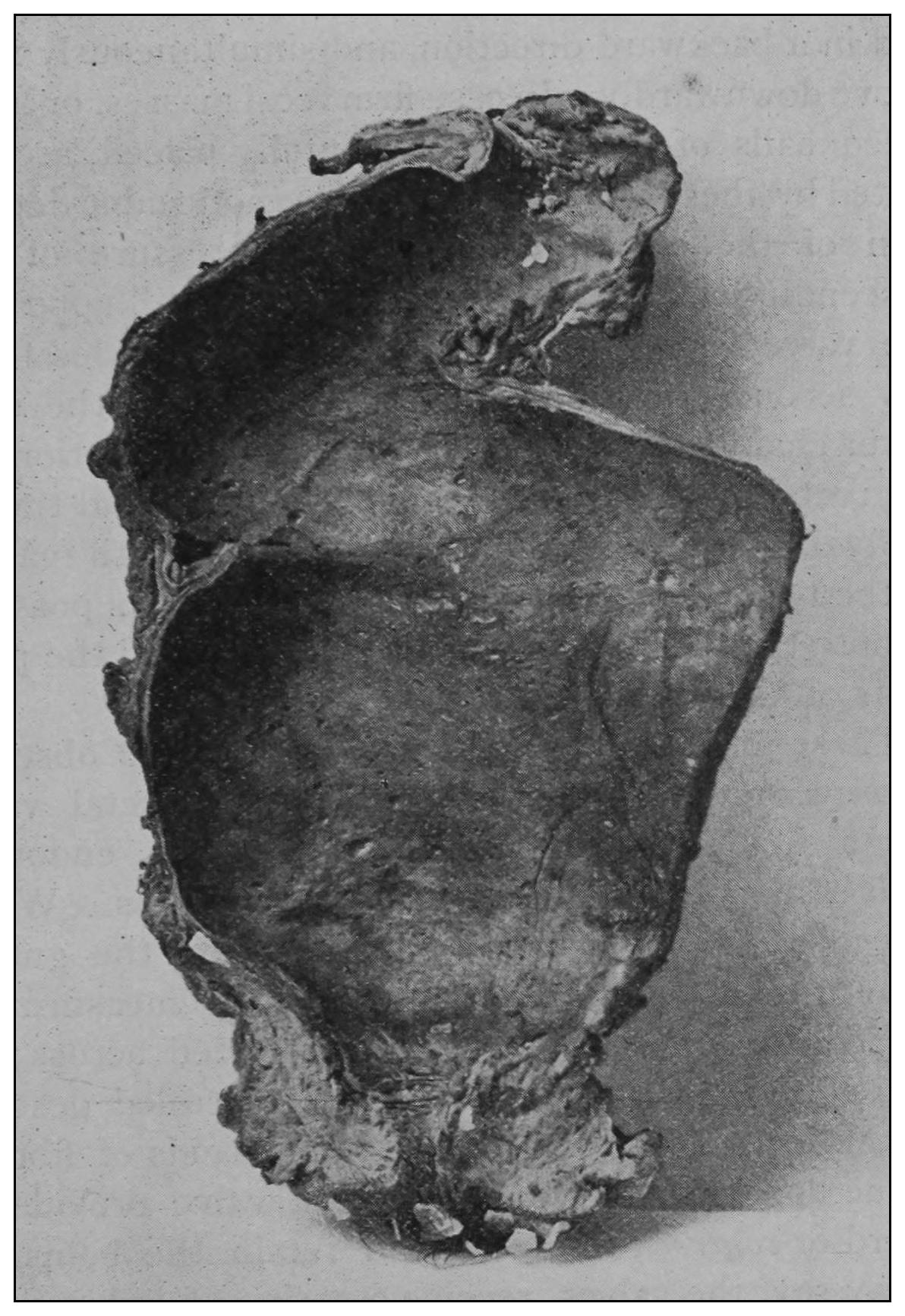
Fig. 27.—Posterior view of a cast-filled rectum. Fig. 28.—Paraffin cast from a rectum.