The Rectal Valve.
Reading edition
That the rectal valve constitutes the most hypsometric of the topographic features of the movable abdominal rectum, is conclusively proven by the photographic pictures. The specimens from which the photographs were taken were prepared by a method consisting in fixing the cadaver in the knee-chest posture and pouring melted paraffin into the atmospherically inflated rectum; when the wax had sufficiently hardened the gut was carefully removed, for a few weeks immersed in alcohol and subsequently dried, varnished, and finally dried and cut into longitudinal, shell-like halves. A comparative study of the photographs of the rectal interiors discovers the fact that the prominence of the rectal valve is increased with the degree of distention of the rectum.
The accompanying drawing was made from a microscopic section taken from the middle of the lower valve of an adult specimen. In the illustration, Fig. 33, it is shown magnified 5 diameters. Beneath the mucosa is noticeable the heavy layer of fibrous tissue which gradually diminishes till it is lost at the valve base. Bundles of circular muscular fibers are seen in the middle of the valve. At its base are seen arteries and veins for its special nutrition. This structural arrangement makes this organ the typic anatomic valve. The evidence of the fibrous tissue in the valve is an original contribution to our knowledge of this subject.
The attached border of each valve spans a little more than half the circumference of the rectum, and its free border projects half across the diameter of the inflated rectum. Each valvular partition projects at nearly a right angle to the wall of the compartment below it and terminates in a sharply defined free border. The free margin of the structure is slightly concave in form and is directed a little obliquely. In life it is noticeable that the free border of the valve is less elastic than the main wall of the valve. The latter will yield to a modicum of traction without disturbing its free border, while traction applied at the margin may at once disturb the entire circumference of the gut at that point. The rectal wall opposite the valve is usually concave. The aperture thus formed between two rectal compartments is irregularly elliptic; it may be called the valve-strait. Usually the greatest diameter of a rectal compartment is directed laterally or else obliquely; it is almost never parallel to the direction of the sacrum. Thus, what has been heretofore considered a cavernous ampulla is seen to be divided into several chambers. There are as many chambers in the rectum as there are rectal valves. The number of rectal valves is variable. Some subjects have but two, others have four, but 90% of persons possess three. The uppermost valve is invariably situated at the juncture of the rectum and the sigmoid flexure, which valve is invariably situated on the left, the next lower is on the right wall, and the lowermost is on the left. The positions of the lower two valves are sometimes anterior and posterior.
The specimen shown in Fig. 30 exhibits the rectum naturally distended with feces. It was found in a deceased member of the demimonde who had been habituated to opium indulgence. She was forty years of age and of robust stature. On opening the abdomen the colon was discovered loaded with a collection of scybalous feces, and the upper chamber of the rectum was partially occupied with a similar deposit. The abdomen was then opened through the pubes and perineum down to the coccyx. The genitourinary organs and contiguous structures were removed, the symphisis forcibly separated and the exposed sigmoid flexure and rectum were photographed as shown in Fig. 29. The sigmoid and rectum were carefully removed and immersed in alcohol. After a fortnight the gut was divided into anterior and posterior halves, as shown in Fig. 30.
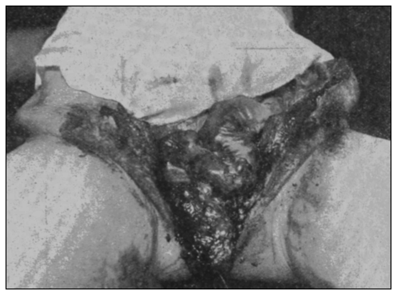
Fig. 29.
—Photograph of a female cadaver showing, after laparo-symphysiotomy and removal of bladder, uterus and adnexa, the upper rectum and sigmoid packed with scybalums.
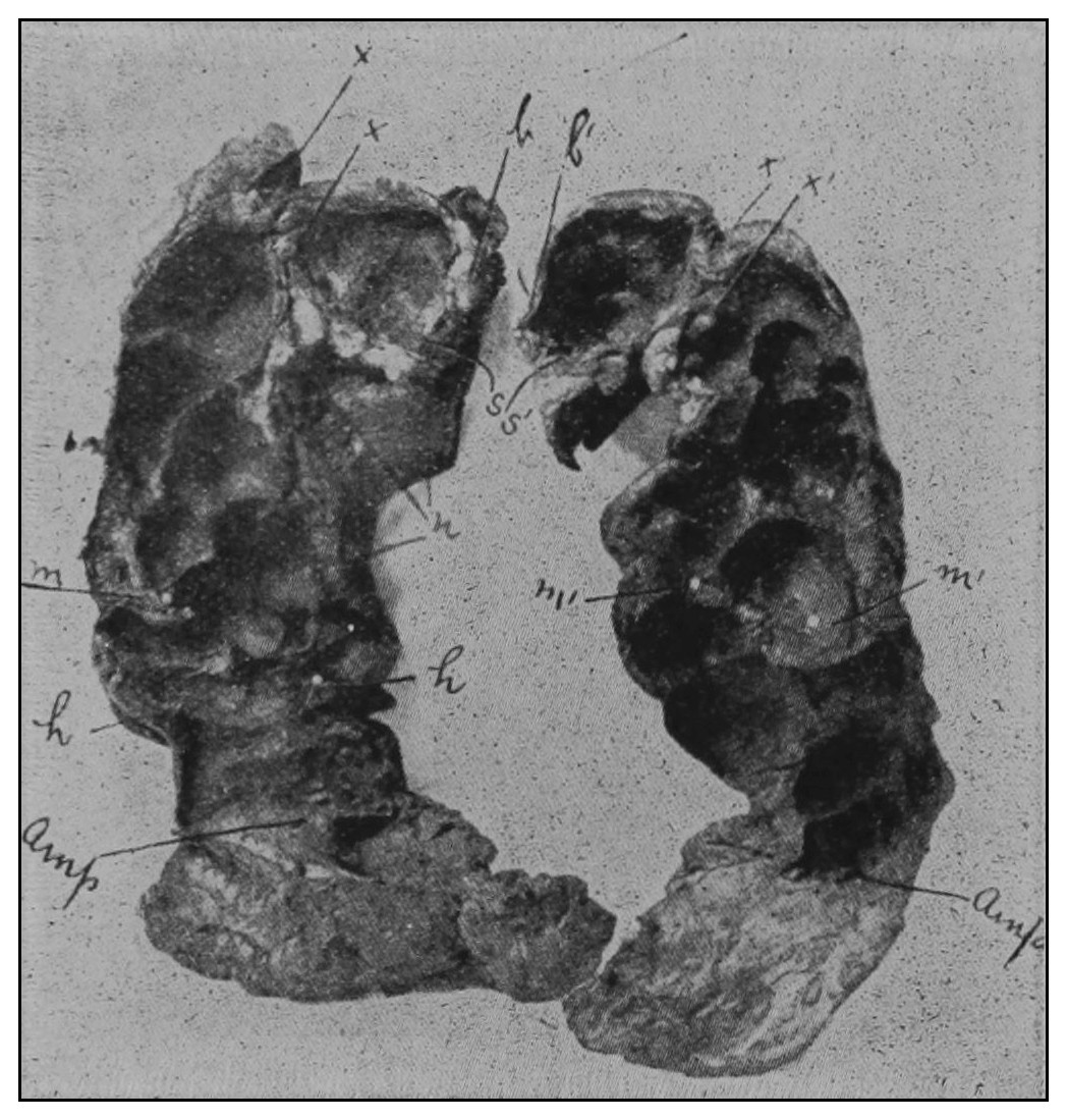
Fig. 30.
—The rectum, the same as is shown in Fig. 29, divided into anterior and posterior halves.
It must be readily seen that the new methods of rectal inflation for rectal inspection and the cast method of dissection, which have determined our newer ideas of the topography of this part, justify that the lowermost chamber be considered the first rectal chamber; the cavernous area beyond the first valve and below the second should be called the second chamber; and the upper chamber the third or perhaps fourth, according to the number of valves. The ancient arbitrary division of the rectum by the anatomists into upper first, middle second and lower third parts should be abandoned because such a method is inaccurate and has no surgical significance. As the arrangement of the fibers of the muscular coats of the abdominal rectum and the attachments of the abdominal rectum provide for extension and contraction of the gut on its axis, as well as expansion of the diameter of the organ, it is obvious that there must be in the same individual a great variation in the distance of any given valve from the levator ani with the variable normal states of the organ. The normal range of movement upward and downward of a given valve is from two to three inches (5.08 to 7.62 cm.).
A discriminating study of the diseases of the rectum requires an arbitrary division of the organ into several quadrants.
Regard for the conveniences of scientific description and considerations of technic and of anatomy justify that the fixed anal rectum be called the anus, and that the movable abdominal rectum be called the rectum. The former is bounded above by the levator ani and below by the ectal sphincter muscle.
From this survey of the topography of the anus and rectum and consideration of the arrangement of the contiguous structures the following salient aphorisms may be deduced:
1. In treating lesions on a level with the sphincter muscles the operator should beware of dividing these structures through the anterior quadrant. In the male the ectal sphincter terminates in the tendinous raphe in common with the transversus perinei. Contraction of the transversus perinei will separate the cut fibers of the ectal sphincter and defeat the desired subsequent union of this muscle. If an incision be carried forward or forward and laterally into the transversus perinei the perineal fascia which doubles over this muscle will be opened and the perineum and peri-urethral structures will be made accessible to infection from the anus and ischiorectal regions. In the female, to carry an incision forward through the anterior quadrant would be unsurgical because the peculiar arrangement of the fibers of the ectal sphincter ani and sphincter vaginae and their relation with the transversus perinei would perhaps conspire to produce vulvoanal or rectovaginal fistulas.
2. A stricture located at the upper end of the fixed rectum and situated in the anal fascia, in the pelvic fascia, or in the fibers of the levator ani muscle, should not be cut in the anterior quadrant nor in the posterior quadrant, but in one or the other, or both, lateral quadrants. An incision through the anterior quadrant on the plane of the levator ani muscle would divide none of its fibers because there are none there and would endanger the urinary organs and vagina in the male and female respectively. An incision made into the posterior quadrant on this plane would fail to increase the diameter of the rectum for the reason that contraction of the fibers of the levator ani would hold in coaptation the lips of the wound in such a manner as to early reestablish the stricture. A skilfully made incision in the lateral quadrants in this region will not endanger the peritoneum. A possible hemorrhage may be readily avoided by digitally ascertaining the situation of the middle hemorrhoidal arteries. And because of the direction of the fibers of the levator muscle a short lateral incision will effectually increase the diameter of the part.
3. The rectal valve must be reckoned with in studying the strictures of the movable abdominal rectum. Linear posterior proctotomy, because of the relation of the peritoneum to the posterior wall of the lower part of the movable rectum, is eminently safe, but will be efficacious only in some special instances—those instances in which the rectal valve involved is posteriorly situated. Semilunar (annular) strictures may be situated at any point in the circumference of the movable rectum, for they are built on the foundations supplied by the rectal valves, and may be safely cut through to the depth of a quarter of an inch (.63 cm.), provided the surgeon be equipped with the proper instruments and provided the rectum be maintained in a state of atmospheric inflation at the time of the operation.
The studies of the topography of the human rectum made in this research employed more than 50 autopsies, on subjects of all ages, and physical examinations of many hundred living persons, and the facts which are set forth above justify the inference, I believe, that none but the topographic designation of the precise situation of the rectal lesion is of reliable surgical significance.
Dissection of many rectums of rabbits, cats, dogs and monkeys proved that these animals are not provided with rectal valves.