The Examination of the Rectum.
Reading edition
It has been complained that the best methods proposed for the inspection of the rectum require so expensive an armamentarium and such painstaking practice on the part of him who would see, that the general practician can not hope to invade with his keen glance this field which is generally regarded a terra incognita. On the contrary, as shall be seen, no artificial means whatsoever are required for a complete ocular inspection of the rectum.
The elevation of the hips which sets in operation that principle of physics which governs the methods of Marion Sims’ vaginal inspection (1845), Van Buren’s rectal inspection (1871), and the methods of the senior Allingham (1882), of Walter J. Otis (1887) and of Howard Kelly (1895), which controls my own proctocolonoscopy (1896), and which suggested Trendelenburg’s posture, is, also, the chief feature of the simplest proctoscopy.
Noninstrumental Inspection of the Rectum.
The essentials to this simplest method are a patient, an assistant and an operator having at least one finger on each hand. The patient is to be put into the knee-chest posture, the assistant is to put and to hold the patient, and the physician’s fingers are to be used to open the anus, all in the following manner, to wit:
1. The patient is to be completely anesthetized as he lies on his back, and then turned toward the assistant and into Sims’ posture.
2. The assistant is to station himself at the patient’s knees. In his left hand he is to grasp the patient’s feet. He is to lean himself against the patient’s knees. He is to pass his right arm under the patient’s hips. Now steadying the feet and bearing himself firmly against the subject’s knees, with his right arm he is to lift the hips and pull the patient into the knee-chest posture, where he is to be balanced on his perpendicular right thigh throughout the whole time of the physician’s manipulations.
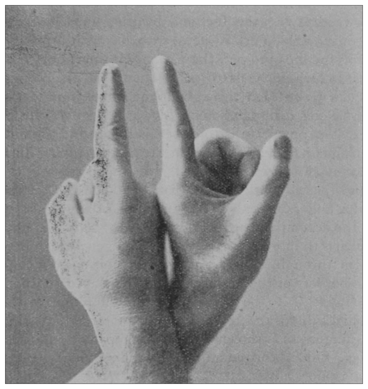
Fig. 1.
—Positions of the hands for the practice of the simplest method of proctoscopy.
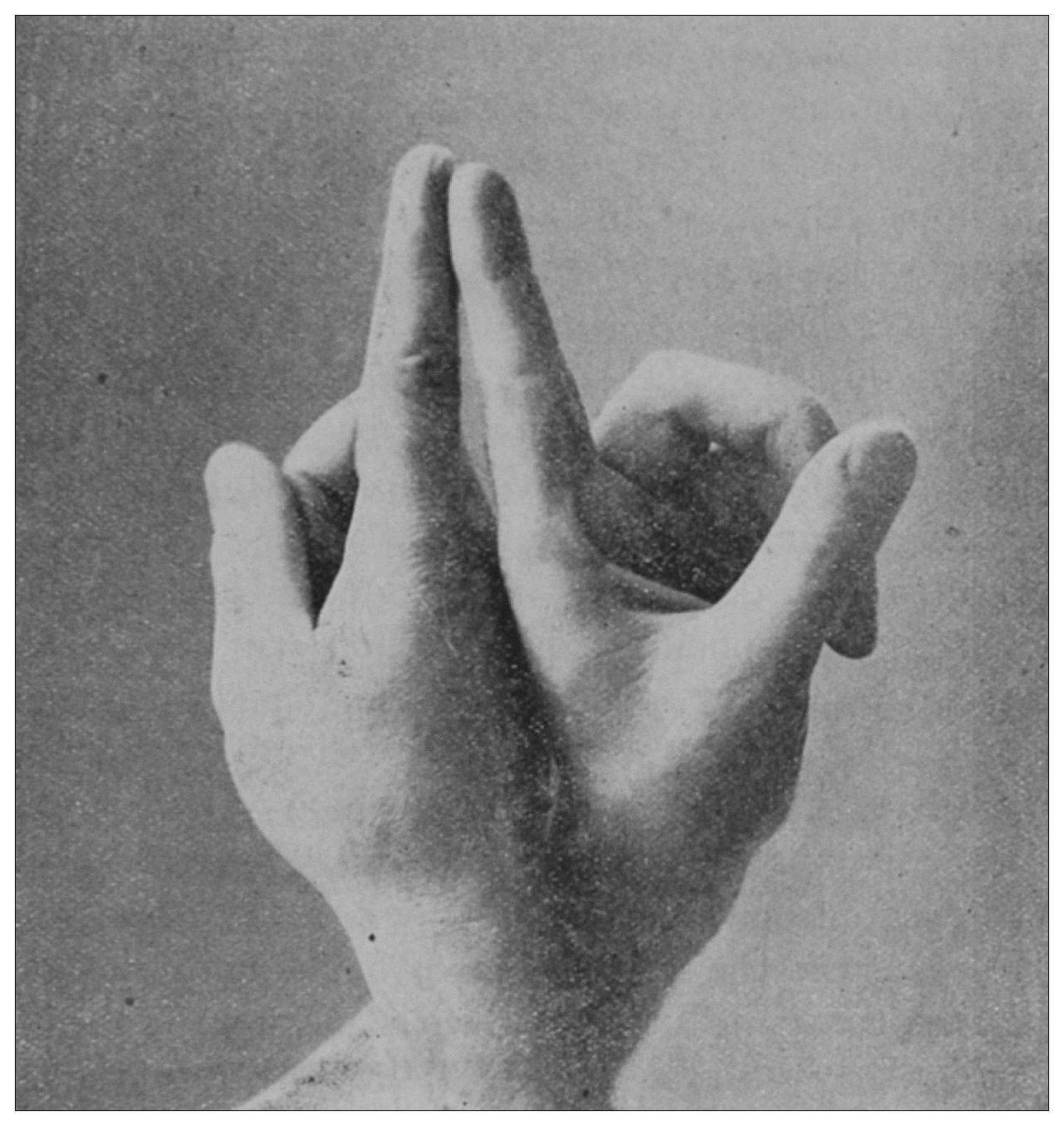
Fig. 2.
—Positions of the fingers for the practice of the simplest method of proctoscopy.
3. The physician is to close his hands and to point each index-finger as shown in the accompanying illustration (Fig. 1). The wrists are to be crossed, the hands placed back against back, and the nails of the index-fingers placed one against the other, as shown in the accompanying illustration (Fig. 2). The physician is to lubricate these fingers and gently insinuate them through the anus and place their ends beyond the borders of the levatores ani. This accomplished, the anus is to be divulsed in the direction of the ischial tuberosities, by the physician forcibly parting his fingers as is shown in the accompanying illustration. Under this manipulation the rectum becomes atmospherically inflated.
Now, provided the physician lowers his head to the level of his fingers and then rises again, or stoops, or moves a little from side to side, he may command under his eye a view of the interior of the atmospherically inflated rectum to the depth of six or eight inches (15.24 or 20.32 cm.), and in some instances he may behold even a part of the sigmoid flexure.
It is possible for the operator to manipulate his patient and to finish his inspection within two and a half or three minutes, provided the patient be in a state of complete anesthesia.
If this method is practised, as I am persuaded it may be with facility by the general practician, I am convinced that the greater number of rectal diseases may be instantaneously diagnosed. But I must declare that here at diagnosis, the achievement of the simplest proctoscopy ends, for the reason that the operator’s hands are so full of his patient he can do nothing at all for the disease which he may have discovered.
Under some conditions and amid some circumstances the rectum will not inflate. If the bladder is much distended; if there is an inordinate hypertrophic rectitis; if there is a close tubular stricture of the rectum; if there is malignant growth or other disease of the rectum by means of which the gut’s coats have become extensively filled and fixed with an organized plastic exudate; if for some reason the extraabdominal pressure is abnormally increased, as it may be by the bearing down of the patient, or by enormous flatus, or by ascites; or if there is an impinging uterus, adrectal growth or extensive infiltrating disease of the contiguous textures, rectal inflation by this method or by any other which is governed by the same principle may be a physical impossibility—but this need not baffle the man bent on seeing by instrumental aid.
Practised as described, when not embarrassed by the exceptions specified, this method will achieve its purpose and reveal to the physician that the transverse diameter of the rectum is variable; that in some places it is not more than an inch (2.54 cm.), in others it is more than four times this diameter.
The rectum may present to the eye of the imaginative observer the appearance of a chain of urinary bladders, communicating one with another by means of irregularly elliptic openings set at varying axes, and bounded by the nonparallel borders of the rectal valves. In the normal rectum the air-pressure smooths the mucous membrane evenly over the entire surface of the gut, as may be observed in the photographic illustrations. The normal mucous membrane of the so-called ampulla appears at first wet and of a shining bluish gray. As it dries, under the influence of gravitation the blue venous tint fades out of the gray and the wall assumes a pink tint. Presently it acquires the appearance of parchment, and sometimes it appears painted at rare intervals with ramifying little arteries which may be crowded and overlapped by the larger companion veins; the latter are less arborescent and more suddenly dive and disappear in the bowel-wall. In time, over all there comes a sheen and the vascular pictures may fade away. These phenomena appear exactly as described only in the healthy rectum. In the diseased organ the color varies much.
Should the operator deviate from the described directions for the manipulation of his fingers and so twist his hands as to divulse the anus in the anteroposterior direction instead of laterally, he invites defeat upon himself, for in the male the fixation of the perineum and the immobility of the coccyx interfere with the requisite dilatation; while, in the female, the extreme mobility of the perineum and particularly the backward displaceability of the coccyx will allow such traction to be made upon the levatores ani as to pull their inner fibers parallel and almost together, and, in consequence, the wider the female’s anus be opened anteroposteriorly the closer is it made to contract laterally to rob one of his view.
Instrumental Inspection.
Certain paraphernalia and much practice in their use are necessary for rapid, complete and painless inspection of the rectum.
The chair which is shown in the illustrations was designed by me to facilitate the placing of the patient in a new posture, which is equivalent to the knee-chest posture.[13]
Fig. 3.—The chair, illumination-apparatus, shoulder-suspender, and small pillow.
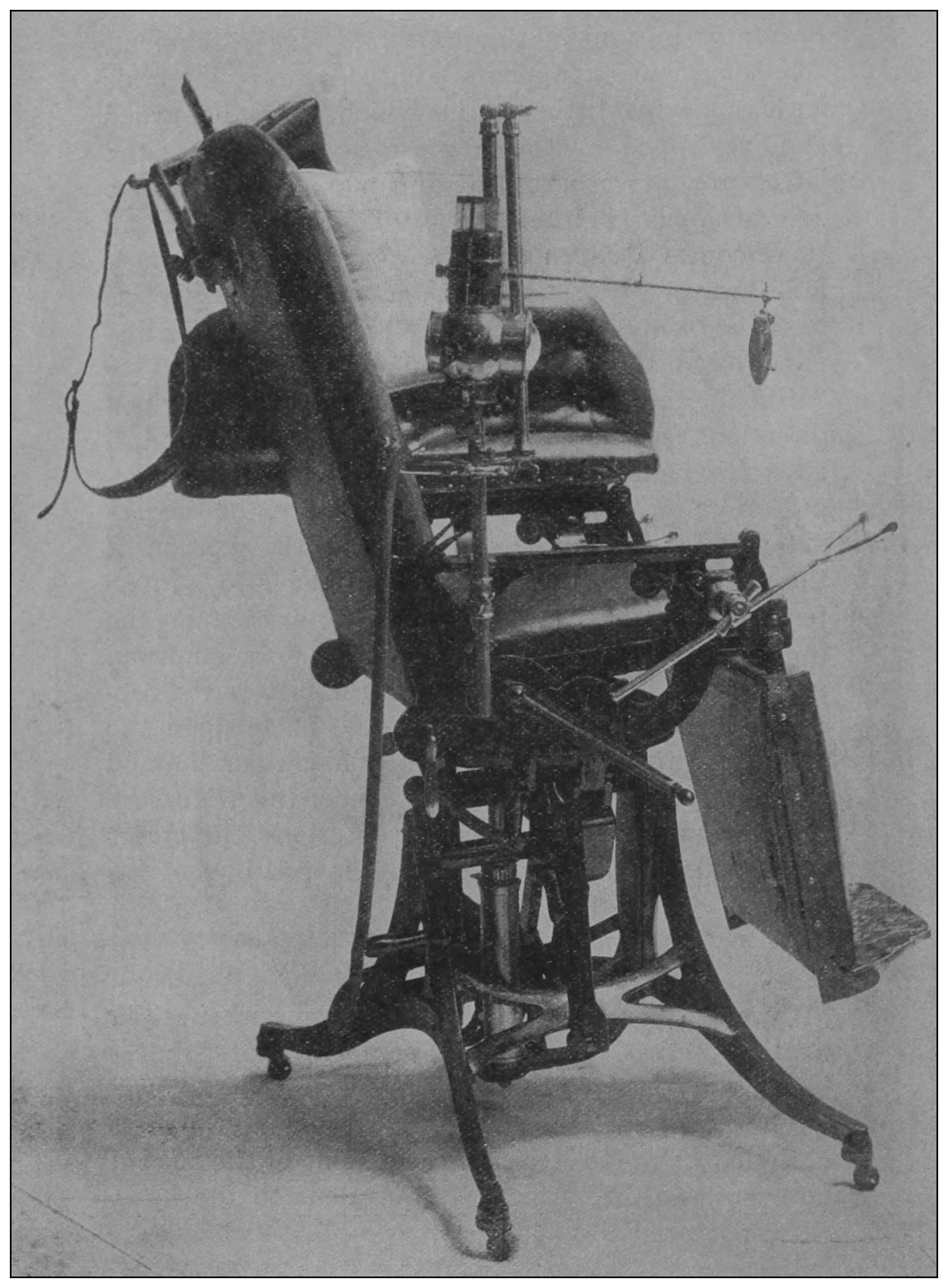
Fig. 4.—The chair in the horizontal posture for anoscopy.
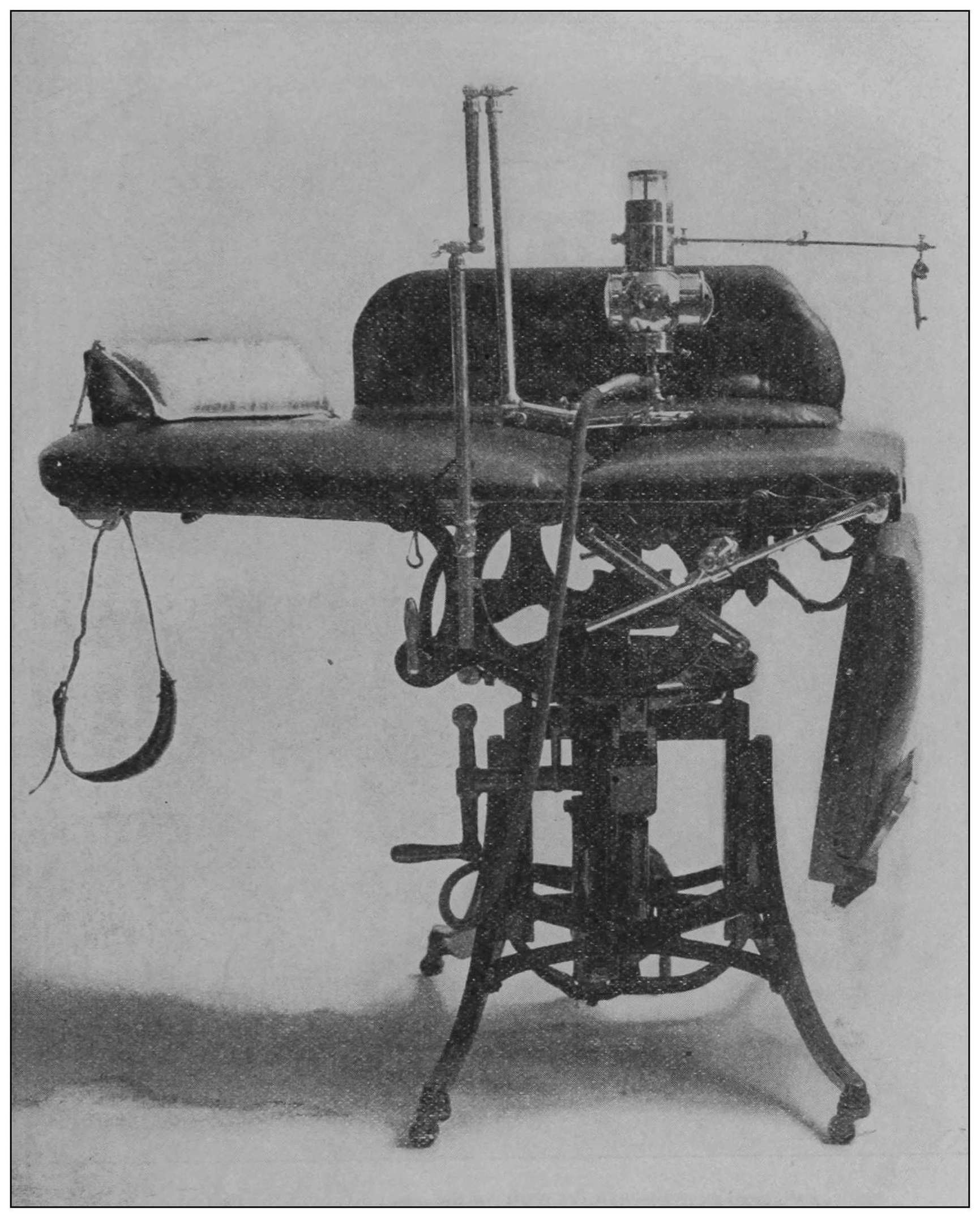
Fig. 5.—The position of the chair for the new posture of the patient.
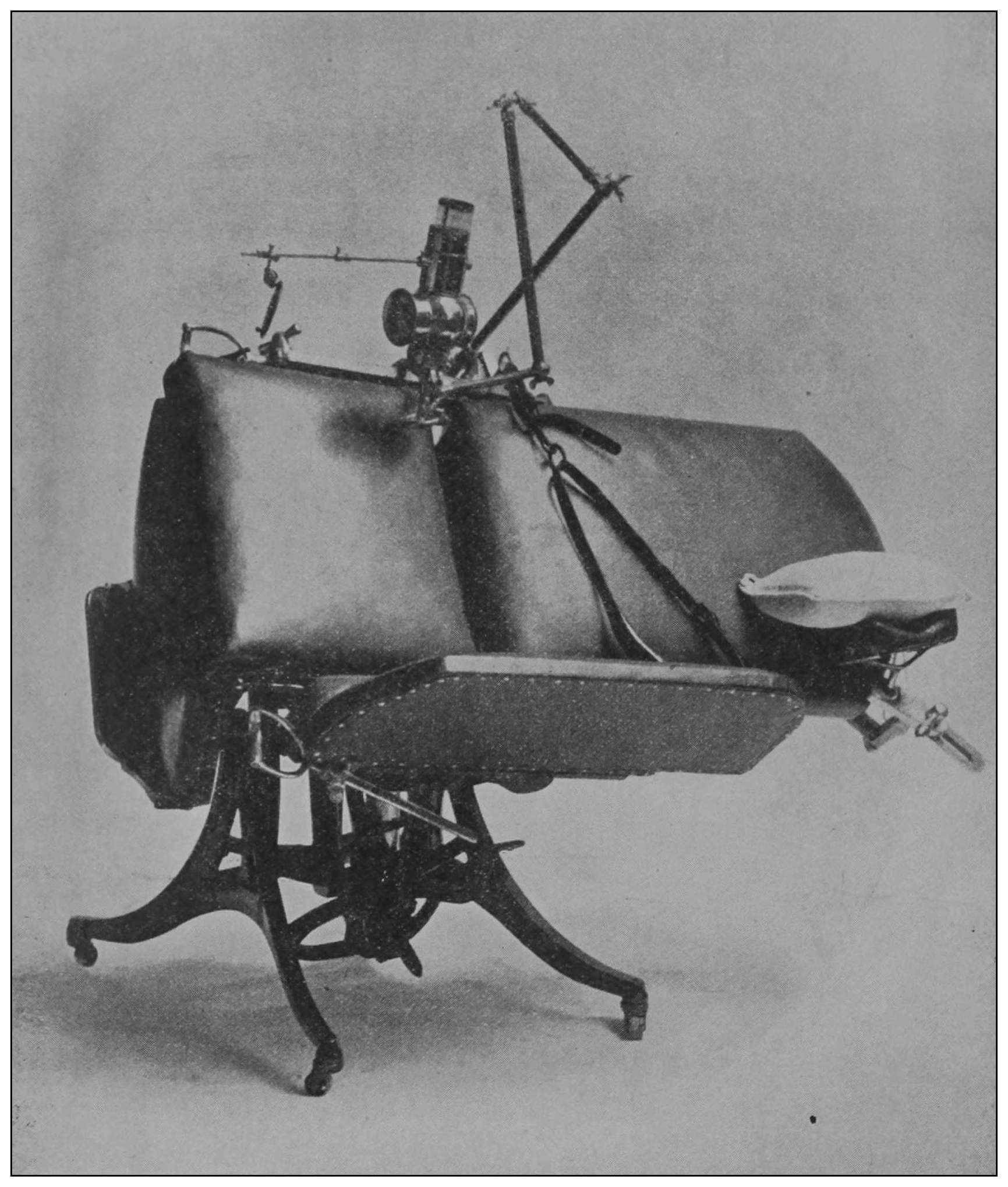
Fig. 3 exhibits the chair and the attached illumination-apparatus in the first position to receive the patient. Fig. 4 shows the chair and illumination attachment in the second position, and Fig. 5 shows the chair and illumination-apparatus in the position for the third step in the procedure. Fig. 3 shows, also, hanging from the head of the chair, a small pillow and the shoulder-suspender.
Fig. 6.—The anoscope.
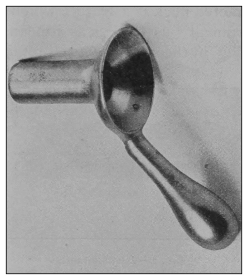
Fig. 7.—The obturator.
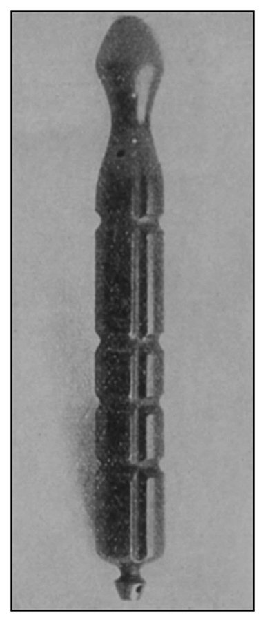
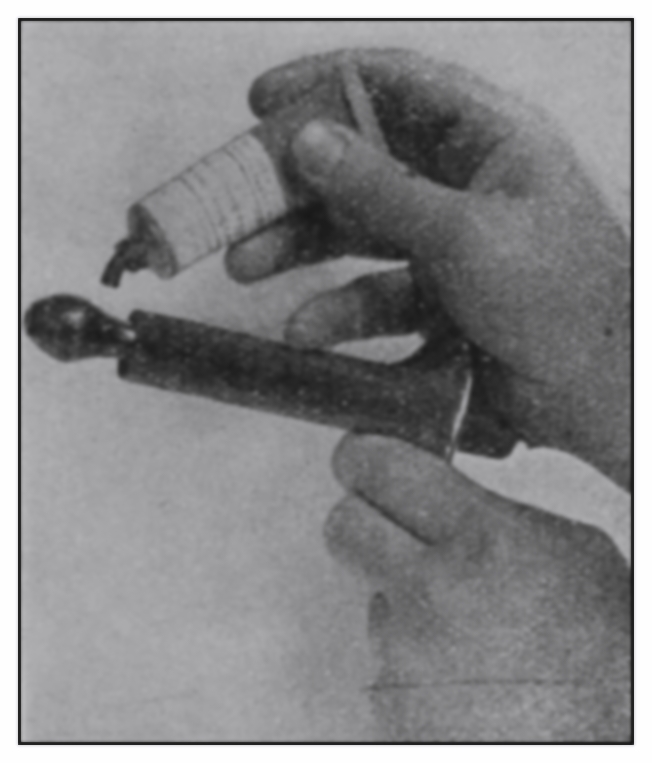
Fig. 8.—The ointment applicator.
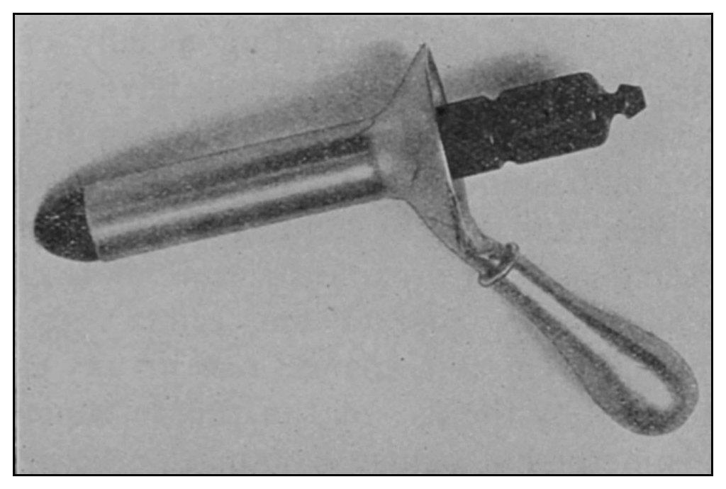
The anoscope (Fig. 6) consists of a short cylindric tube open at the ends. It is two inches (5.08 cm.) in length and seven-eighths of an inch (2.22 cm.) in diameter. The proximal end is provided with a trumpet-shaped expansion and a strong handle. The distinctive feature of the anoscope is the peculiar form of its obturator (Fig. 7), which has a capacity for a multiplicity of uses.
The obturator consists of a hard-rubber cylinder, in the middle of which is fixed a brass tube for the purposes of irrigation. Its surface is fluted in such a manner that it may be made to lock in any of several positions upon a tubercle within the cylinder. These flutes also provide for escape from the rectum of fluids and gases under certain conditions. The contracted neck near the distal end of the obturator provides a cup to facilitate the application of ointments to certain rectal areas.
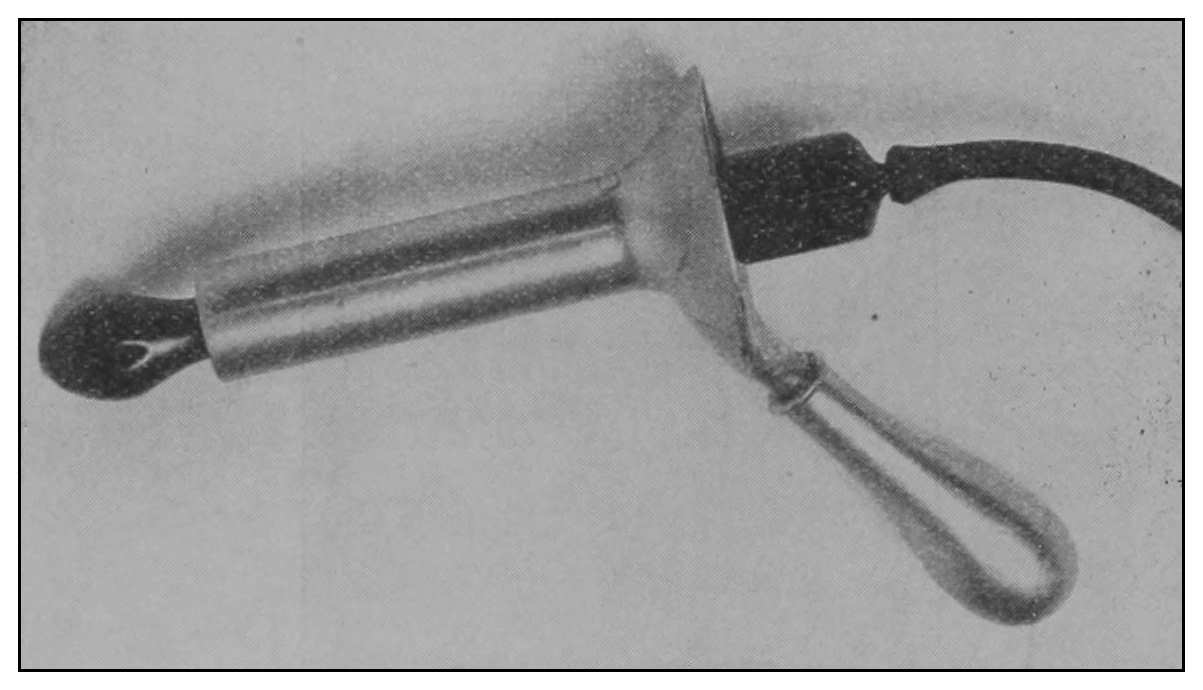
Fig. 9.
—The two-way irrigator.
The contracted neck is a feature which contributes to the instrument’s usefulness as a means for irrigation, providing in the one case a self-retaining direct-flow irrigator, and in the other case when locked in the position shown in Fig. 9, an unobstructed two-way irrigator. Platinum pins connect the centrally-placed brass tube with the surface of the neck of the obturator, which makes the instrument an anal electrode.
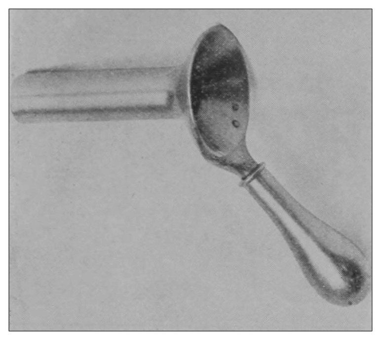
Fig. 10.
—The proctoscope.
The proctoscope (Fig. 10) is of the same diameter as the anoscope, and is four inches (10.16 cm.) in length, which, because of the displaceability of the pelvic floor is usually of sufficient length to reach as high as the promontory of the sacrum, except in some especial instances, the management of which exceptions will be treated of in another place.
Special preliminary preparation of the patient is ordinarily not required, as the usual condition of the rectum is that of emptiness. In some cases, however, it facilitates the inspection if the patient employs rectal lavage an hour before the examination. This injection should not consist of more than one pint of fluid. The excess of this might be accidentally dejected from the sigmoid and obscure the field during examination. The bladder should be emptied, since its distention would necessarily interfere with the accuracy of the examination.
Fig. 11.—The ointment applicator at the time of the placing of the ointment in contact with a diseased area.
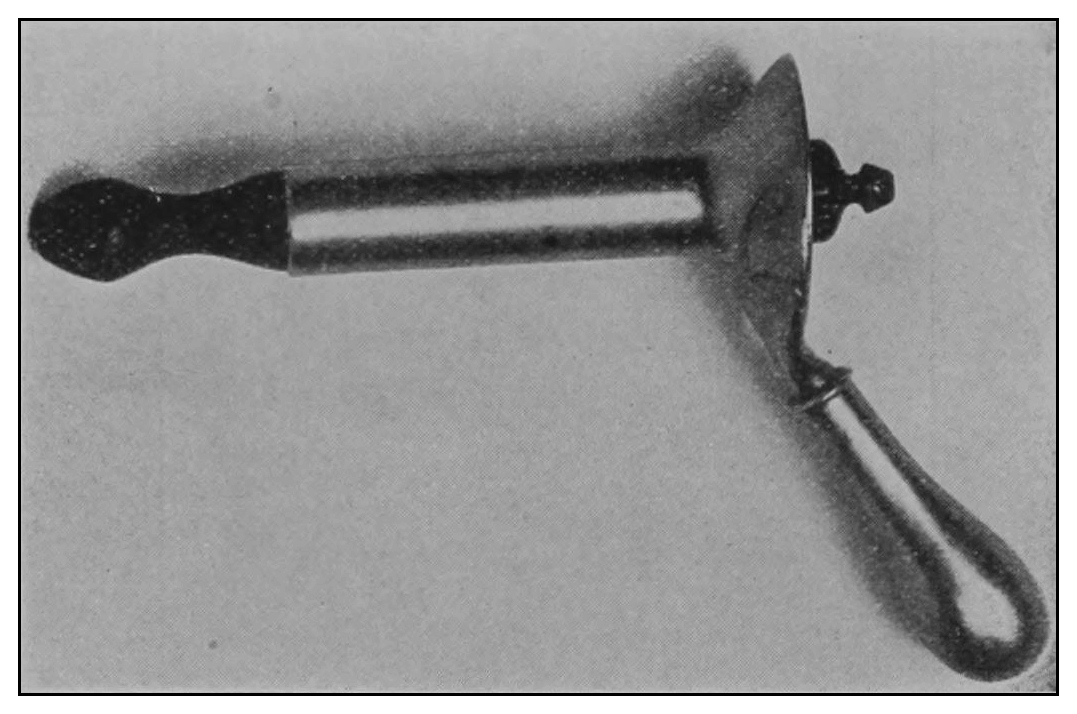
Fig. 12.—The proctoscope ready for introduction.
The Technic.
Step 1.—The patient should be required to sit on the operating-chair with his body turned to the left, facing the knee-board. The right knee should be crossed over the left knee, the left arm should embrace the right border of the chair-back, or it may be folded at the side as for Sims’ posture. The small pillow should be held in the patient’s right hand and against and upon his left shoulder (Fig. 13).
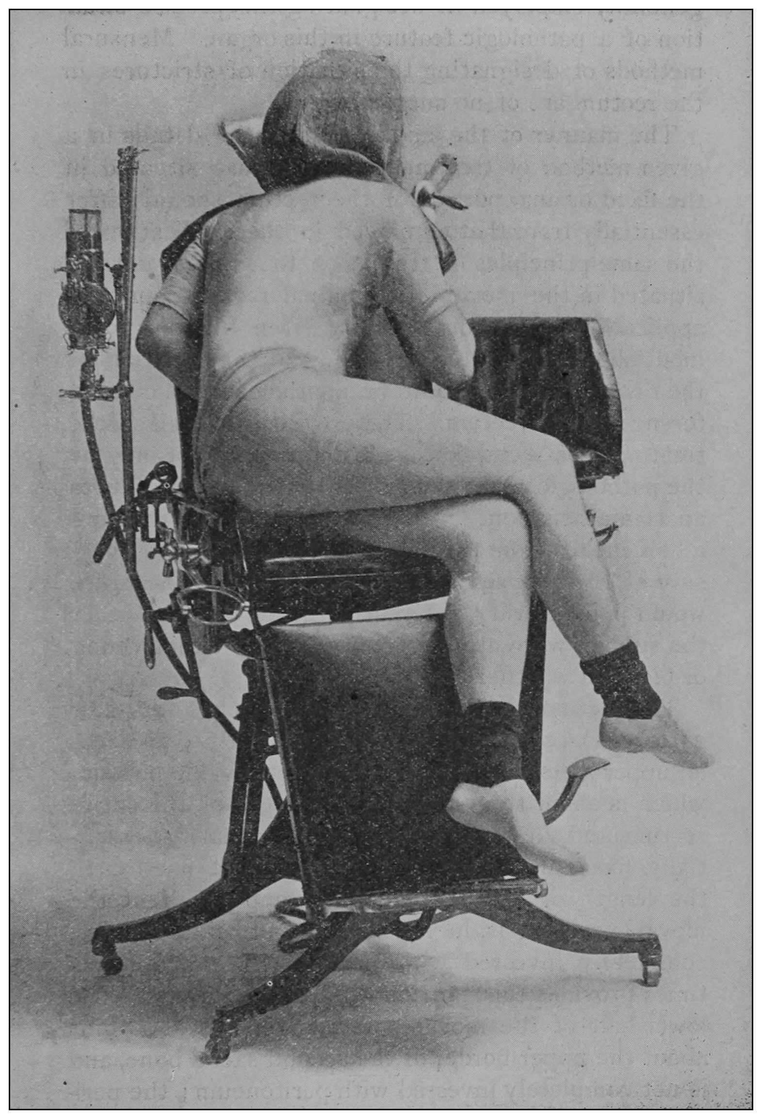
Fig. 13.
—Sitting posture of the patient, the first step toward proctoscopy.
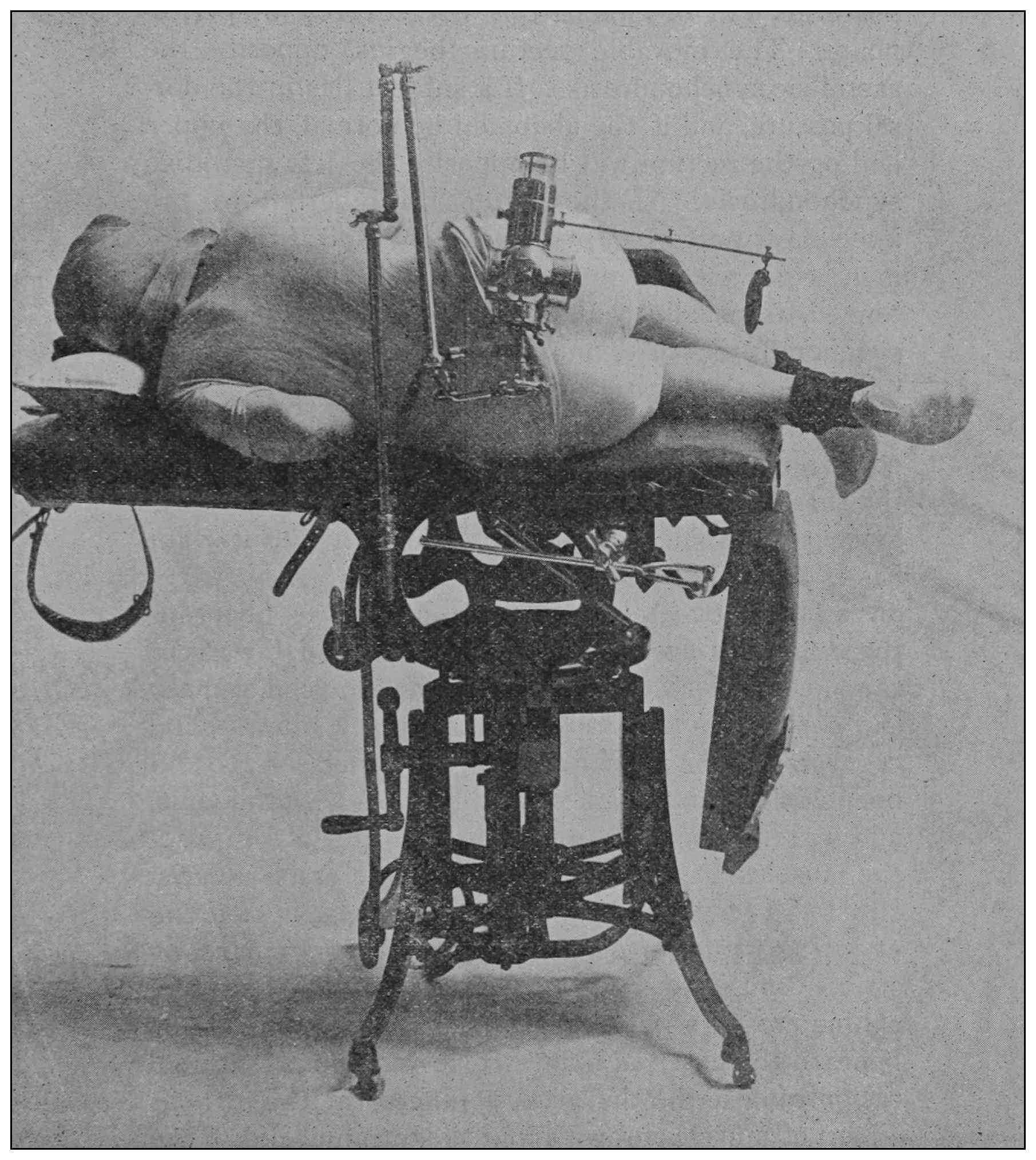
Fig. 14.
—Horizontal posture of the patient for anoscopy.
Step 2 consists in shifting the chair to the horizontal position shown in Figs. 14 and 4, and in adjusting the light-fixture. This movement brings the patient into Sims’ semiprone-semiflexed posture, without requiring any movement whatever on the part of the patient after he is properly seated. In this posture, the external anus and fixed rectum are to be examined.
(a) Digital examination and ocular inspection should now be made of the anal verge, the external anus, and superficial ischiorectal space, at a moment when the patient is relaxed, and, again, when he is bearing down.
(b) Digital examination of the fixed or anal rectum, also, should be made preliminary to the introduction of the anoscope.
(c) The anoscope should be gently pressed into the anus in the direction of its axis till the sphincters relax to receive it. The introduction of an instrument into the rectum may be much facilitated by placing its lubricated end against the ectal sphincter and requiring the patient to bear down; bearing down expands the ectal sphincters, relaxes the levator ani, thins the pelvic floor, or shortens the fixed rectum, and presses the ental sphincter over the instrument—in other words, the patient’s anus is made to climb down upon the speculum. After the introduction of the anoscope, its obturator should be removed, and the inspection made. These observations should be made coincident with the withdrawal of the anoscope. In cases of extremely sensitive ani, a skillfully put hypodermic injection into the sphincters of 10 or 20 minims of ⅒ of 1% solution of cocain may render anoscopy painless.
A desire for precision requires that lesions of the fixed or anal rectum should be noted as occupying a given quadrant, and as situated at a given zone, e.g., a circumscribed disease may be described as situated at the ental sphincter zone, and in the left lateral quadrant.
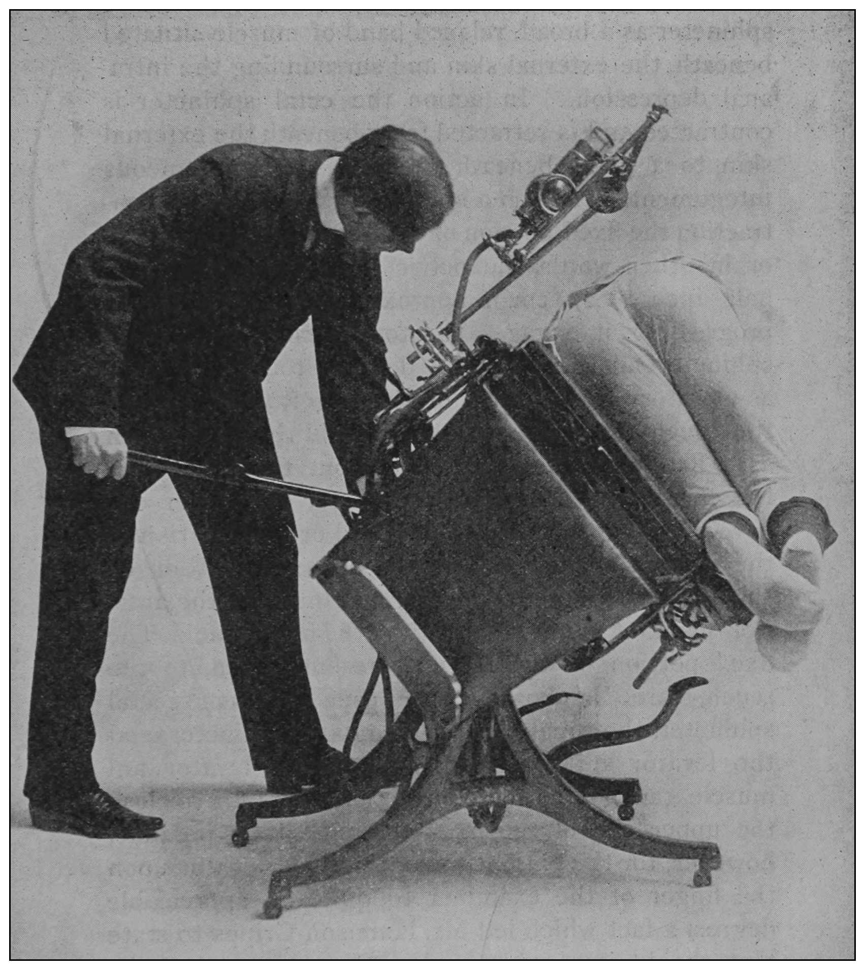
Fig. 15.
—Putting the patient into the new posture.
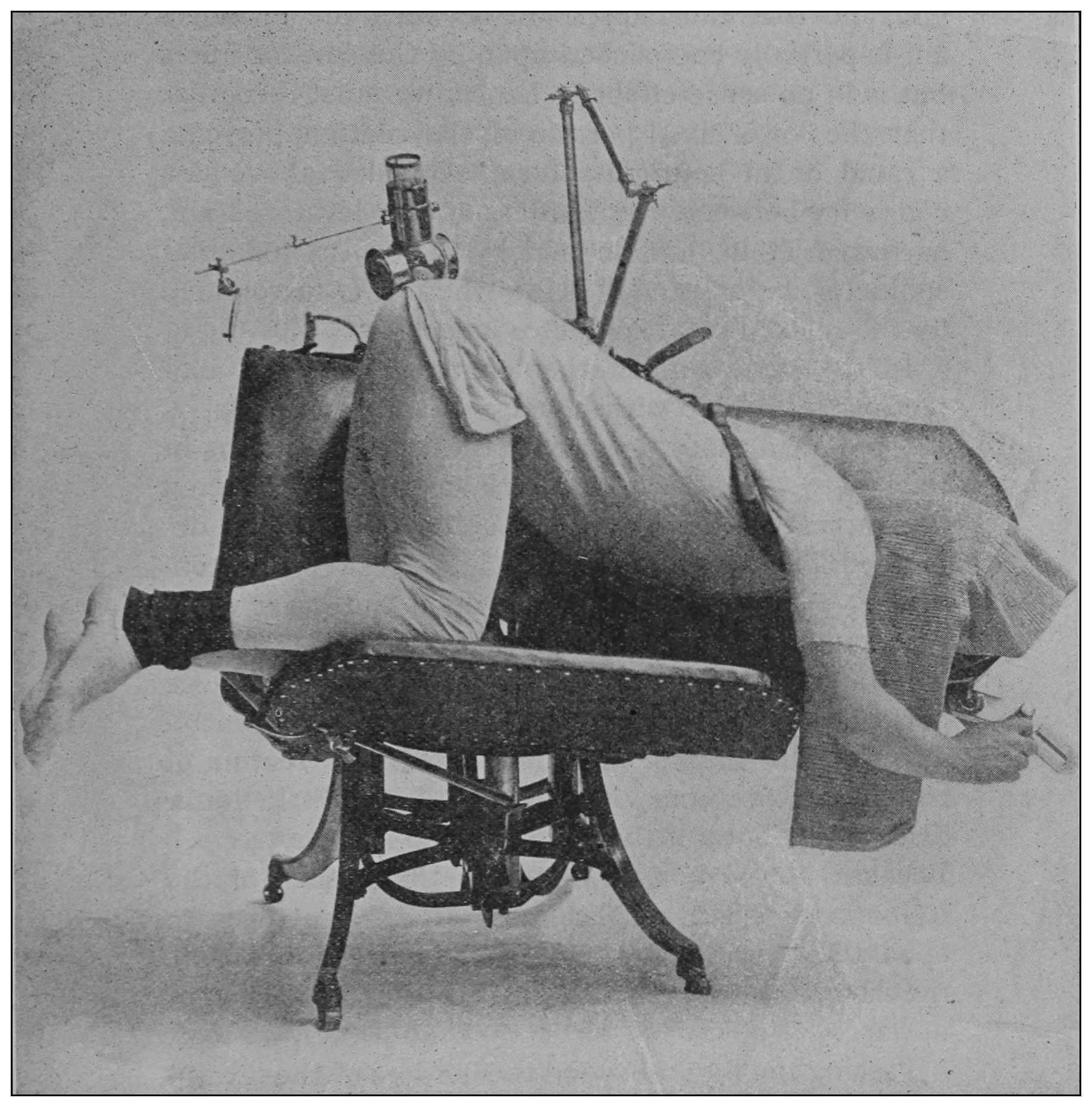
Fig. 16.
—The new posture.
Step 3 (a) requires that the shoulder-suspender should be placed and fixed to the chair, as shown in Figs. 5 and 16, that the knees be drawn up, so that the thighs are at a right angle to the length of the chair-top, and that the chair should be tilted to put the patient in the new posture shown in Fig. 16. The leg foot-board should now be lowered, and the operator’s stool placed in convenient position.
The illumination-apparatus should now be adjusted, as shown in the illustrations. In this new posture, which is equivalent to the knee-chest posture, the abdominal rectum is to be examined.
(b) Introduction of the proctoscope requires supported eversion of the buttocks and steady gentle pressure of the well-lubricated instrument upon the anus and in the direction of the umbilicus, until the sphincters are felt to yield, or the patient may be required to bear down to take the speculum; as the instrument enters the inflatable movable rectum, it should be pointed toward the promontory of the sacrum, and subsequently into the sacral hollow. The withdrawal of the obturator is followed by atmospheric inflation of the rectum.
(c) The operator should observe the degree of rectal distention, the situation and number of the rectal valves, their propinquity to one another when passive, and the relation of one valve to another at the time of the patient’s bearing down. Under pressure of the proctoscope if possible, or the hook if necessary, each valve should be effaced or displaced, and in regular order each of the rectal chambers should be carefully inspected. A proctoscopic mirror may be necessary for viewing the supravalvular surfaces (Fig. 18). The examination being finished:
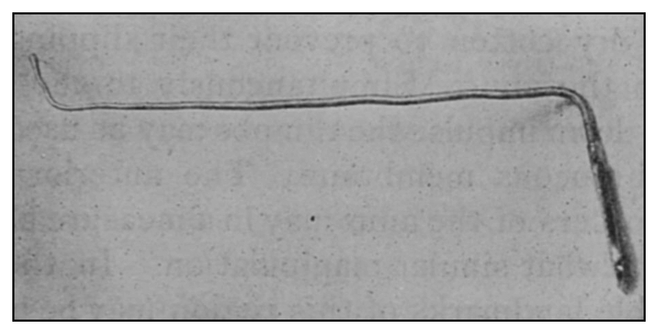
Fig. 17.
—The hook for testing the valves.
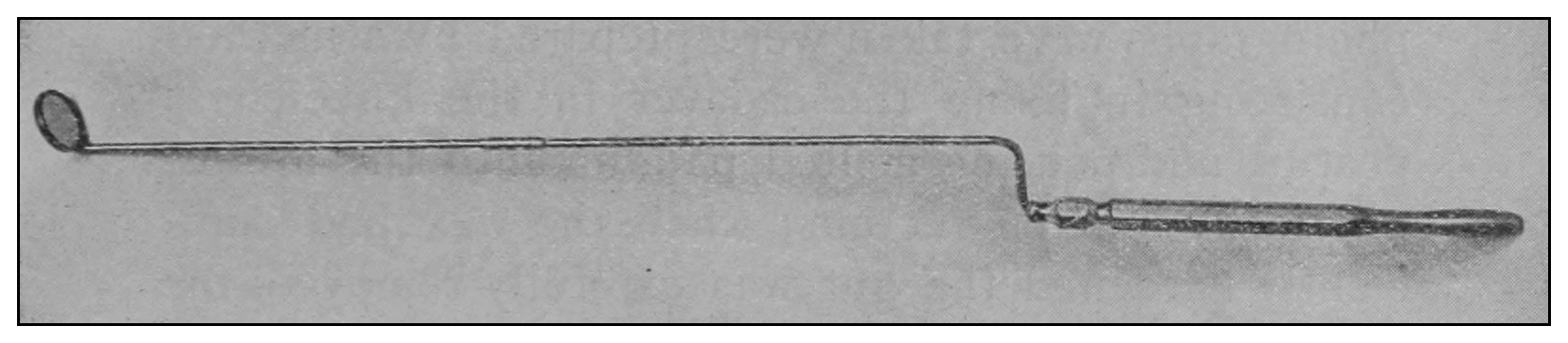
Fig. 18.
—The proctoscopic mirror.
Step 4.—The proctoscope should be withdrawn, the illumination-apparatus fixed in the first position, the leg foot-board lifted to its place, the lever extended, the crank turned and the chair carried back to the horizontal and upright positions, and thus the passive patient may be returned to his feet by the execution in the reverse order of the several steps described.
This method of inspection does not subject the patient to struggle, strain or embarrassment.
Observation by this method has taught me that in nearly all cases of disease at the anus there is congestion or inflammation of the rectal mucous membrane.
Those cases in which there is no apparent lesion at the anus, and which are in a perfunctory way sometimes declared catarrh of the rectum, will at once have the real condition, such as a high up rectal polypus, congenital or organic stricture or ulceration, positively diagnosed, and will be made accessible for intelligent treatment.
New growths and ulcerations may be seen and by means of a long-handled curet scrapings made in order that the microscopist may determine their exact character.
Stricture of the rectum need no longer be regarded as of only doubtful presence, and this method proves positively, even to the casual observer, how fallacious is the method of rectal sounding usually employed for the diagnosis of stricture. I have repeatedly proved to visitors how easy it is for an entering or returning bulb-sound to be caught and held by the rectal valves,[14] and thereby yielding signs generally considered diagnostic of organic stricture of the rectum.
Vesicorectal, vaginorectal and other deep fistulas are often apparent at a glance, but in any case may be discovered by the use of the proctoscopic mirror.
If this method of ocular examination be practised, I am convinced there need be no longer any excuse for calling an undiagnosed disease of the rectum obscure disease, and whatever the disease present this method makes it susceptible of demonstration by the proctologist to the attending physician. There is no necessity that a diagnosis be taken on faith.