Obstipation in Infants.
Reading edition
The infant strains at stool because of the imperfect development of the anatomic features concerned in the mechanism of defecation. These are as follows:
1. The infant’s lower gut is muscularly deficient.
2. Its great length and its mobility within the abdomen are obstructive to defecation.
3. The rectal valves are obstructive.
4. The infant’s anus not being sufficiently expansible is also obstructive to the descent of feces.
The specimens of infant rectums and sigmoids here pictured are all fairly illustrative of the facts upon which the foregoing declarations are based.
The dried specimens shown in the illustrations were prepared by flushing the intestine and then fixing the subject in the genuacromial posture; the anus was then fixed open and melted paraffin was injected under about twelve ounces’ pressure. When the cast had hardened, that portion of the gut which it occupied, was removed. Specimens shown in Figs. 43 and 44 were prepared by placing the subject upon its back and by opening the descending colon; the intestine below was then washed out and the colon perforation fixed at the abdominal wound, which, save for this point, was sewed up, the anus was tied up and as much melted paraffin as would enter under two-pounds’ pressure was forced into the gut; subsequently the sigmoid and rectum were removed as in the other instances. After immersion in alcohol the specimens were varnished.
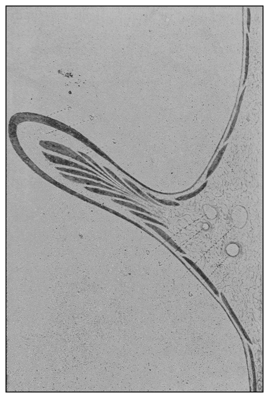
Fig. 33.
—A semilunar valve drawn as seen under a glass magnifying five diameters.
A
, mucous membrane;
B
, fibrous tissue;
C
, bundles of circular muscular fibres;
D
, vein;
E
, artery;
F
, vein;
G
, artery;
H
, areolar and adipose tissue.
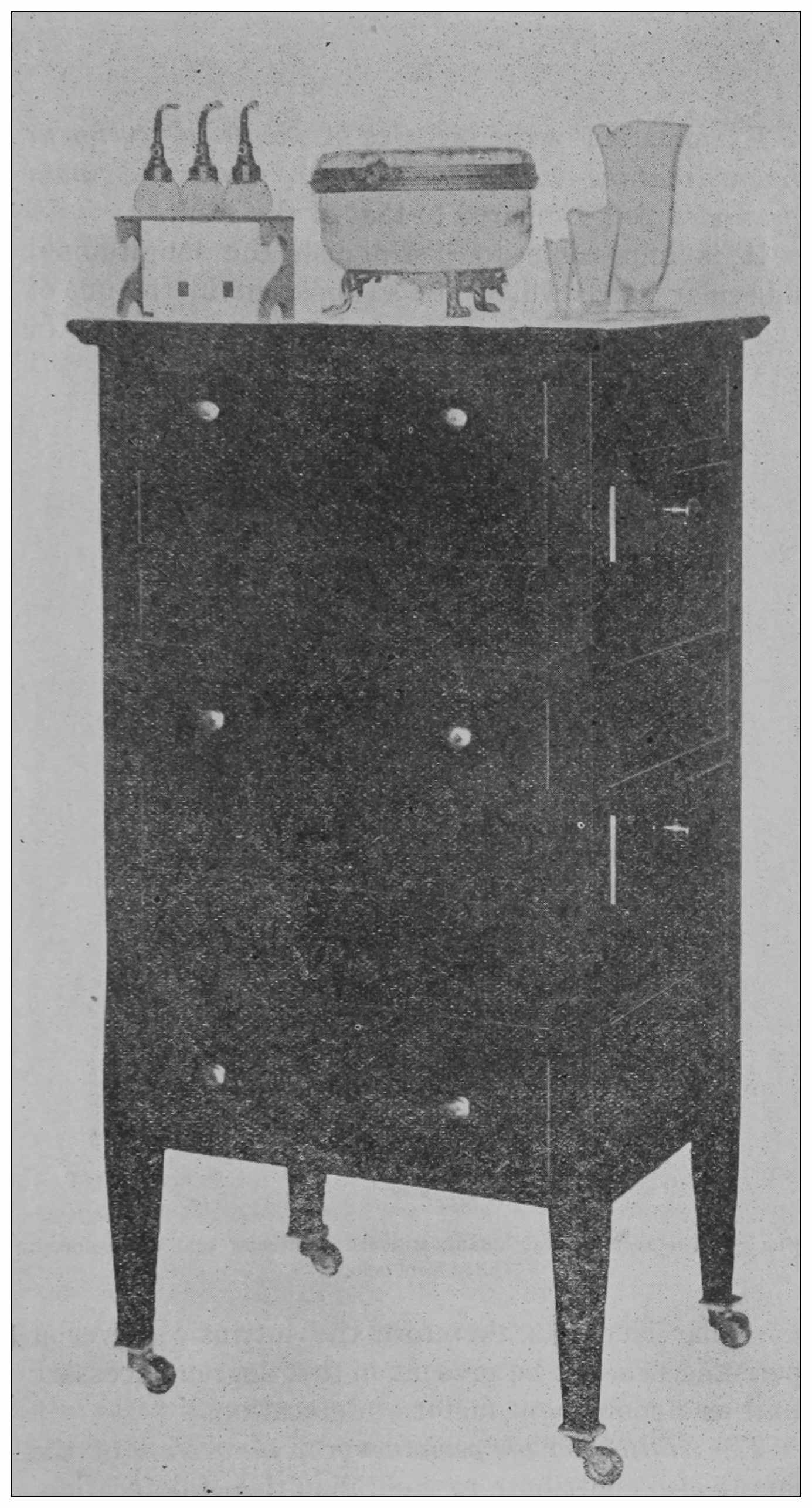
Fig. 34.
—The instrument case; the drawers opening on three sides afford access to any drawer without disturbing the position of the others.
Following are the memoranda of a few of the autopsies made in this study:
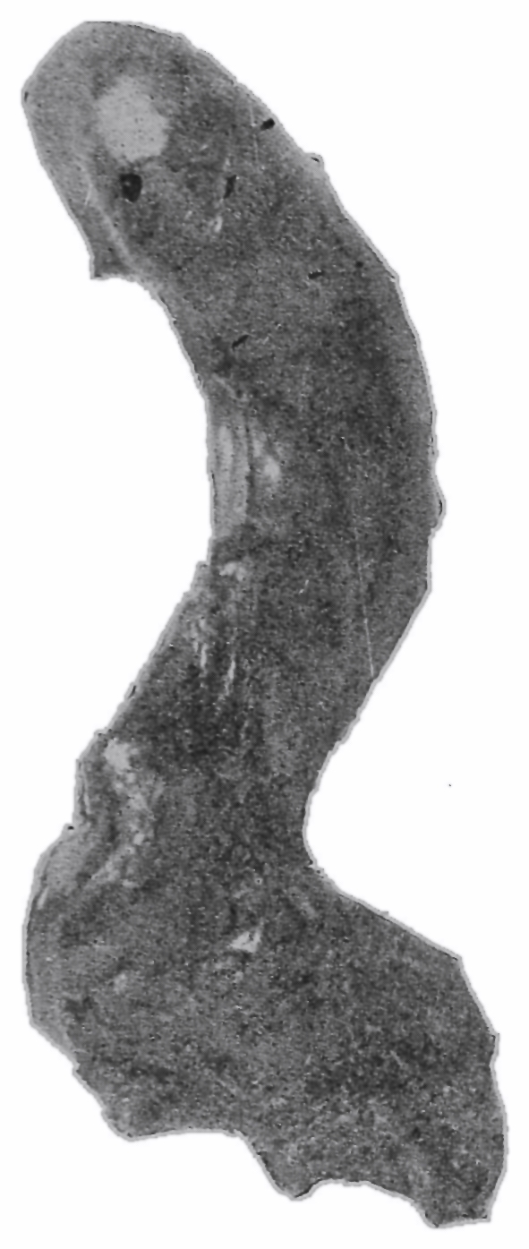
Case 1.—Female, stillborn, height 16 inches (40.64 cm.); circumference at anterior superior spinous process 7½ inches (19.05 cm.); anterior superior spinous process diameter 3 inches (7.62 cm.); ensiform to pubes 4 inches (10.16 cm.); transverse diameter pelvic outlet ¾ inch (1.90 cm.); peritoneum at last vertebra of coccyx. (Fig. 35).
Fig. 35.—The rectum of an infant, stillborn; muscular coat undeveloped. Filled from below.
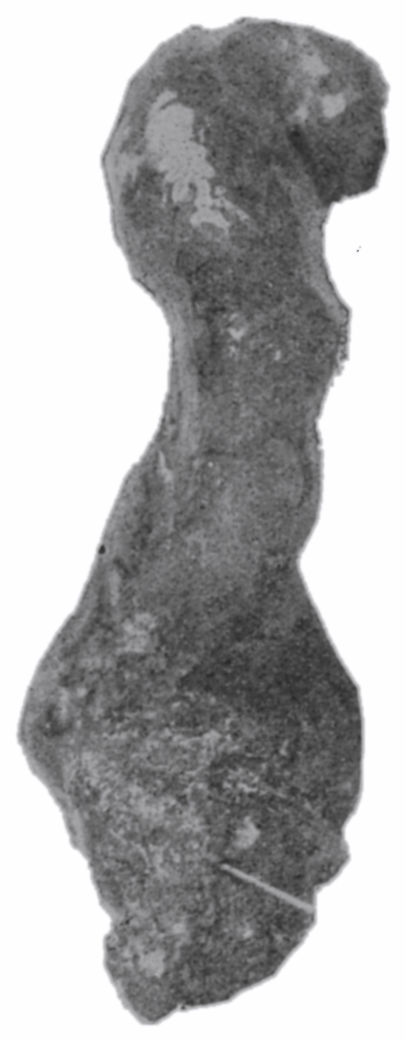
Fig. 36.—Side view of the specimen shown in Fig. 35. Antero-posterior angulation at third sacral vertebra.
Case 2.—Female, aged 1 hour, height 17 inches (43.18 cm.); circumference at anterior superior spinous process 7½ inches (19.05 cm.); anterior superior spinous process diameter 2½ inches (6.35 cm.); ensiform to pubes 4 inches (10.16 cm.); transverse diameter pelvic outlet ½ inch (1.27 cm.); peritoneum at last vertebra of coccyx. (Fig. 37).
Fig. 37.—Front view of the rectum of an infant aged one hour; autopsy while in a state of rigor mortis. The rectum being nearly perpendicular and the sigmoid almost tied in a knot. Filled from below.
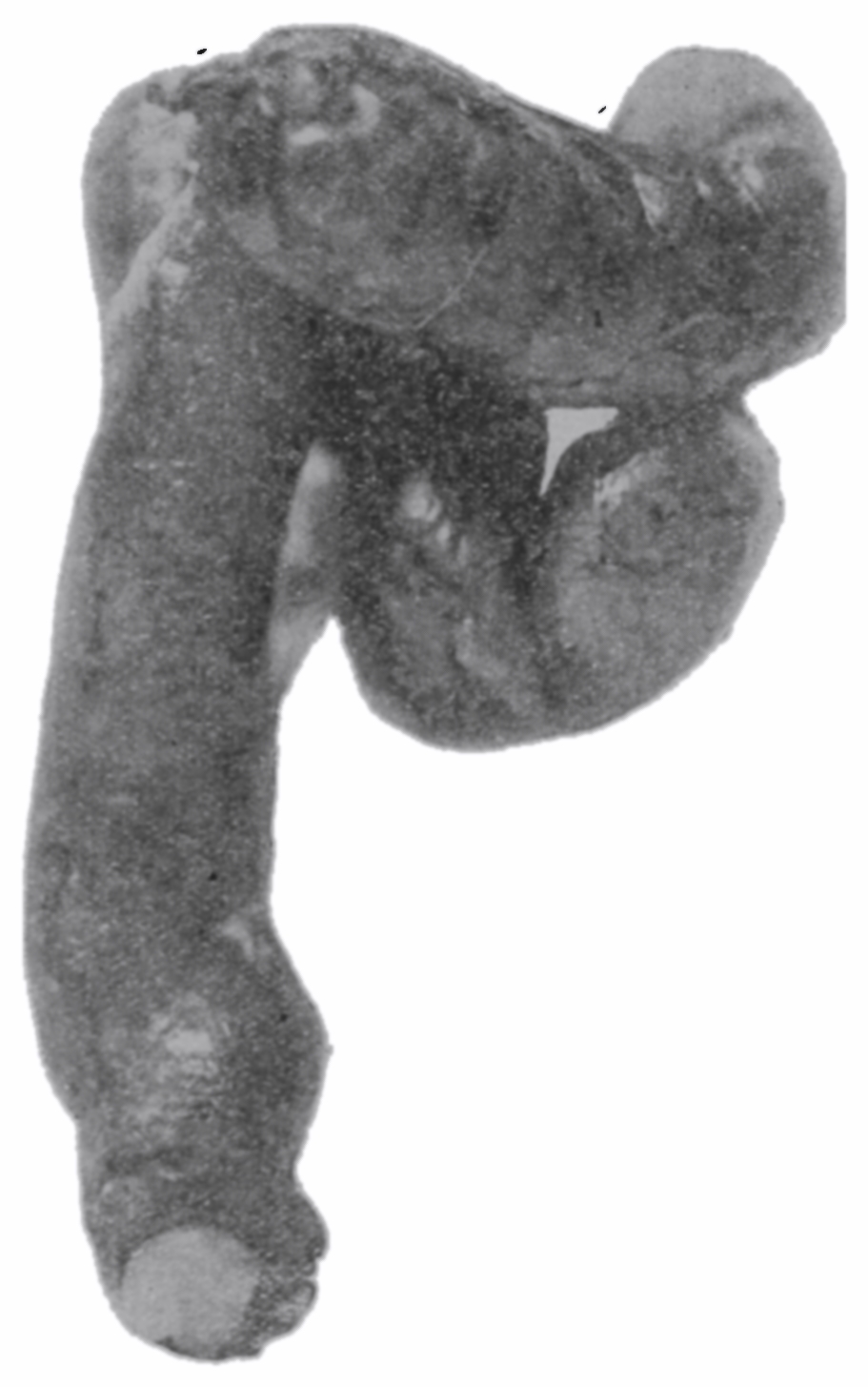
Fig. 38.—Side view of the specimen shown in Fig. 37.
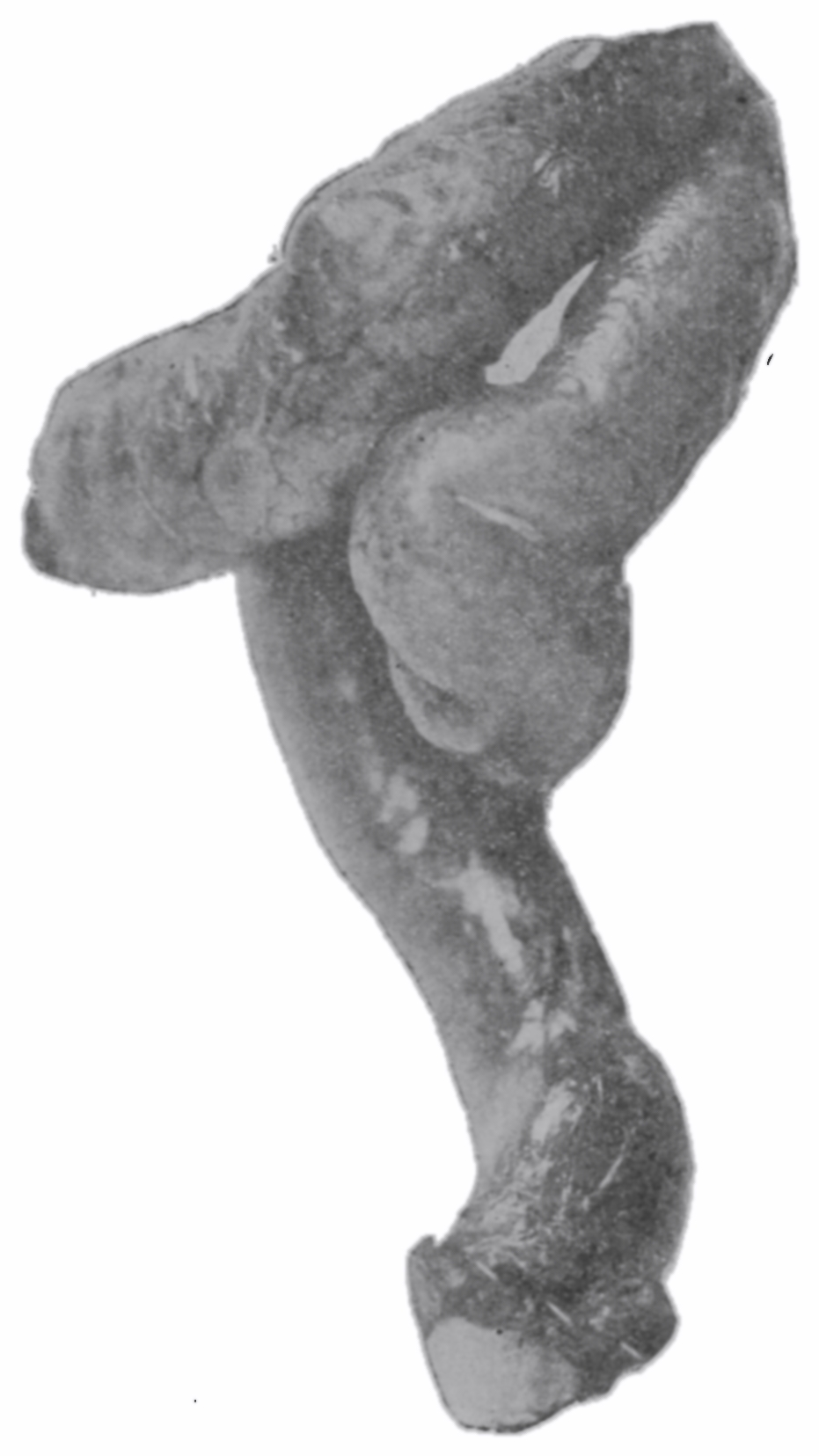
Case 3.—Male, aged 1 month, height 23 inches (58.42 cm.); circumference at anterior superior spinous process 9 inches (22.86 cm.); anterior superior spinous process diameter 3½ inches (8.89 cm.); ensiform to pubes 4 inches (10.16 cm.); transverse diameter pelvic outlet ⅝ inch (1.59 cm.); peritoneum at last vertebra of coccyx.
Fig. 39.—Front view of the rectum of an infant aged one month; autopsy while in the state of rigor mortis. Filled from below. Two-third life-size.
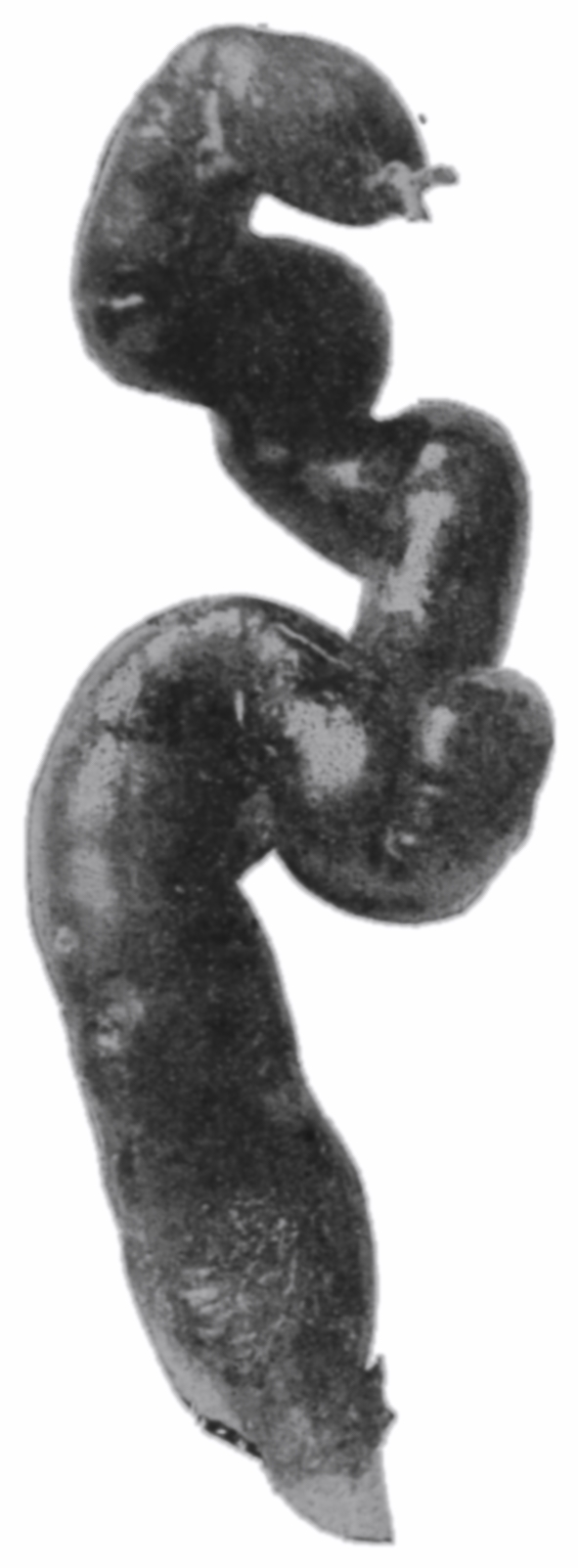
Fig. 40.—Side view of the specimen shown in Fig. 39.
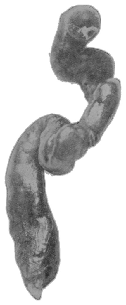
Case 4.—Female, aged 6 weeks, height 24 inches (60.96 cm.); circumference at anterior superior spinous process 10½ inches (26.67 cm.); anterior superior spinous process diameter 3 inches (7.62 cm.); ensiform to pubes 5½ inches (13.97 cm.); transverse diameter pelvic outlet ⅝ inch (1.59 cm.); peritoneum at last vertebra of coccyx. (Fig. 41).
Fig. 41.—Front view of the rectum of an infant aged six weeks. Filled from below.
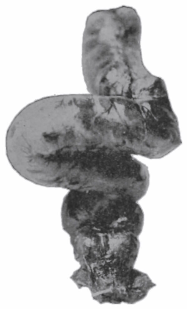
Fig. 42.—Side view of the specimen shown in Fig. 41.
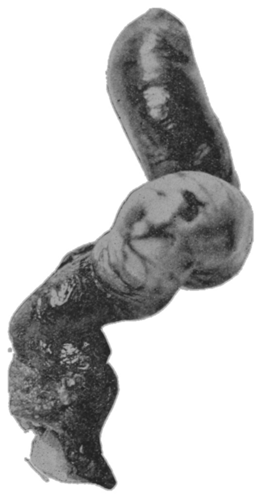
Case 5.—Female, aged 2 months, height 20 inches (50.80 cm.); circumference at anterior superior spinous process 8 inches (20.32 cm.); anterior superior spinous process diameter 3 inches (7.62 cm.); ensiform to pubes 3½ inches (8.89 cm.); transverse diameter pelvic outlet ¾ inch (1.90 cm.); peritoneum at last vertebra of coccyx. (Fig. 49).
Case 6.—Male, aged 6 months, height 24 inches (60.96 cm.); circumference at anterior superior spinous process 10 inches (25.40 cm.); anterior superior spinous process diameter 4 inches (10.16 cm.); ensiform to pubes 5 inches (12.70 cm.); transverse diameter pelvic outlet ¾ inch (1.90 cm.); peritoneum at last sacral vertebra. (Fig. 43).
Fig. 43.—Front view of the rectum of an infant aged six months. Filled from below after disappearance of rigor mortis. Two-thirds life-size.
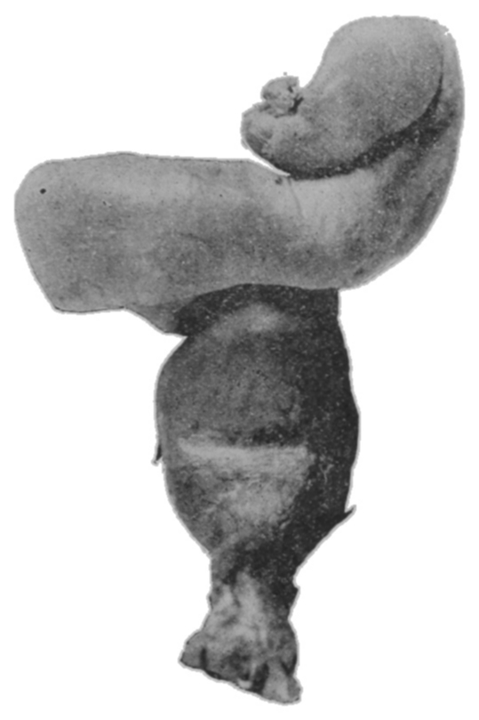
Case 7.—Male, aged 6 months, height 24 inches (60.96 cm.); circumference at anterior superior spinous process 12 inches (30.48 cm.); anterior superior spinous process diameter 4 inches (10.16 cm.); ensiform to pubes 5 inches (12.70 cm.); transverse diameter pelvic outlet ¾ inch (1.90 cm); peritoneum at first bone of coccyx. (Fig. 44).
Fig. 44.—Front view of the rectum of an infant aged six months. Filled from above after disappearance of rigor mortis.
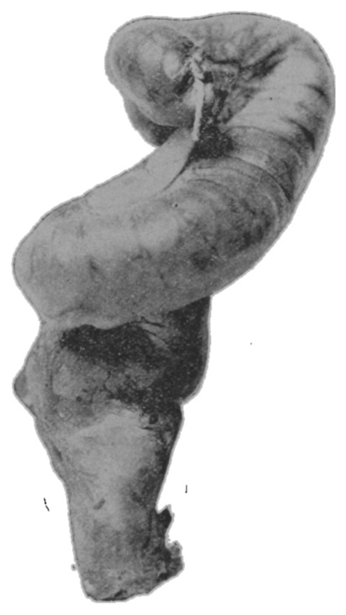
Fig. 45.—Side view of the specimen shown in Fig. 44. The mesentery from sacrum and coccyx to the rectum is fairly well shown, but is shown shorter than in the fresh state, alcohol immersion, varnishing and drying having shrunken it. Observe also how nearly the peritoneum approaches to the anus.
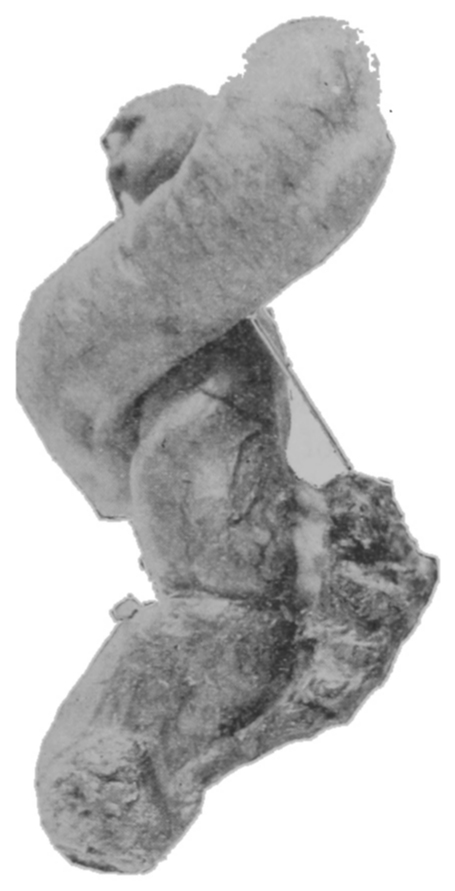
Case 8.—Male, aged 17 months, height 25 inches (63.50 cm.); circumference at anterior superior spinous process 12 inches (30.48 cm.); anterior superior spinous process diameter 4 inches (10.16 cm.); ensiform to pubes 6 inches (15.24 cm.); transverse diameter pelvic outlet 1 inch (2.54 cm.); peritoneum at last sacral vertebra.
Examination of the pictures of the dried specimens reveals that the wall of the infant rectum and sigmoid flexure is thin compared to that of the adult.
It is impossible to distinguish the longitudinal muscular bands which are so apparent in the gut of the adult. The infant gut being very deficient in muscular elements, therefore the intrinsic power of peristalsis cannot be present in that degree necessary to it as a component factor of defecation.
The relations of the peritoneum to the rectum of the infant also contribute to the difficulties of defecation, as also does the relatively great length of the descending colon and sigmoid flexure. In young children the length of sigmoidal mesentery from its attachment to the parietes to its invagination of the lower loop of the sigmoid is often greater than the distance from the promontory of the sacrum to the distal bone of the coccyx. (See Figs. 44 and 45.) From the sigmoidorectal juncture to the beginning of the middle third of the rectum the mesentery rapidly shortens but apparently completely invests the upper third of the rectum. The middle and lower thirds are not so completely invested, and present upon their posterior parts a vertical lane bare of peritoneum, from the borders of which the peritoneum is reflected in lateral directions. This uncovered part of the gut is not applied directly to the sacrum and coccyx. There is a space between which is occupied by loose connective tissue. The distance from the dorsal parietes to the gut is variable here, being from one-half to one-fourth of an inch (1.27 to .63 cm.). At the beginning of the middle third there is usually a gradual decrease in the length of the peritoneal band as it descends, till it is one-eighth or possibly but one-sixteenth of an inch (.32 or .16 cm.) in length at the last bone of the coccyx; it rapidly shortens from this point to its termination. The parietal peritoneum descends over the ischial tuberosities and approaches nearly to the ental sphincter muscle. In the newly-born the peritoneum is situated within one-fourth inch (.63 cm.) of the anal skin.
The disproportionately great length of the descending colon and mesentery of the infant obviously contributes to the possibility of angulation of the gut.
We may see in the presence in the child of lax and long, or relatively long, peritoneal ligaments, and in the great length of the descending colon and the consequent probability of numerous acute angulations in the infant sigmoid and rectum, and in the mobility of these parts within the abdomen, the possibility of development of a perfect adult mechanism for defecation. The essential features of this development are two: (1) the growth of prostate or uterus and their supports which relatively fix the lower rectum; and (2) the downgrowth and outgrowth of the pelvic bones and the consequent conversion of mesenteric peritoneum to parietal peritoneum, which shortens the adult mesentery and in some measure fixes the upper rectum; thus the entire rectum is steadied to facilitate discharge when the mechanism of defecation is set in operation.
Fig. 46.—Diagrammatic of this gut in the empty state, therefore its flexions are not the same as those shown in the photographs. In the partly filled state, which is the condition about the time of defecation, the flexions and positions of these parts would be intermediate to those of the filled and empty conditions. (The dotted lines indicate positions of valves.)
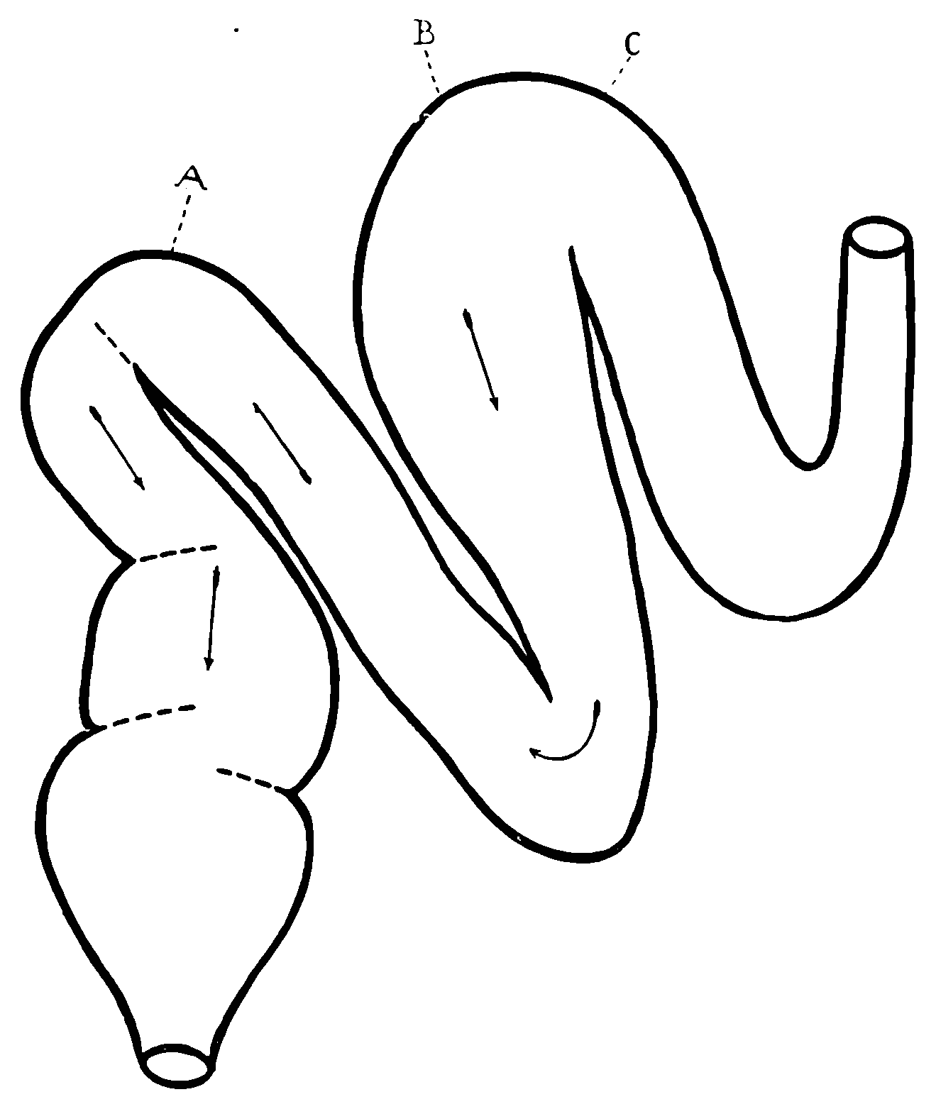
The diagrammatic figures do not exaggerate these obstructive elements. The collateral muscles which assist in the performance of the act of defecation force the feces in the direction of the lower angle in each flexion, and in that flexion whose onward, or, to be paradoxic, whose downward direction for the time points upward (Fig. 46), the auxiliary pressure is in the direction opposite to that of peristalsis.
Fig. 47.—Diagrammatic, showing direction of forces and resistance in infant, in defecation.
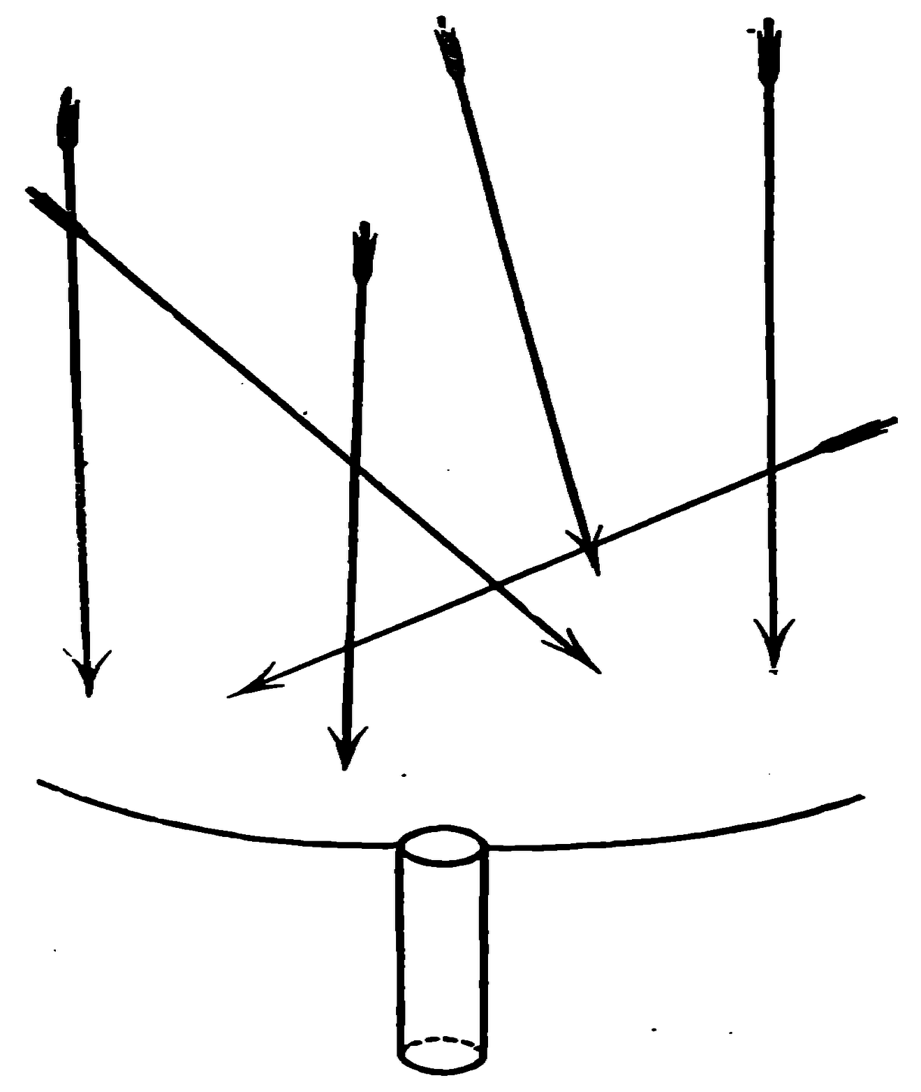
Fig. 48.—Diagrammatic, showing direction of forces and resistance in adult, in defecation.
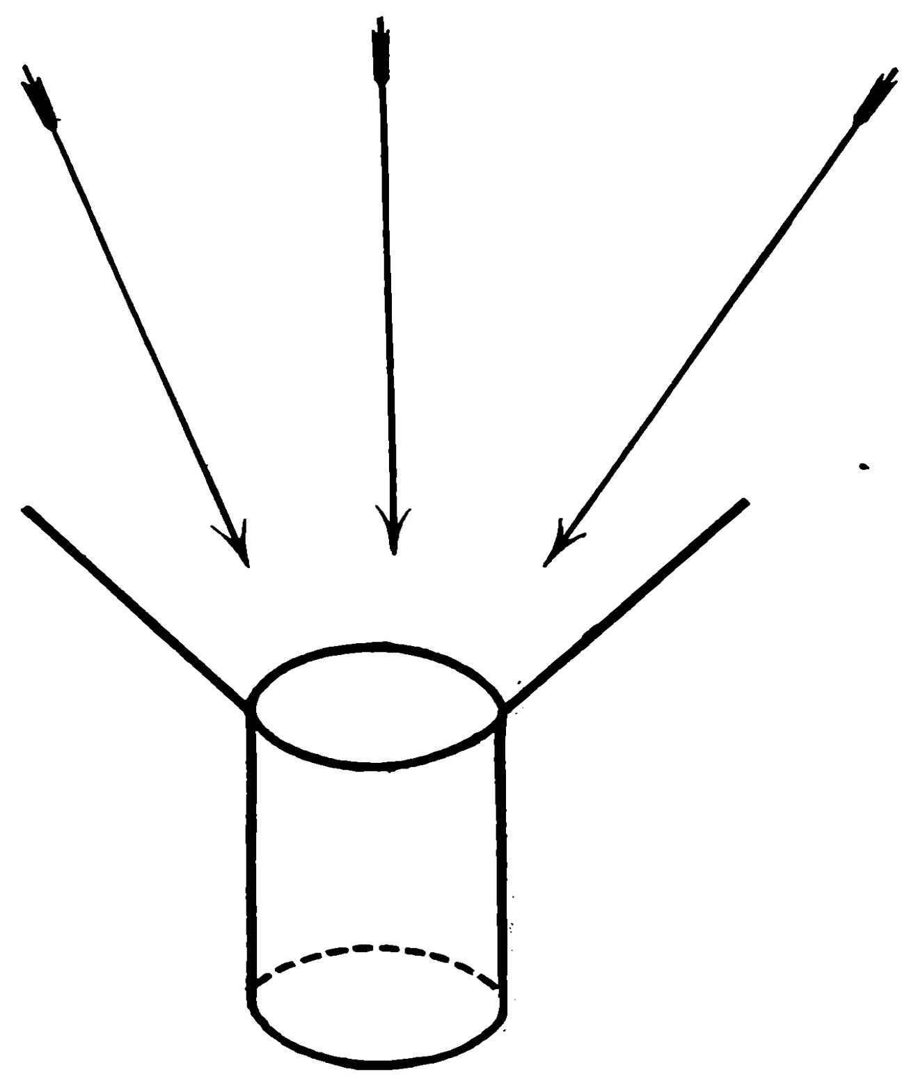
Inspection of the pictures of the dried specimens reveals the angulations referred to, which may be expected to be more numerous the more segmentary the gut’s contents.
The third feature obstructive to defecation in infants is the rectal valve. It is a feature and factor which not only is not recognized, but is one whose very anatomic existence has been persistently disputed by Matthews, Kelsey, and others. It was imperfectly described as an anatomic feature of the rectum by Mr. Houston in the Dublin Hospital Reports in 1830, and in 1887 Dr. Walter A. Otis more practically demonstrated its presence; neither of these gentlemen, however, attributed to it the characteristic element of an anatomic valve, although happily and by chance bestowing upon it its proper name. A number of investigators have discovered this organ and have miscalled it by various names and given it widely varying descriptions; but a greater number still deny its existence and critically strive to dissolve away the imperfect evidence recorded in medical literature.[17]
The pictures here shown of the dried specimens prove that in the infant the valve is such a matter of fact that its existence is no longer a question for debate.
Fig. 49.—The rectum and sigmoid of an infant aged two months. The photograph shows the interior of the anterior half. Portions of two valves are noticed at the middle part.
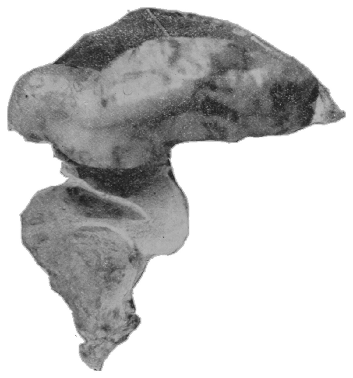
Fig. 50.—The interior view of the opposite posterior half of the rectum shown in Fig. 49, the two valves shown in Fig. 49 being continued toward each extremity of the ampulla. The sigmoid is out of focus.
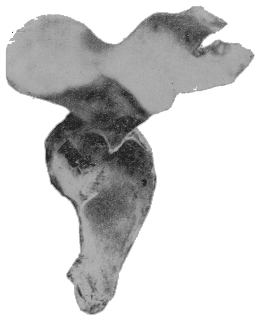
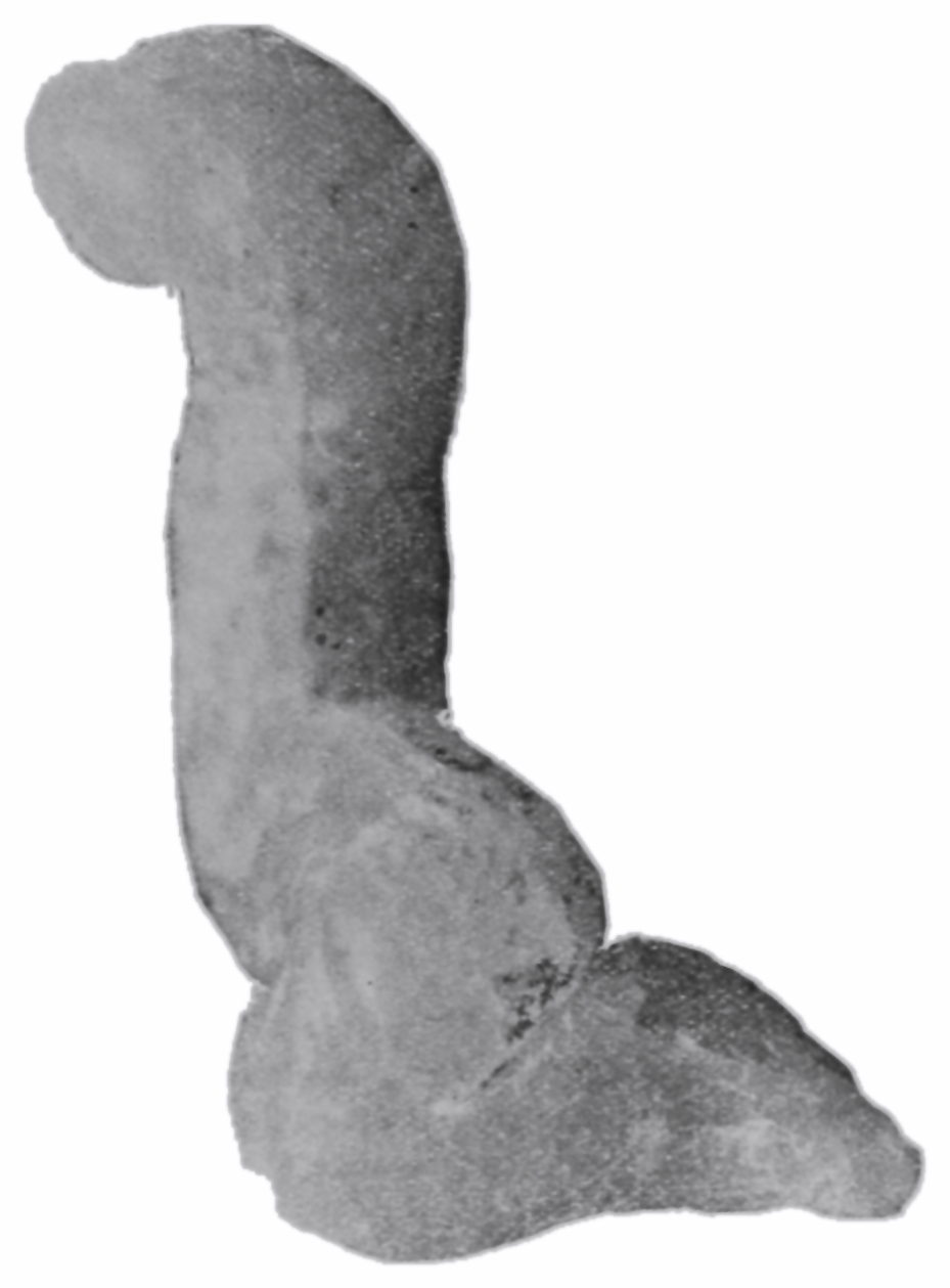
Fig. 51.—The paraffin cast removed from the gut shown in Figs. 49 and 50.
The fetal specimens pictured indicate that the valves are particularly well developed early in embryonic life. The infant specimen shown in Fig. 49 is marked by two valves, situated so close together that the middle portion of the rectum presents its longest diameter at right angles to the main direction of the gut. Another valve may be seen at the juncture of the upper portion and the sigmoid flexure.
It must be readily seen that the presence in the rectum of such a structure as an anatomic valve would be essentially obstructive to the passage of feces.
In studying the physics of the rectum it is important that we recognize that the posterior wall from which the mesentery is reflected is less movable than other parts in the circumference, hence distention of the rectum not only tends to carry a given point of its wall away from the point opposite to it, but also carries it away from a given point cephalad or caudad. So it may be assumed that if the two valves situated at the middle of the cast-distended dried specimen (Fig. 49) are three-eighths of an inch (.95 cm.) separated, and that the two opposite walls of the rectum in the portion bounded by these valves are one inch (2.54 cm.) apart, that in a state of the gut’s collapse or systole the valves would be in contact, and thus afford a very definite obstruction to the descent of semisolid feces.
The bony pelvic outlet in the infant is so contracted that the limits of anal expansion are such as to almost defeat the passage through it of other than fluid feces. It should be remembered that the normal average measurement from ischial tuberosity to ischial tuberosity in the adult is about four inches (10.16 cm.), and it is a fact proven by our observation that the average transverse diameter of the newly-born infant’s pelvic outlet is but a little more than one-half inch (1.27 cm.), the pubococcygeal measurement is even less. Those who are familiar with instrumental divulsion of the adult anus may have observed that two and a quarter inches (5.71 cm.) is the average limit of lateral separation of the anus, the remaining portion of the pelvic outlet being filled with the compressed soft tissues of the ischiorectal space. Therefore it may be estimated that nine-sixteenths at most, of the diameter of the pelvic outlet is the reasonable limit of expansibility of the anus for the passage of feces. Applying this calculation to the infant we find that if the ischial tuberosities are one-half inch (1.27 cm.) separated, that the anal expansibility is but five-sixteenth of an inch (.79 cm.), which is, as we know, the diameter of a No. 22 sound (French scale). Reference to the paraffin cast of the infant gut (Fig. 51) indicates that the average distensibility of the sigmoid flexure and rectal chambers, in which the feces when firm are formed, is four or five times that of the anal expansibility; thus it is readily perceived that compared to the adult the juxtaposition of the ischial tuberosities in the infant supplies a most obstinate obstructive factor in defecation.
To collate in brief:
1. The muscular development of the adult rectum and lower sigmoid is plainly apparent in the plates here exhibited of the fresh specimens. A deficient muscularity is observed in the infant specimens.
2. The peritoneal band of these parts in the adult is observed to be, relatively, very considerably shorter than that in the infant, and the sigmoid flexure is relatively shorter.
3. The rectal valves appear to bear the same proportion to the gut in both adult and infant, but when we remember the difference in muscular development in the two the disproportionately greater resistance of the valve in the infant rectum is obvious.
4. The anal expansibility is remembered as adequate in the adult, and is seen to be deficient in the infant.
Correlative to the facts just stated we must recognize that the adult rectum has resident within its own wall a powerful expulsive muscular mechanism; that the shortening of the mesentery holds the upper rectum steady under the applied auxiliary forces; that the angulations of the normal sigmoid are not necessarily obstructive, though in a desirable measure retardative; that the forward incline of the lower sacrum and coccyx behind, and the development of the uterus and prostate and their inherent supports in front, provide the lower adult rectum with a firm funnel-like arrangement which guides the feces directly upon the os internum of the anus; that the valves divide the feces into portions to facilitate their separate successive discharge, and finally in sequence, that the physiologic descent of the structures of the ischiorectal space reduces this last resistance to the minimum in adult defecation.
Straining, the ruptures and prolapses, obstipation, retention of feces, and the multitudinous consequent ills demand our consideration, though this study forces the conclusion, I believe, that the individual’s escape is ultimately assured by process of development, and that for the normally formed infant, the physician will find the solution of the problem of difficult defecation in the solution of the stool.
Treatment.
Diet, hygroscopic suppositories, and fluid injections which may render more fluid the intestinal contents will favor their descent through the convoluted gut, the valvulated rectum, and the contracted anus.
Massage of the abdomen over the region of the colon aids in (1) the development of the auxiliary abdominal muscles of defecation and of the intrinsic expulsory muscles in the intestinal wall; and further, such manipulations (2) directly propel the gut’s contents along the tortuous course of the bowel and hence (3) reduce, inasmuch as they may overcome the obstructive features of the valve.
If there be an overgrowth of the rectal valve and if it form an almost impassable barrier to the descent of the feces it may be in some measure overcome in infants by the dilatation which may be effected through the means of the gently introduced trained finger. In case the defective valves are present in the form of diaphragmatic strictures or membranous septa with circular aperture, the valve may be safely cut by a method described in another place.
As at birth the pelvic bones are not yet united and as the rami of the ischium and pubes are still quite cartilaginous it is obviously possible that a little energy intelligently directed upon the anus from without will spread the pelvic outlet, whereas the infant labors ineffectually with his own forces applied as they are from within. The nurse, therefore, may be directed to pare away her nail-ends, lubricate her fingers and gently introduce the smallest finger through the anus. Daily gradual dilatation of the infant’s pelvic outlet should be practised, graduating from finger to finger until the required degree of dilatation be reached. The introduction of the finger independent of its dilating uses incites the mechanism of defecation to action.
Rachitic subjects with abnormal contraction of the pelvic outlet demand forcible divulsion of the ischial tuberosities.
Fig. 52.—A three-and-a-half-months fetus. A photograph showing the posterior half of the rectum prepared by the paraffin-cast process. The rectal valves are noticeably prominent at this stage of fetal development.
Fig. 53.—A five-months fetus. A photograph showing a paraffin cast-filled rectum in situ; the other organs having been dissected away.