Defecation.
Reading edition
“Defecation is partly a voluntary and partly a reflex act. But in the infant the voluntary control has not yet been developed; in the adult it may be lost by disease; in an animal it may be abolished by operation; in each case the action becomes wholly reflex. In the normal course of events, the rectum which is empty and quiescent in the intervals of defecation, is excited to contraction as soon as feces begin to enter it through the sigmoid flexure, and the sensations caused by their presence give rise to the desire to empty the bowels. This desire may for a time be resisted by the will, or it may be yielded to. In the latter case the abdominal muscles are forcibly contracted, and the glottis being closed, the whole effect of their contraction is expended in raising the pressure within the abdomen and pelvis.... The sphincter ani is now relaxed by the inhibition of a center in the lumbar portion of the spinal cord, through the activity of which the tonic contraction of the sphincter is normally maintained. This relaxation is partially voluntary, the impulses that come from the brain acting probably through the medium of the lumbar center; but in the dog, after section of the cord in the dorsal region, the whole act of defecation, including contraction of the abdominal muscles and relaxation of the sphincter, still takes place, and here the process must be purely reflex. The contraction of the levatores ani helps to resist overdistention of the pelvic floor and to pull the anus up over the feces as they escape.”[16]
The above paragraph concisely represents the accepted interpretation of the act of defecation. The rectal valve, an individual anatomic organ in itself, must, however, be reckoned with in studying the operations of the integral mechanism of defecation.
Digital Observation of the Nonanesthetized Subject.
In the normal rectum, when the first rectal valve can be reached by the finger, provided it be immediately discovered on the introduction of the finger, a definite but not rigid band of tissue may be felt buried behind the masses of lax mucosa. This band presents at one side that which seems to be a circular aperture of variable diameter; a few seconds’ application of the passive finger detects a gradual relaxation of what first had seemed a ring-like constriction till the finger can make out but an indefinite thickening on a side of the gut-wall. If the finger be held passive, this constriction may again surround the finger; but if so, it does so with a somewhat weaker grasp. Often the presence of the finger within the rectum excites a peristalsis-like contraction, during which the valve usually cannot be detected. When the rectum is filled with water, the valve is sometimes more readily discoverable by the finger, and when discovered, the diameter of the valve-strait is greater than that before noticed, and the grasping tendency of the valve is not so noticeable.
Ocular Observation of the Anesthetized Subject.
If a somewhat emaciated male be selected, one whose rectum is susceptible to atmospheric inflation while in the lithotomy posture, and if the large intestine be freed of its contents, if such a subject be anesthetized and his colon or sigmoid filled with water, and the anus divulsed and held open with the speculum, the water confined to the rectum may be bailed out, and the rectum will then be seen to present the multichambered condition already described. If the inquirer will now place a hand on the abdomen over the sigmoid flexure, and give the part a quick, firm pressure, his watchful eye may observe a downward rush of waters into the rectum, first striking the concave wall formed by the juncture of the upper surface of the uppermost visible valve and the adjoining rectal wall, bounding from this surface to the opposite next lower valve, from which the stream is deflected to a lower point on the opposite wall, by which time all may be obscured by a wave of contracting peristalsis, rhythmically repeated, which may project small quantities of water through the anus. In the intervals of the contractions, the rectal chambers are observed to be either submerged or else atmospherically inflated.
Ocular Observation of the Nonanesthetized Subject.
If a subject under proctoscopy be directed to bear down, it is observable that the empty and atmospherically inflated rectum closes under the compression of the voluntary forces by the concave anterior wall becoming convex, and being carried backward toward the fixed posterior wall, the transverse diameters remaining apparently but little shortened. The compression may be noticed as being applied from before and in a backward direction, and simultaneously from above downwardly. In case firm fecal masses, or lubricated balls of cotton experimentally placed, are dejected by these efforts, there will be noticeable depression of the valve-floor under the pressure of the descending mass. Presently the mass will appear at the valve-edge, where it may seem suspended for a few seconds before it is plunged on into the next lower chamber. Occasional rhythmic contractions of the rectal muscular coats occur, which fill and transitorily obscure the field under observation with masses of the reduplicated mucosa, and it is always possible to note that such contractions have changed the positions of the gut’s contents.
These anatomic findings and physiologic observations warrant the conclusion that the rectal valve has a function. Its histologic elements endow it with essentially passive and active properties. When the muscular elements are relaxed and the gut is either greatly dilated or else in a lesser measure distended, the valve is passively projected across the channel to resist the hurried or uncontrolled descent of the feces. The presence of the bands of fibrous tissue under the free margin of the valve provides a guard or control to receive and retain the bolus, or, I may say, the valves receive a series of boluses, till a sufficient pressure is made to stimulate the complex involuntary mechanism of defecation to an expulsion of the feces or to a reversed peristalsis. The presence of the feces or the involuntary movements incident to their presence, signals the consciousness to cooperative voluntary expulsory effort, or gives warning of the necessity of voluntary resistance. In the event of the exercise of these forces in the direction of resistance there seems to be some reason to believe that an antiperistalsis returns the feces to the sigmoid flexure. The same arrangement which sets in operation the involuntary factors of defecation also provides for the minimum expenditure of energy on the part of the voluntary forces for the reason that the gut’s contents are collected on the surface of the unyielding sacrum and steadied there to receive the pressure of the really expulsory voluntary effort. Such an arrangement of the feces as is mentioned above further facilitates defecation for the reason that the entire contents of the rectum are not rushed upon the anus at once. The surgeon remembers that it is usually impossible to reduce a hernia en masse.
The bundles of circular muscular fibers which constitute the muscular element of the valve evidently belong to the same mechanism and have the same function as those which form the ental sphincter. Their usual state may be that of tonic contraction; they are relaxed by inhibition; when the muscle is in a state of inaction the fibrous bands are projected and support the valves across the channel of the rectum. In health the valve is susceptible, under pressure, of a temporary effacement.
If it be the function of the normal rectal valve to beneficently retard the descent of the feces it is obviously true that it may be the especial property of the valve, in certain other than normal conditions, to maliciously obstruct the descent of the feces.
My experience convinces me that a perfect knowledge of the rectal valve constitutes the key to an understanding of obstipation, rectal stricture, and their sequels.
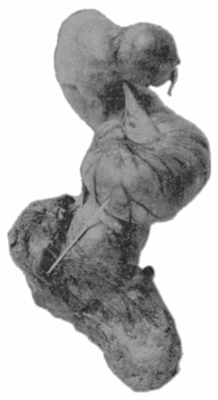
Fig. 31.—From a photograph of an anal end-view of a cast-filled rectum; the average transverse diameter was 3½ inches (8.89 cm.), the vertical diameter was 3 inches (7.62 cm.).
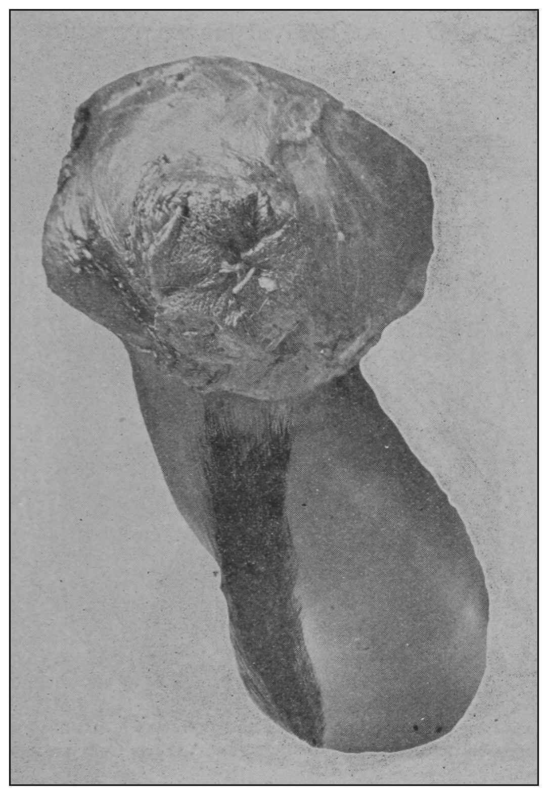
Fig. 32.—From a photograph of an external view of a cast-filled rectum and its mesenteric attachment to the sacrum; taken from an 18-months-old infant.