Part 3 — GLOSSARY OF TECHNICAL TERMS USED IN PART III.
Reading edition
Brown bodyA body formed in a zoœcium by the degeneration of a polypide as a preparation for its regeneration.
Cardiac portion (of the stomach).That part which communicates with the œsophagus.
CollarA longitudinally pleated circular membrane capable of being thrust out of the orifice in advance of the lophophore and of closing together inside the zoœcium above the tentacles when they are retracted.
Dorsal surface(Of zoœcium or polypide) the surface nearest the mouth; (of statoblast) the surface furthest from that by which the statoblast is attached to the funiculus during development.
EctocystThe outer, structureless layer of the zoœcium.
Emarginate (of a zoœcium)Having a thin or defective triangular area in the ectocyst at the tip.
EndocystThe inner, living (cellular) layer of the zoœcium.
EpistomeA leaf-like ciliated organ that projects upwards and forwards over the mouth between it and the anus.
FuniculusA strand of tissue joining the alimentary canal to the endocyst.
Furrowed (of a zoœcium)Having a thin or defective longitudinal linear streak in the ectocyst on the dorsal surface.
GizzardA chamber of the alimentary canal situated at the cardiac end of the stomach and provided internally with a structureless lining.
Intertentacular organA ciliated tube running between the cavity of the zoœcium and the external base of the lophophore.
Keeled (of a zoœcium)Having a longitudinal ridge on the dorsal surface.
LophophoreThe tentacles with the base to which they are attached.
Marginal processes (of statoblast).Chitinous hooked processes on the margin of the swim-ring (q. v.).
ŒsophagusThat part of the alimentary canal which joins the mouth to the stomach.
OrificeThe aperture through which the lophophore can be protruded from or retracted into the zoœcium.
Parietal musclesTransverse muscles running round the inner wall of the zoœcium.
Parieto-vaginal musclesMuscles that surround the orifice, running between the folds of the zoœcium in an oblique direction.
PolypariumThe whole body of zoœcia and polypides which are in organic connection.
PolypideThe tentacular crown, alimentary canal, and retractor muscles of a polyzoon-individual.
Pyloric portion (of the stomach).That part which communicates with the intestine.
Resting budAn external bud provided with food-material in its cells, with a horny external coat and capable of lying dormant in unfavourable conditions.
Retractor musclesThe muscles by the action of which the lophophore can be pulled back into the zoœcium.
StatoblastAn internal bud arising from the funiculus, containing food-material in its cells, covered with a horny coat and capable of lying dormant in unfavourable conditions.
Swim-ringA ring of polygonal air-spaces surrounding the statoblast.
Ventral surface(Of zoœcium or polypide) the surface nearest the anus; (of statoblast) the surface by which the statoblast is attached to the funiculus during development.
ZoariumThe whole body of zoœcia which are in organic connection.
ZoœciumThose parts of the polyzoon-individual which constitute a case or "house" for the polypide.
SYNOPSIS
OF THE
CLASSIFICATION OF THE POLYZOA.
I.
Synopsis of the Subclasses, Orders, and Suborders.
Class POLYZOA.
Small cœlomate animals, each individual of which consists of a polyp-like organism or polypide enclosed in a "house" or zoœcium composed partly of living tissues. The mouth is surrounded by a circle of ciliated tentacles that can be retracted within the zoœcium; the alimentary canal, which is suspended in the zoœcium, is Y-shaped and consists of three parts, the œsophagus, the stomach, and the intestine.
Subclass ENTOPROCTA.
The anus as well as the mouth is enclosed in the circle of tentacles and the zoœcium is not very distinctly separated from the polypide. Some forms are solitary or form temporary colonies by budding.
Most Entoprocta are marine, but a freshwater genus (Urnatella) occurs in N. America, while the Indian genus Loxosomatoides (fig. 34, p. 176) is only known from brackish water.
Subclass ECTOPROCTA.
The anus is outside the circle of tentacles and the zoœcium can always be distinguished from the polypide. All species form by budding permanent communities the individuals in which remain connected together by living tissue.
Order I. GYMNOLÆMATA.
Ectoproctous polyzoa the polypides of which have no epistome; the zoœcia are in nearly all cases distinctly separated from one another by transverse perforated plates.
Most of the Gymnolæmata are marine, but species belonging to two of the three suborders into which they are divided often stray into brackish water, while a few genera that belong to one of these two suborders are practically confined to fresh water. The three suborders are distinguished as follows:—
Suborder A. CHEILOSTOMATA.
The zoœcia are provided with a "lip" or lid hinged to the posterior margin of the orifice (see fig. 33, p. 175). This lid closes automatically outside the zoœcium or in a special chamber on the external surface (the "peristome") when the polypide retracts and is pushed open by the tentacles as they expand. The majority of the zoœcia in each zoarium are more or less distinctly flattened, but some of them are often modified to form "vibracula" and "avicularia."
The Cheilostomata are essentially a marine group, but some species are found in estuaries and even in pools and ditches of brackish water (fig. 33).
Suborder B. CTENOSTOMATA.
The zoœcia are provided with a collar-like membrane which is pleated vertically and closes together above the polypide inside the zoœcium when the former is retracted; it is thrust out of the zoœcium and expands into a ring-shaped form just before the tentacles are extruded. The zoœcia are usually more or less tubular, but in some genera and species are flattened.
The majority of the Ctenostomata are marine, but some genera are found in estuaries, while those of one section of the suborder live almost exclusively in fresh water.
Suborder C. CYCLOSTOMATA.
The zoœcia are provided neither with a lip nor with a collar-like membrane. They are tubular and usually have circular orifices.
The Cyclostomata are exclusively marine.
Order II. PHYLACTOLÆMATA.
Ectoproctous polyzoa the polypides of which have a leaf-shaped organ called an epistome projecting upwards and forwards within the circle of tentacles and between the mouth and the anus. The zoœcia are not distinct from one another, but in dendritic forms the zoarium is divided irregularly by chitinous partitions.
The Phylactolæmata are, without exception, freshwater species.
II.
Synopsis of the Leading Characters of the Divisions of the Suborder Ctenostomata.
Suborder B. CTENOSTOMATA.
The suborder has been subdivided in various ways by different authors. The system here adopted is essentially the same as that proposed in a recent paper by Waters (Journ. Linn. Soc. London, Zool. xxi, p. 231, 1910), but I have thought it necessary to add a fourth division to the three adopted by that author, namely, the Alcyonellea, Stolonifera, and Vesicularina. This new division includes all the freshwater genera and may be known as the Paludicellina. In none of these divisions are the tentacles webbed at the base.
The four divisions may be recognized from the following synopsis of their characteristic features:—
Division I. ALCYONELLEA.
The zoœcia arise directly from one another in a fleshy or gelatinous mass. The polypide has no gizzard. The species are essentially marine, but a few are found in brackish water in estuaries.
Division II. STOLONIFERA.
The zoœcia arise from expansions in a delicate creeping rhizome or root-like structure, the order in which they are connected together being more or less irregular. As a rule (perhaps always) there is no gizzard. The species are marine.
Division III. VESICULARINA.
The zoœcia grow directly from a tubular stem which is usually free and vertical, their arrangement being alternate, spiral or irregular. There is a stout gizzard which bears internal chitinous projections and is tightly compressed when the polypide is retracted. The species are essentially marine, but a few are found in brackish water.
Division IV. PALUDICELLINA, nov.
The zoœcia are arranged in a regular cruciform manner and arise either directly one from another or with the intervention of tubular processes. If the polypide has a gizzard it does not bear internal chitinous projections. Most of the species are confined to fresh water, but a few are found in brackish water or even in the sea.
Although all true freshwater Ctenostomes belong to the fourth of these divisions, species of a genus (Bowerbankia) included in the third are so frequently found in brackish water and in association with one belonging to the fourth, and are so easily confounded with the latter, that I think it necessary to include a brief description of the said genus and of the form that represents it in ponds of brackish water in India.
SYSTEMATIC LIST OF THE INDIAN FRESHWATER POLYZOA.
[The types have been examined in the case of all species, etc., whose names are marked thus, *.]
Order I. GYMNOLÆMATA.
Suborder I. CTENOSTOMATA.
[Division III. Vesicularina.]
[Genus Bowerbankia, Farre (1837).]
[B. caudata subsp. bengalensis*, Annandale (1907).
(Brackish water).]
Division IV. Paludicellina, nov.
Family I. PALUDICELLIDÆ.
Genus 1. Paludicella, Gervais (1836).
? Paludicella sp. (fide Carter).
Genus 2. Victorella, Kent (1870).
26.
V. bengalensis*, Annandale (1907).
Family II. HISLOPIIDÆ.
Genus Hislopia, Carter (1858).
27.
H. lacustris, Carter (1858).
27 a.
H. lacustris subsp. moniliformis*, nov.
Order II. PHYLACTOLÆMATA.
Division I. Plumatellina.
Family 1. FREDERICELLIDÆ.
Genus Fredericella, Gervais (1836).
28.
F. indica*, Annandale (1909).
Family 2. PLUMATELLIDÆ.
Subfamily A. Plumatellinæ.
Genus 1. Plumatella, Lamarck (1816).
29.
P. fruticosa, Allman (1844).
30.
P. emarginata, Allman (1844).
31.
P. javanica*, Kraepelin (1905).
32.
P. diffusa, Leidy (1851).
33.
P. allmani, Hancock (1850).
34.
P. tanganyikæ*, Rousselet (1907).
35.
P. punctata, Hancock (1850).
Genus 2. Stolella, Annandale (1909).
36.
S. indica*, Annandale (1909).
Subfamily B. Lophopinæ.
Genus 1. Lophopodella, Rousselet (1904).
37.
L. carteri* (Hyatt) (1865).
37 a.
L. carteri var. himalayana* (Annandale) (1907).
Genus 2. Pectinatella, Leidy (1851).
38.
P. burmanica*, Annandale (1908).
Order CTENOSTOMATA.
[Division VESICULARINA.
Family VESICULARIDÆ.
Vesicularidæ, Hincks, Brit. Marine Polyzoa, p. 512 (1880).
Zoœcia constricted at the base, deciduous, attached to a stem that is either recumbent or vertical.
Genus BOWERBANKIA, Farre.
Bowerbankia, Farre, Phil. Trans. Roy. Soc. cxxvii, p. 391 (1837). Bowerbankia, Hincks, op. cit. p. 518.
Zoarium vertical or recumbent. Zoœcia ovate or almost cylindrical, arranged on the stem singly, in clusters or in a subspiral line. Polypide with 8 or 10 tentacles.
Bowerbankia caudata, Hincks.
Bowerbankia caudata, Hincks, op. cit. p. 521, pl. lxxv, figs. 7, 8.
This species is easily distinguished from all others by the fact that mature zoœcia have always the appearance of being fixed to the sides of a creeping, adherent stem and are produced, below the point at which they are thus fixed, into a pointed "tail."
Subsp. bengalensis, Annandale.
Bowerbankia caudata, Thornely, Rec. Ind. Mus. i, p. 196 (1907). Bowerbankia caudata, Annandale, ibid. p. 203. Bowerbankia caudata race bengalensis, id., ibid. ii. p. 13 (1908).
The Indian race is only distinguished from the typical form by its greater luxuriance of growth and by the fact that the "tail" of the zoœcia is often of relatively great length, sometimes equaling or exceeding the rest of the zoœcium. The stem, which is divided at irregular intervals by partitions, often crosses and recrosses its own course and even anastomoses, and a fur-like structure is formed in which the zoœcia representing the hairs become much elongated; but upright branches are never formed. The zoarium has a greenish or greyish tinge.
Type in the Indian Museum.
Geographical Distribution.—B. caudata subsp. bengalensis is common in brackish water in the Ganges delta, where it often occurs in close association with Victorella bengalensis, and also at the south end of the Chilka Lake in the north-east of the Madras Presidency. Although it has not yet been found elsewhere, it probably occurs all round the Indian coasts.]
Division PALUDICELLINA, nov.
This division consists of two very distinct families, the species of which are easily distinguished at a glance by the fact that in one (the Paludicellidæ) the zoœcia are tubular, while in the other (the Hislopiidæ) they are broad and flattened. The anatomical and physiological differences between the two families are important, and they are associated together mainly on account of the method of budding by means of which their zoaria are produced.
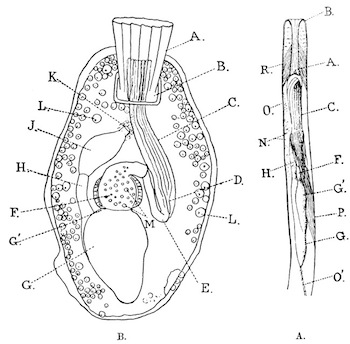
Fig. 35.—Single zoœcia of Victorella and Hislopia (magnified).
A, zoœcium of Victorella pavida, Kent, with the polypide retracted (after Kraepelin). B, zoœcium of Hislopia lacustris, Carter (typical form from the United Provinces), with the collar completely and the tentacles partly protruded. A=collar; B=orifice; C=tentacles; D=pharynx; E=œsophagus proper; F=gizzard; G=stomach; G'=cardiac portion of stomach; H=intestine; J=rectum; K=anus; L=young egg; M=green cysts in gizzard; N=testes; O=ovary; O'=funiculus. The muscles are omitted except in fig. B.
Family PALUDICELLIDÆ.
Paludicellidæ, Allman, Mon. Fresh-Water Polyzoa, p. 113 (1857). Homodiætidæ, Kent, Q. J. Micr. Sci. x, p. 35 (1870). Victorellidæ, Hincks, Brit. Marine Polyzoa, p. 558 (1880). Paludicellidées, Jullien, Bull. Soc. zool. France, x, p. 174 (1885). Paludicellides, Loppens, Ann. Biol. lacustre, iii, p. 170 (1908). Victorellides, id., ibid. p. 171.
Zoarium. The zoarium is recumbent or erect, and is formed typically either of zoœcia arising directly in cruciform formation from one another, or of zoœcia joined together in similar formation with the intervention of tubules arising from their own bases. Complications often arise, however, either on account of the suppression of the lateral buds of a zoœcium, so that the formation becomes linear instead of cruciform, or by the production in an irregular manner of additional tubules and buds from the upper part of the zoœcia. A confused and tangled zoarium may thus be formed, the true nature of which can only be recognized by the examination of its terminal parts.
Zoœcia. The zoœcia are tubular and have a terminal or subterminal orifice, which is angulate or subangulate as seen from above. Owing to this fact, to the stiff nature of the external ectocyst, to the action of circular muscles that surround the tentacular sheath, and to the cylindrical form of the soft inverted part, the orifice, as seen from above, appears to form four flaps or valves, thus .

Polypide. The alimentary canal is elongate and slender as a whole, the œsophagus (including the pharynx) being of considerable length. In Paludicella and Pottsiella the œsophagus opens directly into the cardiac limb of the stomach, which is distinctly constricted at its base; but in Victorella the base of the œsophagus is constricted off from the remainder to form an elongate oval sac the walls of which are lined with a delicate structureless membrane. Victorella may therefore be said to possess a gizzard, but the structure that must be so designated has not the function (that of crushing food) commonly associated with the name, acting merely as a chamber for the retention of solid particles. In this genus the cardiac limb of the stomach is produced and vertical but not constricted at the base. The tentacles in most species number 8, but in Paludicella there are 16.
Resting buds. The peculiar structures known in Europe as "hibernacula" are only found in this family. The name hibernacula, however, is inappropriate to the only known Indian species as they are formed in this country at the approach of summer instead of, as in Europe and N. America, at that of winter. It is best, therefore, to call them "resting buds." They consist of masses of cells congregated at the base of the zoœcia, gorged with food material and covered with a resistant horny covering.
The family Paludicellidæ consists of three genera which may be distinguished as follows:—
I.Orifice terminal; main axis of the zoœcium vertical; zoœcia separated from one another by tubules.
[A.Base of the zoœcia not swollen; no adventitious budsPottsiella.]
B.Base of the zoœcium swollen; adventitious buds produced near the tipVictorella, p. 194.
II.Orifice subterminal, distinctly on the dorsal surface; main axis of the zoœcium horizontal (the zoarium being viewed from the dorsal surface); buds not produced at the tip of the zoœciaPaludicella, p. 192.
Of these three genera, Pottsiella has not yet been found in India and is only known to occur in N. America. It consists of one species, P. erecta (Potts) from the neighbourhood of Philadelphia in the United States.
Victorella includes four species, V. pavida known from England and Germany and said to occur in Australia, V. mülleri from Germany (distinguished by possessing parietal muscles at the tip of the zoœcia), V. symbiotica from African lakes and V. bengalensis from India. These species are closely related.
Paludicella is stated by Carter to have been found in Bombay, but probably what he really found was the young stage of V. bengalensis. A single species is known in Europe and N. America, namely P. ehrenbergi, van Beneden (=Alcyonella articulata, Ehrenberg).
I have examined specimens of all the species of this family as yet known.
Genus 1. PALUDICELLA, Gervais.
Paludicella, Gervais, Compt. Rend. iii, p. 797 (1836). Paludicella, Allman, Mon. Fresh-Water Polyzoa, p. 113 (1857). ? Paludicella, Carter, Ann. Nat. Hist. (3) iii, p. 333 (1859). Paludicella, Jullien, Bull. Soc. zool. France, x, p. 174 (1885). Paludicella, Kraepelin, Deutsch. Süsswasserbryozoen, i, p. 96 (1887). Paludicella, Loppens, Ann. Biol. lacustre, iv, p. 14 (1910).
Zoarium. The nature of the zoarium in this genus is well expressed by Ehrenberg's specific name "articulata," although the name was given under a false impression. The zoœcia arise directly from one another in linear series with occasional side-branches. The side-branches are, however, often suppressed. The zoarium as a whole is either recumbent and adherent or at least partly vertical.
Zoœcia. Although the zoœcia are distinctly tubular as a whole, two longitudinal axes may be distinguished in each, for the tip is bent upwards in a slanting direction, bearing the orifice at its extremity. The main axis is, however, at right angles to the dorso-ventral axis, and the dorsal surface, owing to the position of the aperture, can always be readily distinguished from the ventral, even when the position of the zoœcium is vertical. Each zoœcium tapers towards the posterior extremity. Parietal muscles are always present.
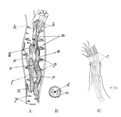
Fig. 36.—Structure of Paludicella ehrenbergi (A and B after Allman).
A=a single zoœcium with the polypide retracted. B=the base of the lophophore as seen from above with the tentacles removed. C=the orifice of a polypide with the collar expanded and the tentacles partly retracted. a=tentacles; c=collar; d=mouth; e=œsophagus; f=stomach; g=intestine; k=parieto-vaginal muscles; p=parietal muscles; o=cardiac part of the stomach; r=retractor muscle; s=funiculus.
Polypide. The most striking features of the polypide are the absence of any trace of a gizzard and the highly specialized form assumed by the cardiac part of the stomach. There are two funiculi, both connecting the pyloric part of the stomach with the endocyst. The ovary develops at the end of the upper, the testis at that of the lower funiculus.
Resting buds. The resting buds are spindle-shaped.
Kraepelin recognized two species in the genus mainly by their method of growth and the number of tentacles. In his P. mülleri the zoarium is always recumbent and the polypide has 8 tentacles, whereas in P. articulata or ehrenbergi the tentacles number 16 and upright branches are usually developed. It is probable, however, that the former species should be assigned to Victorella, for it is often difficult to distinguish Paludicella from young specimens of Victorella unless the latter bear adventitious terminal buds. The gizzard of Victorella can be detected in well-preserved material even under a fairly low power of the microscope, and I have examined specimens of what I believe to be the adult of mülleri which certainly belong to that genus.
It is always difficult to see the collar of Paludicella, because of its transparency and because of the fact that its pleats are apparently not strengthened by chitinous rods as is usually the case. Allman neither mentions it in his description of the genus nor shows it in his figures, and Loppens denies its existence, but it is figured by Kraepelin and can always be detected in well-preserved specimens, if they are examined carefully. If the collar were actually absent, its absence would separate Paludicella not only from Victorella and Pottsiella, but also from all other ctenostomes. In any case, Victorella is distinguished from Paludicella and Pottsiella by anatomical peculiarities (e. g., the possession of a gizzard and the absence of a second funiculus) that may ultimately be considered sufficiently great to justify its recognition as the type and only genus of a separate family or subfamily.
The description of Paludicella is included here on account of Carter's identification of the specimens he found at Bombay; but its occurrence in India is very doubtful.
Genus 2. VICTORELLA, Kent.
Victorella, Kent, Q. J. Micr. Sci. x, p. 34 (1870). Victorella, Hincks, Brit. Marine Polyzoa, p. 559 (1880). Victorella, Kraepelin, Deutsch. Süsswasserbryozoen, i, p. 93 (1887).
Type, Victorella pavida, Kent.
Zoarium. The zoarium consists primarily of a number of erect or semi-erect tubular zoœcia joined together at the base in a cruciform manner by slender tubules, but complications are introduced by the fact that adventitious buds and tubules are produced, often in large numbers, round the terminal region of the zoœcia, and that these buds are often separated from their parent zoœcium by a tubule of considerable length, and take root among other zoœcia at a distance from their point of origin. A tangled mass may thus be formed in which it is difficult to recognize the regular arrangement of the zoœcia that can be readily detached at the growing points of the zoarium.
Zoœcia. The zoœcia when young closely resemble those of Paludicella, but as they grow the terminal upturned part increases rapidly, while the horizontal basal part remains almost stationary and finally appears as a mere swelling at the base of an almost vertical tube, in which by far the greater part, if not the whole, of the polypide is contained. Round the terminal part of this tube adventitious buds and tubules are arranged more or less regularly. There are no parietal muscles.
Polypide. The polypide has 8 slender tentacles, which are thickly covered with short hairs. The basal part of the œsophagus forms a thin-walled sac (the "gizzard") constricted off from the upper portion and bearing internally a thin structureless membrane. Circular muscles exist in its wall but are not strongly developed on its upper part. There is a single funiculus, which connects the posterior end of the stomach with the base of the zoœcium. The ovaries and testes are borne on the endocyst, not in connection with the funiculus.
Resting buds. The resting buds are flattened or resemble young zoœcia in external form.
Victorella, although found in fresh water, occurs more commonly in brackish water and is known to exist in the littoral zone of the sea.
26. Victorella bengalensis, Annandale.
Victorella pavida, Annandale (nec Kent), Rec. Ind. Mus. i, p. 200, figs. 1-4 (1907). Victorella bengalensis, id., ibid. ii, p. 12, fig. 1 (1908).
Zoarium. The mature zoarium resembles a thick fur, the hairs of which are represented by elongate, erect, slender tubules (the zoœcia), the arrangement of the whole being very complicated and irregular. The base of the zoarium often consists of an irregular membrane formed of matted tubules, which are sometimes agglutinated together by a gummy secretion. The zoarium as a whole has a faint yellowish tinge.
Zoœcia. The zoœcia when young are practically recumbent, each being of an ovoid form and having a stout, distinctly quadrate orificial tubule projecting upwards and slightly forwards near the anterior margin of the dorsal surface. At this stage a single tubule, often of great relative length, is often given off near the orifice, bearing a bud at its free extremity. As the zoœcium grows the tubular part becomes much elongated as compared with the basal part and assumes a vertical position. Its quadrate form sometimes persists but more often disappears, so that it becomes almost circular in cross-section throughout its length. Buds are produced near the tip in considerable profusion. As a rule, if they appear at this stage, the tubule connecting them with the parent zoœcium is short or obsolete; sometimes they are produced only on one side of the zoœcium, sometimes on two. The buds themselves produce granddaughter and great-granddaughter buds, often connected together by short tubules, while still small and imperfectly developed. The swelling at the base of the zoœcium, when the latter is fully formed, is small.
Polypide. The polypide has the features characteristic of the genus. The base of the gizzard is surrounded by a strong circular muscle.
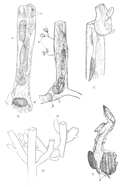
Fig. 37.—Victorella bengalensis (type specimens).
A=single zoœcium without adventitious buds but with a young resting bud (b), × 70 (dorsal view); B=lateral view of a smaller zoœcium without buds, × 70; C=upper part of a zoœcium with a single adventitious bud, × 70; D=outline of the upper part of a zoœcium with adventitious buds of several generations, × 35; E=remains of a zoœcium with two resting buds (b) attached. All the specimens figured are from Port Canning and, except D, are represented as they appear when stained with borax carmine and mounted in canada balsam.
Resting buds. The resting buds (fig. 31, p. 170) are somewhat variable in shape but are always flat with irregular cylindrical or subcylindrical projections round the margin, on which the horny coat is thinner than it is on the upper surface. This surface is either smooth or longitudinally ridged.
Type in the Indian Museum.
This species differs from the European V. pavida in very much the same way as, but to a greater extent than, the Indian race of Bowerbankia caudata does from the typical English one (see p. 189). The growth of the zoarium is much more luxuriant, and the form of the resting buds is different.
Geographical Distribution.—V. bengalensis is abundant in pools of brackish water in the Ganges delta and in the Salt Lakes near Calcutta; it also occurs in ponds of fresh water near the latter. I have received specimens from Madras from Dr. J. R. Henderson, and it is probable that the form from Bombay referred by Carter to Paludicella belonged to this species.
Biology.—In the Ganges delta V. bengalensis is usually found coating the roots and stems of a species of grass that grows in and near brackish water, and on sticks that have fallen into the water. It also spreads over the surface of bricks, and I have found a specimen on a living shell of the common mollusc Melania tuberculata. Dr. Henderson obtained specimens at Madras from the surface of a freshwater shrimp, Palæmon malcolmsonii. In the ponds at Port Canning the zoaria grow side by side with, and even entangled with those of Bowerbankia caudata subsp. bengalensis, to the zoœcia of which their zoœcia bear a very strong external resemblance so far as their distal extremity is concerned. This resemblance, however, disappears in the case of zoœcia that bear terminal buds, for no such buds are borne by B. caudata; and the yellowish tint of the zoaria of V. bengalensis is characteristic. Zoaria of the entoproct Loxosomatoides colonialis and colonies of the hydroid Irene ceylonensis are also found entangled with the zoaria of V. bengalensis, the zoœcia of which are often covered with various species of Vorticellid protozoa and small rotifers. The growth of V. bengalensis is more vigorous than that of the other polyzoa found with it, and patches of B. caudata are frequently surrounded by large areas of V. bengalensis.
The food of V. bengalensis consists largely of diatoms, the siliceous shells of which often form the greater part of its excreta. Minute particles of silt are sometimes retained in the gizzard, being apparently swallowed by accident.
There are still many points to be elucidated as regards the production and development of the resting buds in V. bengalensis, but two facts are now quite clear as regards them: firstly, that these buds are produced at the approach of the hot weather and germinate in November or December; and secondly, that the whole zoarium may be transformed at the former season into a layer of resting buds closely pressed together but sometimes exhibiting in their arrangement the typical cruciform formation. Resting buds may often be found in vigorous colonies as late as the beginning of December; these buds have not been recently formed but have persisted since the previous spring and have not yet germinated. Sometimes only one or two buds are formed at the base of an existing zoœcium (fig. 37 a), but apparently it is possible not only for a zoœcium to be transformed into a resting bud but for it to produce four other buds round its base before undergoing the change. Young polypides are formed inside the buds and a single zoœcium sprouts out of each, as a rule by the growth of one of the basal projections, when conditions are favourable.
Polypides of V. bengalensis are often transformed into brown bodies. When this occurs the orifice closes together, with the collar expanded outside the zoœcium. I have occasionally noticed that the ectocyst of such zoœcia was distinctly thicker and darker in colour than that of normal zoœcia.
Eggs and spermatozoa are produced in great numbers, as a rule simultaneously in the same zoœcia, but individuals kept in captivity often produce spermatozoa only. The eggs are small and are set free as eggs. Nothing is known as regards their development.
Polypides are as a rule found in an active condition only in the cold weather, but I have on one occasion seen them in this condition in August, in a small zoarium attached to a shell of Melania tuberculata taken in a canal of brackish water near Calcutta.
Family HISLOPIIDÆ.
Hislopidées, Jullien, Bull. Soc. zool. France, x, p. 180 (1885). Hislopiidæ, Annandale, Rec. Ind. Mus. i, p. 200 (1907).
Zoarium recumbent, often forming an almost uniform layer on solid subjects.
Zoœcia flattened, adherent; the orifice dorsal, either surrounded by a chitinous rim or situated at the tip of an erect chitinous tubule; no parietal muscles.
Polypide with an ample gizzard which possesses a uniform chitinous lining and does not close together when the polypide is retracted.
Resting bud, not produced.
Only two genera can be recognized in this family, Arachnoidea, Moore, from Central Africa, and Hislopia, Carter, which is widely distributed in Eastern Asia. The former genus possesses an upright orificial tubule and has zoœcia separated by basal tubules. Its anatomy is imperfectly known, but it certainly possesses a gizzard of similar structure to that of Hislopia, between which and Victorella its zoœcium is intermediate in form.
Genus HISLOPIA, Carter.
Hislopia, Carter, Ann. Nat. Hist. (3) i, p. 169 (1858). Hislopia, Stolickza, J. As. Soc. Bengal, xxxviii (2), p. 61 (1869). Norodonia, Jullien, Bull. Soc. zool. France, v, p. 77 (1880). Hislopia, id., ibid. x, p. 183 (1885). Norodonia, id., ibid. p. 180. Echinella, Korotneff, Biol. Centrbl. xxi, p. 311 (1901). Hislopia, Annandale, J. As. Soc. Bengal (new series) ii, p. 59 (1906). Hislopia, Loppens, Ann. Biol. lacustre, iii, p. 175 (1908).
Type, Hislopia lacustris, Carter.
Zoarium. The zoarium consists primarily of a main axis running in a straight line, with lateral branches that point forwards and outwards. Further proliferation, however, often compacts the structure into an almost uniform flat area.
Zoœcia. The zoœcia (fig. 35 B, p. 190) are flat and have the orifice surrounded by a chitinous rim but not much raised above the dorsal surface. They arise directly one from another.
Polypide. The polypide possesses from 12 to 20 tentacles. Its funiculus is rudimentary or absent. Neither the ovaries nor the testes have any fixed position on the lateral walls of the zoœcium to which they are confined.
The position of this genus has been misunderstood by several zoologists. Carter originally described Hislopia as a cheilostome allied to Flustra; in 1880 Jullien perpetuated the error in describing his Norodonia, which was founded on dried specimens of Carter's genus; while Loppens in 1908 still regarded the two "genera" as distinct and placed them both among the cheilostomes. In 1885, however, Jullien retracted his statement that Norodonia was a cheilostome and placed it, together with Hislopia, in a family of which he recognized the latter as the eponymic genus. Carter's mistake arose from the fact that he had only examined preserved specimens, in which the thickened rim of the orifice is strongly reminiscent of the "peristome" of certain cheilostomes, while the posterior of the four folds into which the tentacle sheath naturally falls (as in all ctenostomes, cf. the diagram on p. 191) is in certain conditions rather larger than the other three and suggests the "lip" characteristic of the cheilostomes. If living specimens are examined, however, it is seen at once that the posterior fold, like the two lateral folds and the anterior one, changes its form and size from time to time and has no real resemblance to a "lip."
That there is a remarkable, if superficial, resemblance both as regards the form of the zoœcium and as regards the method of growth between Hislopia and certain cheilostomes cannot be denied, but the structure of the orifice and indeed of the whole organism is that of a ctenostome and the resemblance must be regarded as an instance of convergence rather than of genetic relationship.
The most striking feature of the polypide of Hislopia is its gizzard (fig. 38, p. 201) which is perhaps unique (except for that of Arachnoidea) both in structure and function. In structure its peculiarities reside mainly in three particulars: (i), it is not constricted off directly from the thin-walled œsophageal tube, but possesses at its upper extremity a thick-walled tubular portion which can be entirely closed from the œsophagus at its upper end but always remains in communication with the spherical part of the gizzard; (ii), this spherical part of the gizzard is uniformly lined with a thick chitinous or horny layer which in optical section has the appearance of a pair of ridges; and (iii), there is a ring of long and very powerful cilia round the passage from the gizzard to the stomach. The cardiac limb of the stomach, which is large and heart-shaped, is obsolete. The wall of the spherical part of the gizzard consists of two layers of cells, an outer muscular layer consisting of powerful circular muscles and an inner glandular layer, which secretes the chitinous lining. The inner walls of the tubular part consist of non-ciliated columnar cells, and when the polypide is retracted it lies almost at right angles to the main axis of the zoœcium.
The spherical part of the gizzard invariably contains a number of green cells, which lie free in the liquid it holds and are kept in motion by the cilia at its lower aperture. The majority of these cells can be seen with the aid of a high power of the microscope to consist of a hard spherical coat or cyst containing green protoplasm in which a spherical mass of denser substance (the nucleus) and a number of minute transparent granules can sometimes be detected. The external surface of many of the cysts is covered with similar granules, but some are quite clean.
There can be no doubt that these cysts represent a stage in the life-history of some minute unicellular plant or animal. Indeed, although it has not yet been found possible to work out this life-history in detail, I have been able to obtain much evidence that they are the resting stage of a flagellate organism allied to Euglena which is swallowed by the polyzoon and becomes encysted in its gizzard, extruding in so doing from its external surface a large proportion of the food-material that it has stored up within itself in the form of transparent granules. It may also be stated that some of the organisms die and disintegrate on being received into the gizzard, instead of encysting themselves.
So long as the gizzard retains its spherical form the green cells and its other contents are prevented from entering the stomach by the movements of the cilia that surround its lower aperture, but every now and then, at irregular intervals, the muscles that form its outer wall contract. The chitinous lining although resilient and not inflexible is too stiff to prevent the lumen of the gizzard being obliterated, but the action of the muscles changes its contents from a spherical to an ovoid form and in so doing presses a considerable part of them down into the stomach, through the ring of the cilia.
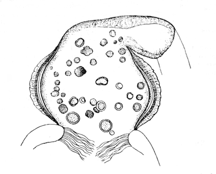
Fig. 38.—Optical section of gizzard of Hislopia lacustris, with contained green cysts, × 240.
The contraction of the gizzard is momentary, and on its re-expansion some of the green cysts that have entered the stomach are often regurgitated into it. Some, however, remain in the stomach, in which they are turned round and round by the action of the cilia at both apertures. They are apparently able to retain their form for some hours in these circumstances but finally disintegrate and disappear, being doubtless digested by the juices poured out upon them by the glandular lining of the stomach. In polypides kept under observation in clean tap-water all the cysts finally disappear, and the fæces assume a green colour. In preserved specimens apparently unaltered cysts are sometimes found in the rectum, but this is exceptional: I have observed nothing of the kind in living polypides. Cysts often remain for several days unaltered in the gizzard.
Imperfect as these observations are, they throw considerable light on the functions of the gizzard in Hislopia. Primarily it appears to act as a food-reservoir in which the green cysts and other minute organisms can be kept until they are required for digestion. When in the gizzard certain organisms surrender a large proportion of the food-material stored up for their own uses, and this food-material doubtless aids in nourishing the polyzoon. Although the cysts in the gizzard are frequently accompanied by diatoms, the latter are not invariably present. The cysts, moreover, are to be found in the zoœcia of polypides that have formed brown bodies, often being actually enclosed in the substance of the brown body. The gizzards of the specimens of Arachnoidea I have examined contain cysts that resemble those found in the same position in Hislopia.
Hislopia is widely distributed in the southern part of the Oriental Region, and, if I am right in regarding Echinella, Korotneff as a synonym, extends its range northwards to Lake Baikal. It appears to be a highly specialized form but is perhaps related, through Arachnoidea, to Victorella.
27. Hislopia lacustris, Carter.
Hislopia lacustris, Carter, Ann. Nat. Hist. (3) i, p. 170, pl. vii, figs. 1-3 (1858). Norodonia cambodgiensis, Jullien, Bull. Soc. zool. France, v, p. 77, figs. 1-3 (1880). Norodonia sinensis, id., ibid. p. 78, figs. 1-3. Norodonia cambodgiensis, id., ibid. x, p. 181, figs. 244, 245 (1885). Norodonia sinensis, id., ibid. p. 182, figs. 246, 247. Hislopia lacustris, Annandale, J. As. Soc. Bengal (new series) iii, p. 85 (1907). Hislopia lacustris, Walton, Rec. Ind. Mus. i, p. 177 (1907). Hislopia lacustris, Kirkpatrick, ibid. ii, p. 98 (1908). Hislopia lacustris, Walton, ibid. iii, p. 295 (1909).
Zoarium. The zoarium forms a flat, more or less solid layer and is closely adherent to foreign objects. As a rule it covers a considerable area, with radiating branches at the edges; but when growing on slender twigs or the stems of water-plants it forms narrow, closely compressed masses. One zoœcium, however, never grows over another.
Zoœcia. The zoœcia are variable in shape. In zoaria which have space for free expansion they are as a rule irregularly oval, the posterior extremity being often narrower than the anterior; but small triangular zoœcia and others that are almost square may often be found. When growing on a support of limited area the zoœcia are smaller and as a rule more elongate. The orifice is situated on a slight eminence nearer the anterior than the posterior margin of the dorsal surface. It is surrounded by a strong chitinous rim, which is usually square or subquadrate but not infrequently circular or subcircular. Sometimes a prominent spine is borne at each corner of the rim, but these spines are often vestigial or absent; they are rarely as long as the transverse diameter of the orifice. The zoœcium is usually surrounded by a chitinous margin, and outside this margin there is often a greater or less extent of adherent membrane. In some zoœcia the margin is obsolete or obsolescent. The dorsal surface is of a glassy transparency but by no means soft.
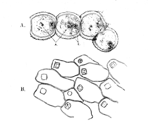
Fig. 39.—Hislopia lacustris.
A=part of a zoarium of the subspecies moniliformis (type specimen, from Calcutta), × 15; A=green cysts in gizzard; E=eggs. B=outline of part of a zoarium of the typical form of the species from the United Provinces, showing variation in the form of the zoœcia and of the orifice, × 15.
Polypide. The polypide has from 12 to 20 tentacles, 16 being a common number.
Type probably not in existence. It is not in the British Museum and Prof. Dendy, who has been kind enough to examine the specimens from Carter's collection now in his possession, tells me that there are none of Hislopia among them.
27 a. Subsp. moniliformis, nov.
Hislopia lacustris, Annandale, J. As. Soc. Bengal (new series) ii, p. 59, fig. 1 (1906).
In this race, which is common in Calcutta, the zoœcia are almost circular but truncate or concave anteriorly and posteriorly. They form linear series with few lateral branches. I have found specimens occasionally on the shell of Vivipara bengalensis, but they are much more common on the leaves of Vallisneria spiralis.
Type in the Indian Museum.
The exact status of the forms described by Jullien as Norodonia cambodgiensis and N. sinensis is doubtful, but I see no reason to regard them as specifically distinct from H. lacustris, Carter, of which they may be provisionally regarded as varieties. The variety cambodgiensis is very like my subspecies moniliformis but has the zoœcia constricted posteriorly, while var. sinensis, although the types were found on Anodonta shells on which there was plenty of room for growth, resemble the confined phase of H. lacustris so far as the form of their zoœcia and of the orifice is concerned.
Geographical Distribution.—The typical form is common in northern India and occurs also in Lower Burma; the subspecies moniliformis appears to be confined to Lower Bengal, while the varieties cambodgiensis and sinensis both occur in China, the former having been found also in Cambodia and Siam. Indian and Burmese localities are:—Bengal, Calcutta (subsp. moniliformis); Berhampur, Murshidabad district (J. Robertson Milne): Central Provinces, Nagpur (Carter): United Provinces, Bulandshahr (H. J. Walton): Burma, Pegu-Sittang Canal (Kirkpatrick).
Biology.—Regarding the typical form of the species Major Walton writes (Rec. Ind. Mus. iii, p. 296):—"In volume i (page 177) of the Records of the Indian Museum, I described the two forms of colonies of Hislopia that I had found in the United Provinces (Bulandshahr). Of these, one was a more or less linear arrangement of the zoœcia on leaves and twigs, and the other, and more common, form was an encrusting sheath on the outer surface of the shells of Paludina. During the present 'rains' (July 1908) I have found many examples of what may be considered a much exaggerated extension of the latter form. These colonies have been on bricks, tiles, and other submerged objects. The largest colony that I have seen so far was on a tile; one side of the tile was exposed above the mud of the bottom of the tank, and its area measured about 120 square inches; the entire surface was almost completely covered by a continuous growth of Hislopia. Another large colony was on a piece of bark which measured 7 inches by 3 inches; both sides were practically everywhere covered by Hislopia."
Major Walton also notes that in the United Provinces the growth of Hislopia is at its maximum during "rains," and that at that time of year almost every adult Paludina in a certain tank at Bulandshahr had its shell covered with the zoœcia. The Calcutta race flourishes all the year round but never forms large or closely compacted zoaria, those on shells of Vivipara exactly resembling those on leaves of Vallisneria.
In Calcutta both eggs and spermatozoa are produced at all times of the year simultaneously in the same zoœcia, but the eggs in one zoœcium often vary greatly in size. When mature they reach relatively considerable dimensions and contain a large amount of food material; but they are set free from the zoœcium as eggs. They lie loose in the zoœcium at a comparatively small size and grow in this position. Nothing is known as regards the development of Hislopia.
Both forms of the species appear to be confined to water that is free from all traces of contamination with brine.
Order PHYLACTOLÆMATA.
The polypide in this order possesses a leaf-like ciliated organ (the epistome) which arises within the lophophore between the mouth and the anus and projects upwards and forwards over the mouth, which it can be used to close. The zoœcia are never distinct from one another, but in dendritic forms such as Plumatella the zoarium is divided at irregular intervals by chitinous partitions. The lophophore in most genera is horseshoe-shaped instead of circular, the part opposite the anus being deeply indented. There are no parietal muscles. The orifice of the zoœcium is always circular, and there is no trace of any structure corresponding to the collar of the ctenostomes. The tentacles are always webbed at the base.
All the phylactolæmata produce the peculiar reproductive bodies known as statoblasts.
The phylactolæmata, which are probably descended from ctenostomatous ancestors, are confined to fresh or slightly brackish water. Most of the genera have a wide geographical distribution, but (with the exception of a few statoblasts of almost recent date) only one fossil form (Plumatellites, Fric. from the chalk of Bohemia) has been referred to the order, and that with some doubt.
It is convenient to recognize two main divisions of the phylactolæmata, but these divisions hardly merit the distinction of being regarded as suborders. They may be called Cristatellina and Plumatellina and distinguished as follows:—
Division I, Plumatellina, nov.—Ectocyst well developed; zoaria without a special organ of progression; polypides contained in tubes.
Division II, Cristatellina, nov.—Ectocyst absent except at the base of the zoarium which is modified to form a creeping "sole"; polypides embedded in a common synœcium of reticulate structure.
The Cristatellina consist of a single genus and probably of a single species (Cristatella mucedo, Cuvier), which is widely distributed in Europe and N. America, but has not been found in the Oriental Region. Eight genera of Plumatellina are known, and five (possibly six) of these genera occur in India.
Division PLUMATELLINA, nov.
The structure of the species included in this division is very uniform as regards the internal organs (see fig. 40 opposite and fig. 47 a, p. 236). The alimentary canal is simpler than that of the Paludicellidæ. A short œsophagus leads directly into the stomach, the cardiac portion of which is produced as a vertical limb almost cylindrical in form and not constricted at the base. This limb is as a rule of greater length than the œsophagus. The pyloric part of the stomach is elongated and narrow, and the intestine short, straight, and of ovoid form. There are no cilia at the pyloric opening. A single funiculus joins the posterior end of the stomach to the wall of the zoœcium, bearing the statoblasts. Sexual organs are often absent.
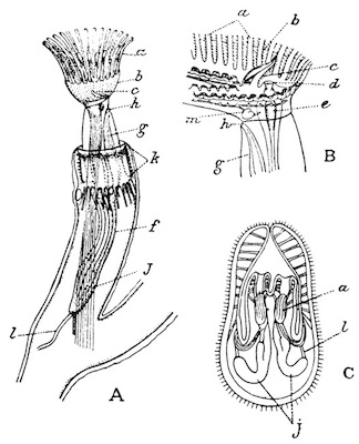
Fig. 40.—Structure of the Plumatellina (after Allman).
A=a zoœcium of Fredericella with the polypide extruded. B=the lophophore of Lophopus (tentacles removed) as seen obliquely from the right side. C=larva of Plumatella as seen in optical section. a=tentacles; b=velum; c=epistome; d=mouth; e=œsophagus; f=stomach; g=intestine; h=anus; j=retractor muscle; k=parieto-vaginal muscles; l=funiculus.
Two families may be recognized as constituting the division, viz., (a) the Fredericellidæ, which have a circular or oval lophophore and simple statoblast without a swim-ring, and (b) the Plumatellidæ, in which the lophophore is shaped like a horseshoe and some or all of the statoblasts are provided with a ring of air-spaces.
Family 1. FREDERICELLIDÆ.
Fredericellidæ, Kraepelin, Deutsch. Süsswasserbryozoen, i, p. 168 (1887).
Zoaria dendritic; zoœcia distinctly tubular, with the ectocyst well developed; statoblasts of one kind only, each surrounded by a chitinous ring devoid of air-spaces; polypides with the lophophore circular or oval when expanded.
The Fredericellidæ consist of a single genus (Fredericella) which includes several closely-allied forms and has a wide geographical distribution.
Genus FREDERICELLA, Gervais (1838).
Fredericella, Allman, Mon. Fresh-Water Polyzoa, p. 110 (1857). Plumatella, ("arrêt de développement") Jullien, Bull. Soc. zool. France, x, p. 121 (1885). Fredericella, Kraepelin, Deutsch. Süsswasserbryozoen, i, p. 99 (1887). Fredericella, Goddard, Proc. Linn. Soc. N. S. Wales, xxxiv, p. 489 (1909).
This genus has the characters of the family. Its status has been much disputed, some authors regarding the shape of the lophophore as of great morphological importance, while Jullien believed that Fredericella was merely an abnormal or monstrous form of Plumatella. The latter belief was doubtless due to the fact that the zoaria of the two genera bear a very close external resemblance to one another and are sometimes found entangled together. The importance of the shape of the lophophore may, however, easily be exaggerated, for, as both Jullien and Goddard have pointed out, it assumes an emarginate form when retracted.
The best known species is the European and N. American F. sultana (Blumenbach), of which several varieties or phases have been described as distinct. This form is stated to occur also in S. Africa. F. australiensis, Goddard[BC] from N. S. Wales is said to differ from this species in having an oval instead of a circular lophophore and in other small anatomical characters; but it is doubtful how far these characters are valid, for the lophophore appears to be capable of changing its shape to some slight extent and has been stated by Jullien to be habitually oval in specimens from France. F. cunningtoni, Rousselet[BD] from Lake Tanganyika has stout zoœcia encrusted with relatively large sand-grains.
The zoaria of Fredericella are usually found attached to solid objects in shallow water, but a form described as F. duplessisi, Ford has been found at a depth of 40 fathoms embedded in mud at the bottom of the Lake of Geneva. F. cunningtoni was dredged from depths of about 10 and about 25 fathoms.
The statoblasts of this genus do not float and often germinate in the parent zoœcium after its polypides have died. They are produced in smaller numbers than is usually the case in other genera of the order. The polypides sometimes undergo a process of regeneration, but without the formation of brown bodies.
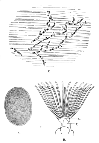
Fig. 41.—Fredericella indica.
A=statoblast, × 120. B=outline of expanded lophophore and adjacent parts, × 75; a=anus, r=rectum. C=outline of zoarium on leaf of water-plant, × 3.
(A and B are from specimens from Igatpuri, C from specimen from Shasthancottah).
28. Fredericella indica, Annandale.
Fredericella indica, Annandale, Rec. Ind. Mus. iii, p. 373, fig. (1909). Fredericella indica, id., ibid. v, p. 39 (1910).
Zoarium. The zoarium is of delicate appearance and branches sparingly. It is often entirely recumbent but sometimes produces short, lax branches that consist of two or three zoœcia only.
Zoœcia. The zoœcia are very slender and almost cylindrical; they are slightly emarginate and furrowed, the keel in which the furrow runs being sometimes prominent. The external surface is minutely roughened and apparently soft, for small grains of sand and other débris cling to it, but never thickly. The ectocyst is practically colourless but not transparent.
Statoblasts. The statoblasts are variable in size and form but most commonly have a regular broad oval outline; sometimes they are kidney-shaped. The dorsal surface is covered with minute star-shaped prominences, which sometimes cover it almost uniformly and are sometimes more numerous in the centre than towards the periphery. The ventral surface is smooth.
Polypide. The lophophore bears about 20-25 tentacles, which are very slender and of moderate length; the velum at their base is narrow; as a rule the lophophore is accurately circular.
Type in the Indian Museum.
The most definite character in which this species differs from F. sultana and F. australiensis is the ornamentation of one surface of the statoblast, both surfaces of which are smooth in the two latter species. From F. cunningtoni, the statoblasts of which are unknown, it differs in having almost cylindrical instead of depressed zoœcia and in not having the zoœcia densely covered with sand-grains.
Geographical Distribution.—Western India (the Malabar Zone): Igatpuri Lake, W. Ghats (alt. ca. 2,000 feet), Bombay Presidency, and Shasthancottah Lake near Quilon, Travancore.
Biology.—In both the lakes in which the species has yet been found it was collected in November. The specimens obtained in Travancore were found to be undergoing a process of regeneration due at least partly to the fact that most of the polypides had perished and that statoblasts were germinating in the old zoœcia. Specimens from the Bombay Presidency, which were obtained a little later in the month, were in a more vigorous condition, although even they contained many young polypides that were not yet fully formed. It seems, therefore, not improbable that F. indica dies down at the beginning of the hot weather and is regenerated by the germination of its statoblasts at the beginning of the cold weather.
At Shasthancottah zoaria were found entangled with zoaria of a delicate form of Plumatella fruticosa to which they bore a very close external resemblance.
Family 2. PLUMATELLIDÆ.
Plumatellidæ, Allman (partim), Mon. Fresh-Water Polyzoa, pp. 76, 81 (1857).
Phylactolæmata which have horseshoe-shaped lophophores and a well-developed ectocyst not specialized to form an organ of progression. Some or all of the statoblasts are provided with a "swim-ring" consisting of symmetrically disposed, polygonal chitinous chambers containing air.
It is convenient to divide the Plumatellidæ as thus defined into subfamilies (the Plumatellinæ and the Lophopinæ), which may be defined as follows:—
Subfamily A. PLUMATELLINÆ.
Zoarium dendritic or linear, firmly fixed to extraneous objects; zoœcia tubular, not fused together to form a gelatinous mass.
Subfamily B. LOPHOPINÆ.
Zoarium forming a gelatinous mass in which the tubular nature of the zoœcia almost disappears, capable to a limited extent of progression along a smooth surface.
Both these subfamilies are represented in the Indian fauna, the Plumatellinæ by two of the three genera known to exist, and the Lophopinæ by two (or possibly three) of the four that have been described. The following key includes all the known genera, but the names of those that have not been recorded from India are enclosed in square brackets.
Key to the Genera of Plumatellidæ.
I.Statoblasts without marginal processes.
A.Zoœcia cylindrical, not embedded in a gelatinous investment (Plumatellinæ).
a. Zoœcia arising directly from one another; no stolon; free statoblast ovalPlumatella, p. 212.
a'. Zoœcia arising singly or in groups from an adherent stolon; free statoblasts oval.Stolella, p. 229.
B.Zoœcia cylindrical, embedded in a structureless gelatinous investment.
Zoœcia arising from a ramifying stolon; statoblasts circular[Stephanella.]
C.Polypides embedded in a hyaline synœcium that conceals the cylindrical form of the zoœcia (Lophopinæ).
c. Polypides upright, their base far removed from that of the zoarium when they are expandedLophopus, p. 231.
c'. Polypides recumbent for the greater part of their length at the base of the zoarium[Australella[BE].]
II.Statoblasts armed (normally) with hooked processes (Lophopinæ).
A.Processes confined to the extremities of the statoblast; zoaria remaining separate throughout lifeLophopodella, p. 231.
B.Processes entirely surrounding the statoblast; many zoaria embedded in a common gelatinous investment so as to form large compound coloniesPectinatella, p. 235.
Subfamily A. PLUMATELLINÆ.
Of the two Indian genera of this subfamily, one (Plumatella) is almost universally distributed, while the other (Stolella) has only been found in the valley of the Ganges. The third genus of the subfamily (Stephanella) is only known from Japan.
It should be noted that zoaria of different species and genera of this subfamily are often found in close proximity to one another and to zoaria of Fredericella, and that the branches of the different species are sometimes entangled together in such a way that they appear, unless carefully separated, to belong to the same zoarium.
Genus 1. PLUMATELLA, Lamarck.
Plumatella, Lamarck, Animaux sans Vert. (ed. 1re) ii, p. 106 (1816). Alcyonella, id., ibid. p. 100. Plumatella, Allman, Mon. Fresh-Water Polyzoa, p. 92 (1857). Alcyonella, id., ibid. p. 86. Plumatella, Hyatt, Comm. Essex Inst. iv, p. 207, pl. viii (1866). Plumatella, Jullien (partim), Bull. Soc. zool. France, x, p. 100 (1885). Hyalinella, id., ibid. p. 133. Plumatella, Kraepelin, Deutsch. Süsswass. Bryozoen, i, p. 104 (1887). Plumatella, Braem, Unter. ü. Bryozoen des süssen Wassers, p. 2 (Bibliotheca Zoologica, ii, 1890).
Zoarium dendritic, recumbent, erect, or partly recumbent and partly erect.
Zoœcia tubular, not confined in a gelatinous synœcium; the ectocyst usually horny.
Statoblasts often of two kinds, free and stationary, the latter without air-cells and as a rule adherent by one surface, the former provided with a well-developed ring of air-cells but without marginal processes, oval in form, never more than about 0.6 mm. in length.
Polypide with less than 65 tentacles.
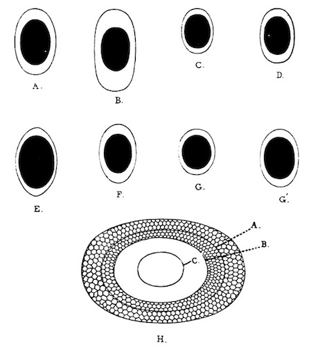
Fig. 42.—Outlines of free statoblasts of Plumatella (enlarged).
A, of P. fruticosa (Calcutta); B, of P. emarginata (Calcutta); C, of P. javanica (Travancore); D, of P. diffusa (Sikhim); E, of P. allmani (Bhim Tal); F, of P. diffusa (Rajshahi, Bengal); G, G', of P. punctata (Calcutta); H, of P. diffusa (Sikhim), statoblast further enlarged: A=outline of capsule; B=limit of swim-ring on ventral surface; C=limit of swim-ring on dorsal surface. [The dark area represents the capsule of the statoblast.]
Certain forms of this genus are liable to become compacted together in such a way as to constitute solid masses consisting of elongate vertical zoœcia closely parallel to one another and sometimes agglutinated by means of a gummy substance. These forms were given by Lamarck in 1816 the name Alcyonella, and there has been much dispute as to whether they represent a distinct genus, distinct species, or merely varieties or phases of more typical forms. It appears to be the case that all species which produce vertical branches are liable to have these branches closely packed together and the individual zoœcia of which they are composed more or less greatly elongated. It is in this way that the form known to Allman as Alcyonella benedeni is produced from the typical Plumatella emarginata. Other forms go further and secrete a gummy substance that glues the upright zoœcia together and forces them to elongate themselves without branching. In these conditions the zoœcia become polygonal in cross-section. It is probable that such forms (e. g., Plumatella fungosa (Pallas)) should rank as distinct species, for the gummy secretion is present in great profusion even in young zoaria in which the zoœcia have not yet assumed a vertical position. No such form, however, has as yet been found in India, and in any case it is impossible to regard Alcyonella as a distinct genus.
Key to the Indian Species of Plumatella.
I.Ectocyst more or less stiff, capable of transverse wrinkling only near the tips of the zoœcia, never contractile or greatly swollen; zoœcia rounded[BF] at the tip when the polypide is retracted. Free statoblasts elongate; the free portion of their swim-ring distinctly narrower at the sides than at the ends.
A.Ectocyst by no means rigid, of a uniform pale colour; zoœcia never emarginate or furrowed, straight, curved or sinuous, elongate, cylindricalfruticosa, p. 217.
B.Ectocyst rigid; zoœcia (or at any rate some of the zoœcia) emarginate and furrowed.
b. Ectocyst darkly pigmented over the greater part of each zoœcium, white at the tip; branching of the zoarium practically dichotomous, profuse, as a rule both horizontal and vertical; zoœcia straight or slightly curved or sinuousemarginata, p. 220.
b'. Ectocyst colourless and hyaline; branching of the zoarium sparse, lateral, irregular, horizontal; zoœcia nearly straight, strongly emarginate and furrowedjavanica, p. 221.
b''. The majority of the zoœcia distinctly L-shaped, one limb being as a rule adherent; ectocyst never densely pigmented.
β. Zoœcia cylindrical, their furrowed keel never prominentdiffusa, p. 223.
β'. Zoœcia (or at any rate some of the zoœcia) constricted or tapering at the base, their emargination and furrow conspicuousallmani, p. 224.
II.Ectocyst stiff; zoœcia truncated when the polypide is retracted. Surface of zoœcia minutely roughened, distinctly annulate on the distal parttanganyikæ, p. 225.
III.Ectocyst swollen and contractile, capable of transverse wrinkling all over the zoœcium; zoœcia never emarginatepunctata, p. 227.
There has always been much difficulty in separating the species of Plumatella, and even now there is no general consensus of opinion as to the number that should be recognized. The difficulty, however, is much reduced if the following precautions are observed:—
(1) If the zoarium appears to be tangled, if the branches intertwine or overlap, or if the zoœcia are closely pressed together, the whole mass should be carefully dissected out. This is necessary not only because zoaria belonging to different species are sometimes found entangled together but also because it is often difficult to recognize the characteristic method of branching and shape of the zoœcia unless it is done.
(2) As large a part as possible of each zoarium should be examined, preferably with a binocular microscope, and allowance should be made for irregularities and abnormalities of all kinds. What must be observed is the rule rather than the exceptions.
(3) When the statoblasts are being examined, care must be taken that they lie flat and that their surface is parallel to that of the nose-piece of the microscope. If they are viewed obliquely it is impossible to see their true outlines and proportions.
(4) In order to see the relative proportions of the capsule and the swim-ring it is necessary that the statoblast should be rendered transparent. This is often difficult owing to the presence of air in the air-cells, but strong nitric acid applied judiciously will render it possible (p. 240).
In supervising the preparation of the plates that illustrate this genus I have impressed upon the artist the importance of representing what he saw rather than what he thought he ought to see, and the figures are very close copies of actual specimens. I have deliberately chosen for representation specimens of Plumatella preserved by the simple methods which are often the only ones that it is possible for a traveller to adopt, for the great majority of naturalists will probably have no opportunity of examining living specimens or specimens preserved by special methods, and the main object, I take it, of this series is to enable naturalists first to distinguish the species described and then to learn something of their habitat and habits.
Geographical Distribution.—Of the seven species included in this key five have been found in Europe (namely P. fruticosa, P. emarginata, P. diffusa, P. allmani, and P. punctata), while of these five all but P. allmani are known to occur in N. America also. P. javanica is apparently peculiar to the Oriental Region, while P. tanganyikæ has only been taken in Central Africa and in the Bombay Presidency.
Types.—Very few of the type-specimens of the older species of Plumatella are in existence. Allman's are neither in Edinburgh nor in London, and Mr. E. Leonard Gill, who has been kind enough to go through the Hancock Collection at Newcastle-on-Tyne, tells me that he cannot trace Hancock's. Those of the forms described by Kraepelin are in Hamburg and that of P. tanganyikæ in the British Museum, and there are schizotypes or paratypes of this species and of P. javanica in Calcutta. The types of Leidy's species were at one time in the collection of the Philadelphia Academy of Science.
Biology.—The zoaria of the species of Plumatella are found firmly attached to stones, bricks, logs of wood, sticks, floating seeds, the stems and roots of water-plants, and occasionally to the shells of molluscs such as Vivipara and Unio. Some species shun the light, but all are apparently confined to shallow water.
Various small oligochæte worms (e. g., Chætogaster spongillæ,[BG] Nais obtusa, Nais elinguis, Slavina appendiculata and Pristina longiseta[BH]), take shelter amongst them; dipterous larvæ of the genus Chironomus often build their protective tubes at the base of the zoaria, and the surface of the zoœcia commonly bears a more or less profuse growth of such protozoa as Vorticella and Epistylis. I have seen a worm of the genus Chætogaster devouring the tentacles of a polypide that had been accidentally injured, but as a rule the movements of the lophophore are too quick to permit attacks of the kind, and I know of no active enemy of the genus. The growth of sponges at the base of the zoaria probably chokes some species, but one form (F. fruticosa) is able to surmount this difficulty by elongating its zoœcia (p. 219). A small worm (Aulophorus tonkinensis) which is common in ponds in Burma and the east of India as far west as Lucknow, often builds the tube in which it lives mainly of the free statoblasts of this genus. It apparently makes no selection in so doing but merely gathers the commonest and lightest objects it can find, for small seeds and minute fragments of wood as well as sponge gemmules and statoblasts of other genera are also collected by it. I know of no better way of obtaining a general idea as to what sponges and phylactolæmata are present in a pond than to examine the tubes of Aulophorus tonkinensis.
I am indebted to Mr. F. H. Gravely, Assistant Superintendent in the Indian Museum, for an interesting note regarding the food of Plumatella. His observations, which were made in Northamptonshire, were unfortunately interrupted at a critical moment, but I have reproduced them with his consent in order that other observers may investigate the phenomena he saw. Mr. Gravely noted that a small green flagellate which was abundant in water in which Plumatella repens was growing luxuriantly, was swallowed by the polypides, and that if the polyparium was kept in a shallow dish of water, living flagellata of the same species congregated in a little pile under the anus of each polypide. His preparations show very clearly that the flagellates were passing through the alimentary canal without apparent change, but the method of preservation does not permit the retractile granules, which were present in large numbers in the cell-substance of the flagellates, to be displayed and it is possible that these granules had disappeared from those flagellates which are present in the recta of his specimens. It is clear, therefore, either that certain flagellates must pass through the alimentary canal of Plumatella unchanged, or that the polyzoon must have the power of absorbing the stored food material the flagellates contain without doing them any other injury.
The free statoblasts of Plumatella are as a rule set free before the cells they contain become differentiated, and float on the surface of the water for some time before they germinate; but occasionally a small polypide is formed inside the capsule while it is still in its parent zoœcium. I have, however, seen only one instance of this premature development, in a single statoblast contained in a small zoarium of P. fruticosa found in Lower Burma in March. The fixed statoblasts usually remain fixed to the support of the zoarium, even when their parent-zoœcium decays, and germinate in situ.
The larva (fig. 40 C, p. 207) that originates from the egg of Plumatella is a minute pear-shaped, bladder-like body covered externally with fine vibratile threads (cilia) and having a pore at the narrow end. At the period at which it is set free from the parent zoœcium it already contains a fully formed polypide or pair of polypides with the tentacles directed towards the narrow end. After a brief period of active life, during which it moves through the water by means of its cilia, it settles down on its broad end, which becomes adhesive; the polypide or pair of polypides is everted through the pore at the narrow end, the whole of this end is turned inside out, and a fresh polyparium is rapidly formed by budding.
29. Plumatella fruticosa, Allman. (Plate III, fig. 1; plate IV, fig. 4; plate V, fig. 1.)
Plumatella fruticosa, Allman, Ann. Nat. Hist. xiii, p. 331 (1844). Plumatella repens, van Beneden (? nec Linné), Mém. Acad. Roy. Belg. 1847, p. 21, pl. i, figs. 1-4. Plumatella fruticosa, Johnston, Brit. Zooph. (ed. 2), p. 404 (1847). Plumatella coralloides, Allman, Rep. Brit. Assoc. 1850, p. 335. Plumatella stricta, id., Mon. Fresh-Water Polyzoa, p. 99, fig. 14 (1857). Plumatella fruticosa, id., ibid. p. 102, pl. vi, figs. 3-5. Plumatella coralloides, id., ibid. p. 103, pl. vii, figs. 1-4. Plumatella repens and P. stricta, Carter, Ann. Nat. Hist. (3) iii, p. 341 (1859). Plumatella lucifuga, Jullien (partim), Bull. Soc. zool. France, x, p. 114 (1885). Plumatella princeps var. fruticosa, Kraepelin, Deutsch. Süsswasserbryozoen, i, p. 120, pl. vii, fig. 148 (1887). Plumatella fruticosa, Braem, Unter. ii. Bryozoen des süssen Wassers, p. 9, pl. i, fig. 15 (Bibl. Zool. ii) (1890). Plumatella repens, Annandale, J. As. Soc. Bengal (new series) iii, 1907, p. 88. Plumatella emarginata, Loppens (partim), Ann. Biol. lacustre, iii, p. 161 (1908). Plumatella fruticosa, Annandale, Rec. Ind. Mus. v, p. 45 (1910).
Zoarium. The zoarium in the typical form has a loose appearance due to the fact that the branches are far apart and the ectocyst by no means rigid. When young the zoarium is adherent, but in well-grown polyparia vertical branches, often an inch or more in length, are freely produced. As a rule they have not the strength to stand upright if removed from the water. Branching is ordinarily lateral and as a rule occurs chiefly on one side of a main branch or trunk. In certain circumstances upright zoœcia are pressed together and reach a great length without branching, and in this form (P. coralloides, Allman) daughter-zoœcia are often produced at the tip of an elongated mother-zoœcium in fan-like formation. A depauperated form (P. stricta, Allman), occurs in which the vertical branches are absent or very short. In all forms internal partitions are numerous and stout.
Zoœcia. The zoœcia are cylindrical and bear a simple keel on their dorsal surface. They are never emarginate or furrowed. In the typical form their diameter is more than half a millimetre, and they are always of considerable length. The ectocyst is thin and never very rigid or deeply pigmented, the colour usually being an almost uniform pale pinkish brown and fading little towards the tip of the zoœcium.
Statoblasts. Both free and stationary statoblasts are formed, but the latter are rare and do not always adhere. They resemble the free statoblasts in general form but have a solid margin instead of a swim-ring and are often minutely serrated round the edge. The free statoblasts are at least considerably, sometimes very elongate; in all zoaria it is possible to find specimens that are more than twice as long as broad. The capsule is relatively large and resembles the swim-ring in outline, so that the free portion of the latter is not much narrower at the sides than at the ends. The sides are distinctly convex and the ends rounded; the swim-ring encroaches little on the surface of the capsule.
Polypide. The tentacles number between 40 and 50 and are not festooned at the base. The stomach is slender and elongate.
Type not in existence.
Systematic Remarks.—P. fruticosa is closely allied to P. repens (European and N. American) but always has much longer statoblasts. Three phases of the species may be distinguished as follows:—
A. (Forma typica). Zoœcia stout in form, not greatly elongate; free branches produced in profusion.
B. (P. stricta, Allman, P. repens, van Beneden). Zoœcia slender; free branches absent or consisting of two or three zoœcia only.
C. (P. coralloides, Allman). Vertical zoœcia pressed together and greatly elongated.
Indian specimens of the typical form agree well with German specimens labelled by Prof. Kraepelin P. princeps var. fruticosa, and specimens of the coralloides phase could hardly be distinguished from similar specimens from Scotland.
Geographical Distribution.—P. fruticosa is widely distributed in Europe and probably in N. America. I have seen Indian specimens from the Punjab (Lahore, Stephenson), from Bombay, from Travancore, from Calcutta and other places in the Ganges delta, from Rajshahi (Rampur Bhoolia) on the R. Ganges, from Kurseong in the E. Himalayas (alt. 4,500 feet), and from Kawkareik in Tenasserim. Statoblasts found on the surface of a pond near Simla in the W. Himalayas (alt. ca. 8,000 feet), probably belong to this species.
Biology.—Allman states that in England P. fruticosa is fond of still and slowly-running water. The typical form and the coralloides phase grow abundantly in the Calcutta tanks, the former often attaining an extraordinary luxuriance. I have found the var. stricta only in water in which there was reason to suspect a lack of minute life (and therefore of food), viz. in Shasthancottah Lake in Travancore, in a swamp in Lower Burma, and in a small jungle stream near the base of the Western Ghats in Travancore. The species is the only one that I have seen in running water in India, and the specimens obtained in the jungle stream in Travancore are the only specimens I have taken in these circumstances. P. fruticosa always grows near the surface or near the edge of water; it is found attached to the stems of bulrushes and other aquatic plants, to floating seeds and logs and (rarely) to stones and bricks. So far as my experience goes it is only found, at any rate in Calcutta, in the cold weather and does not make its appearance earlier than October.
The form Allman called P. coralloides was found by him, "attached to floating logs of wood, together with P. repens and Cordylophora lacustris, and generally immersed in masses of Spongilla fluviatilis." I have always found it immersed in sponges (S. lacustris, S. alba, S. carteri, and S. crassissima), except when the sponge in which it had been immersed had decayed. Indeed, the peculiar form it has assumed appears to be directly due to the pressure of the growing sponge exerted on the zoœcia, for it is often possible to find a zoarium that has been partially overgrown by a sponge and has retained its typical form so long as it was free but has assumed the coralloides form where immersed.[BI] In Shasthancottah Lake, Travancore, I found specimens of the stricta phase embedded in the gelatinous mass formed by a social rotifer and to some extent assimilated to the coralloides form.
30. Plumatella emarginata, Allman. (Plate III, fig. 2; plate IV, figs. 1, 1 a.)
Plumatella emarginata, Allman, Ann. Nat. Hist. xiii, p. 330 (1844). Plumatella emarginata, Johnston, Brit. Zooph. (ed. 2), p. 404 (1847). Alcyonella benedeni, Allman, Mon. Fresh-Water Polyzoa, p. 89, pl. iv, figs. 5-11 (1857). Plumatella emarginata, id., ibid. p. 104, pl. vii, figs. 5-10. Plumatella lucifuga, Jullien, Bull. Soc. zool. France, x, figs. 89, 90, p. 114 (1885). Plumatella princeps var. emarginata, Kraepelin (partim), Deutsch. Süsswasserbryoz. p. 120, pl. iv, fig. 108, pl. v, fig. 123 (1887). Plumatella emarginata, Braem, Unter. ii. Bryoz. süssen Wassers, p. 9, pl. i, figs. 12, 14 (Bibl. Zool. ii) (1890). Plumatella emarginata, Annandale (partim), J. As. Soc. Bengal, (new series) iii, 1907, p. 89. Plumatella princeps, Loppens (partim), Ann. Biol. lacustre, iii, p. 162, fig. 7 (1908). Plumatella emarginata, Annandale, Rec. Ind. Mus. v, p. 47 (1910).
Zoarium. The zoarium often covers a considerable area on flat surfaces and is sometimes entirely recumbent. More usually, however, the younger part is vertical. In either case the branching is practically dichotomous, two young zoœcia arising almost simultaneously at the tip of a mother-zoœcium and diverging from one another at a small angle. When the zoarium becomes vertical, rigid branches of as much as an inch in length are sometimes produced in this way and, arising parallel to one another, are pressed together to form an almost solid mass (=Alcyonella benedeni, Allman). In such cases the basal zoœcium or at any rate the basal part of each upright branch is considerably elongated. In recumbent zoœcia the main branches often radiate outwards from a common centre.
Zoœcia. The zoœcia are of almost equal width throughout, slender, and moderately elongate when recumbent. Their ectocyst is stiff; they are emarginate at the tip and more or less distinctly furrowed on the dorsal surface, the keel in which the furrow runs not being prominent. The orifice is often on the dorsal surface even in upright branches. Each zoœcium is of a dark brown or almost black colour for the greater part of its length but has a conspicuous white tip which is extended down the dorsal surface in the form of a triangle, its limits being rather more extensive than and parallel to those of the emargination.
Statoblast. The majority of the free statoblasts are elongate and truncate or subtruncate at the extremities, the sides being as a rule straight and parallel. In every polyparium specimens will be found that are between twice and thrice as long as broad. The capsule is, however, relatively much broader than the swim-ring, often being nearly circular, and there is therefore at either end a considerable extent of free air-cells, while the extent of these cells at the sides of the capsule is small. The air-cells cover a considerable part of the dorsal surface of the capsule. Fixed statoblasts are usually found in old colonies, especially at the approach of the hot weather. They have an oval form and are surrounded by a membranous margin on which traces of reticulation can often be detected. As a rule statoblasts of both types are produced in considerable but not in excessive numbers.
Polypide. There are about 40 tentacles, the velum at the base of which extends upwards for a considerable distance without being festooned. The stomach is elongate and slender and narrowly rounded at the base.
The method of branching, the coloration of the zoœcia and the form of the free statoblast are all characteristic. Luxuriant or closely compressed zoaria of P. diffusa often bear a superficial resemblance to those of P. emarginata, but the resemblance disappears if they are carefully dissected out. Indian specimens of P. emarginata agree closely with European ones.
Geographical Distribution.—P. emarginata is a common species in Europe, N. America, and southern Asia and probably also occurs in Africa and Australia. I have examined specimens from Calcutta, Rangoon, and Mandalay in Indian territory, and also from Jalor in the Patani States (Malay Peninsula) and the Talé Noi, Lakon Sitamarat, Lower Siam. Gemmules found by Apstein (Zool. Jahrb. (Syst.) xxv, 1907, p. 201) in plankton from the Colombo lake may belong to this species or to any of the others included by Kraepelin in his P. princeps.
Biology.—In Ireland Allan found P. emarginata in streams and rivulets, but it also occurs in European lakes. In India I have only found it in ponds. It prefers to adhere to the surface of stones or bricks, but when these are not available is found on the stems of water-plants. In the latter position the form called Alcyonella benedeni by Allman is usually produced, owing to the fact that the upright branches are crowded together through lack of space, very much in the same way (although owing to a different cause) as those of P. fruticosa are crowded together in the coralloides phase, to which the benedeni phase of P. emarginata is in many respects analogous.
Although it is essentially a cold-weather species in Calcutta, P. emarginata is sometimes found in a living condition during the "rains." Zoaria examined at this season, however, contains few living polypides, the majority of the zoœcia having rotted away and left fixed statoblasts only to mark their former position.
31. Plumatella javanica, Kraepelin.
Plumatella javanica, Kraepelin, Mitt. Nat. Mus. Hamb. xxiii, p. 143, figs. 1-3 (1903). Plumatella emarginata var. javanica, Loppens, Ann. Biol. lacustre, iii, p. 162 (1908). Plumatella javanica, Annandale, Rec. Ind. Mus. v, p. 50 (1910). Plumatella allmani var. dumortieri, id. (partim) (nec Allman), ibid. p. 49.
This species is related to P. emarginata, from which it may be distinguished by the following characters:— Zoarium. The zoarium is always entirely recumbent and branches sparingly; its method of branching does not approach the dichotomous type but is lateral and irregular. Linear series of zoœcia without lateral branches are often formed.
Zoœcia. The zoœcia are slender and often very long; they are strongly emarginate and furrowed, and the keel that contains the furrow is conspicuous. The ectocyst is hyaline and as a rule absolutely colourless.
Statoblasts. The free statoblasts are variable in length, sometimes distinctly elongate, sometimes elongate only to a moderate degree; they are rounded at the extremities and have the sides slightly or distinctly convex outwards. The capsule is relatively large, and the free portion of the swim-ring is not much broader at the ends than at the sides. The fixed statoblasts are elongate and surrounded by an irregularly shaped chitinous membrane, which is often of considerable extent. The whole of the dorsal surface is covered with what appear to be rudimentary air-spaces some of which even contain air.
The transparent glassy ectocyst and strong furrowed keel of this species are very characteristic, but the former character is apt to be obscured by staining due to external causes, especially when the zoarium is attached to dead wood. The shape of the free statoblasts is too variable to be regarded as a good diagnostic character, but the fixed statoblasts, when they are to be found, are very characteristic in appearance. P. javanica appears to be closely related to Allman's P. dumortieri, with which stained zoaria are apt to be confused. The character of the ectocyst is, however, different, and the free part of the swim-ring is distinctly narrower at the sides of the free statoblasts. Dr. Kraepelin has been kind enough to send me one of the types.
Types in the Hamburg and Indian Museums.
Geographical Distribution.—Java, Penang, India. Indian localities are:—Bengal, Calcutta; Berhampore, Murshidabad; R. Jharai, Siripur, Saran district, Tirhut: E. Himalayas, Kurseong, Darjiling district (alt. 4,500 feet): Madras Presidency, canal near Srayikaad, Travancore. Mr. C. W. Beebe has recently sent me a specimen taken by him in the Botanical Gardens at Penang.
Biology.—Very little is known about the biology of this species. Kraepelin took it in Java on the leaves of water-lilies. It is not uncommon during the cold weather in the Calcutta Zoological Gardens on floating seeds and sticks and on the stems of bulrushes; in Travancore I took it in November on the submerged leaves of Pandani growing at the edge of a canal of slightly brackish water. Mr. Hodgart, the collector of the Indian Museum, found it in the R. Jharai on the stems of water-plants at a time of flood in the "rains." In Calcutta it is often found entangled with P. fruticosa and P. emarginata.
32. Plumatella diffusa, Leidy. (Plate IV, fig. 2.)
Plumatella diffusa, Leidy, P. Ac. Philad. v, p. 261 (1852). Plumatella diffusa, Allman, Mon. Fresh-Water Polyzoa, p. 105 (1857). Plumatella diffusa, Hyatt, Comm. Essex Inst. iv, pl. viii, figs. 11, 12 (1866). Plumatella diffusa, id., ibid. v, p. 107, fig. 12 (1868). Plumatella repens, Jullien, Bull. Soc. zool. France, x, fig. 37 (lapsus for 73), p. 110 (1885). Plumatella diffusa, id., ibid. figs. 155, 157, pp. 130, 131. Plumatella allmani var. diffusa, Annandale, Rec. Ind. Mus. v, p. 49 (1910).
Zoarium. The zoarium often covers a considerable area on flat surfaces and is sometimes found crowded together on the stems of plants. In the latter case the arrangement of the main branches is distinctly radiate. Upright branches occur rarely and never consist of more than three zoœcia. The characteristic method of branching is best represented by the following diagram:—
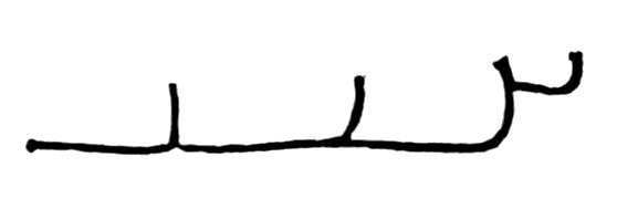
Fig. 43.
The partitions are stout and numerous.
Zoœcia. The great majority of the zoœcia in each zoarium are distinctly L-shaped, the long limb being usually adherent. The vital organs of the polypide are contained in the vertical limb, while the horizontal one, in mature polyparia, is packed full of free statoblasts. The zoœcia are cylindrical and as a rule obscurely emarginate and furrowed. The ectocyst is stiff; it is never deeply pigmented but is usually of a transparent horn-colour at the base of each zoœcium and colourless at the tip, the contrast between the two portions never being very strong. The basal portion is rough on the surface, the distal portion smooth.
Statoblasts. Free statoblasts are produced in very great profusion and fixed statoblasts are also to be found as a rule. The latter resemble those of P. emarginata. The free statoblasts are never very large or relatively broad, but they vary considerably as regards size and outline. The capsule is large, the sides convex outwards and the extremity more or less broadly rounded. The air-cells are unusually large and extend over a great part of the dorsal surface of the statoblast.
Polypide. The polypide is shorter and stouter than that of P. emarginata and as a rule has fewer tentacles.
The most characteristic feature of this species is the form of the zoœcia, which differ greatly from those of any other Indian species but P. allmani. In the latter they are distinctly "keg-shaped" (i. e., constricted at the base and swollen in the middle), and the zoarium never spreads out over large surfaces in the way in which that of P. diffusa does.
Type—? in the Philadelphia Academy of Sciences.
Geographical Distribution.—This species was originally described from North America (in which it is apparently common) and occurs also in Europe. I have seen Indian specimens from the following localities:—Bengal, Calcutta and neighbourhood; Rajshahi (Rampur Bhulia): E. Himalayas, Gangtok, Native Sikhim (alt. 6,150 feet) (Kirkpatrick, Stewart): Punjab, Lahore (Stephenson).
Biology.—P. diffusa in Lower Bengal is a cold-weather species. It is remarkable for the enormous number of gemmules it produces and is usually found either on floating objects such as the stems of certain water-plants, or on stones or bricks at the edge of ponds.
33. Plumatella allmani, Hancock. (Plate IV, figs. 3, 3 a.)
Plumatella allmani, Hancock, Ann. Nat. Hist. (2) v, p. 200, pl. v, fig. 3-4, pl. iii, fig. 2-3 (1850). Plumatella allmani, Allman, Mon. Fresh-Water Polyzoa, p. 106, fig. 16 (1857). Plumatella elegans, id., ibid. p. 107, pl. viii, figs. 6-10. Plumatella lucifuga ("forme rampante") Jullien, Bull. Soc. zool. France, x, p. 114 (1885).
This species is closely allied to P. diffusa, from which it differs in the following characters:—
(1) The zoarium never covers a large area and as a rule grows sparingly and mainly in two directions.
(2) The zoœcia are more irregular in shape, not so distinctly elbowed, smaller; they have a much more prominently keeled ridge. The great majority of them are constricted at the base and taper towards the orifice. In young zoaria they are almost colourless but in older ones there is a band of not very dense pigment round the base of the vertical limb.
(3) The free statoblasts are comparatively large and usually show a tendency to taper at the extremities, often being almost rhomboidal in form. The swim-ring does not extend so far over the dorsal surface as it does in those of P. diffusa; the "cells" of which it is composed are small.
Type not in existence.
I have seen every gradation between this form and Allman's P. elegans.
Geographical Distribution.—P. allmani is apparently a rare species to which there are few references in literature. It was originally described from England and is stated by Jullien to occur in France. I have found specimens only in the lake Bhim Tal (alt. 4,500 feet) in the W. Himalayas.
Biology.—The original specimens were found by Hancock on stones. My own were growing on the leaves of water-plants, usually on the under side. When the zoœcia were forced to stretch across from one leaflet to another they assumed the sinuous form characteristic of Allman's P. elegans.
34. Plumatella tanganyikæ, Rousselet.
Plumatella tanganyikæ, Rousselet, Proc. Zool. Soc. London, 1907 (i), p. 252, pl. xiv, figs. 1-4. Plumatella bombayensis, Annandale, Rec. Ind. Mus. ii, p. 169, figs. 1, 2 (1908). Plumatella bombayensis, id., ibid. v, p. 51 (1910).
Zoarium. The whole colony is recumbent but branches freely and at short intervals in a horizontal plane, so that the zoœcia become crowded together and the branches sometimes overlap one another. The zoarium often covers a considerable area, but growth seems to be mainly in two directions. When growing on the stems of water-plants the branches are often parallel and closely pressed together but remain recumbent in this position. A stout membrane sometimes extends between branches and individual zoœcia.
Zoœcia. The walls of the zoœcia are thick, stiff, and more or less darkly but not opaquely pigmented; the external surface, although not very smooth, is always clean. The two most noteworthy characters of the zoœcia are (i) their truncated appearance when the polypide is retracted, and (ii) the conspicuous, although often irregular external annulation of their walls. The tip of each zoœcium, owing to the fact that the invaginated part of the ectocyst is soft and sharply separated from the stiffened wall of the tube, terminates abruptly and is not rounded off gradually as is the case in most species of the genus; sometimes it expands into a trumpet-like mouth. The annulation of the external surface is due to numerous thickened areas of the ectocyst which take the form of slender rings surrounding the zoœcium; they are most conspicuous on its distal half. On the dorsal surface of the base of each zoœcium there is a conspicuous furrowed keel, which, however, does not usually extend to the distal end; the latter is oval in cross-section. The zoœcia are short and broad; their base is always recumbent, and, when the zoarium is attached to a stone or shell, often seems to be actually embedded in the support; the distal part turns upwards and is free, so that the aperture is terminal; the zoœcia of the older parts of the zoarium exhibit the specific characters much more clearly than those at the growing points.
Polypide. The lophophore bears 20 to 30 tentacles, which are long and slender; the velum at their base extends up each tentacle in the form of a sharply pointed projection, but these projections do not extend for more than one-fifth of the length of the tentacles. Both the velum and the tentacular sheath bear numerous minute tubercles on the external surface. The base of the stomach is rounded, and the whole of the alimentary canal has a stout appearance.
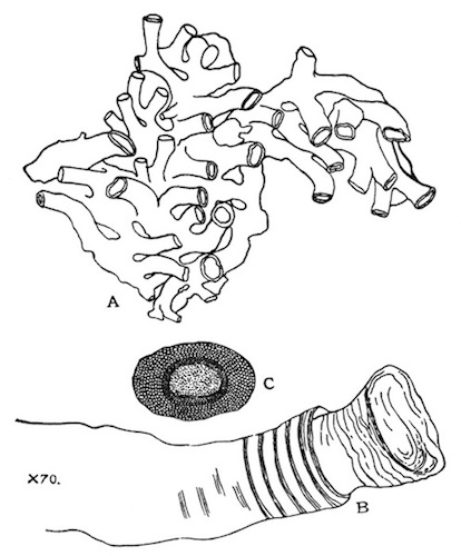
Fig. 44.—Plumatella tanganyikæ from Igatpuri Lake.
A=outline of part of zoarium from a stone, × 16; B=outline of the tip of a single zoœcium, × 70; C=free statoblast, × 70.
Statoblasts. Both fixed and free statoblasts are produced, but not in very large numbers. The latter are broadly oval and are surrounded by a stout chitinous ring, which often possesses irregular membranous projections; the surface is smooth. The free statoblasts are small and moderately elongate, the maximum breadth as a rule measuring about 2/3 of the length; the capsule is relatively large and the ring of air-cells is not very much broader at the ends than at the sides; the dorsal surface of the central capsule is profusely tuberculate. The outline of the whole structure is often somewhat irregular.
In deference to Mr. Rousselet's opinion expressed in a letter I have hitherto regarded the Bombay form of this species as distinct from the African one, and there certainly is a great difference in the appearance of specimens taken on the lower surface of stones in Igatpuri Lake and of the types of P. tanganyikæ, one of which is now in the collection of the Indian Museum. The dark colour of the former, however, and their vigorous growth appear to be directly due to environment, for these characters disappear to a large extent in specimens growing on the stems of water-plants in the same lake. Indeed, such specimens are exactly intermediate between the form "bombayensis" and the typical form of the species. P. tanganyikæ is closely allied to P. philippinensis, Kraepelin, from the island of Luzon, but the latter has a smooth and polished ectocyst devoid of annulations, and zoœcia of a more elongate and regular form.
Types of the species in the British and Indian Museums, those of P. bombayensis in the latter collection.
Geographical Distribution.—P. tanganyikæ is only known as yet from L. Tanganyika in Central Africa and from Igatpuri in the Bombay Presidency.
Biology.—In both localities the zoaria were found in shallow water. In L. Tanganyika they were encrusting stones and shells, while at Igatpuri they were fixed for the most part to the lower surface of stones but were also found on the stems of water-plants. My specimens from the Bombay Presidency were taken, on two separate occasions, at the end of November. At that date the zoaria were already decaying and large blanks, marked out by fixed statoblasts, were often observed on the stones. Probably, therefore, the species flourishes during the "rains."
35. Plumatella punctata, Hancock. (Plate IV, fig. 5.)
Plumatella punctata, Hancock, Ann. Nat. Hist. (2) v, p. 200, pl. iii, fig. 1, and pl. v, figs. 6, 7 (1850). Plumatella vesicularis, Leidy, P. Ac. Philad. vii, p. 192 (1854). Plumatella vitrea, Hyatt, Comm. Essex Inst. iv, pl. ix, figs. 1, 2 (1866). Plumatella punctata, Allman, Mon. Fresh-Water Polyzoa, p. 100, fig. 15 (1857). Plumatella vesicularis, id., ibid. p. 101. Plumatella vitrea, Hyatt, Proc. Essex Inst. v, p. 225, figs. 18, 19 (1868). Plumatella vesicularis, id., ibid. p. 225. Hyalinella vesicularis, Jullien, Bull. Soc. zool. France, x, p. 133, figs. 165-172 (1885). Hyalinella vitrea, id., ibid. p. 134, figs. 173-179. Plumatella punctata, Kraepelin, Deutsch. Süsswasserbryozoen, i, p. 126, pl. iv, figs. 115, 116; pl. v, figs. 124, 125; pl. vii, figs. 153, 154 (1887). Plumatella vesicularis, Braem, Unters. ü. Bryozoen süssen Wassers, p. 8, pl. i, fig. 8 (Bibl. Zool. ii) (1890). Hyalinella punctata, Loppens, Ann. Biol. lacustre, iii, p. 163 (1908). Plumatella punctata, Annandale, Rec. Ind. Mus. v, p. 52 (1910).
Zoarium. The zoarium is entirely recumbent and often appears to form an almost uniform flat layer instead of a dendritic body. Sometimes, however, it is distinctly linear, with lateral branches produced irregularly at considerable distances apart.
Zoœcia. The zoœcia differ from those of all other species in having a greatly swollen, soft ectocyst which can be transversely wrinkled all over the zoœcium by the action of the muscles of the polypide and is distinctly contractile. It is mainly owing to the swollen and almost gelatinous nature of the ectocyst that the dendritic character of the zoarium is frequently concealed, for the method of branching is essentially the same as that of P. diffusa, although the zoœcia are not so distinctly elbowed. The ectocyst is colourless or faintly tinted with brown; as a rule it is not quite hyaline and the external surface is minutely roughened or tuberculate. The zoœcia are not emarginate or furrowed.
Statoblasts. Stationary statoblasts are not found. The free statoblasts are variable and often asymmetrical in outline, but the free portion of the swim-ring is always of nearly equal diameter all round the periphery and the capsule relatively large. Some of the statoblasts are always broad in comparison with their length.
Polypide. The polypide is comparatively short and stout. European specimens are said to have from 30 to 40 tentacles, but Indian specimens have only from 20 to 30.
Shrunken specimens of the less congested forms of this species closely resemble specimens of P. repens, but the statoblasts are more variable in shape and the ectocyst, even in such specimens, is thicker. Living or well-preserved specimens cannot be mistaken for those of any other species. Jullien regarded P. punctata as the type of a distinct genus (Hyalinella) but included in Plumatella at least one form (P. "arethusa") which probably belongs to this species. Kraepelin distinguishes as "varieties" two phases, a summer phase ("var. prostrata") and an autumn phase ("var. densa"). The former often forms linear series of considerable length with only an occasional side-branch, while in the autumn phase branching is so profuse and the branches are so closely pressed together that the zoarium comes to resemble a uniform gelatinous patch rather than a dendritic growth. A phase resembling the European autumn form is the commonest in Calcutta and I have also found one intermediate between this and Kraepelin's "var. prostrata," neither having any seasonal significance in India.