Part 3 — GLOSSARY OF TECHNICAL TERMS USED IN PART III. (continued 2)
Reading edition
Geographical Distribution.—P. punctata is widely distributed in Europe and N. America, but in the Oriental Region it has only been found in Calcutta and the neighbourhood.
Biology.—In this part of India P. punctata flourishes both during the "rains" and in winter. I have found specimens in June and July and also in December and January. The majority of them were attached to bricks, but some were on the roots of duckweed, the stems of water-plants, and the tips of creepers falling into water. The species is often found together with Stolella indica and also with other species of its own genus. It is most common, in the neighbourhood of Calcutta, in that part of the town which is near the Salt Lakes, and occurs in ponds the water of which is slightly brackish.
Genus 2. STOLELLA, Annandale.
Stolella, Annandale, Rec. Ind. Mus. iii, p. 279 (1909). Stolella, id., ibid. v, p. 53 (1910).
Type, Stolella indica, Annandale.
Zoarium. The zoarium consists of groups of zoœcia (or occasionally of single zoœcia) joined together by an adherent rhizome. There is no gelatinous investment.
Zoœcia. The adult zoœcia resemble those of Plumatella except in being sometimes more or less upright.
Polypide and Statoblasts. The polypide and statoblasts resemble those of Plumatella. Fixed as well as free statoblasts occur.
This genus is closely allied to Plumatella, from which it is probably derived. The root-like tube from which the zoœcia arise is formed by the great elongation of the basal part of a zoœcium, and the zoaria closely resemble those of P. punctata, for it is not until several zoœcia have been produced that the characteristic mode of growth becomes apparent.
Stolella has only been found in India and is monotypic[BJ].
36. Stolella indica, Annandale. (Plate V, figs. 3, 4.)
Stolella indica, Annandale, Rec. Ind. Mus. iii, p. 279, fig. (1909). Stolella indica, id., ibid. v, p. 53 (1910).
Zoarium. The zoarium is adherent and linear, having neither lateral nor vertical branches.
Zoœcia. The zoœcia are short and slender, erect or nearly so, distinctly emarginate and furrowed. Their ectocyst is soft, colourless and transparent but minutely roughened on the surface.
Polypide. The tentacles number from 30 to 35 and are rather short and stout, sometimes being slightly expanded at the tips. The stomach is comparatively short and abruptly truncated posteriorly.
Statoblasts. Both free and fixed statoblasts are found, and both are variable in form, the latter varying in outline from the circular to the broadly oval. The free statoblasts resemble those of Plumatella punctata, but are sometimes rather more elongate.
Type in the Indian Museum.
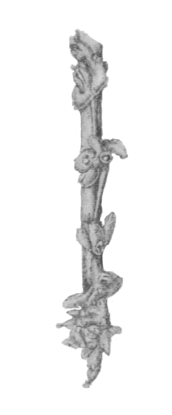
Fig. 45.—Zoarium of Stolella indica on stem of water-plant (from Calcutta), × 6.
Geographical Distribution.—So far as we know, this species is confined to the Indo-Gangetic Plain. Major Walton found it at Bulandshahr in the United Provinces, and it is not uncommon in the neighbourhood of Calcutta.
Biology.—The zoaria of S. indica are usually fixed to the roots of duckweed or to the stems of other plants. They are often found together with those of P. punctata. A slight infusion of brackish water into the ponds in which it lives does not seem to be inimical to this species, but I have found it in ponds in which nothing of the kind was possible. It flourishes during the "rains" and, to judge from specimens kept in an aquarium, is very short-lived. Major Walton found it growing over a zoarium of Hislopia lacustris.
Subfamily B. LOPHOPINÆ.
The zoaria of this subfamily are never dendritic but form gelatinous masses which, except in Australella, are cushion-shaped or sack-like. With the possible exception of Australella, they possess to a limited extent the power of moving along vertical or horizontal surfaces, but it is by no means clear how they do so (see p. 172). The statoblasts are remarkable for their large size, and it is noteworthy that Australella, which is intermediate in structure between the Plumatellinæ and the Lophopinæ, possesses statoblasts of intermediate size. The swim-ring is always well developed, and fixed statoblasts are unknown.
Only two genera (Lophopodella and Pectinatella) have been definitely proved to occur in India, but a third (Lophopus[BK]) is stated to have been found in Madras. Should it be met with it will easily be recognized by the upright position of its polypides when their tentacles are expanded and by the fact that the statoblasts never bear marginal processes.
Genus 3. LOPHOPODELLA, Rousselet.
Lophopodella, Rousselet, Journ. Quek. Micr. Club (2) ix, p. 45 (1904). Lophopodella, Annandale, Rec. Ind. Mus. v, p. 54 (1910).
Type, Pectinatella carteri, Hyatt.
Zoarium. The zoarium consists of a circular or oval mass of no great size. Polyparia do not form compound colonies.
Polypides. The polypides lie semi-recumbent in the mass and never stand upright in a vertical position.
Statoblasts. The statoblasts are of considerable size and normally bear at both ends a series of chitinous processes armed with double rows of small curved spinules.
As a rule the genus is easily recognized by means of the statoblasts, but sometimes the processes at the ends of these structures are absent or abortive and it is then difficult to distinguish them from those of Lophopus. There is, however, no species of that genus known that has statoblasts shaped like those of the Indian species of Lophopodella.
Three species of Lophopodella, all of which occur in Africa, have been described; L. capensis from S. Africa, which has the ends of the statoblast greatly produced, L. thomasi from Rhodesia, in which they are distinctly concave, and L. carteri from E. Africa, India and Japan, in which they are convex or truncate.
The germination of the gemmule and the early stages in the development of the polyparium of L. capensis have been described by Miss Sollas (Ann. Nat. Hist. (8) ii, p. 264, 1908).
37. Lophopodella carteri (Hyatt). (Plate III, figs. 4, 4a.)
Lophopus sp., Carter, Ann. Nat. Hist. (3) iii, p. 335, pl. viii, figs. 8-15 (1859). ? Lophopus sp., Mitchell, Q. J. Micr. Sci. London (3) ii, p. 61 (1862). Pectinatella carteri, Hyatt, Comm. Essex Inst. iv, p. 203 (footnote) (1866). Pectinatella carteri, Meissner, Die Moosthiere Ost-Afrikas, p. 4 (in Mobius's Deutsch-Ost-Afrika, iv, 1898). Lophopodella carteri, Rousselet, Journ. Quek. Micr. Club, (2) ix, p. 47, pl. iii, figs. 6, 7 (1904). Lophopus carteri, Annandale, Rec. Ind. Mus. ii, p. 171, fig. 3 (1908). Lophopodella carteri, id., ibid. v, p. 55 (1910).
Zoarium. The zoarium as a rule has one horizontal axis longer than the other so that it assumes an oval form when the polypides are expanded; when they are retracted its outline is distinctly lobular. Viewed from the side it is mound-shaped. The polypides radiate, as a rule in several circles, from a common centre. The ectocyst is much swollen, hyaline and colourless.
Polypide. The polypide has normally about 60 tentacles, the velum at the base of which is narrow and by no means strongly festooned. The stomach is yellow or greenish in colour. The extended part of the polypide measures when fully expanded rather less than 3 mm., and each limb of the lophophore about the same.
Statoblast. The statoblast is variable in shape and size but measures on an average about 0.85 × 0.56 mm. The ends are truncate or subtruncate; the capsule is small as compared with the swim-ring and as a rule circular or nearly so. The processes at the two ends are variable in number; so also are their spinules, which are arranged in two parallel rows, one row on each side of the process, and are neither very numerous nor set close together; as a rule they curve round through the greater part of a circle and are absent from the basal part of the process.
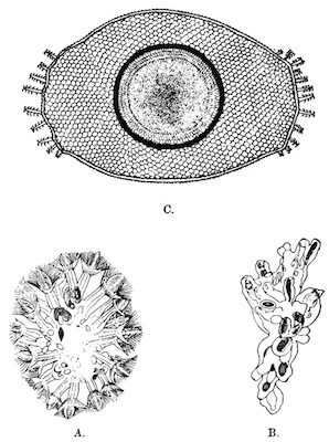
Fig. 46.—Lophopodella carteri (from Igatpuri Lake).
A=outline of a zoarium with the polypides expanded, as seen from below through glass to which it was attached, × 4; B=outline of a zoarium with the polypides highly contracted, as seen from above, × 4; C=statoblast, × 75.
37 a. Var. himalayana.
Lophopus lendenfeldi, Annandale (nec Ridley), J. As. Soc. Bengal, (n. s.) iii, 1907, p. 92, pl. ii, figs. 1-4 (1907). Lophopus lendenfeldi var. himalayanus, id., Rec. Ind. Mus. i, p. 147, figs. 1, 2 (1907). Lophopus himalayanus, id., ibid. ii, p. 172, fig. 4 (1908).
This variety differs from the typical form in having fewer tentacles and in the fact that the marginal processes of the statoblast are abortive or absent.
Pectinatella davenporti, Oka[BL] from Japan is evidently a local race of L. carteri, from the typical form of which it differs in having the marginal processes of the statoblast more numerous and better developed. The abortive structure of these processes in var. himalayana points to an arrest of development, for they are the last part of the statoblast to be formed.
Types. The statoblasts mounted in Canada balsam by Carter and now in the British Museum must be regarded as the types of the species named but not seen by Hyatt. The types of the var. himalayana are in the Indian Museum and those of the subspecies davenporti presumably in the possession of Dr. Oka in Tokyo.
Geographical Distribution.—The typical form occurs in Bombay, the W. Himalayas and possibly Madras, and its statoblasts have been found in E. Africa; the var. himalayana has only been taken in the W. Himalayas and the subspecies davenporti in Japan. Indian localities are:—Bombay Presidency, Igatpuri Lake, W. Ghats (alt. ca. 2,000 feet); the Island of Bombay (Carter): W. Himalayas, Bhim Tal, Kumaon (alt. 4,500 feet).
Biology.—L. carteri is found on the lower surface of stones and on the stems and leaves of water-plants, usually in lakes or large ponds. Although the zoaria do not form compound colonies by secreting a common membrane or investment, they are markedly gregarious. The most closely congregated and the largest zoaria I have seen were assembled amongst a gelatinous green alga of the genus Tolypothrix[BM] (Myxophyceæ) that grows on the vertical stems of a plant at the edge of Igatpuri Lake; it is noteworthy that in this case the alga seemed to take the place of the common investment of Pectinatella burmanica, in which green cells are present in large numbers (p. 237). The zoaria of L. carteri are able to change their position, and I found that if a number of them were placed in a bottle of water they slowly came together at one spot, thus apparently forming temporary compound colonies. Before a movement of the whole zoarium commences its base becomes detached from its support at the anterior end (fig. 32, p. 172), but the whole action is extremely slow and I have not been able to discover any facts that cast light on its exact method of production. At Igatpuri statoblasts are being produced in considerable numbers at the end of November, but many young zoaria can be found in which none have as yet been formed.
The larva of a fly of the genus Chironomus is often found inhabiting a tube below zoaria of L. carteri. It is thus protected from its enemies but can protrude its head from beneath the zoarium and seize the small animals on which it preys.
Genus 4. PECTINATELLA, Leidy.
Cristatella, Leidy, P. Ac. Philad. v, p. 265 (1852). Pectinatella, id., ibid., p. 320. Pectinatella, Allman, Mon. Fresh-Water Polyzoa, p. 81 (1857). Pectinatella, Hyatt, Proc. Essex Inst. v, p. 227, fig. 20 (1867). Pectinatella, Kraepelin, Deutsch. Süsswasserbryozoen, i, p. 133 (1887). Pectinatella, Oka, Journ. Coll. Sci. Tokyo, iv, p. 89 (1891).
Type, Pectinatella magnifica, Leidy.
This genus is closely allied to Lophopodella, from which it is often difficult to distinguish young specimens. Adult zoaria are, however, always embedded together in groups in a gelatinous investment which they are thought to secrete in common[BN], and the statoblasts are entirely surrounded by processes that bear curved spinules at their tips only. The polypides have the same semi-recumbent position as those of Lophopodella but are larger than those of any species of Lophopodella or Lophopus yet known. The statoblasts are larger than those of any other Plumatellidæ.
The type-species was originally found in N. America but has since been taken in several localities in continental Europe. Except this and the Indian form only one species is known, namely P. gelatinosa from Japan. P. magnifica has circular statoblasts with long marginal processes, while in P. gelatinosa the statoblasts are subquadrate and in P. burmanica almost circular, both Asiatic forms having very short marginal processes.
The compound colonies formed by Pectinatella are often of great size. Those of P. gelatinosa are sometimes over 2 metres in length, while those of P. burmanica in the Sur Lake appeared to be only limited as regards their growth by the shallowness of the water in which the reeds to which they were attached were growing. Some were observed that were over 2 feet long.
38. Pectinatella burmanica, Annandale. (Plate III, fig. 5.)
Pectinatella burmanica, Annandale, Rec. Ind. Mus. ii, p. 174, fig. 5 (1908). Pectinatella burmanica, id., ibid. v, p. 56 (1910). Pectinatella burmanica, id., Spol. Zeyl. vii, p. 63, pl. i, fig. 3 (1910).
Zoarium. The zoaria are circular or nearly so except when about to undergo division, in which case they are constricted in the middle. As a rule they measure nearly an inch (2 cm.) in diameter. The polypides have a definite arrangement in each zoarium, being divided into four groups, each of which has a fan-like form. In the first place they are separated into two main divisions in a line running through the centre of the zoarium, and secondly each main division is separated into two subordinate ones in a line running across the other at right angles. The number of zoaria joined together in a single compound colony is very variable; sometimes there are only about half a dozen and sometimes several hundreds. The common investment in living colonies is often as much as two inches thick and has a translucent dark greenish colour due to the presence in it of green cells.
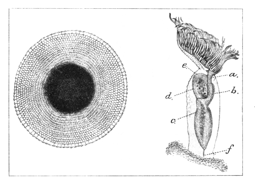
Fig. 47.—Pectinatella burmanica.
A=polypide with the lophophore expanded, × 15; a=œsophagus; b=cardiac limb of stomach; c=stomach; d=rectum; e=anus; f=funiculus. [The muscles are omitted and the external tubercles are only shown on part of the polypide. The specimen is from the Sur Lake, Orissa.] B=statoblast from Ceylon, × 35.
Polypide. The polypide can be extruded for a distance of at least 5 mm. Its whole external surface is covered with minute tubercles. There are about 90 tentacles, which are long and slender, the velum at their base being narrow and almost straight. The stomach is of considerable stoutness.
Statoblast. The statoblasts are of large size, measuring from 1 to 1.75 mm. in diameter. In form they are almost circular, but one side is always slightly flattened. The marginal processes are very short and bear a single pair of hooks at the tip. The capsule is circular and small as compared with the free part of the swim-ring.
Type in the Indian Museum.
P. burmanica is evidently a near relation of P. gelatinosa, Oka, from Japan, differing from that species in the shape of the statoblasts and in having much longer tentacles. The arrangement of the polypides in the zoarium and the general structure of the statoblasts are very similar in the two species.
Geographical Distribution.—P. burmanica was originally described from a swamp at Kawkareik in the Amherst district of Tenasserim but has also been found in the Sur Lake near Puri in Orissa. Dr. A. Willey obtained specimens from a pool by the roadside between Maradankadewela and Galapitagala, at the foot of Ritigala, N. Central Province, Ceylon.
Biology.—The first specimen obtained was a statoblast fixed to a tube of the oligochæte worm Aulophorus tonkinensis taken at Kawkareik in March. At the same time young zoaria, which did not yet possess a common investment, were found on a leaf growing on a twig which drooped into the water. Large compound colonies were taken in Orissa in October. They completely encased the stems of reeds, thus forming hollow cylinders, but slipped from their supports when the reeds were pulled out of the water. In life they resembled gelatinous algæ rather than animals and exhibited a striking similarity to masses of zoaria of Lophopodella carteri surrounded by such algæ. Some of the colonies were evidently dying and contained few polypides in a living condition, but many statoblasts; others were in a flourishing condition and were producing larvæ and statoblasts simultaneously.
A piece of a colony full of larvæ was placed before midday in an aquarium, which was kept in a shady verandah. Large numbers of larvæ were set free almost immediately. They measured about 2 mm. in length and were distinctly pear-shaped; each contained a pair of polypides, which occupied a comparatively small part of the interior, the whole of the broader half being hollow. The larvæ swam slowly, broad-end-first, by means of the cilia with which their surface was covered, occasionally gyrating on their long axis and always adopting an erratic course. Towards evening they showed signs of settling down, frequently touching the glass of the aquarium with their broad ends and sometimes remaining still in this position for some minutes. Many attempts were, however, made before fixation was completed, and this did not occur until after nightfall. By next morning every larva was fixed to the glass and had everted its two polypides. Unfortunately I was not able to trace the development further, but young compound colonies were found in which the secretion of the common investment had just commenced. The zoaria in these colonies measured about 1 cm. in diameter and already contained many polypides each.
Oka has described the development from the statoblast of the allied Japanese species. He found that each statoblast produced in the first instance a single polypide, and that the statoblasts, which were produced in autumn, lay dormant through the winter and germinated in spring. As the Sur Lake begins to undergo desiccation as soon as the "rains" cease, the statoblasts in it probably do not germinate until the break of the next "rains" about the middle of June. I have had dried statoblasts in my possession for over two years. Their cellular contents appear to be in good condition, although the cells show no signs of development; but they have not germinated in my aquarium, in which some of them have now been kept for more than six months.
The green cells of the common investment are peculiar bodies that deserve further study than it has yet been possible to devote to them. Each cell is of ovoid form, varying somewhat in size but as a rule measuring about 0.03 × 0.008 mm. There can be no doubt that these bodies represent a stage in the life-history of an alga[BO]. Diatoms, bacilli and other minute plants are often present in the membrane as well as the characteristic green cells, but do not form a constant feature of it.
APPENDIX TO THE VOLUME.
Hints on the Preparation of Specimens.
To preserve Spongillidæ.—Spongillidæ must be preserved dry or in very strong alcohol. Formalin should not be used.
To clean siliceous sponge spicules.—Place small fragments of the dried sponge (if alcohol is present, the reaction is apt to be violent) in a test tube, cover them with strong nitric acid and boil over the flame of a Bunsen burner or small spirit lamp until the solid particles disappear. Add a large quantity of water to the acid and filter through pure cellulose filter-paper, agitating the liquid repeatedly. Pass clean water in considerable quantities through the filter-paper and dry the latter carefully; place it in a spirally coiled wire and ignite with a match, holding the wire in such a way that the spicules released by the burning of the paper fall into a suitable receptacle. They may then be picked up with a camel's-hair brush and mounted in Canada balsam.
To examine the skeleton of a Spongillid.—Cut thin hand-sections with a sharp scalpel, dehydrate if necessary, and mount in Canada balsam.
To prepare gemmules for examination.—Place the gemmules dry in a watch-glass with a few drops of strong nitric acid. When gas is given off freely add water in considerable quantities. Remove the gemmules with a camel's-hair brush to clean water, then to 50%, 70%, 90% and absolute alcohol in succession, leaving them for an hour in each strength of spirit. Clear with oil of cloves and mount in Canada balsam.
To ascertain the presence of bubble-cells in the parenchyma of a Spongillid.—Tease up a small piece of the sponge with a pair of needles, mount under a thin cover-slip in strong spirit, and examine under a high power of the microscope.
To preserve Hydra in an expanded condition.—Place the polyp in a watch-glass of clean water and wait until its tentacles are expanded. Heat a few drops of commercial formaldehyde and squirt the liquid while still hot at the Hydra, which will be killed instantaneously. Remove it to a solution of formaldehyde and spirit of the following formula:—
Commercial formaldehyde1 part.
Absolute alcohol3 parts.
Distilled water7 parts.
Then pass the Hydra through 50% and 70% alcohol and keep in 90%.
To examine the capsules of the nettle-cells.—Place a living Hydra in a small drop of water on a slide and press a thin cover-slip down upon it.
To preserve freshwater polyzoa in an expanded condition.—Place the polyzoa in a glass tube full of clean water and allow them to expand their tentacles. Drop on them gradually when they are fully expanded a 2% aqueous solution of cocaine, two or three drops at a time, until movement ceases in the tentacles. Then pour commercial formaldehyde into the tube in considerable quantities. Allow the whole to stand for half an hour. If it is proposed to stain the specimens for anatomical investigation, they should then be removed through 50% and 70% to 90% alcohol. If, on the other hand, it is desired to keep them in a life-like condition they may be kept permanently in a solution of one part of commercial formaldehyde in four parts of water. Care must be taken that the process of paralyzing the polypides is not unduly prolonged, and it is always as well to preserve duplicate specimens in spirit or formalin with the lophophore retracted.
To prepare statoblasts for examination.—Place the statoblasts for a few minutes in strong nitric acid. Then remove the acid with water, pass through alcohol, clear with oil of cloves, and mount in a small quantity of Canada balsam under a cover-slip, taking care that the statoblasts lie parallel to the latter.
ADDENDA.
The following addenda are due mainly to an expedition to the lakes of Kumaon in the W. Himalayas undertaken by Mr. S. W. Kemp in May, 1911.
PART I.
Genus SPONGILLA.
Subgenus EUSPONGILLA (p. 69).
1 a. Spongilla lacustris, subsp. reticulata (p. 71).
Specimens were taken in the lake Malwa Tal (alt. 3600 feet) in Kumaon, while others have recently been obtained from the Kalichedu irrigation-tank in the Pagnor talug of the Nellore district, Madras (G. H. Tipper).
4. Spongilla cinerea (p. 79).
Specimens were taken in Naukuchia Tal (alt. 4200 feet) in Kumaon. They have a pale yellow colour when dry. This sponge has not hitherto been found outside the Bombay Presidency.
Subgenus EUNAPIUS (p. 86).
8. Spongilla carteri (p. 87).
Specimens were taken in Bhim Tal (alt. 4450 feet) and Sat Tal (alt. 4500 feet). Some of them approach the variety cava in structure.
Subgenus STRATOSPONGILLA (p. 100).
12. Spongilla bombayensis (p. 102).
Add a new variety:—
13 a. Var. pneumatica, nov.
This variety differs from the typical form in the following characters:—
(i.) The sponge forms a flat layer of a pale brownish colour as a rule with short and very delicate vertical branches.In one specimen it takes the form of an elegant cup attached, only at the base, to a slender twig.
(ii.) The gemmules are covered, outside the spicules, by a thick pneumatic coat of irregular formation and with comparatively large air-spaces.
(iii.) The gemmule-spicules are regularly sausage-shaped.
Types in the Indian Museum.
Habitat. Naukuchia Tal (alt. 4200 feet), Kumaon, W. Himalayas (S. W. Kemp).
Genus EPHYDATIA (p. 108).
After Ephydatia meyeni, p. 108, add:—
Ephydatia fluviatilis, auct.
? Ephydatia fluviatilis, Lamouroux, Encyclop. Méthod. ii, p. 327 (1824). Spongilla fluviatilis, Bowerbank (partim), Proc. Zool. Soc. London, 1863, p. 445, pl. xxxviii, fig. 1. Ephydatia fluviatilis, J. E. Gray (partim), Proc. Zool. Soc. London, 1867, p. 550. Meyenia fluviatilis, Carter (partim), Ann. Nat. Hist. (5) vii, p. 92, pl. vi, fig. 11 a, b (1881). Ephydatia fluviatilis, Vejdovsky, Abh. k. Böhm. Gesellschaft Wiss. xii, p. 24, pl. i, figs. 1, 2, 7, 10, 14, 19 (1883). Ephydatia fluviatilis, id., P. Ac. Philad. 1887, p. 178. Meyenia fluviatilis var. gracilis, Potts, ibid., p. 224. Meyenia robusta, id., ibid., p. 225, pl. ix, fig. 5. Ephydatia fluviatilis, Weltner, Arch. Naturg. Berlin, 1895 (i) p. 122. Ephydatia robusta, Annandale, Journ. As. Soc. Bengal, 1907, p. 24, fig. 7. Ephydatia fluviatilis, Weltner, in Brauer's Süsswasserfauna Deutschlands xix, Süsswasserschwämme, p. 185, figs. 316, 317 (1909). Ephydatia fluviatilis, Annandale, P. U. S. Mus. xxxviii, p. 649 (1910).
[Many more references to this common species might be cited, but those given above will be sufficient.]
This species only differs from E. meyeni in the following characters:—
(i.) there are no bubble-cells in the parenchyma;
(ii.) there is less spongin in the skeleton, which is less compact;
(iii.) the gemmule-spicules are longer, the shafts being as a rule longer than the diameter of the rotulæ;
(iv.) the gemmules are armed with a single row of regularly arranged spicules embedded in pneumatic tissue with minute air-spaces.
The sponge is a variable one and several "varieties" have been described from different parts of the world. My Indian specimens come nearest to the form described by Potts as Meyenia robusta, but have rather more slender skeleton-spicules and more elongate gemmule-spicules. The latter also appear to be less frequently "monstrous."
Type ?
Geographical Distribution.—E. fluviatilis is widely distributed in Europe and occurs in N. America,[BP] S. Africa (var. capensis, Kirkpatrick), Australia, and Japan. Specimens were obtained by Mr. Kemp from several lakes in Kumaon, namely Naukuchia Tal (alt. 4200 feet), Bhim Tal (4450 feet), Sat Tal (4500 feet), and Naini Tal (6300 feet). The gemmules from Bhim Tal referred by me to E. robusta (Potts) also belong to this species.
Biology. The external form of the sponge is due in great part to its environment. Specimens on small stones from the bottom of the Kumaon Lakes consist of thin disk-like films, often not more than a few centimetres in diameter and a few millimetres thick: others, growing on thin twigs, are elevated and compressed, resembling a cockscomb in appearance, while others again form nodules and masses of irregular form among the branches of delicate water-weeds. Some of these last are penetrated by zoaria of Fredericella indica.
Weltner has published some very interesting observations on the seasonal variation of minute structure in European representatives of the species (Arch. Naturg. Berlin, lxxiii (i), p. 273 1907) and has discussed the formation of the abnormal spicules that sometimes occur (ibid. lxvii (Special Number), p. 191, pls. vi, vii, figs. 27-59, 1901).
Genus CORVOSPONGILLA (p. 122).
After Corvospongilla burmanica, p. 123, add a new species:—
Corvospongilla caunteri, nov.
Sponge forming thin films of considerable area not more than 3 or 4 mm. thick, of a bright green colour, moderately hard but friable. The surface smooth; oscula inconspicuous, surrounded by shallow and ill-defined radiating furrows; a very stout basal membrane present.
Skeleton reticulate but almost devoid of spongin, the reticulations close but formed mainly by single spicules; skeleton-fibres barely distinguishable. A close layer of spicules lying parallel to the basal membrane.
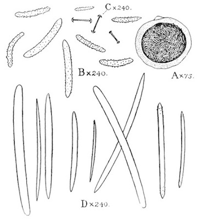
Fig. 48.—Corvospongilla caunteri (type, from Lucknow).
A=Gemmule; B=gemmule-spicules; C=flesh-spicules; D=Skeleton-spicules.
Spicules. Skeleton-spicules variable in size and shape, almost straight, as a rule smooth, moderately stout, blunt or abruptly pointed; sometimes roughened or spiny at the tips, often sharply pointed. Flesh-spicules minute, few in number, with smooth, slender shafts which are variable in length, never very strongly curved; the terminal spines relatively short, not strongly recurved. Gemmule-spicules amphistrongylous or amphioxous, irregularly spiny, slender, of variable length.
Gemmules free in the substance of the sponge, spherical or somewhat depressed, very variable in size but never large, having a thick external pneumatic coat in which the air-spaces are extremely small and, inside this coat, a single rather sparse layer of spicules lying parallel to the gemmule. A single depressed aperture present.
Type in the Indian Museum.
Habitat. Hazratganj, Lucknow; on piers of bridge in running water (J. Caunter, 29-30. iv. 11).
The structure of the gemmules of this species differs considerably from that in any other known species of the genus, in which these structures are usually adherent and devoid of a true pneumatic coat. In some of the gemmules before me this coat measures in thickness about 1/9 of the total diameter of the gemmule. C. caunteri is the first species of Corvospongilla to be found in the Indo-Gangetic plain.
PART II.
Genus HYDRA (p. 147).
25. Hydra oligactis (p. 158).
Mr. Kemp found this species common in Bhim Tal in May. His specimens, which were of a reddish-brown colour in life, appear to have been of more vigorous constitution than those taken by Major Stephenson in Lahore. Some of them had four buds but none were sexually mature.
PART III.
Genus FREDERICELLA (p. 208).
28. Fredericella indica (p. 210).
This species is common in some of the Kumaon lakes, in which it grows, at any rate at the beginning of summer, much more luxuriantly than it does in the lakes of the Malabar Zone in autumn, forming dense bushy masses on the under surface of stones, on sticks, &c. The vertical branches often consist of many zoœcia. Mr. Kemp took specimens in Malwa Tal, Sath Tal, and Naini Tal (alt. 3600-6300 feet).
Genus PLUMATELLA (p. 212).
30. Plumatella emarginata (p. 220).
Mr. Kemp took bushy masses of this species in Malwa Tal and Bhim Tal.
32. Plumatella diffusa (p. 223).
This species is common in Malwa Tal and Bhim Tal in May.
33. Plumatella allmani (p. 224).
Mr. Kemp only found this species in Malwa Tal, in which (at any rate in May) it appears to be less abundant than it is in Bhim Tal in autumn. Mr. Kemp's specimens belong to the form called P. elegans by Allman.
34. Plumatella tanganyikæ (p. 225).
Specimens taken by Mr. Kemp, somewhat sparingly, in Bhim Tal and Sath Tal in May exhibit a somewhat greater tendency towards uprightness of the zoœcia than those I found in autumn in Igatpuri lake. The ectocyst is, in the former specimens, of a deep but bright reddish-brown. The zoaria are attached to twigs and small stones.
Genus STOLELLA (p. 229).
After Stolella indica, p. 229, add a new species:—
Stolella himalayana, nov.
This species may be distinguished from S. indica by (i) its entirely recumbent zoœcia, and (ii) the lateral branches of its zoarium.
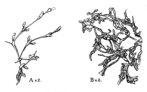
Fig. 49.—Stolella himalayana (types, from the Kumaon lakes).
A. The greater part of a young zoarium. B. Part of a much older zoarium.
Zoarium entirely recumbent, consisting of zoœcia joined together, often in groups of three, by slender, transparent, tubular processes. These processes are often of great relative length; they are formed by a modification of the posterior or proximal part of the zoœcia, from which they are not separated by a partition, and they increase in length up to a certain point more rapidly than the zoœcia proper. A zoœcium often gives rise first to an anterior daughter-zoœcium, the proximal part of which becomes elongate and attenuated in due course, and then to a pair of lateral daughter-zoœcia situated one on either side. As a result of this method of budding a zoarium with a close superficial resemblance to that of Paludicella is at first produced, but as the colony increases in age and complexity this resemblance largely disappears, for the zoœcia and their basal tubules grow over one another and often become strangely contorted (fig. 49).
Zoœcia elongate and slender, flattened on the ventral, strongly convex on the dorsal surface; rather deep in proportion to their breadth; the ectocyst colourless, not very transparent except on the stolon-like tubular part; dorsal keel and furrow as a rule absent; orifice unusually inconspicuous, situated on a tubercle on the dorsal surface.
Polypide stout and short; the tip of the fundus of the stomach capable of very complete constriction; the retractor muscles unusually short and stout.
Statoblasts. Only free statoblasts have been observed. They resemble those of S. indica, but are perhaps a little longer and more elongate.
Types in the Indian Museum.
The discovery of this species makes it necessary to modify the diagnosis of the genus, the essential character of which, as distinguishing it from Plumatella, is the differentiation of the proximal part of some or all of the zoœcia to form stolon-like tubules. From Stephanella, Oka, it is distinguished by the absence of a gelatinous covering, and by the fact that all the zoœcia are attached, at least at the base, to some extraneous object.
Habitat. Malwa Tal, Kumaon (alt. 3600 feet), W. Himalayas (Kemp, May 1911).
Biology. Mr. Kemp took three specimens, all attached to the lower surface of stones. They contained few statoblasts and were evidently in a condition of vigorous growth. Between the lateral branches new polyparia were developing in several instances from free statoblasts, each of which appeared to contain two polypides.
ALPHABETICAL INDEX.
All names printed in italics are synonyms.
When more than one reference is given, the page on which the description occurs is indicated by thickened numerals.
alba (Euspongilla) (Spongilla), 8, 9.
alba (Spongilla), 4, 22, 63, 76.
alba var. bengalensis (Spongilla), 4, 22, 63, 77.
alba var. cerebellata (Spongilla), 22, 63, 76.
alba var. marina (Spongilla), 77.
Alcyonella, 212.
Alcyonellea, 185.
allmani (Plumatella), 7, 8, 9, 23, 188, 224, 246.
allmani var. diffusa (Plumatella), 223.
allmani var. dumortieri (Plumatella), 222.
attenuata (Hydra), 148, 158.
aurantiaca (Hydra), 148.
aurea (Pectispongilla), 9, 22, 63, 106.
aurea var. subspinosa (Pectispongilla), 63, 107.
benedeni (Alcyonella), 220.
bengalensis (Bowerbankia), 189.
bengalensis (Membranipora), 23.
bengalensis (Spongilla), 77.
bengalensis (Victorella), 4, 8, 9, 23, 187, 195.
blembingia (Ephydatia), 54.
bogorensis (Ephydatia), 54.
bombayensis (Plumatella), 225.
bombayensis (Spongilla), 22, 63, 100, 102, 241.
bombayensis (Stratospongilla) (Spongilla), 8, 9.
Bowerbankia, 187, 189.
brunnea (Hydra), 148.
burmanica (Corvospongilla), 8, 22, 64, 122.
burmanica (Pectinatella), 8, 10, 23, 188, 235.
calcuttana (Spongilla), 96.
cambodgiensis (Norodonia), 202.
Carterella, 108.
carteri (Eunapius) (Spongilla), 7, 8, 9, 10.
carteri (Eunapius), 87.
carteri (Lophopodella), 7, 8, 23, 188, 232, 233.
carteri (Lophopus), 232.
carteri (Pectinatella), 231, 232.
carteri (Spongilla), 4, 22, 63, 86, 87, 241.
carteri var. cava (Spongilla), 22, 63.
carteri var. himalayana (Lophopodella), 23, 188.
carteri var. lobosa (Spongilla), 22, 63.
carteri var. mollis (Spongilla), 22, 63.
caudata (Bowerbankia), 189.
caudata subsp. bengalensis (Bowerbankia), 23, 189.
caunteri (Corvospongilla), 243.
cava (Spongilla), 88.
cerebellata (Spongilla), 76.
ceylonensis (Irene), 22, 140.
Cheilostomata, 184.
Chlorella, 50.
cinerea (Euspongilla) (Spongilla), 9.
cinerea (Spongilla), 22, 63, 72, 79, 241.
clementis (Stratospongilla) (Spongilla), 53.
coggini (Stratospongilla) (Spongilla), 53.
colonialis (Loxosomatoides), 23.
contecta (Spongilla), 95.
coralloides (Plumatella), 217.
Corvospongilla, 64, 122, 243.
crassior (Spongilla), 98.
crassissima (Eunapius) (Spongilla), 9.
crassissima (Spongilla), 4, 22, 63, 98.
crassissima var. crassior (Spongilla), 23, 63.
crateriformis (Meyenia), 83.
crateriformis (Ephydatia), 83, 84.
crateriformis (Euspongilla) (Spongilla), 8, 9.
crateriformis (Meyenia), 83.
crateriformis (Spongilla), 22, 63, 83.
Cristatella, 235.
Cristatellina, 206.
Ctenostomata, 184, 185, 187, 189.
Cyclostomata, 184.
decipiens (Spongilla), 54, 96, 97.
diffusa (Plumatella), 7, 8, 9, 23, 188, 223, 245.
diœcia (Hydra), 158.
Dosilia, 64, 110.
Echinella, 199.
elegans (Plumatella), 224.
Eleutheroblastea, 146, 147.
emarginata (Plumatella), 4, 8, 9, 10, 23, 188, 218, 220, 245.
emarginata var. javanica (Plumatella), 221.
Entoprocta, 183.
Ephydatia, 64, 108, 242.
erinaceus (Spongilla), 114.
Eunapius, 63, 86, 241.
Euspongilla, 63, 67, 69, 241.
filamentata (Syncoryne), 22, 140.
fluviatilis (Ephydatia), 109, 242.
fluviatilis (Meyenia), 242.
fluviatilis (Spongilla), 108, 242.
fluviatilis var. gracilis (Meyenia), 242.
fortis (Ephydatia), 52, 53.
fragilis (Spongilla), 95, 96.
fragilis subsp. calcuttana (Eunapius) (Spongilla), 9.
fragilis subsp. calcuttana (Spongilla), 22, 63.
fragilis subsp. decipiens (Spongilla), 22, 63.
Fredericella, 188, 208, 245.
Fredericellidæ, 188, 208.
friabilis (Spongilla), 87.
fruticosa (Plumatella), 4, 7, 8, 9, 23, 188, 217, 218.
fusca (Hydra), 158, 159.
Gecarcinucus, 10.
gemina (Eunapius) (Spongilla), 8.
gemina (Spongilla), 22, 63, 97.
glomerata (Spongilla), 95.
grisea (Hydra), 148, 149.
Gymnolæmata, 184, 187.
Halichondrina, 65.
hemephydatia (Euspongilla) (Spongilla), 8.
hemephydatia (Spongilla), 22, 63, 82.
hexactinella (Hydra), 148.
himalayana (Lophopodella), 233.
himalayana (Stolella), 246.
himalayanus (Lophopus), 233.
Hislopia, 187, 199.
Hislopidées, 199.
Hislopiidæ, 187, 199.
Homodiætidæ, 191.
Hyalinella, 212.
Hydra, 146, 147, 245.
Hydraidæ, 147.
Hydridæ, 146, 147.
hydriforme (Polypodium), 142.
Hydrozoa, 146.
indica (Ephydatia), 83.
indica (Fredericella), 9, 23, 188, 209, 245.
indica (Spongilla), 22, 63, 100.
indica (Stolella), 4, 9, 23, 188, 229.
indica (Stratospongilla), (Spongilla), 9.
javanica (Plumatella), 4, 8, 9, 23, 188, 221, 222.
kawaii (Limnocodium), 141.
lacroixii (Membranipora), 23.
lacustris (Cordylophora), 141.
lacustris (Euspongilla), 69.
lacustris (Hislopia), 4, 8, 9, 23, 187, 199, 202, 204.
lacustris (Spongilla), 63, 67, 69.
lacustris subsp. moniliformis (Hislopia), 9, 23, 187.
lacustris subsp. reticulata (Spongilla), 4, 8, 9, 22, 63, 71, 241.
lacustris var. bengalensis (Spongilla), 77.
lapidosa (Corvospongilla), 9, 22, 64, 124.
lapidosa (Spongilla), 124.
latouchiana (Trochospongilla), 4, 8, 9, 22, 64, 115.
leidyi (Trochospongilla), 115.
lendenfeldi (Lophopus), 233.
lendenfeldi var. himalayanus (Lophopus), 233.
lobosa (Spongilla), 89.
Lophopinæ, 188, 211, 231.
Lophopodella, 8, 188, 231.
Lophopus, 8, 232.
lordii (Spongilla), 95.
loricata (Spongilla), 122.
loricata var. burmanica, (Spongilla), 122.
lucifuga (Plumatella), 217, 220, 224.
magnifica (Pectinatella), 235.
meyeni (Ephydatia), 7, 9, 17, 22, 64, 108.
meyeni (Spongilla), 108.
Meyenia, 108, 113.
microsclerifera (Euspongilla) (Spongilla), 53.
mollis (Spongilla), 88.
moniliformis (Hislopia), 204.
monœcia (Hydra), 158.
morgiana (Spongilla), 95.
mülleri (Ephydatia), 109, 243.
mülleri subsp. meyeni (Ephydatia), 109.
Norodonia, 199.
oligactis (Hydra), 7, 22, 146, 158, 159, 245.
orientalis (Hydra), 148, 149.
ottavænsis (Spongilla), 95.
pallens (Hydra), 148.
Paludicella, 187, 192.
Paludicellidæ, 187, 191.
Paludicellidées, 191.
Paludicellides, 191.
Paludicellina, 186, 187, 190.
paulula (Spongilla), 120.
pavida (Victorella), 194, 195.
Pectinatella, 188, 235.
pectinatellophila (Dactyloccopsis), 238.
Pectispongilla, 63, 106.
pennsylvanica (Trochospongilla), 9, 22, 64, 118.
pennsylvanica (Tubella), 118.
pentactinella (Hydra), 149.
philippinensis (Euspongilla) (Spongilla), 53.
phillottiana (Trochospongilla), 4, 8, 9, 22, 64, 117.
Phylactolæmata, 185, 188, 206.
Plumatella, 188, 208, 212, 245.
Plumatellidæ, 188, 211.
Plumatellina, 188, 206.
Plumatellinæ, 188, 211, 212.
plumosa (Dosilia), 8, 9, 22, 64, 111.
plumosa (Ephydatia), 111.
plumosa (Meyenia), 111.
plumosa (Spongilla), 111.
pneumatica (Stratospongilla) (Spongilla), 241.
polypus (Hydra), 148, 159.
Polyzoa, 183.
princeps (Plumatella), 220.
princeps var. emarginata (Plumatella), 220.
princeps var. fruticosa (Plumatella), 217.
proliferens (Euspongilla) (Spongilla), 8, 9, 10.
proliferens (Spongilla), 4, 8, 22, 63, 72.
Proterospongia, 27.
punctata (Hyalinella), 228.
punctata (Plumatella), 9, 188, 227.
repens (Plumatella), 217, 223.
reticulata (Spongilla), 71.
rhætica (Hydra), 158.
robusta (Ephydatia), 109, 242.
robusta (Meyenia), 242.
roeselii (Hydra), 158.
ryderi (Microhydra), 141.
schilleriana (Sagartia), 2, 22, 140.
schilleriana subsp. exul (Sagartia), 22.
sibirica (Spongilla), 95.
sinensis (Norodonia), 202.
sinensis (Stratospongilla) (Spongilla), 53.
socialis (Hydra), 158.
sowerbii (Limnocodium), 141.
Spongilla, 63, 67, 86, 241.
Spongilladæ, 65.
Spongillidæ, 65.
Stolella, 188, 229, 246.
Stolonifera, 185.
Stratospongilla, 63, 100, 241.
stricta (Plumatella), 217.
subspinosa (Pectispongilla), 107.
sumatrana (Stratospongilla) (Spongilla), 53.
tanganyikæ (Limnocnida), 142.
tanganyikæ (Plumatella), 9, 23, 188, 225, 246.
Trachospongilla, 64, 113.
Trachyspongilla, 108.
travancorica (Euspongilla) (Spongilla), 9.
travancorica (Spongilla), 22, 63, 81.
trembleyi (Hydra), 148.
Tubella, 64, 113, 120.
ultima (Spongilla), 22, 63, 105.
ultima (Stratospongilla) (Spongilla), 9.
Vesicularidæ, 189.
Vesicularina, 186, 187, 189.
vesicularis (Hyalinella), 228.
vesicularis (Plumatella), 227, 228.
vesparioides (Tubella), 8, 22, 64, 120.
vesparium (Tubella), 54.
vestita (Bimeria), 22, 139.
Victorella, 189, 194.
Victorellidæ, 191.
Victorellides, 191.
viridis (Hydra), 147.
vitrea (Hyalinella), 228.
vitrea (Plumatella), 227, 228.
vulgaris (Hydra), 4, 8, 9, 10, 22, 130, 146, 148, 149, 158.
whiteleggei (Cordylophora), 141.
yunnanensis (Euspongilla) (Spongilla), 53.
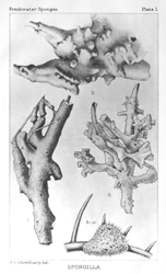
PLATE I.
Specimens of Spongilla preserved in spirit.
Figs. 1-3.S. (Euspongilla) alba var. bengalensis (nat. size) from ponds of brackish water at Port Canning in the delta of the Ganges. Fig. 1 represents the type-specimen of the variety, and was taken in the winter of 1905-6. Figs. 2 and 3 represent specimens taken in the same ponds in the winters of 1907 and 1908 respectively.
Fig. 4.Spongilla sp. (? abnormal form of S. (Eunapius carteri)) from an aquarium in Calcutta (× 10).
SPONGILLA
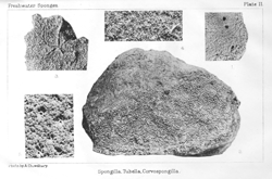
PLATE II.
Photographs of dried specimens of Spongilla, Tubella, AND Corvospongilla.
Fig. 1.Part of a large specimen of S. (Eunapius) carteri from Calcutta, to show the conspicuous rounded oscula (reduced).
Fig. 2.Gemmules of S. (Stratospongilla) bombayensis on a stone from the edge of Igatpuri Lake, Bombay Presidency (nat. size).
Fig. 3.Part of one of the type-specimens of S. (Stratospongilla) ultima from Cape Comorin, Travancore, to show the star-shaped oscula (slightly enlarged).
Fig. 4.Part of the type specimen of T. vesparioides (external membrane destroyed), to show the reticulate skeleton and the numerous gemmules (nat. size).
Fig. 5.Part of a schizotype of C. burmanica to show the elevated oscula (nat. size).
Spongilla, Tubella, Corvospongilla.
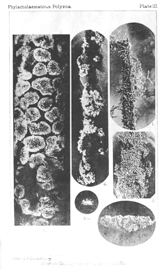
PLATE III.
Photographs of specimens of Plumatella, Lophopodella, and Pectinatella.
Fig. 1.Specimen in spirit of P. fruticosa (typical form) on the leaf of a bulrush from a pond in the Calcutta Zoological Gardens (nat. size).
Fig. 2.A small zoarium of the benedeni phase of P. emarginata from Rangoon (nat. size). Part of the mass has been removed at one end to show the structure. The specimen was preserved in spirit.
Fig. 3.Part of a large zoarium of P. diffusa on a log of wood from Gangtok, Sikhim (nat. size). An enlarged figure of another part of the same specimen is given in fig. 2, Pl. IV. The specimen was preserved in spirit.
Figs. 4, 4 a.Specimens of L. carteri from Igatpuri Lake, Bombay, preserved in formalin. Fig. 4 represents a mass of polyparia surrounded by a green gelatinous alga on the stem of a water-plant; fig. 4a an isolated polyparium with the polypides fully expanded from the under surface of a stone in the same lake. Both figures are of natural size.
Fig. 5.Part of a compound colony of P. burmanica on the stem of a reed from the Sur Lake, Orissa (nat. size, preserved in formalin).
Plumatella, Lophopodella, Pectinatella.
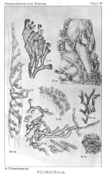
PLATE IV.
Specimens of Plumatella.
Fig. 1.Vertical branch of a polyparium of P. emarginata from Calcutta, to show method of branching (× 8). The specimen was preserved in formalin, stained with hæmalum, and after dehydration and clearing, mounted in canada balsam.
Fig. 1 a.Part of a young, horizontal zoarium of P. emarginata from Rangoon (× 4, preserved in spirit).
Fig. 2.Part of a zoarium of P. diffusa from Gangtok, Sikhim (× 4). See Pl. III, fig. 3.
Figs. 3, 3 a.Specimens in spirit of P. allmani from Bhim Tal (lake), W. Himalayas. Fig. 3 represents a mature polyparium; fig. 3 a a young polyparium to which the valves of the statoblast (×) whence it had arisen are still attached.
Fig. 4.Part of a zoarium of the coralloides phase of P. fruticosa (from Calcutta) preserved in spirit, as seen on the surface of the sponge in which it is embedded (× 3).
Fig. 5.Part of the margin of a living polyparium of P. punctata from Calcutta (× 8) with the polypides fully expanded.
PLUMATELLA.
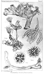
PLATE V.
Specimens of Plumatella, Stolella, and Pectinatella.
Fig. 1.Part of a zoarium of the coralloides phase of P. fruticosa (× 10) from Calcutta. The specimen, which was preserved in spirit, had been removed from a sponge of Spongilla carteri.
Fig. 2.Terminal branch of a polyparium of P. punctata from Calcutta (× 30). The specimen was preserved in formalin, stained with hæmatoxylin, and finally mounted in canada balsam.
Fig. 3.Part of an adult polyparium of S. indica from the United Provinces (× 30). The specimen was preserved in formalin, stained with hæmalum, and finally mounted in canada balsam. The lower zoœcium contains a mature free statoblast, the upper one a fixed one.
Fig. 4.The growing point of a young polyparium of the same species from Calcutta (× 30), to show the method of formation of the stolon that connects the different groups of zoœcia. The specimen had been treated in the same way as that represented in fig. 3.
Figs. 5, 5 a.Zoaria from a compound colony of P. burmanica from the Sur Lake, Orissa (× 2). The specimens, which were preserved in formalin, are represented as seen from the adherent surface of the colony.
Plumatella, Stolella, Pectinatella..