Chapter XXX
Reading edition
II. MEDULLOSEAE.
The term Medulloseae was first employed by Goeppert and Stenzel[236] for a family of Palaeozoic plants that appears to have reached its maximum development in the Permian period: the oldest representatives so far discovered are of Upper Carboniferous age. Our knowledge of the family is chiefly derived from a study of the anatomical characters of stems, and it is therefore on this basis that any grouping of genera or species should be attempted. Although there is little information with regard to the reproductive organs of Medullosa, the type-genus, it is certain that the Medulloseae are Pteridosperms differing from members of that group included in the Lyginopterideae in the presence of more than one stele in the stem, in the habit of the fronds, and in the structure of the rachis, as also in the structure of the seeds, though these organs bear a fairly close resemblance to the seeds of Lyginopteris and Heterangium. The fronds of the Lyginopterideae are of the Sphenopteris type while in the case of such species of Medullosa as afford evidence of connexion between stems and leaves the latter have the characters of Neuropteris, Alethopteris, Odontopteris, Linopteris, and other form-genera usually included in the Neuropterideae. Dr Lotsy[237] speaks of Lyginopteris and Heterangium as members of the Sphenopteridophylla and assigns species of Medullosa either to the Neuropteridophylla or to the Pecopteridophylla, the latter subdivision including species with fronds of the Alethopteris type. There is, however, little doubt that other forms of leaves, such as Odontopteris and possibly Taeniopteris, were borne on Medullosan stems. It is undesirable except in the absence of more trustworthy criteria to make use of so protean a feature as leaf-form as a basis of classification. The name Neuropterideae has been frequently employed for Pteridosperms other than the Lyginopterideae on the ground that the foliage of Medullosa is represented by species assigned to form-genera included in the Neuropterideae. It is, however, preferable to restrict the family-name Neuropterideae to fronds and to speak of the second family of Pteridosperms as the Medulloseae, including the genera Medullosa, Sutcliffia, and Rhexoxylon.
MEDULLOSA.
Some species of Medullosa probably resembled in habit Angiopteris evecta and the larger Marattias; they had short and relatively thick stems clothed with the large decurrent bases of long compound fronds superficially like those of some recent Ferns and the leaves of the Cycad Bowenia. It is probable that, as Zeiller[238] has pointed out, the fronds of Medullosa and of other Pteridosperms had a greater tendency than those of true Ferns to a dichotomy of the rachis. In other types the stems reached a considerable length and leaves and branches were separated by several feet of bare stem. The large size of the leaf-stalks in proportion to the diameter of the stem as shown by such species as Medullosa anglica and M. Leuckarti (fig. 416) suggests either a short and thick main axis or, in the case of long stems bearing scattered leaves, a plant that supported itself partially at least by a habit of growth comparable with that of tropical Aroids or other lianes. While Medullosa anglica with its contiguous leaf-bases affords an example of the first type, the occurrence of stems of a Permian species, M. stellata, 3½ metres long without branches or leaf-scars, suggests the habit of a liane; similarly a specimen of Medullosa Leuckarti in the Chemnitz Museum bearing a few spreading petioles but little narrower than the stem and given off at a wide angle would seem to favour the view that some species were ill adapted to be mechanically self-supporting plants. The longest piece of stem that has come under my notice is a specimen of M. stellata in the Chemnitz Museum reaching a length of nearly 8 metres: some species attained a diameter of about 50 centimetres.
Medullosa is always polystelic: the plan of the vascular system varies considerably as regards both the number and form of the steles, but there is a uniform type of structure within the limits of each stele that recalls the single stele of Heterangium. The steles consist of a central region composed of primary xylem, originally surrounded by phloem, which in its mesarch or exarch structure agrees with the vascular tissue of some species of Gleichenia or Lygodium. To this central region a cambium added secondary xylem and phloem either in the form of a cylinder of uniform breadth, or more frequently the centrifugally developed xylem exceeded in amount the secondary conducting tissue added to the inner side of the primary region. Apart from anatomical details a Medullosan stem with its several steles, each with secondary tissue, embedded in parenchymatous ground-tissue resembles the stems of some Dicotyledonous climbers such as Thinouia scandens, species of Serjania and Paullinia[239].
Anatomically the main features of the stelar system of Medullosa, neglecting the secondary xylem and phloem, are in closer agreement with the stems of Ferns than with those of any other plants. It has been shown that the genus Heterangium bears a close resemblance to Gleichenia in the structure of the primary stele (fig. 418, C): one of the oldest types of Medullosa, M. anglica, may be described as a Heterangium with three steles and may be compared with a dictyostelic Fern in which the irregular vascular framework is made up of three main strands. In certain types of Medullosa (fig. 416) the ground-plan of the vascular system recalls that of a solenostelic Fern, while in others the greater complexity suggests comparison with such Ferns as Matonia, Angiopteris, Psaronius, or Cyathea; ‘it is as though Nature were at the Carboniferous moment in the midst of a series of amazing engineering experiments, most of which were either buried deep in Palaeozoic oblivion, or permitted to survive only as vestigial relics and atavistic ghosts’[240]. Though many Medullosae resemble Ferns there is an important difference between the two groups in the origin of the various plans of Medullosan stelar systems: in Ferns the leaf is the determining factor in the evolution of stelar arrangement, while in Medullosa the occasional interruption of a solenostele or the development of an apparently complex dictyostele are features independent of the leaf and leaf-traces. In the structure of the secondary xylem and phloem and in root-structure Medullosa agrees with recent Cycads. The genus is in short a generalised type with filicinean and cycadean affinities. In the possession of seeds borne on modified pinnae of compound fronds, Medullosa resembles both Cycas and the Lyginopterideae. The seeds exhibit a fairly close agreement with those of Lyginopteris, Heterangium and recent Cycads, but they appear to have advanced further towards the cycadean type than is the case with the closely related seeds of the Lyginopterideae. The microsporophylls are very imperfectly known but they were undoubtedly much less advanced and more fern-like than the megasporophylls.
The genus Medullosa is recorded from the Permian strata of Saxony, France, and Bohemia[241]; also from the Coal Measures of England, and the discovery of petrified petioles of Myeloxylon, the type borne on Medullosan stems in European species, may be taken as evidence of the existence of the genus in North America during the Carboniferous period[242].
The name Medullosa was applied by Cotta[243] to three types, Medullosa elegans, M. stellata, and M. porosa, from the Rothliegende of the Chemnitz district. The first of these was recognised by Brongniart[244] as a distinct genus for which he proposed the designation Myeloxylon and this was afterwards identified by Renault, Williamson, and other palaeobotanists as a petiole and not a stem. Further reference is made to Myeloxylon on a later page. Cotta spoke of Medullosa as the most puzzling of the genera dealt with in his ‘Dendrolithen,’ and in spite of the many additions to our knowledge the position of this Palaeozoic genus is still a fertile source of speculation. The generic designation Medullosa is applied to stems, with or without petioles; petioles or rachises of fronds that frequently occur apart from stems are referred to the genus Myeloxylon. The leaves of Medullosa include several well-known species of Carboniferous and Permian genera such as Alethopteris, Neuropteris and others that have in recent years been transferred from the Filicales to the Pteridosperms. In a few instances seeds have been found in organic connexion with Medullosan foliage, and there can be no reasonable doubt that Trigonocarpus, some forms of Rhabdocarpus, Pachytesta, and other seeds represent the integumented megasporangia of Medullosa or some closely allied genus.
Before attempting to summarise the salient features of Medullosa a description of a few selected types will serve to place us in a better position to consider the genus as a whole. The British species are placed first on the ground that they are both geologically the oldest though, historically, the most recently described, representatives of the genus; and in the organisation of the stem they are simpler than the continental species. Their resemblance to Heterangium serves to some extent to bridge the gap between the majority of species of Medullosa and the simpler types of Pteridosperms represented by Heterangium and Lyginopteris.
Medullosa anglica Scott[245].
Prior to the discovery of this species the genus Medullosa had not been recorded from Britain. A section in the Williamson collection recognised by Scott as that of a Medullosa had been identified by Williamson as a large Heterangium stem. An undescribed specimen was found by Arber[246] in the Binney collection at Cambridge which afforded some additional information as to the structure of the roots.
The specimens on which Scott’s thorough description is based were obtained by Messrs Wilde and Lomax from the Lower Coal Measures of Lancashire. The stem of this oldest species has the habit of a tree-fern and is almost completely invested by the stout decurrent bases of the petioles of large spirally disposed compound fronds with a phyllotaxis of ⅖, the leaves of the same orthostichy being separated from one another by a vertical distance of approximately 10 cm.
A transverse section of a slightly flattened stem is shown in fig. 416, A, the bases of three petioles give to it an angular form. Its dimensions are approximately 10 × 4 cm. The ground-tissue of two of the petioles is continuous with that of the stem, while that of the third leaf-stalk is cut through near its separation from the stem and its adaxial face is already defined by a hypodermal band of stereome, d. The surface of the stem is characterised by fine longitudinal ribs caused by the slightly projecting stereome in the outer cortex, and from the narrow furrows between the leaf-bases adventitious roots emerge in vertical series. The position of an interfoliar furrow is shown by a small arrow in fig. 416, A. There are three steles, 2–3 cm. × 6–10 mm. in diameter: each agrees very closely in structure with the single stele of Heterangium. Medullosa anglica may be described as a polystelic Heterangium and as having the same relation to Heterangium as regards the stelar system as Primula auricula bears to the monostelic Primula. The central core of the stele (the black patches in the diagram, fig. 416, A) consists of an anastomosing system of tracheal groups embedded in an irregular parenchymatous reticulum. The large primary tracheids reach a diameter of 150μ and have multiseriate pitting: at the periphery of the primary xylem there is a more definite grouping of tracheids as in Heterangium, and the slightly internal (mesarch) protoxylem elements are associated with scalariform and densely spiral tracheids (fig. 416, B, C) narrower than the more internal reticulate elements. The secondary xylem is manoxylic as in Cycads, tracheids in 2–4 radial series alternating with medullary rays 1–3 cells broad and usually of considerable depth (fig. 416, B). The principal rays are continuous with the parenchymatous matrix of the central core. Thick-walled tubular elements, no doubt of the nature of sieve-tubes, form a conspicuous feature in the phloem.
The three steles occasionally divide and fuse with one another. The tissue between the steles is crushed and disorganised and in the living plant was probably small in amount. In the imperfectly preserved inner cortical region there is a sinuous band of secondary parenchyma (periderm; fig. 416, A, c) developed from a deep-seated phellogen; in older stems this formed the superficial tissue after the fall of the leaves. There is no definite boundary between the cortex of the stem and the petiole-bases except when the hypoderm cuts across the cortex preparatory to the separation of a leaf-stalk. The stem-cortex and the ground-tissue of the petioles consist of parenchyma with numerous secretory canals, not sacs only as in Heterangium, and are abundantly supplied with scattered vascular bundles of collateral and exarch structure.
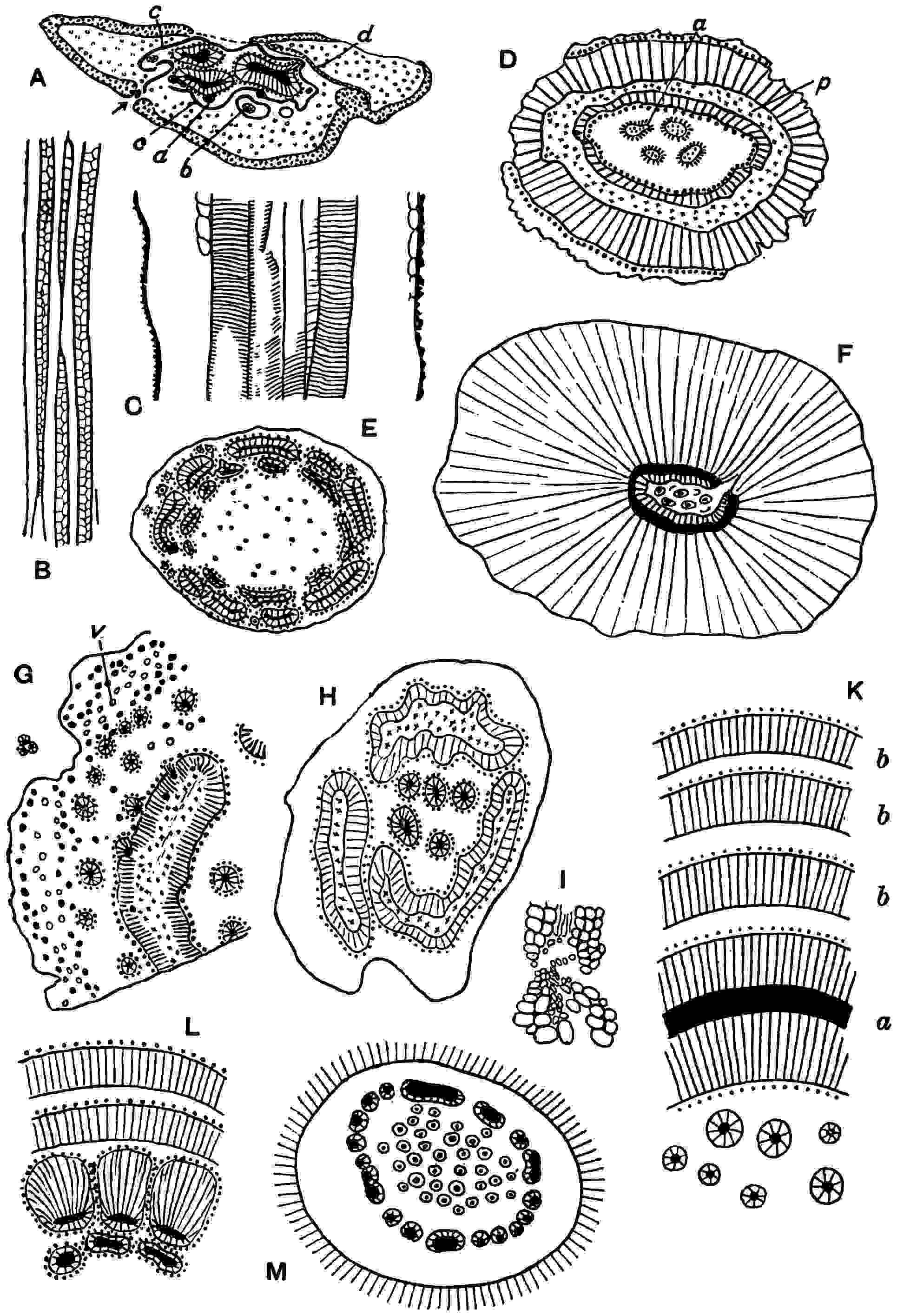
Fig. 416.
Medullosa
stems. A-C,
Medullosa anglica
; A, transverse section;
a
, accessory vascular strand;
b
, accessory strand enclosed by periderm;
c
, band of periderm encircling steles;
d
, sclerenchyma between leaf and stem. B and C, longitudinal sections. (After Scott.) D,
Medullosa stellata
;
a
, star-rings;
p
, ‘partial pith.’ (After Weber and Sterzel.) E,
Medullosa Solmsi
. (After Weber and Sterzel.) F, Medullosa stellata, from a specimen in the British Museum (No. 13767). G,
Medullosa stellata
var.
cortica
;
v
, leaf-bundles. (After Weber and Sterzel.) H, I,
Medullosa Leuckarti
. (After Weber and Sterzel and Solms-Laubach.) K,
Medullosa stellata
var.
gigantea
;
a
, concentric stele;
b
,
b
, later cylinders of centrifugal tissue. (Adapted from Weber and Sterzel.) L,
Medullosa Solmsi
var.
lignosa
. M,
Medullosa porosa
. (L, M, after Weber and Sterzel.)
The leaf-traces are furnished by the peripheral tracheal groups at the free surface of the primary portion of each stele: each trace is at first concentric and consists of primary xylem with one or more protoxylem strands near the outer surface and is completely or partially enclosed by secondary xylem and phloem. In the course of its passage to the leaf a leaf-trace loses its secondary tissues, which were added by the cambium during the traverse of the zone of secondary wood, and divides into small collateral bundles consisting mainly of spiral and scalariform tracheids. The collateral bundles accompanied by some narrow fibres are of the Myeloxylon type (fig. 420), the xylem being wholly centripetal. In the behaviour of the leaf-traces and in the vascular system of the petioles Medullosa differs from Heterangium and Lyginopteris. Each leaf-base is supplied by sets of vascular strands which pass into it from the stem at different levels; a large leaf-base reaching 4 cm. in diameter receives as many as 70–80 bundles. The hypoderm is like that first described in the French species Myeloxylon Landriotii[247] and often spoken of as the Sparganum type of hypoderm. The branching of the rachises points to a compound frond, and the occurrence of numerous linear pinnules with revolute margins (fig. 420, D) in association with the stem suggests that the ultimate segments were of the Alethopteris form. This inference receives confirmation from the occurrence of petrified specimens of undoubted Alethopteris rachises with the structure of Myeloxylon. It is practically certain that the leaves borne on the stems of Medullosa anglica are those long known as Alethopteris lonchitica (Vol. ii. A, p. 553, fig. 364).
An interesting feature in the stems is the occurrence of cortical vascular strands (fig. 416, A, a, b), reaching a diameter of 7 mm., containing scattered tracheids in a parenchymatous core surrounded by secondary xylem and phloem. These cauline bundles are almost identical both in structure and distribution with the accessory steles in the stem of a recent Cycas, and the agreement is emphasised by the presence of short square-ended tracheids in the primary xylem.
The roots branch freely and may attain a diameter of more than 1 cm.: they are generally triarch and the triangular primary xylem is enclosed by secondary xylem except opposite the protoxylem. The cortex is like that of Lyginopteris roots and a conspicuous double layer of superficial tissue is another feature common to both (cf. fig. 410). The exceptionally well preserved specimens described by Arber[248] show very clearly the thick zone of periderm which forms the covering of older roots, and in some of the sieve-tubes groups of dark brown patches show the form and arrangement of the sieve-plates.
Reproductive organs. We have as yet no precise information in regard to the reproductive organs of Medullosa anglica, but there can be little or no doubt that the fronds bore seeds that have long been known under the generic name of Trigonocarpus. Many years ago Mr Hemingway noticed the almost constant association of the fronds of Alethopteris lonchitica with Trigonocarpus, and Dr Kidston’s discovery[249] of seed-bearing Neuropteris pinnae considerably strengthened the evidence derived from mere association. The structure of Trigonocarpus is described later (p. 117) in a section devoted to reproductive organs attributed to Medullosa. Nothing is known as to the microspore-bearing organs.
While in the structure of each of the steles Medullosa anglica agrees very closely with Heterangium, it differs from that genus in the presence of three steles and in the structure of the petioles which are much less fern-like than the simpler petioles of Heterangium and Lyginopteris. From the continental species the British species is distinguished by its simpler stelar system, though there is a close correspondence as regards individual steles.
Medullosa pusilla Scott.
This species, briefly referred to by Scott in 1909[250] and fully described in a recent paper[251], is founded on material from the Lower Coal Measures of Colne, Lancashire. It agrees in essential features with Medullosa anglica, but differs in the following particulars: the linear dimensions of the stem are about one quarter those of a typical stem of the older species; the leaf-traces possess little or no secondary xylem and the relatively large decurrent leaf-bases have a narrower and simpler hypoderm. The stem has a tri-stelar vascular system enclosed in a ring of internal periderm, and each stele (3 mm. in diameter) consists of a roughly triangular strand of reticulate tracheids and a small amount of scattered parenchyma. The protoxylem is either exarch or, as in M. anglica, mesarch, the exact position being difficult to determine in the available material. The secondary xylem closely resembles that of M. anglica.
Scott suggests the possibility that Alethopteris decurrens may be the foliage of Medullosa pusilla. It is possible that there is no specific difference between M. pusilla and M. anglica, but on the present evidence the employment of a distinctive name is desirable.
Medullosa centrofilis de Fraine.
This species was founded by Miss de Fraine[252] on a petrified stem from the Lower Coal Measures of Lancashire. The maximum diameter of the flattened stem including four decurrent leaf-bases is 5 cm. The vascular system consists of an outer group of four steles, reduced to three by fusion in the upper part of the specimen, enclosing a central smaller stele or star-ring (fig. 417). It is the presence of the star-ring that distinguishes this type from the other two British species and forms a connecting link with certain continental Medullosae. The peripheral steles agree with the steles of M. anglica but, as in M. pusilla, there is some doubt as to the exarch or mesarch position of the protoxylem. In the structure of the xylem the central stele conforms to the rest of the vascular system and a strand of protoxylem is preserved that is almost certainly exarch. There is evidence that the peripheral steles occasionally anastomose, but the central stele follows an independent course at least in the piece of stem examined. Leaf-traces are furnished by the primary xylem of the outer steles, and they appear to be without secondary tracheids as in M. pusilla. A zone of secondary cortex encloses the vascular system as in the other British stems: it is pointed out by Miss de Fraine[253] that this tissue, usually described as a deep-seated periderm, must have differed from cork in that there is no sign of drying up or decay in the tissues external to it. The leaf-bases are of the usual Myeloxylon type. In size this species is intermediate between Medullosa anglica and M. pusilla.
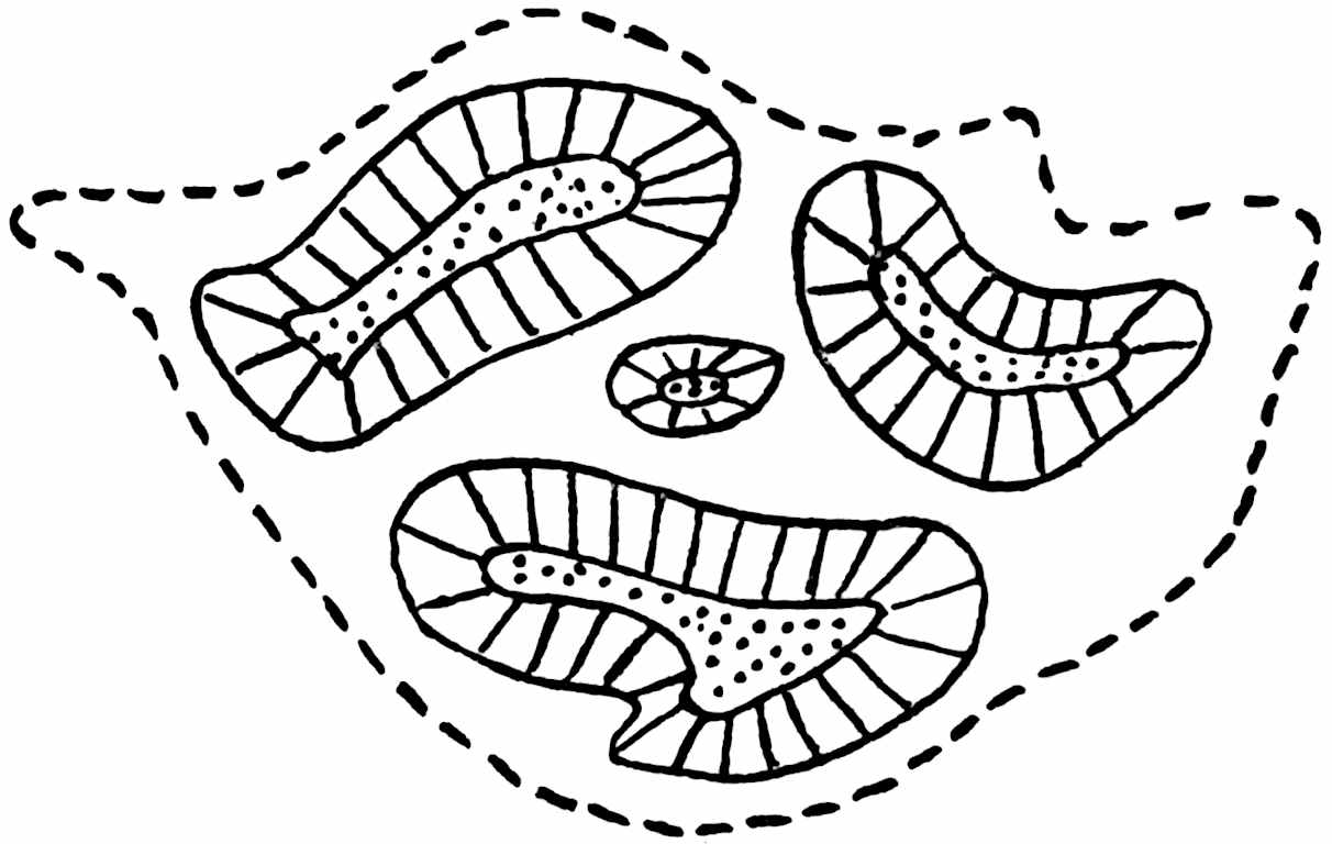
Fig. 417.
Medullosa centrofilis.
Transverse section showing the deep-seated ‘periderm’ (broken line) surrounding four steles. (After de Fraine.)
Medullosa stellata Cotta.
Cotta[254] described Medullosa stellata as a stem characterised by the occurrence of several many-rayed stellate columns (‘vielstrahlige Sternsäule’) in a pith enclosed by a double cylinder of secondary xylem. The so-called pith is the central ground-tissue of the stem and the double ‘striated ring’ of Cotta is a cylindrical stele identical in structure with each of the steles of Medullosa anglica but having a tubular form instead of forming a relatively broad and short band (cf. fig. 416, D and A). Goeppert[255] in his Permian Flora gave a detailed account of the species, some of his sections being cut from Cotta’s material, and by the employment of varietal epithets emphasised the range of variation within the limits of the type. Goeppert and Stenzel[256] and, several years later, Weber and Sterzel[257] adopted the same plan as a convenient method of drawing attention to differences in anatomical characters. As Schenk[258] pointed out, there is a considerable risk in the case of small pieces of stems of attaching excessive importance to structural variations, and it is by no means improbable, as he said, that differences which are the expression of states of preservation or stages in development have been incorrectly regarded as distinguishing marks of individual plants. It is, however, convenient to recognise some of the more striking deviations from the type-species by speaking of the different forms as varieties though, as Weber and Sterzel fully admit, such varieties and even some of the species must be looked upon as provisional. Weber and Sterzel give expression to the provisional nature of their grouping by classifying the species with their varieties into form-cycles. Under the form-cycle Medullosa stellata five more or less well defined forms are recognised, the type-species being Medullosa stellata var. typica[259].
Medullosa stellata var. typica.
Part of a transverse section of a cylindrical stem is represented diagrammatically in fig. 416, D. Very little of the cortex is preserved: a parenchymatous axial region with scattered secretory canals contains four oval or cylindrical vascular steles, the stellate columns of Cotta or star-rings of later authors. These are of the same nature as the small central stele in the English Medullosa centrofilis. The central region of the stem in this specimen is completely surrounded by a narrow cylinder of inversely orientated secondary xylem and phloem (fig. 416, D), the phloem being on the inner side of the xylem. Beyond the xylem is a parenchymatous band containing scattered groups of primary xylem tracheids with spiral, scalariform, and reticulate pitting, and this zone, which is usually designated the ‘partial pith,’ is succeeded by a second and broader, normally orientated, cylinder of secondary xylem and phloem. In this section the two concentric cylinders separated by the partial pith form a solenostele like that of several recent Ferns except in the presence of secondary tissue. The term ‘partial pith’ applied to the tissue between the two cylinders of secondary tissue is misleading: this tissue (fig. 416, D, p) is the primary xylem of the stele and is homologous with the primary portion of the stele of Heterangium and of the steles of M. anglica. In many sections the continuity of the tubular stele is broken. In a section in the British Museum cut from one of Cotta’s specimens[260], 6 × 3·5 cm. in diameter to the outer edge of the vascular tissue, the cylindrical stele is interrupted at two places. An example of the interrupted type of stele is shown in fig. 416, F, and in fig. 416, H: the latter belongs to a distinct species. The complete type of cylindrical stele is exceptional and occurs occasionally at different levels in the stem. An important point is that the frequent breaks in the cylinder are not connected with the exit of leaf-traces and do not, therefore, correspond to the foliar gaps in the solenostele or dictyostele of a Fern.
The secondary xylem is of the cycadean type (fig. 418, B, D) like that of Heterangium and Lyginopteris and several other stems. Each of the star-rings in the axial region consists of a parenchymatous core with scattered primary tracheids enclosed by secondary vascular tissue (fig. 418, B). The star-ring shown in fig. 418, B, from a Chemnitz stem illustrates the characteristic cycadean character of the secondary xylem with broad medullary rays: some of the innermost elements are in contact with the primary tracheids. The phloem is rendered conspicuous by the black contents in some of the elements. Both the star-rings and the larger peripheral steles are constructed on the same plan and agree with the steles of M. anglica. The star-rings occasionally branch and anastomose with one another and with the encircling stele. The star-ring in fig. 416, D at a is about to give off a small strand.
Leaf-traces are furnished by the primary xylem at the edge of the ‘partial pith’ of the outer stele: as a leaf-trace passes outwards through the outer cylinder of secondary xylem the cambium invests it with secondary xylem and phloem, but as it passes through the cortex of the stem it becomes reduced to its primary elements, and by successive branching gives rise to small collateral bundles which enter the petioles. The piece of stem shown in fig. 416, G, illustrates the exit of leaf-traces from the stele and their subsequent division into several small bundles, v, which are scattered in the cortex with strands of sclerenchyma. In a specimen identified with Medullosa stellata, Schenk[261] found part of a leaf-base attached to the stem: its vascular system was of the Myeloxylon type, the bundles being identical with those in the cortex of the stem seen in fig. 416, G.
In some stems of M. stellata the outer, centrifugally developed, portion of the main stele is very much broader than in the example represented in fig. 416, D. The diagrammatic sketch reproduced in fig. 416, F, represents a section of a Chemnitz specimen in the British Museum[262] in which the axial region containing several star-rings is almost enclosed by an inner zone of secondary xylem, and beyond the narrow primary xylem (black in the sketch) the rest of the block consists exclusively of secondary xylem 5·5 cm. broad. This example illustrates a common tendency in Medullosa towards a large excess of centrifugal over centripetal secondary vascular tissue. A similar specimen of Medullosa stellata is figured by Mougeot[263] from the Vosges showing a considerable development of centrifugal xylem comparable with that in the British Museum stem. Weber and Sterzel[264] describe stems of Medullosa stellata showing slight periodic swellings which it is suggested, though there is no evidence in support of the opinion, may be connected with reproductive organs.
Medullosa stellata var. corticata[265]. The specimen referred to this variety, represented in fig. 416, G, has already been quoted as affording data with regard to the origin and behaviour of the leaf-traces. In this type of stem the outer portion of the main stele is narrower than in M. stellata var. typica and the stele never forms a complete tube. The star-rings in the centre of the stem are more numerous than in the type-species of the genus. In the axial region of some stems included in the form-cycle to which M. stellata belongs there may be flatter and tangentially elongated vascular strands in addition to the cylindrical star-rings; these are termed plate-rings.
In Medullosa stellata var. lignosa[266] the outer xylem reaches a breadth of 4 cm. and the star-rings are reduced to one. The form M. stellata var. gigantea[267] (fig. 416, K) is of special interest as an example of a stem reaching a diameter of nearly 50 cm. and having as many as 43 large and small star-rings in the axial region. A large tubular stele like that of the type-species (fig. 416, D) surrounds the central region, but in this form the cylindrical stele a is succeeded by concentric cylinders of normally orientated xylem and phloem (fig. 416, K, bb) produced by successive cambiums either cortical or pericyclic in origin. This type of stem presents a striking resemblance to stems of Cycas and Macrozamia except in the possession of a double cylindrical stele consisting of both centripetal and centrifugal secondary xylem and phloem separated by a zone of primary xylem (partial pith).
Medullosa gigas Renault.
This species was founded on a piece of stem from the Permian of Autun[268], consisting almost entirely of secondary xylem, which Brongniart had previously placed in his genus Palaeoxylon[269]. The secondary xylem reaches a diameter of 45–50 cm. and in the portion of the central region preserved there are a few vascular strands like the star-rings of other species. The considerable development of secondary xylem indicates a form of stem similar to some forms of M. stellata (e.g. fig. 416, F), but as the available data are insufficient for accurate determination Renault’s specific name is retained. Renault describes the internal xylem cylinder (i.e. the centripetal xylem) as very slightly developed or as hardly visible, a feature in which the French specimen shows a nearer approach to the structure of a recent Cycad.
Medullosa porosa Cotta.
The second of Cotta’s species[270], which has been fully investigated by Weber and Sterzel, is constructed on the same plan as that of M. stellata, but the stem is distinguished by the greater number of star-rings and, more especially, by the presence of an outer system of vascular strands in the axial region (fig. 416, M): these form a frequently interrupted cylinder of anastomosing strands characterised by the feeble development of secondary xylem and phloem or by the absence of this tissue on the outer face of the strands. The component parts of this outer series occasionally fuse with the internal star-rings.
This type has a large axial region containing several very small star-rings enclosed by two concentric zones of separate plate-rings (fig. 416, E) each consisting of a complete flattened cylinder of secondary xylem and phloem enclosing primary xylem. As the complete cylindrical stele of the stem of Medullosa stellata shown in fig. 416, D, was compared with the solenostele of a Fern, so in this stem (fig. 416, E) the vascular cylinder may be compared at least superficially with a dictyostele. From the inner circle of plate-rings strands are given off in the form of star-rings and these pass through the gaps in the outer system, eventually breaking up in the cortex into numerous collateral bundles. In another form of this species, var. lignosa (fig. 416, L), the axial region is enclosed by a circle of plate-rings like those in the type-form, but these are succeeded by a circle of very asymmetrically developed and large steles with the outer xylem and phloem much broader than the inner. Moreover in this form additional cylinders of normally orientated vascular tissue are added as in M. stellata var. gigantea and in some recent Cycads. It is noteworthy that the secondary wood of Medullosa Solmsi is rather more compact than in other species, a feature in which it to some extent agrees with the South African genus Rhexoxylon.
Medullosa Leuckarti Goeppert and Stenzel.
In this species[273], also from the Permian of Saxony, the central region including some star-rings is surrounded by sinuous flattened concentric steles (snake-rings) agreeing anatomically with the steles of other species and characterised by the comparatively small breadth of the secondary xylem and phloem (fig. 416, H). Leaf-traces are given off, as in M. anglica and other species, from the outer edge of the primary xylem. In some forms there is a single set of snake-rings; in others there is a double series. Fig. 418, D, shows part of the secondary xylem of a stele of this species from Chemnitz: the tracheids are in some places continuous with the primary xylem, and on the outer edge of the secondary wood is a cylinder of phloem. A section of Medullosa Leuckarti figured by Goeppert and Stenzel[274] shows some radial rows of very thick-walled elements in the secondary phloem which they describe as bast sclerenchyma, but Solms-Laubach[275] believes them to be sieve-tubes. Precisely similar elements are figured by Scott[276] in M. anglica and as this author suggests the thick walls are probably not an original feature. The structure of the primary xylem is more clearly seen in fig. 418, C, and the relation between primary and secondary xylem is shown in fig. 416, I, where the position of the protoxylem may be either exarch or mesarch. The protoxylem is only occasionally recognisable but some of the peripheral primary tracheal groups are undoubtedly mesarch. External to the stele, a part of which is reproduced in fig. 418, D, are strands of stereome elements and beyond them a band of radially elongated cells that may be ‘periderm’: still farther out there are some imperfectly preserved vascular bundles that are leaf-traces. This species is important as affording a complete demonstration of the organic connexion between the stem and petioles of the Myeloxylon Landrioti type which indicate that the fronds were probably Alethopteroid.
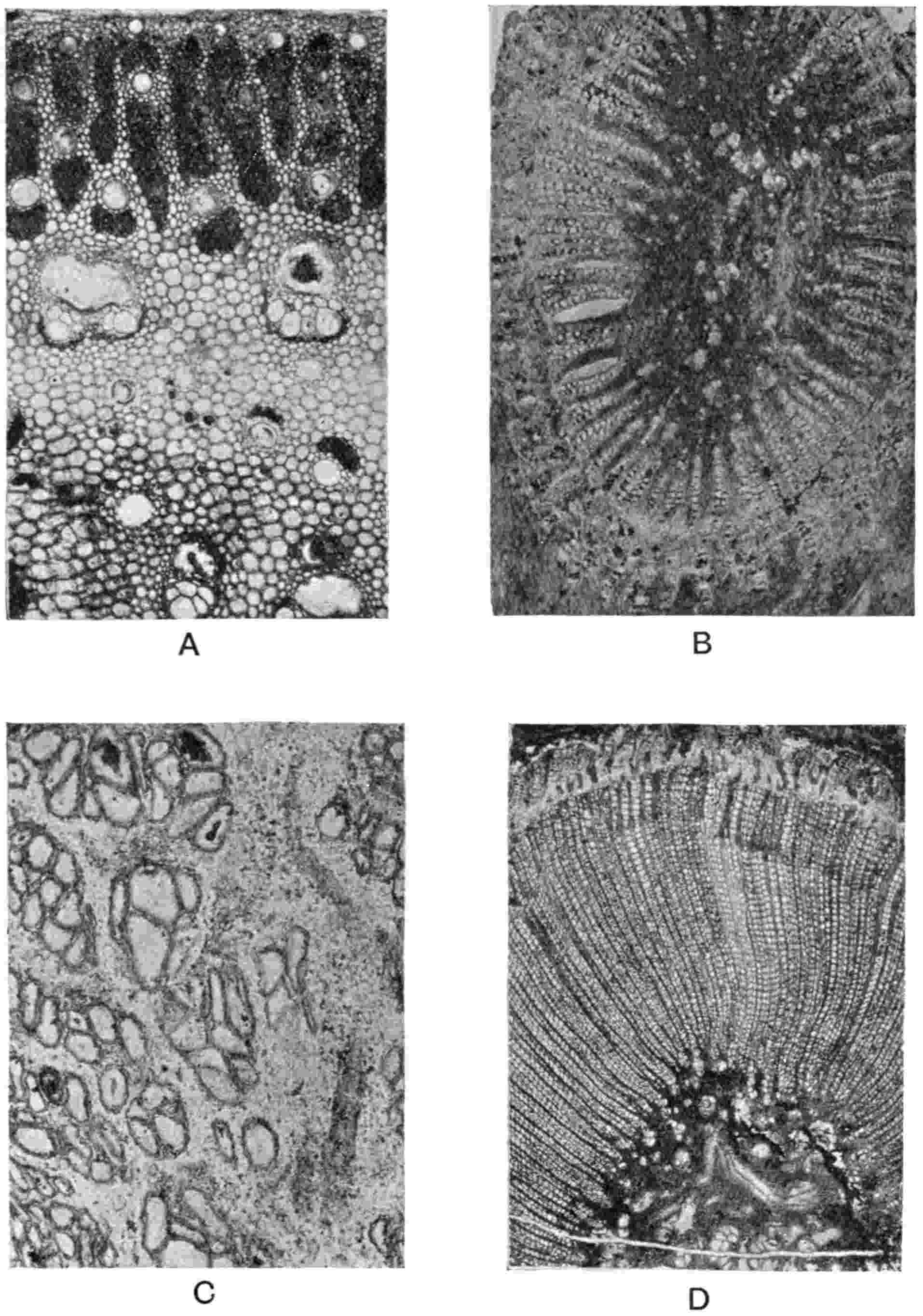
Fig. 418.
A,
Myeloxylon radiatum
, part of petiole. B,
Medullosa stellata
; star-ring; × 14. C, D,
Medullosa Leuckarti
; C, primary xylem; D, part of primary stelar tissues and secondary xylem and phloem; × 6. (A–D, Kidston Coll., 1014, 1307, 1238.)
The specimen on which the diagrammatic drawing reproduced in fig. 419 is based has been investigated by Weber and Sterzel[277] and by Solms-Laubach[278]. The figure is a slightly simplified version of that given by Weber and Sterzel; it represents the stem of Medullosa Leuckarti as a transparent object, the two lower transverse sections, B and C, being seen in perspective through the longitudinal faces. The steles are shaded obliquely in the longitudinal sections, and in the three transverse sections, A, B, C, the primary xylem (partial pith) is black and the enclosing secondary vascular tissue radially shaded. The whole block is 9 cm. in length and 6 cm. broad. Only a part of the axial region is shown internal to the peripheral snake-rings and in it are the star-rings S, S, b, and c. Outside the main steles is the narrow cortex R and portions of leaf-bases I–IV. The lowest section, C, shows part of a peripheral snake-ring with a slight swelling at f on its inner side which, as seen in sections B and A, foreshadows the separation of the star-ring S and the consequent break in the continuity of the snake-ring (d, e, sect. B). In section A the gap is closed: in the longitudinal section between B and A the star-ring S is seen to form two branches, a and b, the branch a closing the gap between d and e in section B. These sections demonstrate the formation of a star-ring from the main peripheral stele and the formation of additional star-rings by branching.
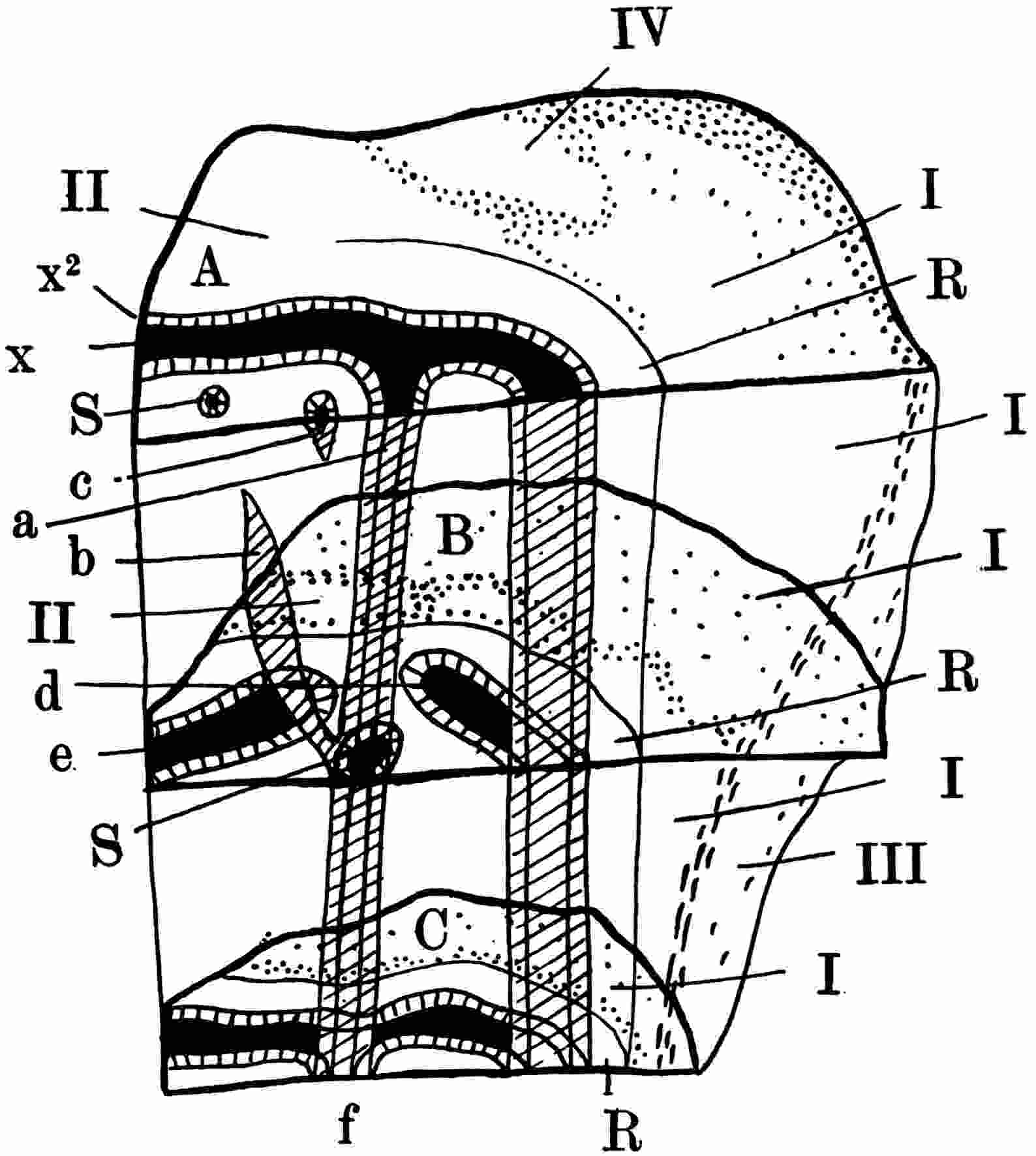
Fig. 419.
Medullosa Leuckarti.
Diagrammatic sketch of stem showing longitudinal and transverse sections: for explanation see text. (After Weber and Sterzel.)
Numerous vascular bundles destined for the leaves are scattered in the cortex. The course of the decurrent leaf-base I is shown on the longitudinal faces, its boundary being marked by crowded stereome strands (of the Myeloxylon Landrioti type); other leaf-bases are represented by II, III, and IV.
In habit Medullosa Leuckarti differs from such a type as M. stellata in its relatively shorter and stouter stem and in the shorter internodes.
Leaves and Reproductive Organs.
i. Leaves. It has already been stated that in some cases petioles occur in organic connexion with Medullosan stems, notably in M. anglica and M. Leuckarti: in the exceptionally rich collection in the Chemnitz Museum, which forms a fitting memorial of the work of the late Prof. Sterzel, there is a stem of M. Leuckarti bearing large petioles of the type known as Myeloxylon radiatum. The occurrence of vascular bundles in the cortex of other species of stem identical with those in the attached petioles points to a uniform type of leaf-structure so far as regards the petioles and rachises of Medullosa. While it is clearly unnecessary to distinguish by a special generic title the petrified portions of fronds known to belong to certain species of stems, the frequent occurrence of detached petioles necessitates some distinctive term. The name employed is Myeloxylon: the genus was instituted by Brongniart in 1849 for Cotta’s species Medullosa elegans the petiolar nature of which was suspected by Binney in 1872.
Myeloxylon
(Medullosan leaf-stalks).
1832. Medullosa elegans Cotta.
1865. Stenzelia Goeppert.
1876. Myelopteris Renault[279];
1877. Aulacopteris Grand’Eury[280].
There is a very close agreement in general anatomical structure between the numerous specimens of Myeloxylon from the Permian strata of Saxony and France and the Coal Measures and Millstone Grit of England[281]; the genus is also recorded from the Upper Carboniferous of Kansas[282]. Two well-defined types instituted by Renault are, however, readily distinguished by the form of the hypodermal stereome strands. Myeloxylon may be defined as follows: Oval or cylindrical branched axes, reaching a diameter of 15 cm., bearing pinnae having the characters of Alethopteris, Neuropteris, Odontopteris, and some other genera that were formerly classed as Ferns. Below a single-layered epidermis, in which stomata have been recognised, occur a few layers of parenchyma: this superficial tissue, which is rarely preserved, is succeeded by a hypodermal region consisting of parenchymatous tissue and numerous vertical groups of narrow thick-walled fibres arranged as radial plates or circular, oval, or reniform strands (the Sparganum type of cortex). In the hypoderm as in the ground-tissue generally secretory canals, often accompanied by stereome, are a characteristic feature. The vascular system is represented by a considerable number of collateral bundles scattered through the ground-tissue and especially abundant in the outer region: the bundles sometimes assume a more or less regular disposition in concentric circles. Each bundle consists of a small group of xylem tracheids, for the most part spiral or scalariform, though reticulately pitted elements are by no means rare, with a single protoxylem group on the outer face next the phloem (fig. 420, B, C). As a rule the xylem is wholly centripetal, but occasionally the exarch structure becomes mesarch by the occurrence of a few centrifugal tracheids. The phloem, rarely preserved (fig. 420, B), consists of narrow sieve-tubes with parenchyma, and the bundle as a whole is often partially enclosed by a sheath of fibres.
Superficially the anatomical structure is similar to that of the petioles of Angiopteris or Marattia, and both Williamson[283] and Renault placed Myeloxylon in the Ferns; but the collateral form of the vascular bundles, the position of the protoxylem, and the arrangement of the hypoderm tissues, are cycadean features.
Myeloxylon radiatum (Renault).
This type is characterised by the radially elongated stereome of the hypoderm. Prof. Zeiller[284], who has given a very clear and concise description of Myeloxylon, is disposed to regard Cotta’s Medullosa elegans as a specifically distinct form on the ground that there are two concentric zones of stereome in the hypoderm; but this feature is shown only in one of Cotta’s figures, and Weber and Sterzel[285] point out that a doubting of the hypoderm zone may be caused by accidental juxtaposition of two faulted pieces of peripheral tissue. The drawing reproduced in fig. 420, A, shows the structural plan of an unusually large petiole from the Permian of Autun: a portion of the outer tissue is seen in fig. 418, A. The vascular bundle, fig. 420, C, from a Millstone Grit specimen[286], shows the centripetal nature of the xylem and fragments of phloem in the outer half of the bundle, with imperfectly preserved fibres abutting on the xylem. The characteristic hypoderm is shown also in fig. 418, A; the double xylem strand on the left illustrates a common feature caused by the branching of vascular bundles. Several secretory canals are scattered in the ground-tissue. The pinnules of Myeloxylon radiatum, or at least of some specimens, have been shown by Renault to be of the Neuropteris type.
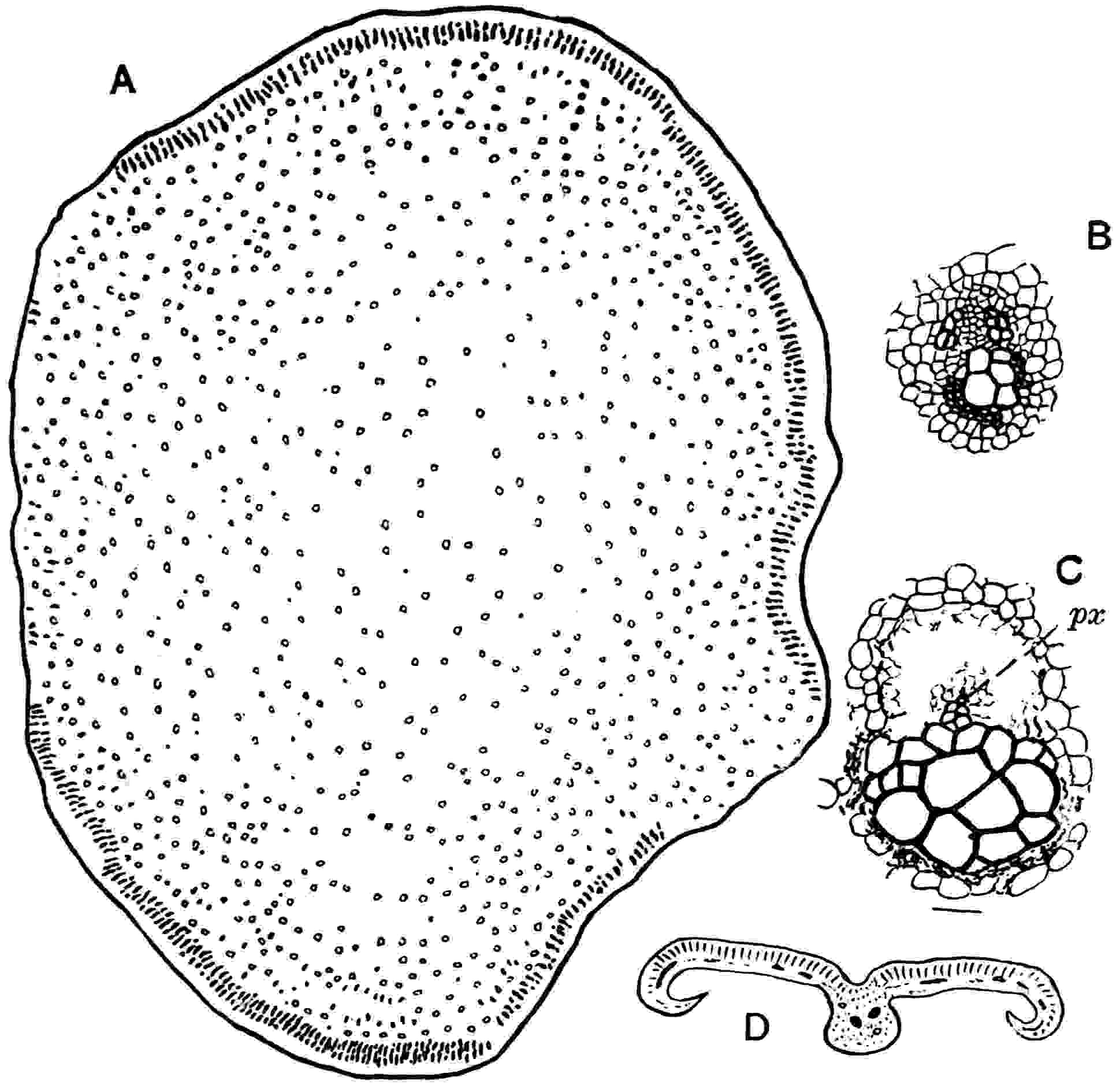
Fig. 420.
A, B,
Myeloxylon radiatum
; A, petiole ⅔ nat. size; B, vascular bundle. C,
Myeloxylon
sp. vascular bundle;
px
, protoxylem. D,
Medullosa anglica
; section of pinnule. (A, B, after Zeiller; D, after Scott.)
Myeloxylon Landrioti (Renault)[287].
In this species the distinguishing feature is the occurrence of the hypodermal stereome in the form of circular, oval, or reniform strands in place of the radial plates of M. radiatum. It is this form of petiole that was borne by the stems of Medullosa anglica and M. Leuckarti. In M. anglica the pinnules (fig. 420, D) are of the Alethopteris type, almost certainly A. lonchitica. Renault and Zeiller have described French specimens of Myeloxylon Landrioti bearing pinnules like those of Alethopteris aquilina and A. Grandini.
Myeloxylon topekense (Penhallow).
The occurrence of Myeloxylon petioles in the New World was recorded by the late Prof. Penhallow[288] who founded this species on some imperfectly petrified specimens from Upper Carboniferous strata at Topeka, Kansas. Enough material was available to show the Myeloxylon characters, but the preservation is too imperfect to admit of a complete diagnosis. The hypodermal stereome shows a tendency to form tangentially extended strands in place of the more circular or radially elongated groups in the European species.
In addition to Alethopteris, Neuropteris (including Cyclopteris) and Odontopteris fronds, which are known to possess rachises with the Myeloxylon features, there is reason to believe that the Permian Callipteris fronds and possibly some of the older Taeniopteris leaves may also belong to Medullosa[289]. It is, however, unsafe to assume that the occurrence of Myeloxylon petioles necessarily denotes the existence of Medullosa. The French stem Colpoxylon aeduense Brongn.[290] bore leaves with the same general anatomical features as those of a typical Myeloxylon, and there can be little doubt that other genera of the Medulloseae also possessed fronds constructed on the same plan as those known to have been borne by Medullosa. An interesting illustration of an injured organ that had produced a wound-cambium is afforded by a Myeloxylon petiole from the Coal Measures described by Mr Holden[291].
Reproductive Organs.
Reference was made in volume ii. to the reasons which led to the removal of several genera of Carboniferous and Permian fronds from the Filicales to the Pteridosperms, and in Chapter xxix. of the present volume certain species of Sphenopteris are described as the foliage of Lyginopteris and Heterangium. It is with such genera as Neuropteris, Alethopteris, Linopteris and others that we are now more especially concerned, as they represent some of the types of leaves borne by Medullosa and other members of the Medulloseae. The absence of any specimens among the large number of these common genera bearing undoubted sporangia aroused suspicion as to the correctness of the generally accepted view that these fern-like fossils were the leaves of Palaeozoic Ferns. Subsequently the suspicion based on negative evidence was confirmed by researches into the anatomical structure of the leaf-bases, petioles, and fragments of pinnae attached to and associated with stems of Medullosa. It is only in a few cases that actual organic connexion between reproductive organs and Medullosan leaves has been demonstrated, but from such facts as are established it is safe to make the general statement that stems of Medullosa—a generic term that undoubtedly includes plants which, had we a fuller knowledge of them as complete individuals, would be assigned to more than one generic type—possessed fronds simulating in habit those of certain Ferns with some of the pinnae bearing seeds often of considerable size and in all cases of complex structure, agreeing in many respects with those of existing Cycads, while other fronds, or in some cases it may be other pinnae, bore microsporangia similar in form to the sporangia of Ferns.
a. Microsporangia. Neuropteris. A. Neuropteris heterophylla[292].
Several examples of supposed fertile specimens of Neuropteris are recorded in palaeobotanical literature, but it was not until 1887 that any satisfactory specimen was discovered. In that year Kidston[293] described a specimen of Neuropteris heterophylla from the Lower Coal Measures of Scotland in which slender forked branchlets bear small bodies at their tips some of which appear to represent four-valved organs (fig. 421, D), though the imperfect state of preservation renders impossible any definite pronouncement as to their structure. To the specimen are attached a few sterile pinnules, showing that it is a portion of a frond of N. heterophylla characterised by the substitution of reproductive organs for pinnules. The subsequent discovery of seeds attached to pinnae of the same species afforded strong presumptive evidence, almost amounting to proof, of the microsporangial nature of the Scotch specimen. For this specimen, although no precise diagnosis is possible, Dr P. Bertrand[294] has proposed the generic name Neurotheca. In 1911 the Abbé Carpentier[295] described some small ovoid bodies, 1–1·5 mm. long, from the Coal Measures of France arranged in groups of 4 to 6 and in some cases said to be borne on a slender pedicel which he found in association with N. heterophylla and compared with sporangia described by Lesquereux from the Coal Measures of Arkansas as Sorocladus stellata[296]. These supposed microsporangia have recently been assigned by Bertrand[297] to Sphenophyllum.
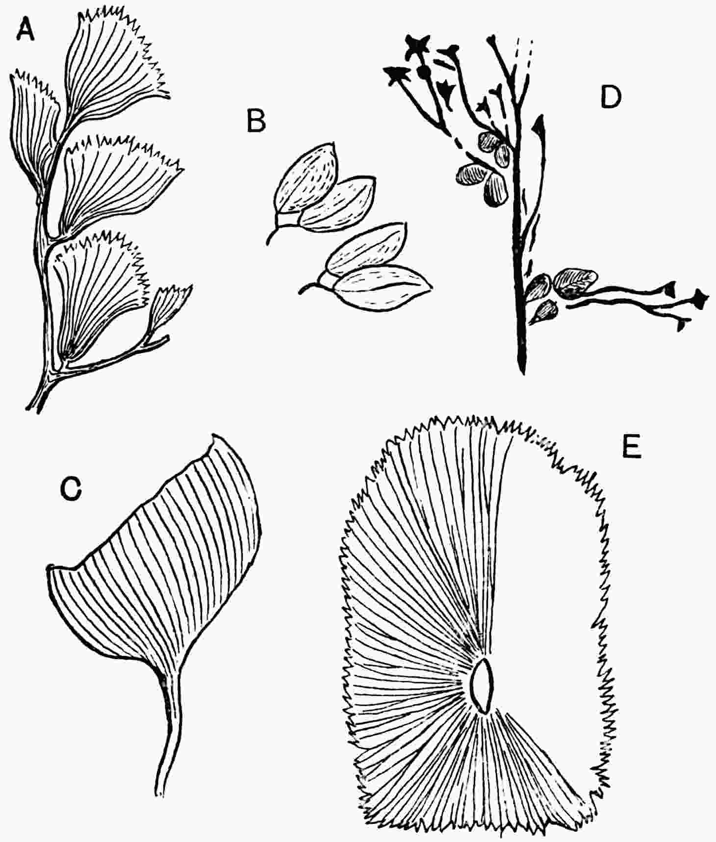
Fig. 421.
A. Potoniea adiantiformis Zeill. (very slightly enlarged).
B. Microsporangia of Potoniea (enlarged).
C. Linopteris obliqua; side-view of microsporophyll.
D. Neuropteris heterophylla, pinnules and microsporangia (?).
E. Linopteris obliqua, surface-view of microsporophyll.
(A, C, E, after P. Bertrand; B, after Carpentier; D, after Kidston.)
B. Neuropteris gigantea, etc. Potoniea, Zeiller.
In 1899 Zeiller[298] instituted the name Potoniea for some peculiar fertile leaves found in the Coal Measures of Heraclea consisting of a branched axis with cuneate segments, 7–10 × 6–8 mm., bearing numerous fusiform bodies, 1–1·5 mm. long, at the upper edge (fig. 421, A). These marginal bodies he regarded as sporangia and expressed the opinion that Potoniea may be the fructification of some form of Neuropteris, Mariopteris or Alethopteris. Carpentier[299] afterwards described similar though rather larger examples from the Pas-de-Calais coal-field as microsporophyll fragments of some Pteridosperm: he stated that similar specimens had been found by Kidston in England. In a later work Carpentier[300] described the sporangia as crowded in groups (fig. 421, B) in the substance of the thick lamina of Potoniea, and he connected the fertile segments with Neuropteris gigantea, N. pseudogigantea Pot. and Linopteris obliqua (Bunb.). Similar specimens are said to have been found in Holland and Silesia. Bertrand[301] also records the association of Potoniea with Neuropteris gigantea and N. pseudogigantea: he describes some specimens as belonging to N. gigantea Sternb. while others, distinguished only by small differences, he attributes to N. pseudogigantea. Kidston[302] has recently drawn attention to the inconstancy of the characters mentioned by Potonié as distinguishing features of N. pseudogigantea, and he shows good cause for referring the examples so named to N. gigantea. The fertile lamina is almost orbicular in surface-view and attached to a slightly excentric pedicel; the microsporangia are borne on the lower surface and probably in groups as described by Carpentier.
Neuropteris Carpentieri Kidston. Kidston[303] has recently described some fertile leaflets under this name from the Westphalian series of South Staffordshire which he identifies with French examples referred by Carpentier[304] to Potoniea adiantiformis Zeill. The fertile pinnules are thick and sub-cyclopteroid in form; the upper surface bears densely packed, narrow and long, microsporangia, 4 × 0·5 mm., containing more or less spherical microspores 45–60μ in diameter; the ventral face of the lamina on the removal of the spores shows several strong veins. With these are associated sterile pinnules of the Neuropteroid type, and examples are described intermediate between the sterile and fertile leaflets. Kidston believes the specimens to be microsporophylls of some species of Neuropteris, but as the material does not suffice for identification with any known species a new name is proposed. It is pointed out that in some states of preservation the leaflets resemble Goeppert’s Permian species Dictyothalamus Schrollianus[305].
Alethopteris. Linopteris.
No specimens of Alethopteris fronds have so far been described that afford any information as to the nature of the microsporangia, and we have no means of knowing whether they were borne on naked pedicels as in Neuropteris heterophylla, or on modified pinnules as in N. gigantea.
Zeiller in 1888[306] described some fertile pinnules of Dictyopteris Schützii Roem. from Commentry bearing two rows of long sporangia: he subsequently transferred this species to the genus Linopteris[307] and expressed the opinion that the sporangia occur singly and not in groups as he originally believed. Zeiller compares the fertile pinnules with the type Crossotheca. Bertrand[308], as the result of examining similar specimens, has suggested that the fringe of pendulous bodies regarded by Zeiller as sporangia may be tooth-like lobes of modified pinnules which served to protect microsporangia borne on the lower surface of the lamina. The nature of the impressions is not clear, though there is little doubt that they are microsporophylls. The fertile pinnae of Linopteris obliqua (Bunb.) described by Carpentier[309] and Bertrand[310] closely resemble the microspore-bearing organs which have been referred to Neuropteris gigantea; they have the characters of Potoniea and consist of oval laminae similar to the sterile pinnules but about half their size: the lamina was attached excentrically to a slender stalk (fig. 421, C, E) and traversed by numerous occasionally anastomosing veins. No actual microsporangia have been discovered in organic connexion with the lamina.
b. Megasporangia. Neuropteris.
Kidston’s discovery of undoubted seeds attached to pinnae of Neuropteris[311] marked an important step in our more exact knowledge of the morphology of Medullosan sporophylls. Specimens from the ironstone balls (Coal Measures) of Coseley near Dudley showed seeds attached to portions of pinnae bearing pinnules of Neuropteris heterophylla. The seeds are approximately 3 cm. long and from 1·10 to 1·40 cm. broad; oblong and gradually tapering from the middle to a slightly curved and obtuse apical snout (fig. 422). The outer surface shows numerous longitudinal ribs which no doubt represent hypodermal fibres. The preservation of the seeds, which appear to be circular in section, is not such as to render possible a description of structural features. In their asymmetrical form the seeds agree with the genus Platyspermum as recently defined by Arber, but Kidston’s specimens are in all probability radiospermic. Kidston compares the Neuropteris seeds with Rhabdocarpus tunicatus as figured from the Commentry coalfield[312] and with specimens from Gard named by Grand’Eury R. subtunicatus[313].
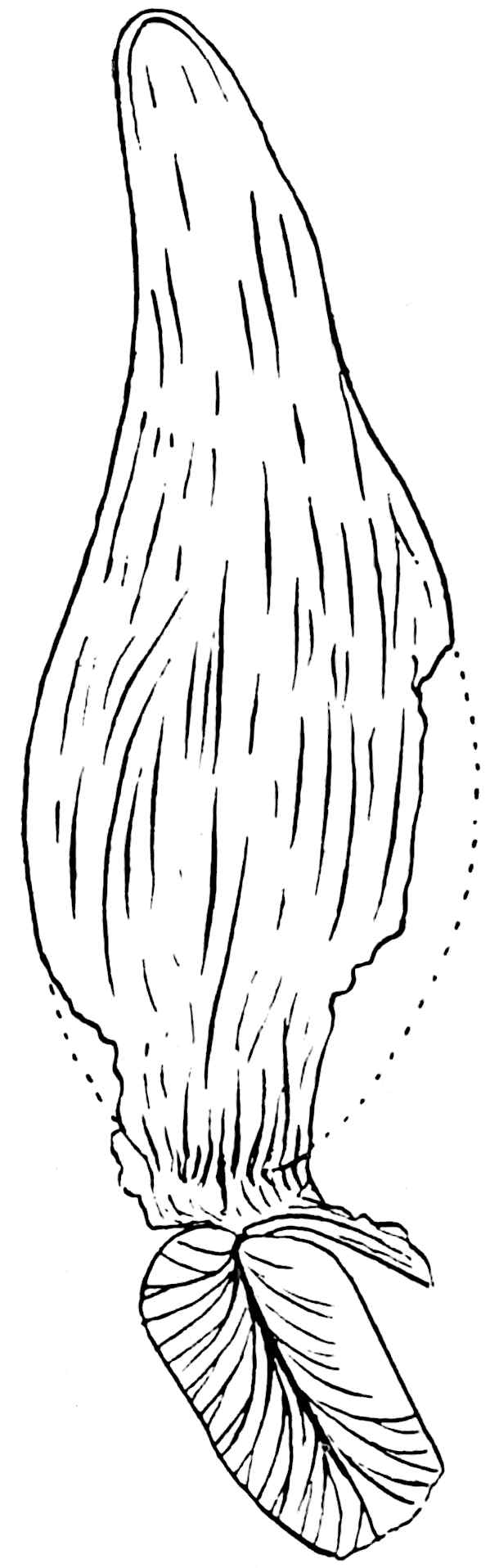
Fig. 422.
Neuropterocarpus Kidstoni
(Arber). (After Kidston; × 3.)
Additional proof of the occurrence of seeds on Neuropteris fronds is furnished by examples from the Coal Measures of Holland described by Kidston and Jongmans[314]: these seeds are of the same general type as those from Coseley but nearly twice as large, and they were borne at the tips of a dichotomously branched pedicel of Neuropteris obliqua. Grand’Eury in 1904[315] recorded the association of radiospermic seeds with Neuropteris fronds though no case of actual attachment was found. It is, however, noteworthy that he speaks of the frequent association with Neuropteris of seeds characterised by six or rarely twelve longitudinal keels, a feature recalling the sclerotesta of Trigonocarpus and allied seeds. Until petrified specimens are available it is impossible to refer the seeds of Neuropteris to a generic type founded on structural features: the seeds described by Kidston are, as he says, very similar in external characters to species assigned to Rhabdocarpus, and there can be little doubt as to the generic identity of the Neuropteris seeds and some of the impressions referred to Rhabdocarpus which are characterised by a similarity in form, an apical snout that gives an asymmetrical appearance to the specimens and the presence of numerous longitudinal striations[316]. It is, however, by no means certain that these seeds possessed the morphological features of Rhabdocarpus as described by Brongniart in petrified examples from St Étienne[317].
The seeds of Neuropteris may, as Kidston suggests, agree anatomically more closely with Pachytesta[318], a type that Grand’Eury associates with Alethopteris fronds. With a view to avoid the danger of incorrectly identifying petrified specimens and impressions that cannot be proved to belong to the same generic type, I have suggested the restriction of the name Rhabdocarpus[319] to seeds that do not furnish evidence as to internal structure, and the employment of the designation Rhabdospermum for seeds that conform to those described by Brongniart as Rhabdocarpus. The seeds of Neuropteris may be of the Rhabdospermum type or they may agree anatomically more closely with Trigonocarpus or Pachytesta; they are members of either the Trigonocarpales or the Cardiocarpales, probably the former group. Dr P. Bertrand[320] assigns to Neuropteris gigantea some seeds of the Hexapterospermum (= Hexagonocarpus[321]) type which occur in association with cupule-like organs. These supposed cupules resemble the Potoniea leaflets with microsporangia also referred to the same species of Neuropteris; they are characterised by a laciniate edge and may be compared with the Indian fossil described by Zeiller as Ottokaria bengalensis[322] (fig. 433). Dr Arber[323] and Dr P. Bertrand[324] have independently proposed the generic name Neurospermum for the seeds of Neuropteris heterophylla and N. obliqua in preference to Rhabdocarpus: the former author speaks of the seeds of N. heterophylla as Neurospermum Kidstoni. The generic term Neuropterocarpus used by Grand’Eury in 1904[325], though not defined by him, has priority and avoids the adoption of a new designation for seeds attached to Neuropteris fronds. In his definition of Neurospermum Arber makes no reference to the obliquity of the apical snout that is clearly shown in fig. 422. There is no evidence that Neuropterocarpus possessed a cupular investment comparable with that of Lagenostoma. Additional instances of the association of seeds with Neuropteris fronds are recorded by Renier from the Belgian Coal Measures, also by Bertrand and Chodat from France: Renier found seeds associated with N. Schlehani Stur and with the same species of frond Bertrand found impressions of oval ribbed seeds. Prof. Chodat[326] has figured some fragments of Neuropteris pinnules referred to N. auriculata Brongn. from the Stephanian of France which show small seed-like bodies apparently in organic connexion with the lamina; but the specimens are too imperfect to afford any satisfactory evidence as to the nature of the reproductive organs.
Lotsy[327] has expressed the opinion that the bodies attached to Neuropteris pinnae described by Kidston and other authors as seeds may possibly be vegetative buds, but if this were the case one would expect to find some evidence of the bud-nature in some at least of the specimens that have already been found.
It would seem that the microsporophylls of Neuropteris were constructed on different plans, some being of the type described by Zeiller and other observers as Potoniea, while others bore sporangia on pinnae without any accompanying laminae; but our knowledge of the latter form represented by Kidston’s specimen of Neuropteris heterophylla (fig. 421, D) is very incomplete. On the other hand the seeds appear to have been characterised by features suggesting a close affinity to Trigonocarpus and pointing to membership of the same family.
Alethopteris.
Although no specimens have been discovered showing actual connexion between fronds and seeds, it is practically certain that Alethopteris leaves, or at least some species of the genus, bore seeds of the Trigonocarpus type. The association of Trigonocarpus Parkinsoni with Alethopteris lonchitica is too frequent to be fortuitous and there is further evidence afforded by certain anatomical resemblances. In France other species of Alethopteris, e.g. Alethopteris Serlii and A. Grandini, occur in association with Pachytesta[328], a large seed similar to Trigonocarpus, and Trigonocarpus is found in the Pas-de-Calais coal-field with Alethopteris Serlii.
TRIGONOCARPUS. Brongniart.
The generic name Trigonocarpum was instituted by Brongniart in 1828[329] for ovoid longitudinally ribbed ‘fruits’ from Upper Carboniferous strata, the type-species being named T. Parkinsoni. The generic name is often altered to Trigonocarpon: Williamson[330], who adopted this form, states that Brongniart substituted Trigonocarpon for Trigonocarpum in his Tableau[331], but in that work the original termination is used, the form Trigonocarpon, probably the result of a slip, appearing only in the index. In his later work on seeds Brongniart adopted the name Trigonocarpus, and in recent years this has been widely employed. Among other species named by Brongniart are two previously referred by Sternberg to Palmacites. Several examples of Brongniart’s genus were described by Lindley and Hutton, and in their description of T. Noeggerathii the statement is made that a fractured specimen demonstrated that ‘the fossil in its ordinary state is an interior part divested of fleshy covering’[332]; this suspicion of the true nature of the nut-like fossils was afterwards proved correct by the investigations of Hooker and Binney[333] and by the later work of Williamson. The specimens on which the genus was founded are casts of seed-cavities and it is in this state that the seeds are usually preserved, often in large numbers, in the sandstones of the Coal Measures, as in the block shown in fig. 423 from the famous quarry at Peel near Bolton, Lancashire. Another type of preservation is represented by the seeds figured by Lindley and Hutton as Carpolithes alata[334], but the generic identity of the two states was not recognised until the discovery of petrified material afforded the clue. Figs. 424, 1, 425 illustrate the appearance of Trigonocarpus when preserved as a carbonised impression showing a thick fleshy envelope enclosing an oval kernel with a hard wall prolonged upwards as a longer or shorter micropyle. Casts of the seed-cavity are represented in figs. 423; 424, 2, 3. The surface of these casts occasionally shows one or more short cylindrical projections which are probably extensions of the sand or mud into holes formed in the testa by boring insects. The view that Trigonocarpus seeds are ‘obviously Palm fruits’ was not accepted by Hooker and Binney who inclined to regard them as the seeds of Conifers and compared them especially with the similar nuts of Ginkgo seeds (cf. fig. 631, C). It was Mr Wild[335] who was first struck by the association of Trigonocarpus and the petioles of Medullosa (Myeloxylon) and by some resemblances in structure between the testa and the hypoderm of the petioles; though, as Scott and Maslen[336] point out, the agreement is not so close as Wild believed, his view of a possible connexion between the reproductive and vegetative organs has been confirmed. Williamson extended our knowledge of the genus by his account of Trigonocarpus olivaeformis Lind. and Hutt., a form that is specifically identical with T. Parkinsoni Brongn. This author also drew attention to the close resemblance between Brongniart’s three genera Trigonocarpus, Hexapterospermum, Tripterospermum and expressed doubts as to the possibility of founding specific differences on casts of the Trigonocarpus type without the evidence of anatomy. Our knowledge of the structure of Trigonocarpus has in recent years been considerably extended by the researches of Oliver, Scott and Maslen, and Salisbury.
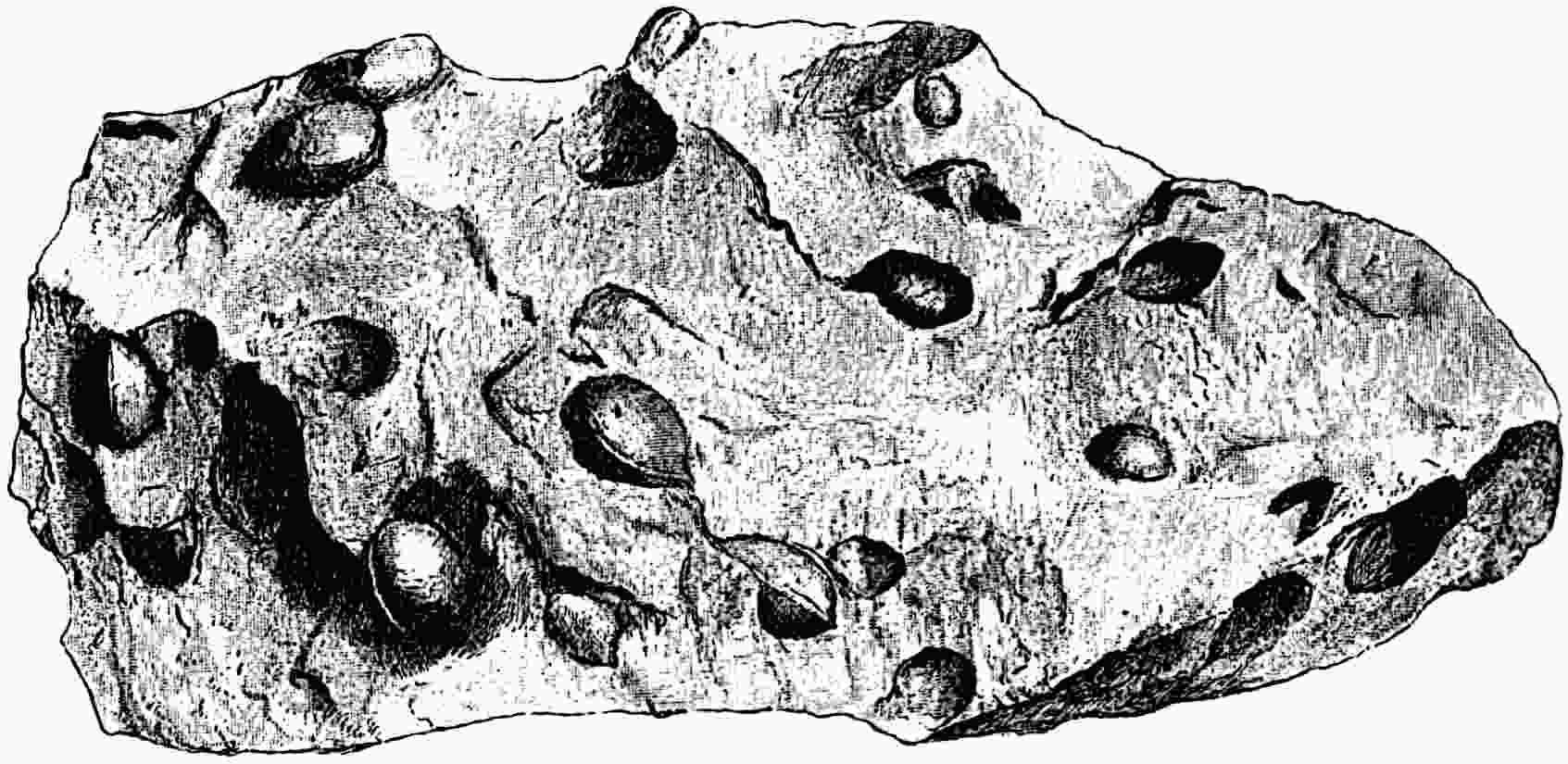
Fig. 423.
Casts of the seed-cavity of
Trigonocarpus Parkinsoni
. (Manchester Museum. ½ nat. size.)
Trigonocarpus Parkinsoni[337] Brongniart.
The seeds of this species like all examples of the genus are radiospermic, that is radially symmetrical in contrast to the flattened or platyspermic seeds. The complete seed is elongate oval in form when preserved as an impression (fig. 425, A) and reaches a length of 4–5 cm.: the casts of the seed-cavity are ovoid and provided with three prominent ridges (fig. 424, 2, 3). The testa forms a thick covering differentiated into three regions, an outer flesh or sarcotesta, a sclerous shell or sclerotesta, and an inner flesh. Transverse sections show that the sclerotesta has three sharp longitudinal keels with corresponding furrows on the inner face, and between each pair of main ribs are 2–3 less prominent ridges, usually 12 in all (fig. 426). The sarcotesta consists of thin-walled parenchyma passing externally into a more lacunar tissue with a palisade-like hypoderm: the sclerotesta consists of thick cells which interlace and form an efficient protective shell. Both the sarcotesta and sclerotesta are continued into the apical region as the wall of the long micropyle, the sarcotesta being prolonged beyond the sclerotesta at the apex of the integument[338]. The micropyle is triangular in section and may exceed in length the whole seed (figs. 425; 426, A). Its form as seen in transverse section (fig. 426, B) suggests the presence of wings: this appearance may be deceptive and due to pressure or, more probably, it represents an original feature. The seed-body, that is the portion enclosed by the integument, consists of the nucellus, represented by a few crushed layers of cells, bounded by a well-defined epidermis; the nucellus is separated from the integument from the base of the seed upwards, an important feature in which this and some other Palaeozoic seeds differ from Lagenostoma and the seeds of recent Cycads which are characterised by an integument adnate to the nucellus up to the level of the shoulder; the seeds of the Conifer Phyllocladus afford an example of separation of integument and nucellus as in Trigonocarpus. The innermost layer of the nucellus consists mainly of tracheal tissue investing the large megaspore (fig. 426, A, C, m) which is preserved as a contracted membrane detached from the nucellus after the death of the seed. At the summit of the nucellus is a relatively small pollen-chamber (fig. 426, A, Pc) like a broad and low cupola bearing a terminal beak which extended some distance into the micropylar tube. No microspores have been found in this species, but Oliver[339] records the occurrence of multicellular microspores in Trigonocarpus pusillus. The pedicel of the seed had a central strand of sclerous tissue penetrated by a concentric vascular bundle which gives off six strands to supply the sarcotesta (fig. 426, C, v) and then passes into the nucellus where it forms a tracheal sheath (fig. 426, A, nt) surrounding the lower part of the megaspore and at a higher level breaks up into anastomosing strands of tracheids which reach up to the plane of insertion of the pollen-chamber.
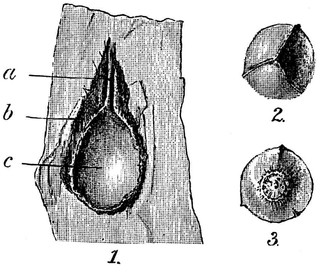
Fig. 424.
Trigonocarpus.
1. Section of imperfect seed showing a micropyle;
b
, sclerotesta;
c
, cast of seed-cavity; 2, 3, apical and basal view of seed-cast. (Approximately nat. size.) M. S.
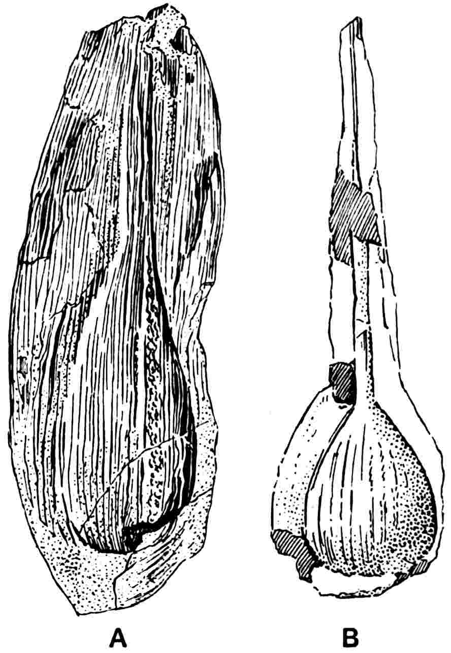
Fig. 425.
Trigonocarpus Parkinsoni.
A. Impression showing the nucule and sarcotesta. (= Carpolithes alata Lind. and Hutt.)
B. Specimen with long micropyle. (A, nat. size; from the Lower Coal Measures of Kilmarnock; B, nat. size; Middle C. M. of Yorkshire. Kidston Coll., 1579 and 1062.)
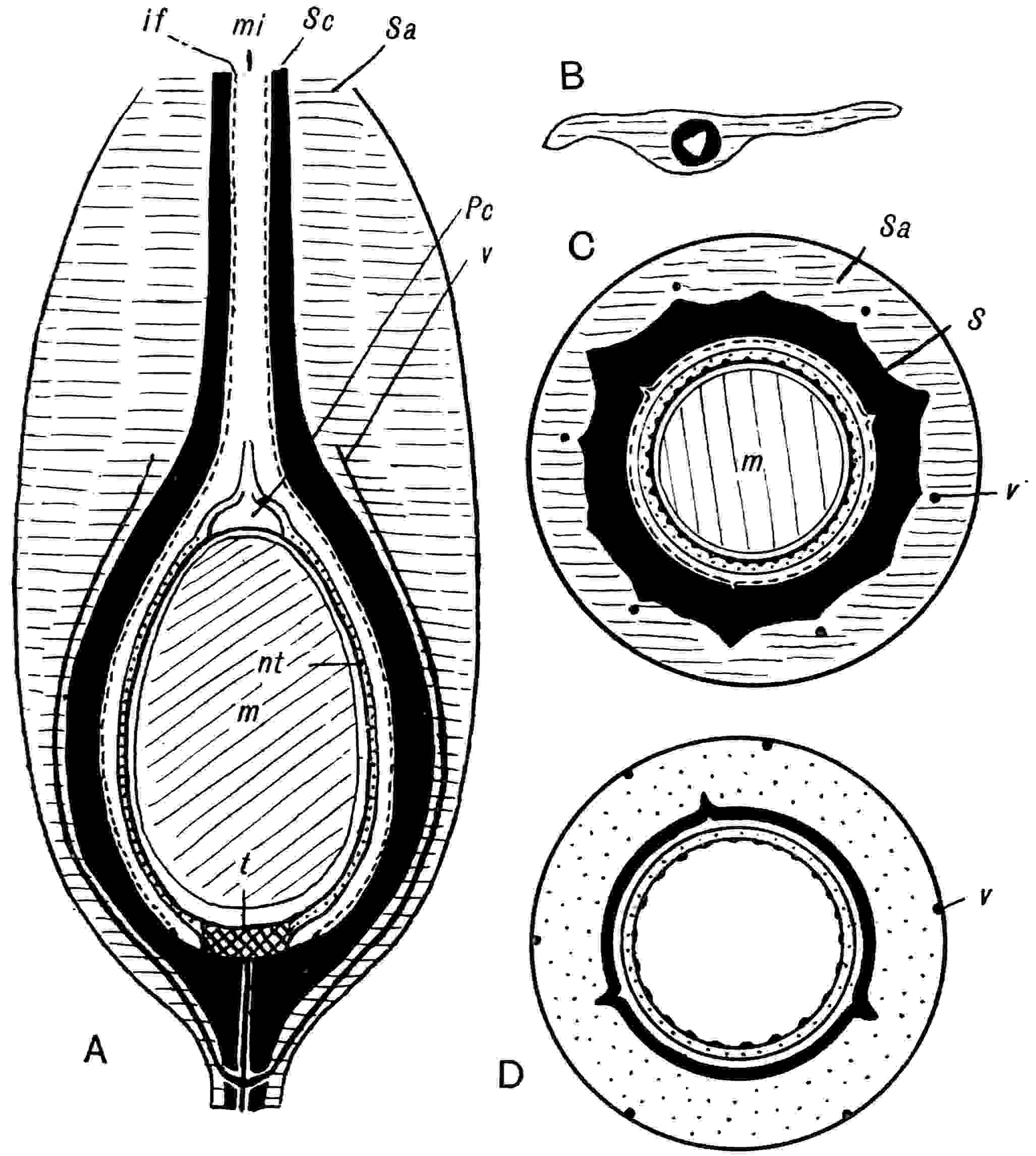
Fig. 426.
Trigonocarpus Parkinsoni
. A, longitudinal section;
Sa
,
Sc
,
if
, sarcotesta, sclerotesta, inner flesh;
mi
, micropyle;
Pc
, pollen-chamber;
v
, vascular bundles;
m
, megaspore and prothallus;
t
, tracheal disc;
nt
, tracheids in the nucellus. B, transverse section of the upper region showing the sarcotesta, sclerotesta (black) and the micropyle. C, transverse section of
T. Parkinsoni
. D, transverse section of
Trigonocarpus shorensis
. (A, C, after Scott; B, after Wild; D, after Salisbury.)
A second species described by Scott and Maslen as Trigonocarpus Oliveri has been further investigated by Salisbury who finds that it is an 8-angled seed which cannot be retained in the genus Trigonocarpus: its systematic position ‘must for the present remain uncertain[340].’ Dr Arber has recently described a new species of Trigonocarpus, T. Moyseyi[341], from the Nottingham Coal-field (Middle Coal Measures), similar to T. Parkinsoni but much broader in proportion to its length: this species is founded on an impression without structure.
The species Trigonocarpus Dawesi Lind. and Hutt.[342], from the Middle Coal Measures of Lancashire, was founded on casts differing in their large dimensions from those of T. Parkinsoni: specimens referred to this species were described by Fiedler[343] from Saxony in 1857 and Lesquereux[344] figures similar casts from the Upper Carboniferous and Permian rocks of North America.
Trigonocarpus shorensis Salisbury.
This species, founded on specimens from the Lower Coal Measures of Shore, Lancashire[345], may exceed 4 cm. in length and has a breadth of 2·5 cm. In general plan it agrees with T. Parkinsoni but there are certain well-marked differences: the micropyle is much shorter; the thick sarcotesta, attaining a breadth of 6 mm. at the base of the micropyle, is characterised by the presence of six peripherally placed vascular bundles (fig. 426, D, v) in contrast to the deeply embedded bundles of T. Parkinsoni. Below the epidermis of the sarcotesta is a hypoderm formed of radially disposed plates of sclerous tissue similar to that of Myeloxylon and different from the palisade-like hypoderm of the type-species. Within the sarcotesta is a hard shell, the sclerotesta, characterised by three prominent ribs extending from base to apex and three shorter ribs which reach from the chalaza to about a third of the length of the seed. The fact that the sarcotesta and sclerotesta pass gradually into one another is a point in favour of the view that the integument is a single structure. There appears to be good evidence of the restriction of an inner flesh to the micropylar region, whereas this tissue in T. Parkinsoni was probably continuous over the whole inner face of the sclerotesta. The sarcotesta is lacunar in its outer part as in some other types of Palaeozoic seeds, a feature probably connected with floating efficiency. Trigonocarpus shorensis occurs in association with Myeloxylon petioles, and there is a resemblance between the seed and the vegetative organs in the structure of the hypoderm as also in the structure of the secretory sacs which are particularly numerous in this species. Salisbury draws attention to the close resemblance between the form of T. shorensis and the seeds found in organic connexion with pinnae of Neuropteris obliqua[346].
The species T. corrugatus described by Renault[347] bears a close resemblance to T. shorensis.
Other Genera founded in part on Reproductive Organs which may belong to the Medulloseae.
Codonotheca, Schützia, Whittleseya, Dolerophyllum, Ottokaria, Strobilites.
CODONOTHECA. Sellards.
Codonotheca caduca Sellards. This genus was founded on some spore-bearing bodies from the Coal Measures of Illinois[348]: nothing is known as to the plant which bore them, but Sellards is inclined to associate them with Neuropteris decipiens Lesq.[349], a species abundant in the same coal-field. Whatever may have been the parent-plant it is probable, as the author of the genus believes, that Codonotheca is the microspore-bearing organ of a Pteridosperm. As shown in fig. 427, 5, the form is that of a stalked cup consisting in the basal portion of a stout axis, the peripheral tissue of which is believed to have been fleshy, containing an axial rod of conducting tissue running up to the floor of the cup, c, and then dividing into six vascular strands, each of which forks into two branches. The upper part is composed of six linear segments united basally to form the sloping surface of the cup. On the inner face of each segment is a more or less well-defined depression covered with large elliptical spores ·29–·31 mm. long by ·18–·19 mm. broad (fig. 427, 6, 8). The presence of a median ridge (fig. 427, 8) indicates a bilateral origin. ‘There is no grouping of the spores or other indication of the location of the sporangia, which were doubtless more or less completely immersed in the tissue, the dividing wall disappearing at maturity.’ The spores are seen in fig. 427, 2, 3, on the inner face of the lobes. Some of the specimens have a fairly long pedicel: in the example shown in fig. 427, 1, the fleshy part of the basal portion is not preserved, only the more resistant vascular core. In a later account of these organs Sellards speaks of several lying by the side of a central stalk to which he thinks they were originally attached by slender pedicels. In view of Dr Benson’s interpretation of the morphology of Telangium it is permissible to suggest that if a central sporangium in such a synangium as that of Codonotheca developed a megaspore and the peripheral sporogenous lobes were sterilised, the result would be an arrangement not unlike the apical region of the seed Physostoma, the tentacles of which have been homologised with the canopy of Lagenostoma. There are obvious difficulties in the way of this, perhaps strained, comparison: the larger size of the spore-bearing linear segments of Codonotheca led Sellards to regard each as a synangium rather than a single sporangium. But precise information as to the structure of the American fossils is not as yet available. If the association of Codonotheca with Neuropteris fronds has any significance it would favour a reference of these organs to the Medulloseae. In the absence of anatomical data it is impossible in some cases to distinguish microspore-bearing organs of the Codonotheca type from small seeds enclosed in a lobed cupule or even seeds with a lobed integument: a case in point is the New Brunswick species Pterispermostrobus bifurcatus Stopes[350].
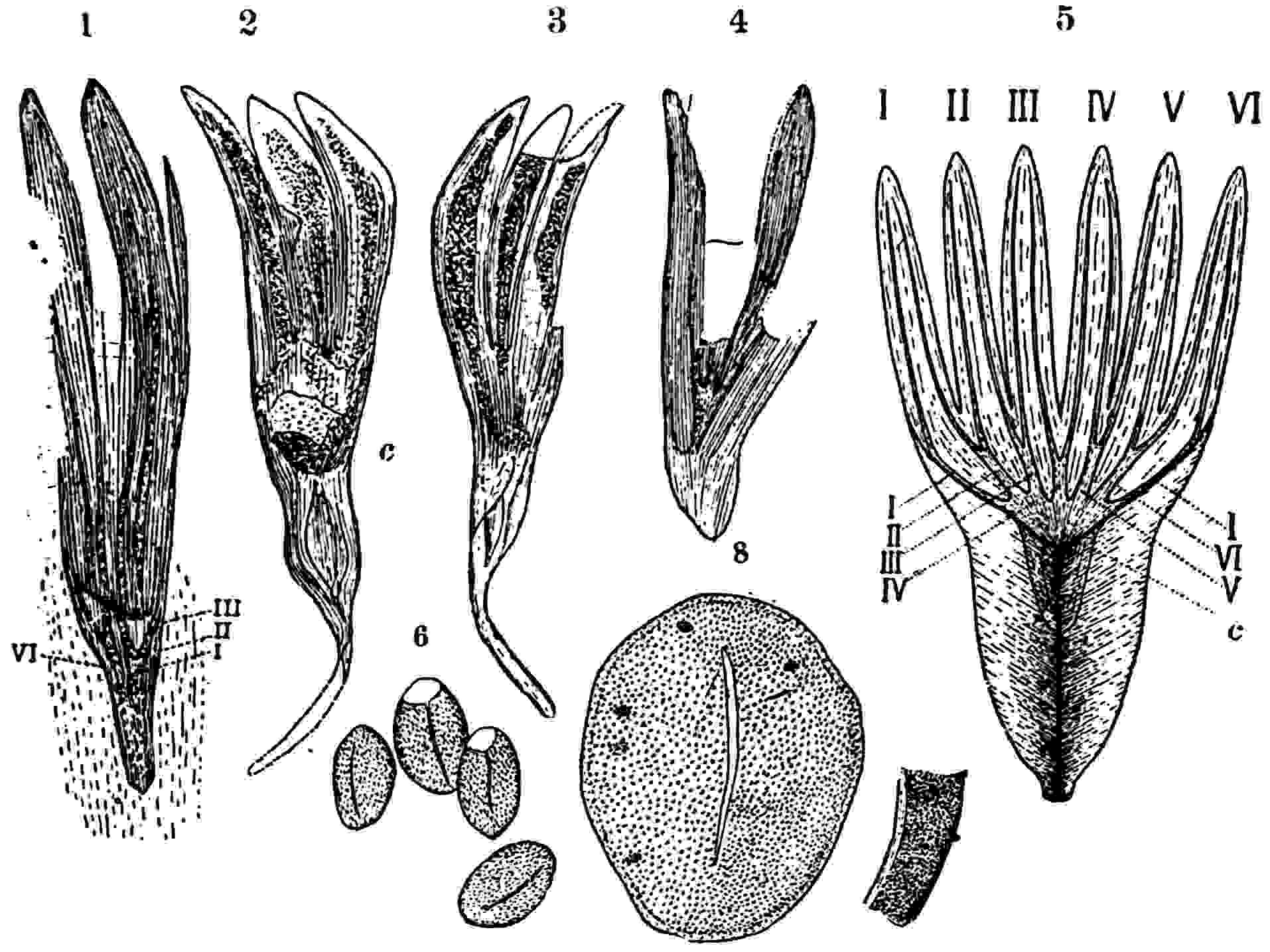
Fig. 427.
Codonotheca caduca
. 1. The vascular tissue preserved as a conical base which was originally surrounded by fleshy tissue (
cf.
5); I–III, VI, vascular bundles (× 2). 2, 3. Segments with spores;
c
, floor of cup (nat. size). 4. Vascular tissue as a cone in the middle of the fleshy base (nat. size). 5. Plan of the whole organ opened out flat (nat. size). 6. Spores (× 28). 8. A single spore showing median slit (× 84). (After Sellards.)
A Spitzbergen, Culm, fossil recently described by Nathorst[351] as Codonotheca (?) pusilla is briefly referred to under the genus Pterispermostrobus.
SCHÜTZIA. Geinitz.
This generic name was instituted by Geinitz[352] for some Permian fossils obtained by Bergmeister Schütz and regarded by the author of the genus as probably fertile branches of some Conifer. A more complete account was published by Goeppert[353] in his ‘Permian Flora,’ where the name Anthodiopsis Beinertiana occurs on the Plates, printed before the publication of Geinitz’s description, but in the text the specimens are referred to Schützia anomala.
Schützia anomala Geinitz.
The type-species, recorded from Bohemia and Silesia, is represented by fertile shoots consisting of a thick main axis bearing apparently two-ranked though probably spirally disposed short lateral branches, each of which terminates in a receptacle with numerous crowded linear-lanceolate bracts superficially resembling a partially expanded inflorescence of a Composite. Goeppert believed that the branches bore seeds and he refers to this species a number of detached, longitudinally striated and bluntly terminated, seeds. The same author describes other specimens from the same localities associated with Schützia anomala, which he names Dictyothalamus Schrollianus[354]: in habit these agree closely with Schützia but the receptacles, the reticulate appearance of which suggested the generic name Dictyothalamus, bear a large number of small bodies regarded as seeds. The preservation of the fossils is not such as to enable us to determine their true nature but it is probable that Schützia and Dictyothalamus are not generically distinct. In his description of Dictyothalamus Goeppert suggests that the two associated types may be the male and female shoots of one plant, but he speaks of seeds in both cases. Schimper[355], who unites Dictyothalamus with Schützia, regards the latter as female and the former as male.
Schützia Bennieana Kidston.
This species, described by Kidston[356] from the Calciferous series of Scotland, differs from S. anomala in its much more slender axis and in the relatively narrower and less globular clusters of bract-like appendages. The principal axis bears three lateral branches with terminal clusters of acute and narrow linear scale-leaves. No seeds were found in association with the specimens.
Schützia permiensis (Renault).
Renault founded this Permian species as Antholithus permiensis[357] on a specimen from Lodève; it consists of an incomplete inflorescence 6·4 cm. long bearing four lateral branches with stalks 1·5 to 2 cm. long terminated by clusters of small oval bracts 5 mm. long. Renault compares the fossil with the recent Conifers Glyptostrobus and Tsuga, but it exhibits a much closer resemblance to Schützia anomala.
The genus Schützia, originally described from Permian strata, is recorded also from Westphalian strata in North Africa[358] as well as from Lower Carboniferous rocks in Scotland. The data at present available are insufficient to determine the morphological nature of the fertile branches: the evidence adduced by Goeppert in support of the occurrence of seeds is not convincing and the interpretation of the bract-like appendages is still an open question; they may have formed a cupular investment to seeds, but in the Scotch species the general appearance rather suggests that they may be microspore-bearing organs comparable with those of Codonotheca[359]. There are no adequate grounds for supposing Schützia to belong to the Coniferales, a view advanced by some authors; it is much more likely to represent the fertile shoots of a Pteridosperm.
WHITTLESEYA. Newberry.
The genus Whittleseya, referred by many authors to the Ginkgoales, has no substantial claim to be regarded as allied to that group: its position is still uncertain, but the recent discovery of fertile specimens suggests the probability of a relationship to Potoniea and an identification of Whittleseya as another form of microsporophyll of a Pteridosperm.
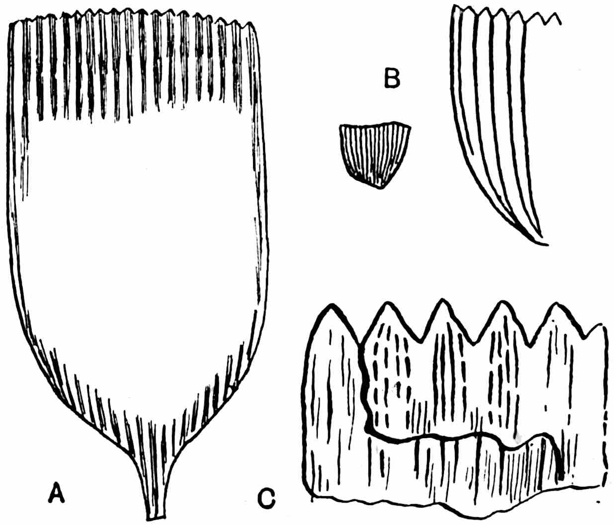
Fig. 428.
A, C. Whittleseya elegans. A, single leaflet, from Pennsylvania. (After Lesquereux; ⅚ nat. size.) C, diagrammatic sketch of part of a leaflet showing the teeth, the striated texture of the carbonised layer, and the vein-like markings on a lower surface. (After Thomas; enlarged.)
B. Whittleseya brevifolia. (After White; the smaller figure ⅚ nat. size.)
The generic name was given by Newberry[360] to some leaves, or possibly leaflets, originally described by C. Whittlesey from the Coal Measures of Ohio. Whittleseya is represented by species from several North American localities[361] in Ohio, Pennsylvania, Arkansas, Nova Scotia, and New Brunswick[362]; it occurs in Silesia and has recently been found in the English Coal Measures[363]. The genus is confined to Upper Carboniferous strata.
The leaves are fairly thick; the lamina is oblong, cuneate, broadly triangular or linear, usually rounded and truncate (fig. 428, A, C), generally dentate at the distal end, the proximal portion being gradually or abruptly contracted and occasionally prolonged into a short pedicel. The veins or ribs are parallel to the sides of the lamina and except near the base unbranched.
Whittleseya elegans Newberry.
The type-species, from North America and Europe, is characterised by its shovel-like lamina from 3 to 6 cm. long closely resembling in shape some lepidopterous scales; the surface is ribbed, each rib corresponding to a tooth on the distal margin; on each of the parallel ridges are 4–5 longitudinal lines indicating either veins or stereome strands (fig. 428, A, C). The examination of preparations made by Dr Kidston from a leaflet of this species enables me to add a few facts with regard to the microspores. The spores, which cover almost the whole surface of the lamina, show a tendency to a more or less definite arrangement in longitudinal rows. Two types of cuticularised membrane are represented among the associated fragments: in some pieces of cuticle the cells are short and have straight walls while in others the preservation is inferior and the cells appear to be longer and narrower. One or both of these membranes probably belong to the sporangia. The oval slit, which is a striking feature on several of the spores (fig. 429), points to their bilateral nature and dehiscence along the major axis. A comparison of these spores with those obtained by Kidston from the English species Whittleseya fertilis reveals a very close agreement both in size and shape and confirms the identification of the Staffordshire specimens as leaflets of Whittleseya. The large size of the microspores and the gaping oval aperture in some of them are features in which they agree closely with the spores of Dolerophyllum fertile described by Renault[364]. In both cases the spores tend to be arranged in long groups and they are practically identical in form and in the nature of the exine; those of Dolerophyllum are 280μ long while those of W. fertilis reach a length of 220μ. In some of the Whittleseya spores the exine has split as in the specimen shown in fig. 429, but in others there are two curved lines along which dehiscence has begun, a character in which the spores appear to be identical with those of Dolerophyllum described by Renault who speaks of dehiscence by means of an operculum. There is, I venture to think, little doubt as to the very close affinity of the two types. The systematic position of Dolerophyllum is not certainly established; if the generic identity of the leaves described as D. Berthieri Ren. and the petrified specimens named D. fertile is assumed, it is a legitimate inference that the genus is founded on fertile pinnules of a Pteridosperm with foliage of the Neuropteris or Cyclopteris form. It would seem probable that both Whittleseya and Dolerophyllum fertile are microspore-bearing leaflets of Pteridosperms, possibly of some Medullosan plants. The leaflets of Whittleseya agree in form fairly closely with those of Potoniea adiantiformis Zeill. described on a previous page[365] as the male organs of a Pteridosperm.
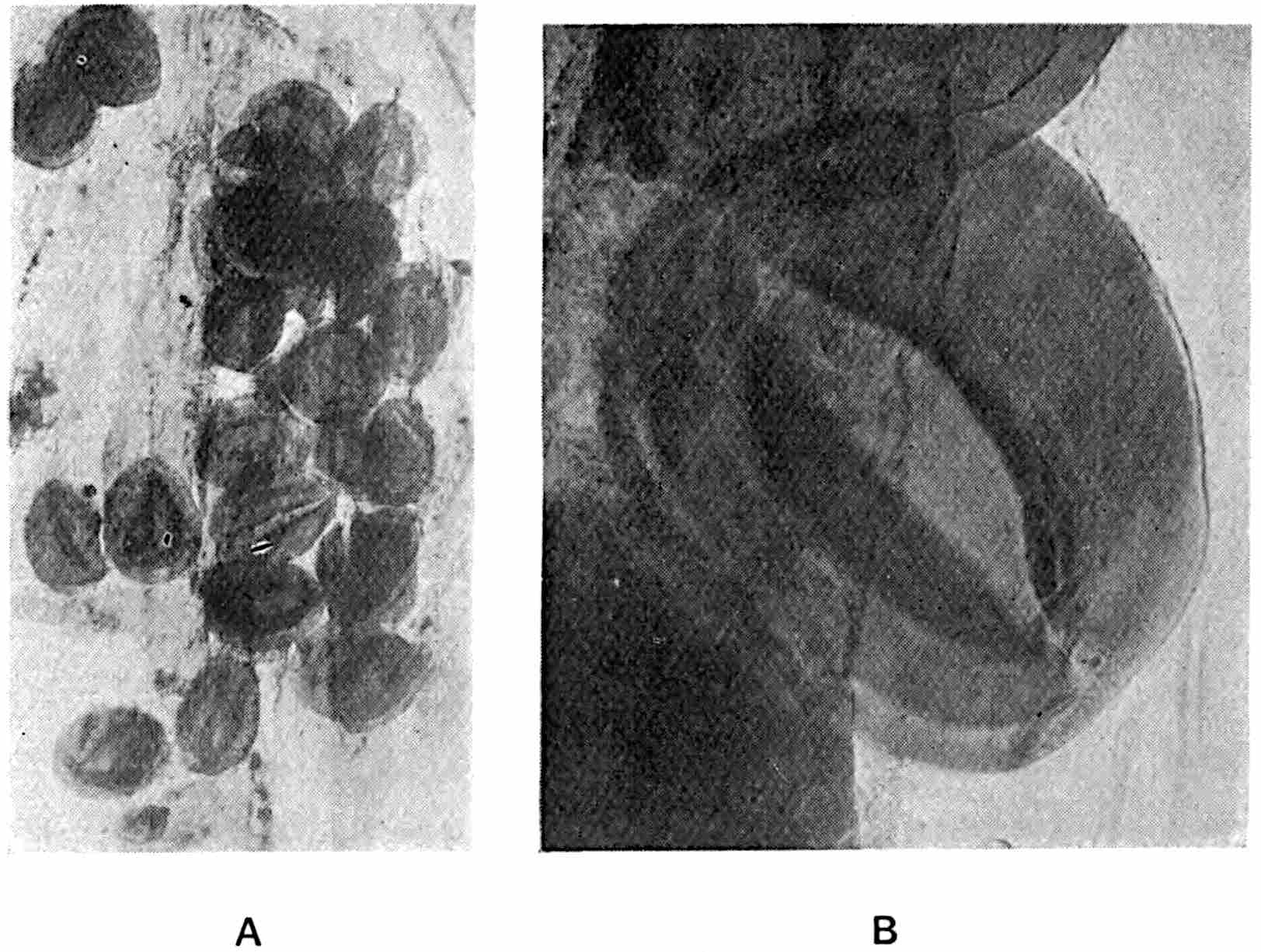
Fig. 429.
Microspores of
Whittleseya elegans
. A, a group of spores; B, a single spore. (Preparations made from an American specimen, No. 2314, in Dr Kidston’s Collection.)
The specimens described by Lesquereux from Pennsylvania as W. integrifolia and W. undulata are less satisfactory than W. elegans. The Arkansas species W. microphylla[366], characterised by the obcuneate form of the lamina, is said to occur not only as detached leaflets but in loose bunches at the ends of slender axes, a circumstance favourable to the suggestion, based on the recently described English specimens, that the Whittleseya leaves may be fertile pinnules of a Pteridosperm frond. Among other species attributed to Newberry’s genus is W. brevifolia Wh. from Nova Scotia[367] with much smaller broadly triangular leaves 7 mm. long exclusive of the petiole and 8 mm. broad at the distal end (fig. 428, B). Dr Matthew[368] has also described a Canadian species W. concinna from New Brunswick in beds assigned by Dr Stopes[369] to the Westphalian series.
Whittleseya fertilis Kidston.
Since the discovery of Whittleseya elegans in the Coal Measures of Staffordshire recorded by Mr Thomas, Dr Kidston has published an account of some specimens from the same district under the name Whittleseya (?) fertilis[370]: these consist of smaller cuneate scale-leaves or leaflets 1·4–2·4 cm. long and 8–9 mm. broad; the lamina has a dentate upper margin and is longitudinally ribbed. The scales occur in superposed pairs, closely fitting but not organically connected, at least in the state in which they are preserved; each pair forms a sporangium-like case enclosing numerous spores but the actual sporangia or synangia have not been preserved. Kidston describes the spores as 210–222μ in length, elliptical, and characterised in many cases by an oval slit; they are practically identical with the microspores of W. elegans.
DOLEROPHYLLUM. Saporta.
This name was proposed[371] primarily for a large ovoid petrified bud composed of rolled Cyclopteroid leaves from Permian rocks in the Ural Mountains, which had been previously described by more than one writer under different names and regarded as a young shoot of a Palm or other Monocotyledon. Eichwald[372], who published good drawings, called the fossil Noeggerathia Goepperti. Saporta connected with this species some leaf-impressions from the Permian of Bohemia described by Goeppert[373] as Noeggerathia cyclopteroides: in his family Dolerophylleae[374] the French author included other leaves which are probably not closely related to the type-species, Dolerophyllum Goepperti. The Dolerophylleae are spoken of by Saporta and Marion[375] as Progymnosperms. Before the publication of Saporta’s note Grand’Eury had instituted the genus Doleropteris[376] and the family Doleropteroideae; in the former he included several forms of leaves agreeing generally with Goeppert’s Noeggerathia cyclopteroides. Zeiller[377] adopts Grand’Eury’s designation for the Russian fossil in preference to Dolerophyllum, a choice justified by considerations of priority; but the latter name is retained in this account as it was assigned by Saporta to the specimen of greatest botanical interest, namely Dolerophyllum Goepperti, and because it does not suggest affinity to Ferns.
Dolerophyllum Goepperti (Eichwald).
The type-species is from the Zechstein of Orenburg in the Urals and no specimens having precisely the same structure have been found elsewhere. Eichwald assigned it, with leaf-impressions of various kinds, to the Noeggerathieae and named it Noeggerathia Goepperti: it had previously been described by Kutorga[378] as Aroides crassispatha and Unger[379] included it among the Palms as Palaeospathe aroidea. The species has been described also by Saporta and Marion and by Renault[380]. The following account is based on sections cut from a specimen in the British Museum[381] which, though assigned on the label (within a query) to France and named Dolerophyllum Berthieri, is undoubtedly Eichwald’s species from East Russia.
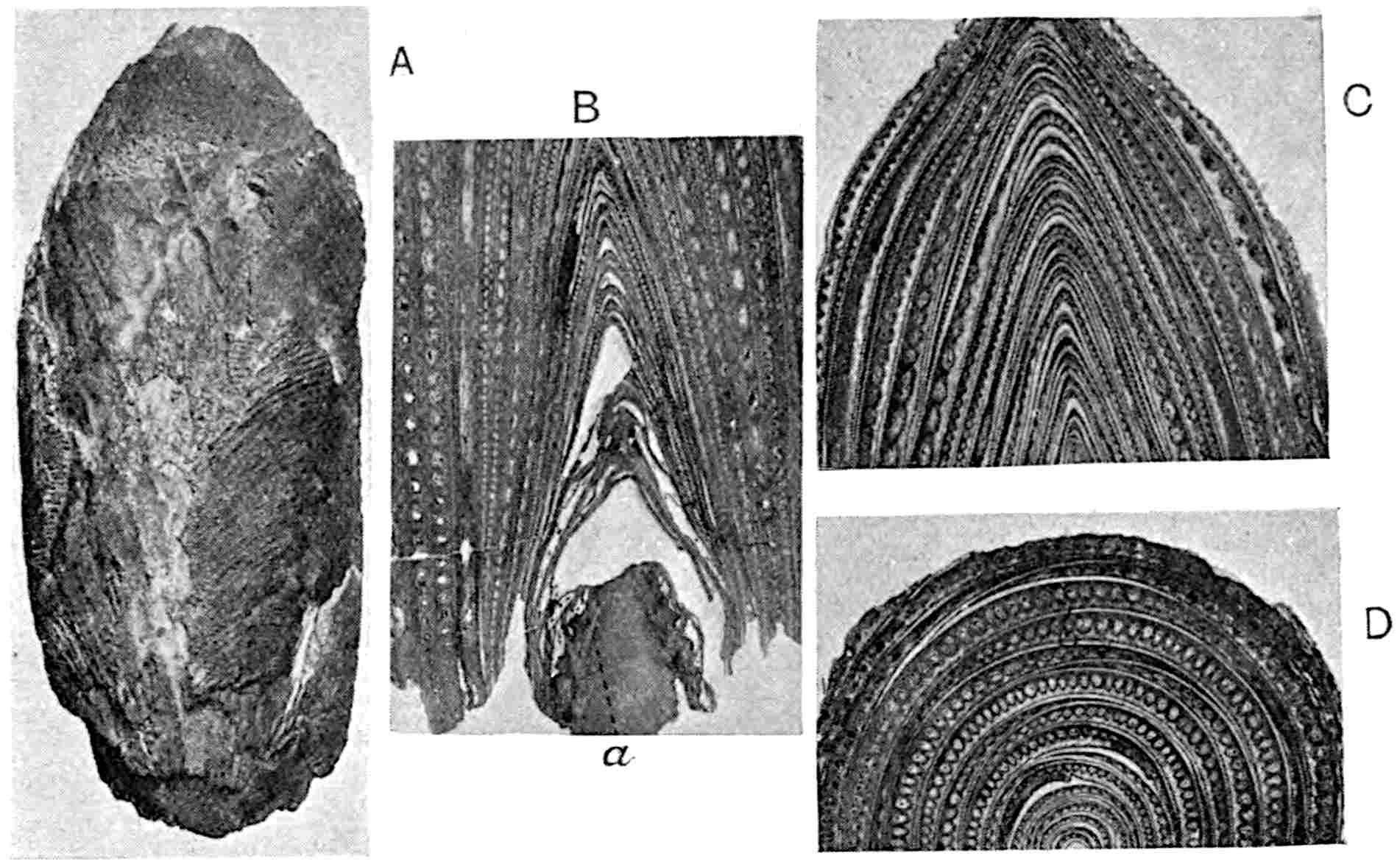
Fig. 430.
Dolerophyllum Goepperti
. Bud in surface-view, A; in longitudinal section, B, C; in transverse section, D. (British Museum; A, ⅔ nat. size.)
The specimen (fig. 430) is 9 cm. long and 4·2 cm. broad: at the slightly contracted and broken base is a piece of immature axis (fig. 430, B, a) 12 mm. in diameter overtopped by a mass of closely packed leaves encircling one another like the bulb-scales of an Onion (fig. 430 A, C)[382]. Most of the leaves included in the bud were attached to the axis below the broken base. The curved, dichotomously branched, veins are seen on some of the pieces of lamina on the surface of the bud (fig. 430, A). The considerable breadth of the leaves is demonstrated by the longitudinal and transverse sections. In fig. C most of the laminae can be traced through the whole height of each of the steep-sided arches: a few overlapping margins are seen in fig. D. The veins are for the most part imperfectly preserved and appear as clear spaces at regular intervals in the brown mesophyll. The axis of the shoot consists of homogeneous parenchyma except near the sloping sides where narrow dark bands (fig. 430, B, a) mark the position of desmogen-strands of thin-walled elongated elements representing an early stage in the development of vascular bundles some of which have already produced spiral tracheids. Short secretory cells accompany the immature conducting elements. The lamina slightly exceeds 2 mm. in thickness in the broadest part: the mesophyll is composed of large parenchymatous cells of elliptical or spherical form often loosely attached owing to the well-developed system of intercellular spaces. The lower epidermis, assuming that the outer face of the rolled leaves is the morphologically lower surface, forms a uniform layer of palisade cells characterised by their free conical ends (fig. 431, A, B, e) which in some oblique sections appear as sharply pointed papillae with almost filiform apices; but while the cells were doubtless papillose like those of the epidermis of a velvety petal, the pointed form is due in part to the greater distinctness of the dark contents as compared with the lighter cell-walls. The upper epidermis is much less distinct; it consists of smaller flattened cells with occasional stomata. Renault[383] figures a specimen with stomata in a better state of preservation. The vascular bundles are rendered conspicuous by large secretory cells on the lower side, in the larger veins in the form of an arc or irregular group (fig. 432, A), but in the finer veins as single cells (fig. 431, A, B). These sacs resemble the tannin cells accompanying the veins in a leaf of Ginkgo (cf. fig. 631, G). The xylem-elements are of two kinds, (i) elongated spiral and scalariform conducting elements, forming a vertical plate of a few rows in the larger veins (figs. 431, 432) or a small compact group in the more slender veins (fig. 413, A, B); (ii) much larger isodiametric cells with reticulate or spiral thickening resembling the transfusion-tracheids of Conifers or, perhaps more closely, similar elements in the leaves of Lepidodendron. These short tracheids are especially abundant on the flanks of the conducting tracheids (figs. 431, t; 432, A, t), but they sometimes form a complete investment. In the obliquely cut vein reproduced in fig. 431, D, the transfusion-tracheids are abundant: a few are enlarged in fig. 431, E. In the smaller veins (fig. 431, A, B) they are represented by the larger elements, t, on the sides of the conducting strands. The protoxylem lies close to the upper edge in the middle line (px, figs. 431, A; 432, A); it is difficult to determine its precise position, but it would seem to be slightly internal, the bundle being not quite endarch. No phloem was recognised in the British Museum specimen, but it presumably occurred, if present, where the black patch is shown in fig. 432, A. Renault describes some phloem in sections which he examined.
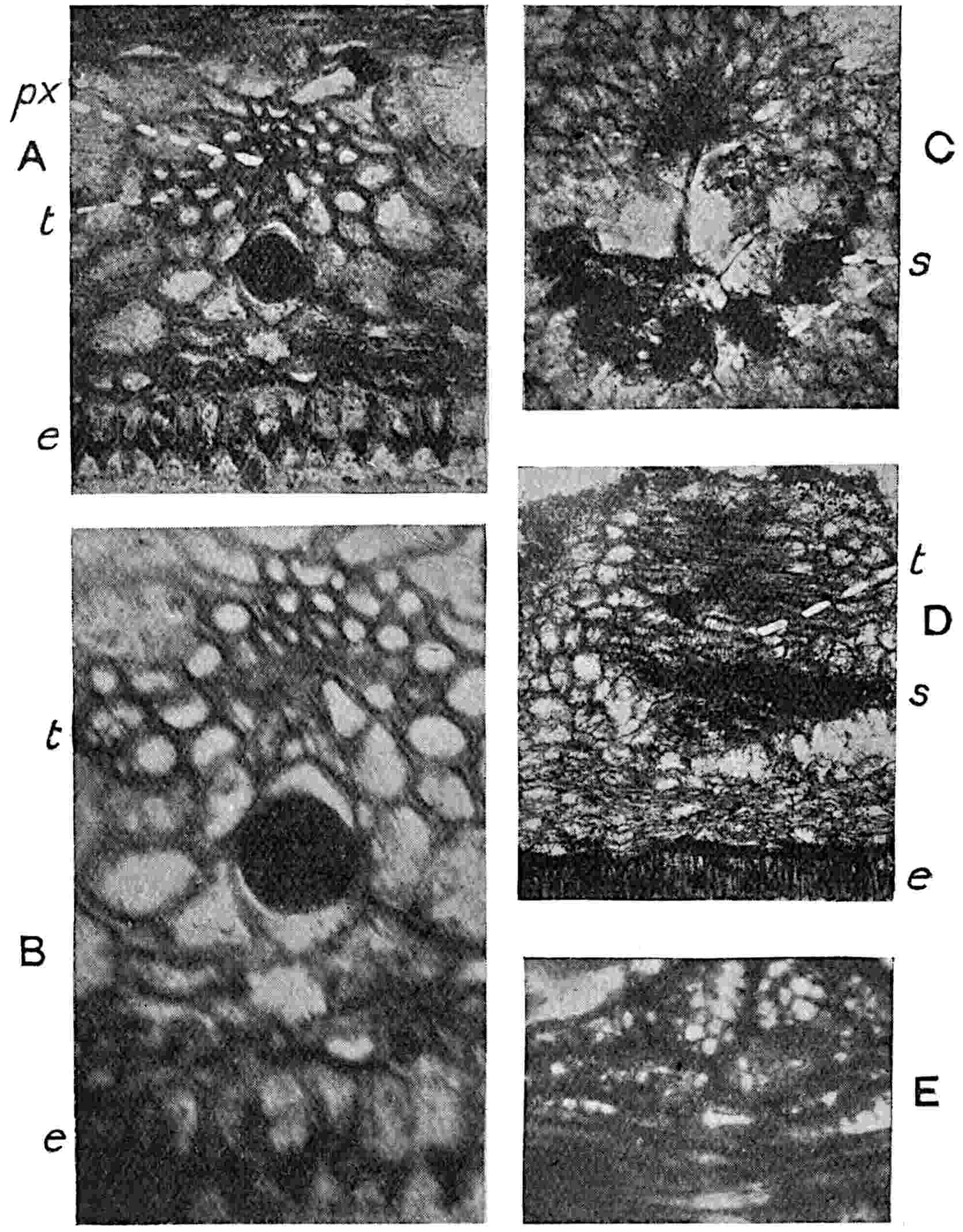
Fig. 431.
Dolerophyllum Goepperti
. Transverse sections of leaves.
e
, epidermis;
s
, secretory cells;
t
, transfusion-tracheids;
px
, protoxylem. (British Museum.)
The mesophyll next the upper surface is in most cases represented by spaces between the veins which give a crenulated outline to the parenchyma (fig. 430, C, D); in some places the spaces contain remains of very loose and crowded cells suggesting the original presence of very lacunar tissue or possibly of thin-walled storage-cells. The confinement of stomata to what is assumed to be the upper surface may, as Renault and others have suggested, indicate leaves which floated on water, an inference opposed to the view that the gaps in the mesophyll mark the position of water-tissue.
No specimens have been described which enable us to correlate with certainty mature leaves or foliage-shoots with the petrified bud. It is, however, not improbable that the impression from Mount Pelé near Epinac named by Renault Dolerophyllum Berthieri[384] may be correctly referred to the same genus. The type-specimen consists of an axis, whether a rachis of a compound leaf or a shoot with simple leaves cannot be determined, bearing partially overlapping more or less orbicular leaves 18–20 cm. in diameter, with a Cyclopteris venation. Among other leaves of unknown affinity referred to the same genus attention is drawn to Dolerophyllum pseudopeltatum (Grand’Eury)[385] with an orbicular lamina reaching in some specimens 22 × 19 cm. Specimens of Dolerophyllum pseudopeltatum are figured by Renault from the Commentry coal-field[386], some of which reach a diameter of 12 cm. The only British specimen of a leaflet of this type which I have seen is one in Dr Kidston’s collection from the Stephanian series, Glamorganshire. It is probable that some at least of the impressions assigned to Dolerophyllum or Doleropteris would be more appropriately included in Cyclopteris or Cardiopteris and may have been borne on the axis of large Pteridosperm fronds. Grand’Eury[387] has also called attention to the difficulty of distinguishing the larger Cyclopteris leaflets from Dolerophyllum. Some of the Cyclopteroid leaflets figured by Roehl[388] on Neuropteris fronds differ but slightly from those of D. pseudopeltatum. The shoot showing large leaf-scars figured by Saporta and Marion[389] as probably the axis of a Dolerophyllum may well be a piece of Cordaites.
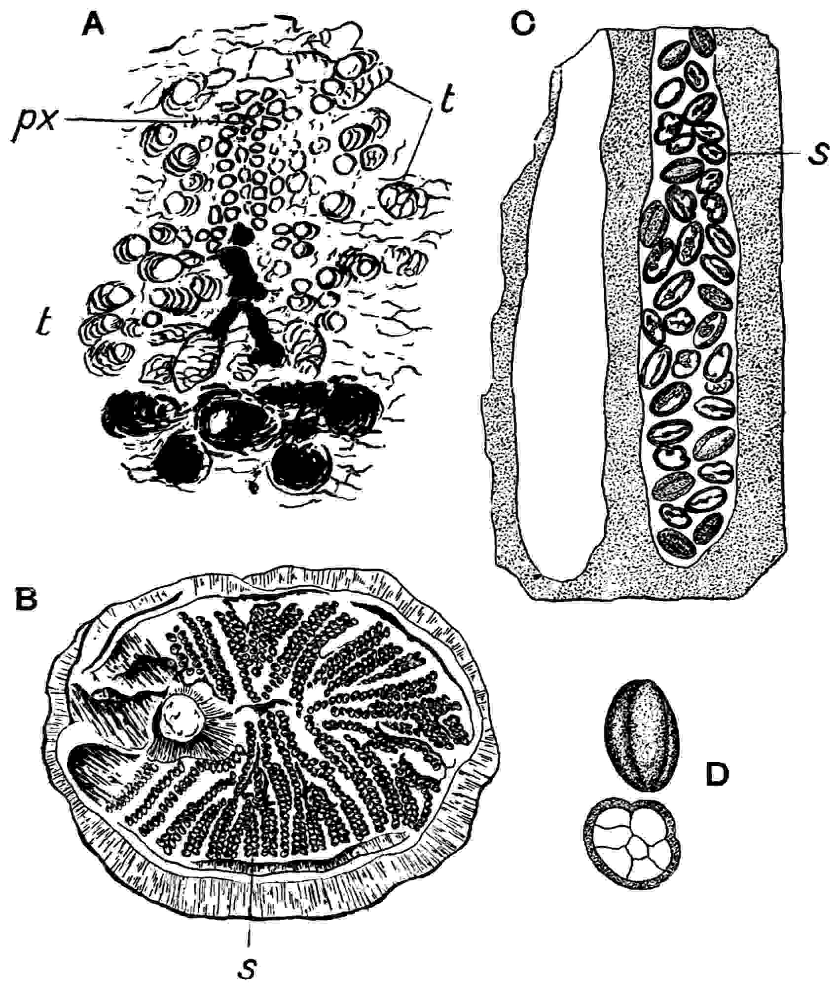
Fig. 432.
A.
Dolerophyllum Goepperti
, section of vein. B.
Dolerophyllum Berthieri
;
s
, microspores. C.
Dolerophyllum fertile
. D.
Dolerophyllum fertile
, microspores. (After Renault.)
Microsporophylls assigned to Dolerophyllum.
Certain problematical fossils found in association with the sterile leaves of Dolerophyllum Berthieri have been described by Renault as the male organs of that species. These are elliptical discs, 6 × 5 cm., with an excentrically placed stalk: embedded in a carbonised lamina are numerous rows of elliptical bodies, 410μ × 280μ, characterised by two curved longitudinal grooves on the surface and regarded by Renault as pollen-grains. The chains of these microspores radiate outwards from the neighbourhood of the stalk and cover most of the surface of the disc (fig. 432, B). Some silicified pieces of similar spore-bearing discs from Grand’ Croix named Dolerophyllum fertile[390] afford additional information as to these remarkable reproductive organs. The earlier account of this species by Renault is confirmed by Solms-Laubach[391] who examined the original sections. The peltate fleshy discs preserved as incomplete specimens consist of lacunar parenchyma 15–18 mm. thick traversed at right angles to the surface by numerous loculi (fig. 432, C), circular or oval in transverse section, containing large numbers of microspores, s, similar in size and form to those on the carbonised discs of the Mt Pelé specimen. Vascular strands occur between and parallel to the spore-chambers. The spores contain 8–10 cells (fig. 432, D) and Renault believes that dehiscence of the exine occurred along the two deep grooves which mark the limits of an operculum. He emphasises the peculiar structure of the microspores by speaking of them as prepollinia: in size and in the presence of internal cells (? male prothallus) they resemble the spores found in the pollen-chamber of a seed described by Renault as Aetheotesta elliptica[392] which he thinks may belong to a member of the Dolerophylleae. It has also been suggested that Codonospermum may be a seed of Dolerophyllum[393]. An unconvincing specimen described by Saporta and Marion[394] as a seed-bearing bract is regarded by them as referable to Dolerophyllum, but the evidence for any connexion is far from satisfactory.
There is nothing definite to be said with regard to the affinity of Dolerophyllum Goepperti or the microsporophylls represented by D. fertile and the specimens associated with D. Berthieri. Renault considers that both sterile and fertile specimens belong to the same genus, which he assigns to a position between Pteridophytes and Cycads. As Solms-Laubach says, the evidence supplied by the structure of the veins of D. Goepperti in favour of a cycadean alliance is not convincing. The type of vernation is unlike that of any known Cycad or indeed of any Gymnosperm: the large size of the leaves is another though weaker objection to this comparison, as the pinnae of Bowenia (fig. 391) and especially those of some species of Zamia (fig. 388), are of equal or larger dimensions. If, as seems probable, the xylem-strands are mesarch that is a point of contact with recent Cycads, but the bundle as a whole bears but a remote resemblance to that of a cycadean leaf and is much more like the veins of Ginkgo. The bud shown in fig. 430, A, is probably a young shoot and not merely a large compound leaf. If it were an unexpanded frond of Neuropteris bearing Cyclopteris pinnules we should expect to find indications of scattered desmogen-strands such as would occur in the Myeloxylon type of rachis. The resemblance to most forms of Cordaites is by no means close though a few leaves referred to that genus (e.g. C. circularis, fig. 468, B) are similar to those of Dolerophyllum[395].
The male organs are unlike those of any other plant: they may be described as sporophylls with microsporangia or perhaps synangia embedded in the mesophyll and containing microspores similar to those of some Pteridosperms or true Gymnosperms. Attention has been called to the close resemblance of the spores shown in fig. 432, C, D to those recently discovered by Kidston and referred to the genus Whittleseya (fig. 429), and it is very probable that the striking similarity is an index of affinity.
Ottokaria. Zeiller.
Ottokaria bengalensis Zeiller. A specimen of doubtful affinity from the Lower Gondwana (Karharbari beds) of Passerabhia, India, was originally described by Zeiller[396] as Feistmantelia bengalensis, but in a postscript he substituted the name Ottokaria on the ground that Feistmantelia had previously been employed by Lester Ward. Fig. 433 is drawn from the original specimen: it consists of a stalk attached in a slightly excentric position to an almost orbicular lamina, 2·5 cm. in diameter, with subacute marginal teeth and traversed by numerous radially disposed striations. Zeiller compares the fossil with Whittleseya elegans and Rhipidopsis ginkgoides and assigns it with some hesitation to the Salisburieae. An examination of the type-specimen led me to form the opinion that it may be a cupular organ of a Pteridosperm that enclosed a seed. The lamina is slightly concave and has the form of a shallow cup; moreover the surface-features resemble those of a bract rather than the regularly veined lamina of a foliage-leaf. The specimen bears a very close resemblance to one figured by Bertrand[397] as the cupule of Hexapterospermum modestae which he connects with fronds of Neuropteris gigantea.
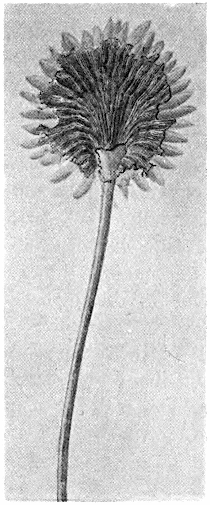
Fig. 433.
Ottokaria bengalensis.
(Nat. size; drawn from the type-specimen.)
Ottokaria occurs in association with fronds of Glossopteris indica and with the large seeds described by Zeiller[398] as Cardiocarpus indicus. I have lately obtained some evidence in favour of assigning Feistmantel’s seeds Carpolithes Milleri[399] to the genus Glossopteris: among several specimens from the Lower Gondwana rocks of India I found an example showing a seed partially covered by a scale-leaf in its natural position which appears to be identical with scale-leaves of Glossopteris. It may be that the specimen represented in fig. 433 belongs to Cardiocarpus indicus, though this is a mere guess: my belief is that Ottokaria is a cupular organ that enclosed the base of a seed borne on a Pteridosperm. There is little doubt that as additional data are obtained it will be found that Pteridosperms played no inconsiderable part in the vegetation of Gondwana Land.
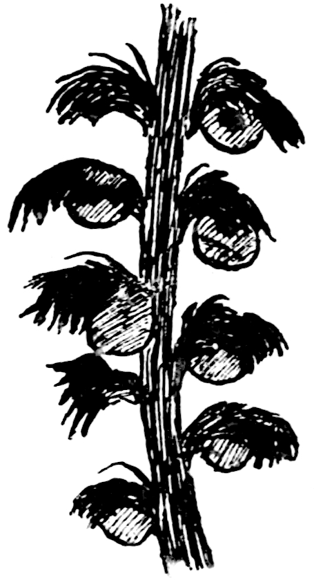
Fig. 434.
Strobilites Milleryensis.
(Natural size; after Renault.)
Strobilites. Schimper and Mougeot.
Strobilites Milleryensis (Renault).
This species, from the Permian of France, was placed by Renault in Cycadospadix[400], but having regard to the fact that it differs essentially in habit from Mesozoic examples of that genus the provisional name Strobilites[401] is suggested. The type-specimens are long and narrow spikes or loose strobili, 8–16 cm. long and 2–2·6 cm. broad; a stout axis bears spirally disposed bracts 8–10 mm. long attached by a slender decurrent pedicel expanded distally into a fan-shaped laciniate lamina with a convex upper face, and there are said to be two seeds attached to the sides of each pedicel (fig. 434). The oval seeds appear to be platyspermic and resemble Samaropsis fluitans Daws. Two of the strobili figured by Renault are attached at right angles to a second axis, a habit suggesting comparison with that of a large compound frond. Renault is inclined to regard these fertile shoots as cycadean and suggests a possible connexion with the Permian stems Ptychoxylon or Poroxylon, both of which are known to have produced fairly numerous branches. In habit the spikes are similar to some of the longer examples of Cordaianthus, but their preservation is not sufficiently good to afford accurate information as to the relation of seed to sporophyll. Strobilites Milleryensis is, perhaps, more likely to be the fertile branch of a compound frond of a Pteridosperm, and it is significant that the seeds have been found in association with Callipteris leaves.
COLPOXYLON. Brongniart.
Colpoxylon aeduense Brongniart. The genus was founded by Brongniart[402] on a piece of stem 15 cm. in diameter from the Permian of the Autun district and regarded by him as a distinct type, with certain resemblances to recent Cycads. A thick section in the British Museum, 13 cm. in diameter (fig. 435, A), illustrates the main anatomical features described by Renault[403], to whom our knowledge of the genus is chiefly due. There are two large steles of irregular outline closely resembling those of Medullosa Leuckarti (cf. fig. 416, H); each consists of a band of secondary xylem with broad medullary rays and a narrow zone of phloem enclosing a central region composed of parenchyma, in which strands of primary tracheids, both reticulate and spiral, pursue a more or less horizontal course, associated with a few small groups of vertical xylem-strands at the inner edge of the secondary wood. The manoxylic nature of the wood is clearly shown in fig. 436; the continuous ink-line marks the position of the cambium and the dots show the internal protoxylem. Homogeneous parenchyma surrounds the steles and beyond this is crushed tissue containing large secretory canals and nests of stereome fibres either as separate groups or in contact with the canals (fig. 435, C). In the same peripheral tissue occur scattered collateral vascular bundles (fig. 435, D) identical with those of Myeloxylon. The outer cortex of the stem is marked off from the more homogeneous inner region by a fairly distinct line where there is some indication of periderm. The anatomical features are clearly shown in fig. 436, a photograph from a section in Dr Kidston’s collection. At a is an imperfectly preserved vascular bundle with a crescentic group of secondary xylem which is probably a leaf-trace that has just emerged from the secondary cylinder. Renault speaks of these more or less circular strands as possibly connected with reproductive shoots, but it is more probable that they are homologous with the strands in the pericycle and inner cortex of Medullosa and represent leaf-traces before division into smaller collateral strands. Renault describes the stem as possessing seven vascular cylinders in the apical region and suggests branching of the main axis as the cause of the increase in number: there is, however, no evidence to support such correlation. The two steles seen in fig. 435, A, become merged at a lower level into a single stele of sinuous form (fig. 435, B).