Chapter XXX · (continued 2)
Reading edition
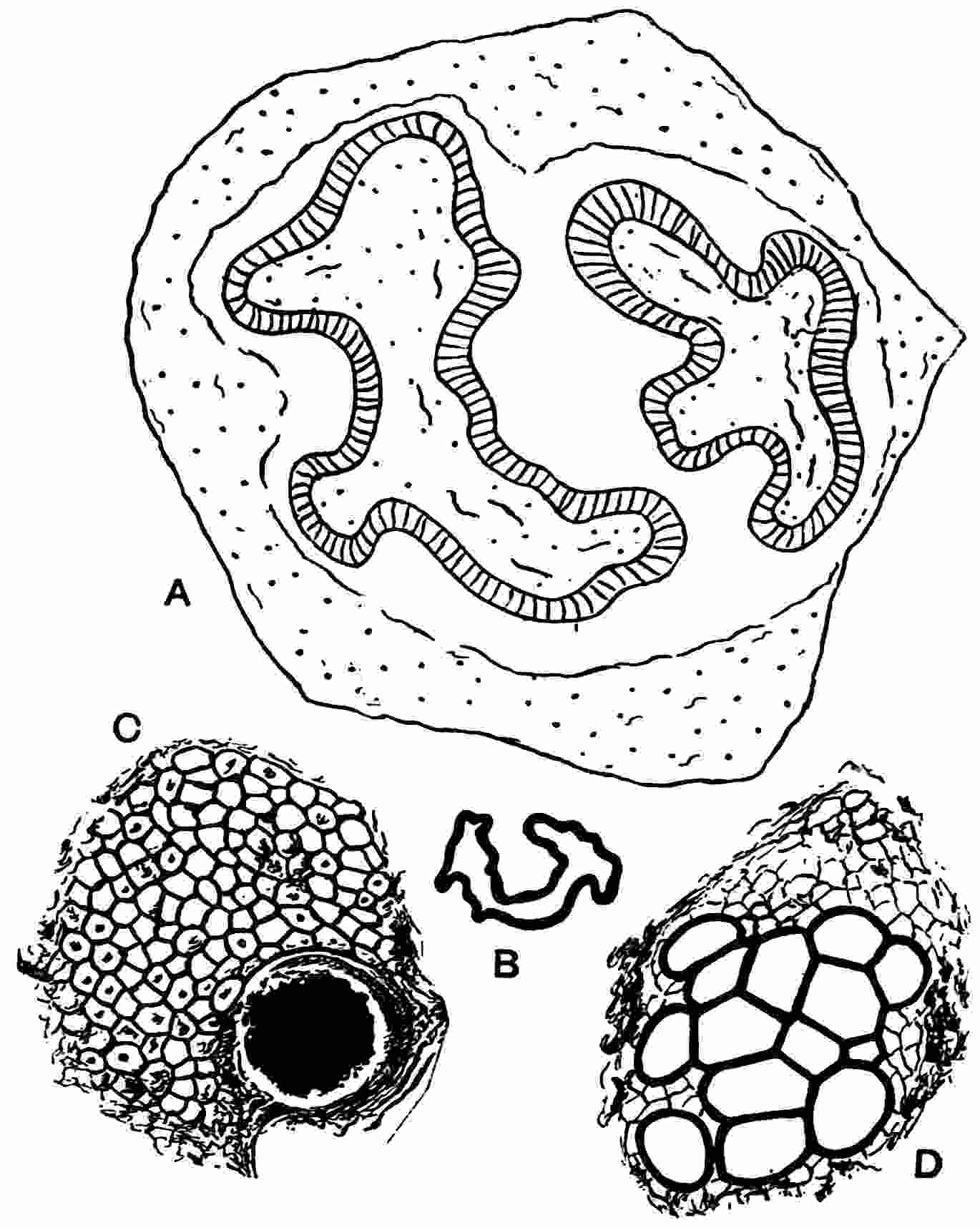
Fig. 435.
Colpoxylon aeduense
. A. Diagram of a transverse section of a specimen in the British Museum. (V. 9393. ½ nat. size.) B. Stele; much reduced from Renault’s figure. C, D. Secretory canal with fibres and collateral vascular bundle (Kidston Coll., 1946).
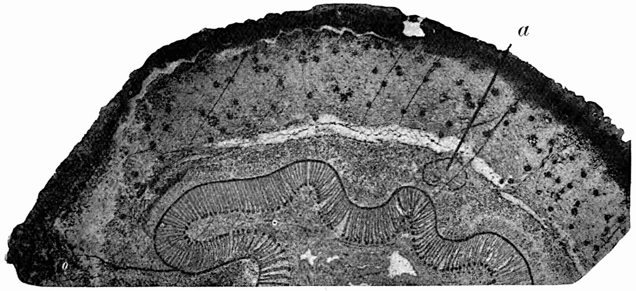
Fig. 436.
Colpoxylon aeduense
. Transverse section of half of the stem;
a
, stele of branch. Slightly enlarged. (Kidston Coll., 1946.)
Beyond the facts furnished by the leaf-trace bundles in the outer cortex and the occurrence of two large scars about 5 cm. in breadth on a stem figured by Renault, we have no positive information as to the form of the leaves or the structure of the reproductive organs. There is little doubt that the fronds were large and compound like those of most species of Medullosa. There is, however, some slight evidence that Alethopteris Grandini Brongn. and seeds of the Pachytesta type (fig. 497) were borne on Colpoxylon stems; this rests solely on the association in the Loire coal-basin[404] of Alethopteris fronds with stems presenting structural resemblances to Colpoxylon aeduense.
The striking resemblance between Colpoxylon and Medullosa Leuckarti has led certain authors[405] to propose the substitution of Medullosa for Colpoxylon. The resemblances though close are hardly sufficient to warrant this course. In Colpoxylon the stelar system is simpler; there is no central region with star- or plate-rings as in Medullosa Leuckarti but, as in Medullosa anglica, the vascular tissue consists only of large steles without a medullary system. Colpoxylon differs from M. anglica in the reduction in some parts of the stem of the vascular system to a single stele and, moreover, the primary portion of the steles is much more parenchymatous in structure and contains more irregularly anastomosing tracheal strands than is the case in M. anglica.
The alteration in the pattern formed by the vascular system at different levels in some Medullosan stems, especially in Colpoxylon, may be compared with the varying disposition of the vascular strands in the thick dorsiventral rhizomes of Polypodium heracleum Kunz. and P. quercifolium L. In the rhizome of P. heracleum there are two vascular systems, an outer, cortical, system in the form of a hollow cylinder composed of a lattice-work with polygonal meshes from which branches are given off to the roots, and a more complex medullary system that is concerned with the emission of leaf-traces. As shown by a series of drawings reproduced in an account by Klein[406] of the anatomy of these species of Polypodium, the inner system of steles consists of two cylinders connected towards the upper surface of the stem by a rounded arch of vascular strands; nearer the leaf-base the two cylinders meet and eventually a larger cylinder is produced partly from the upper halves of the two cylinders of the previous section and in part from the connecting arch: the remains of the two smaller cylinders become connected with the outer vascular system. These and other changes suggest comparison with Colpoxylon as also with the stelar changes in the stem of Ptychoxylon. The comparison cannot be carried beyond the grosser features and is chiefly interesting as affording a further illustration of a similarity in plan between some recent Ferns and extinct Pteridosperms and other Palaeozoic genera.
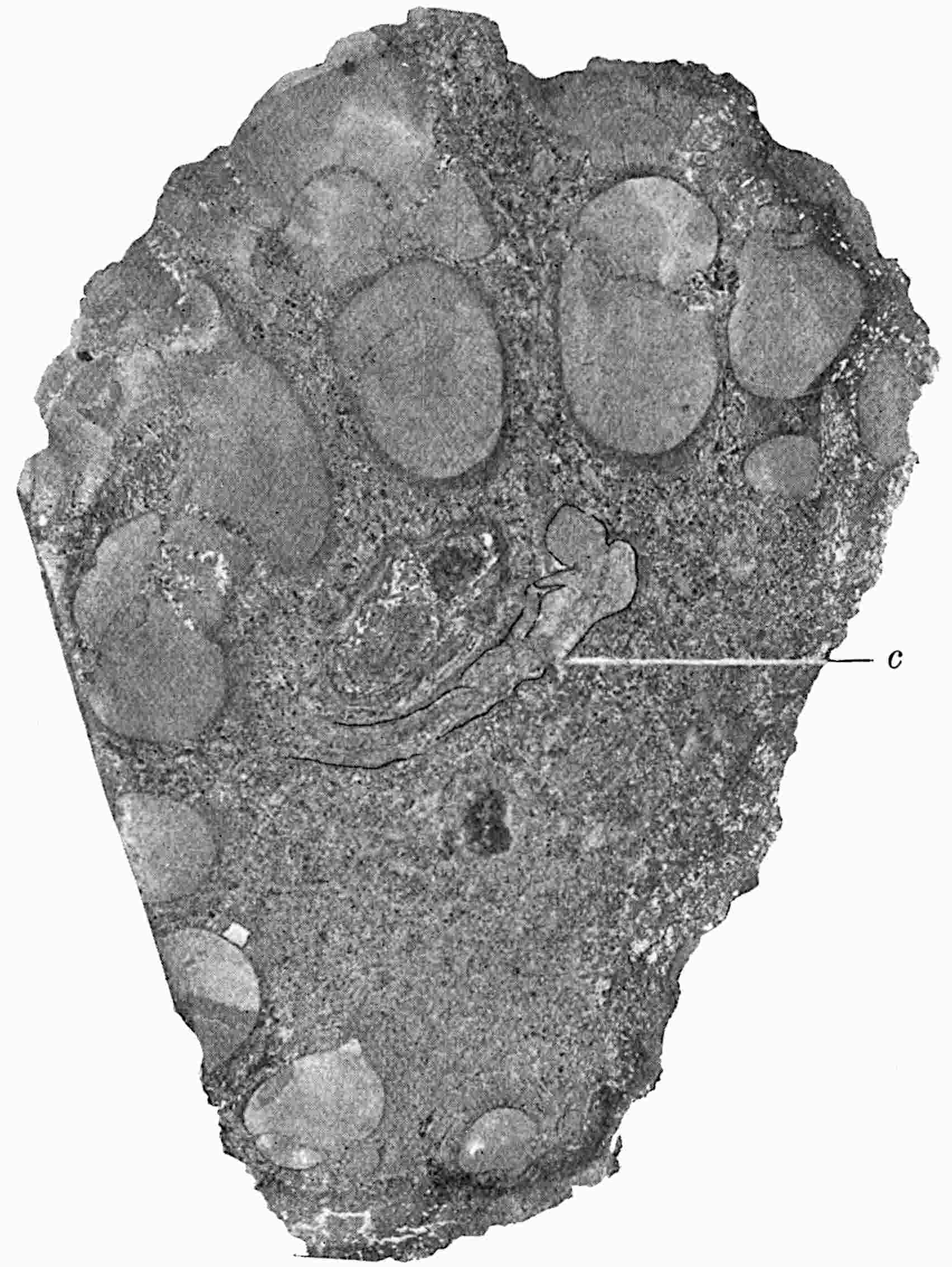
Fig. 437.
Rhexoxylon africanum
. Transverse section of stem showing peripheral steles and central (
c
) stele. (After Bancroft; × 2.)
RHEXOXYLON. Bancroft.
Rhexoxylon africanum Bancroft. The genus Rhexoxylon was instituted for a new type of stem represented by a single incomplete specimen from the Karroo series of South Africa: its precise geological horizon is not known but it may be referred provisionally to the lower or Palaeozoic portion of the series. Though our knowledge of the morphological features of the type-species is far from complete owing in part to the method of preservation of the specimen and in part to the destruction of the outer portion of the vascular tissue and the whole of the cortex, Miss Bancroft’s careful description[407] demonstrates the existence of characters which justify the employment of a new generic name. Rhexoxylon is more nearly related to the Medulloseae than to any other group and is particularly interesting as the first recorded example of this group from the Southern Hemisphere.
Fig. 437 shows a transverse section (7 × 5 cm.) of the stem. The ground-tissue consists of fairly large-celled parenchyma with sclerous nests and a few bands of periderm. At the periphery of the stem are radially disposed groups of vascular tissue varying in size and to some extent in shape. Unfortunately the stem is incomplete and it is impossible to say how much vascular or other tissue originally existed beyond the present corroded edge. The vascular groups, or steles as they may legitimately be called, follow a vertical course through the length of the block (6·5 cm.) and afford only slight evidence of branching or anastomosing. A close examination of the steles shows that they consist of portions of two series, an inner and outer set; there is also a curved vascular band in the central ground-tissue (fig. 437, c) and some isolated and scattered patches of vascular elements. Each stele of the inner series is made up of two parts, an outer smaller and normally orientated group of secondary xylem and a larger inversely orientated inner group of identical structure. A single stele of the inner series is shown in fig. 438, B, C; the larger inner portion consists of slightly divergent rows of tracheids and uniseriate medullary rays and is separated from the smaller portion by a narrow space, a, occupied by crushed tissue which may correspond to the ‘partial pith’ or primary xylem of a Medullosan stele. The two groups of xylem are no doubt the products of two cambium arcs, the protoxylem of each group being situated on the flat inner face. The cambium and phloem are represented only by crushed brown cells on the curved outer edge of the xylem. The separate individuality of the two portions of each stele is indicated not only by the presence of the ‘partial pith’ but by the discontinuity of the tissue at the ends of the narrow space. The tracheids seen at b, fig. C, are in oblique longitudinal section and are probably being detached to form a leaf-trace. This type of stele may be compared with the steles of Medullosa Solmsi (fig. 416, L) but those of Rhexoxylon differ in the lack of continuity of the secondary xylem round the narrow band of crushed primary xylem. The other steles of the inner ring exhibit the same dual nature though with local modifications. In the stele seen in fig. 438, B, there is a close approach to a continuous cylinder of secondary xylem especially on the right-hand side. External to the inner series are several portions of normally orientated secondary xylem-groups (fig. 437): these probably represent a second series of steles separated from the inner series by a narrow crushed arc of tissue on which the protoxylem strands of the outer groups abut. The xylem of the outer steles agrees in its normal orientation with the outer and smaller part of the inner steles and, as there is no accompanying group of inversely orientated xylem corresponding to the larger mass of secondary xylem of the inner series, the outer strands are designated partial steles. The central stele consists of two curved irregular bands composed of vertically and obliquely running tracheids: the central part of this stele consists of crushed tissue that probably represents primary xylem like that between the two parts of each of the peripheral steles.
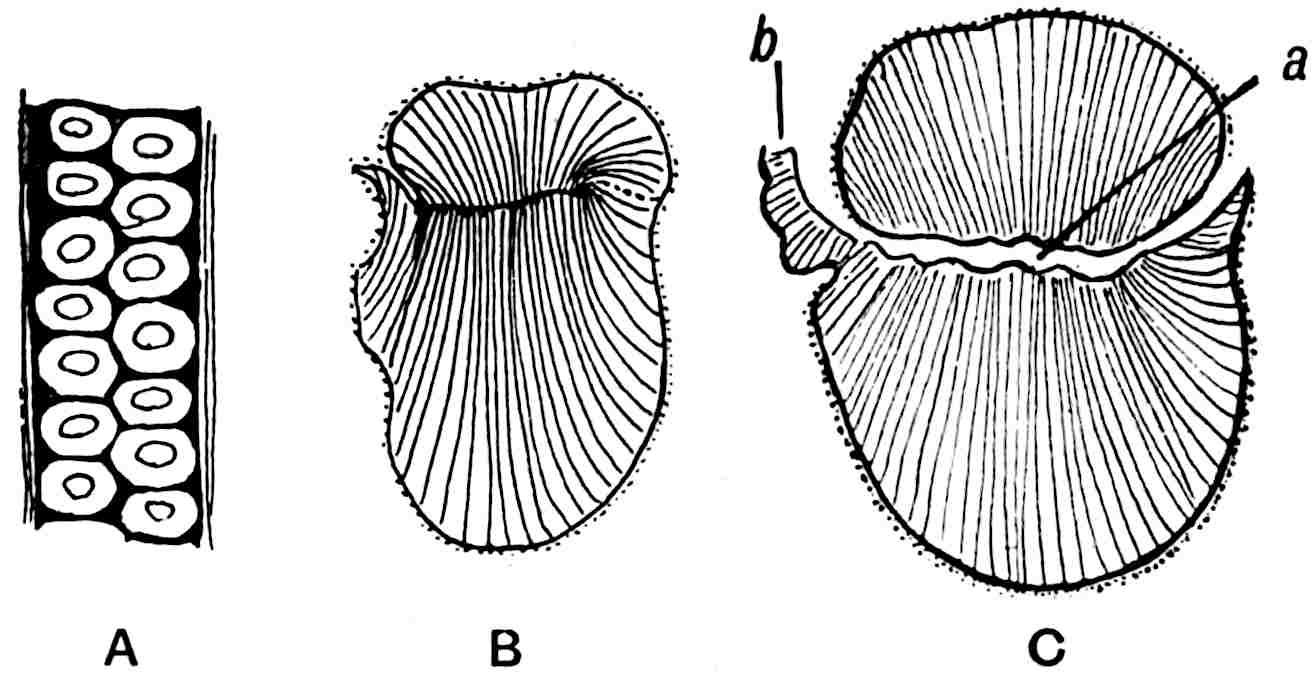
Fig. 438.
Rhexoxylon africanum
. A, radial wall of tracheid; B, C, peripheral steles;
a
, primary portion of stele;
b
, lateral strand. (After Bancroft.)
Rhexoxylon differs from the usual Medullosan type in the structure of the secondary xylem which is composed of tracheids with an Araucarian form of pitting: there are usually two alternate rows of contiguous pits (fig. 438, A) and occasionally one or three rows. The medullary rays are uniseriate and 3 to 15 cells in depth, a feature characteristic of coniferous wood and not of the wood of the Medulloseae.
In the absence of more complete information as to the anatomical structure of this stem and of all information as to the leaves or reproductive organs it is impossible to fix with precision the systematic position of the genus. It is, however, clear that Rhexoxylon is closely connected with Medullosa in certain features though in the structure of the wood it exhibits important peculiarities. The imperfectly known stem Cladoxylon Kidstoni[408] shows a fairly close agreement with the African plant in the form of the steles (fig. 460) but the pitting is scalariform.
SUTCLIFFIA. Scott.
Sutcliffia insignis Scott.
The generic name Sutcliffia was given by Dr Scott[409] to a peculiar type of stem from the Lower Coal Measures of Shore, Lancashire, in recognition of the valuable services rendered to Palaeobotany by the late Mr W. H. Sutcliffe, the owner of the colliery from which several new types of plants have been obtained. Two stems are recorded, the type-specimen and a more recently discovered stem, also from Shore, described by Miss de Fraine[410], which differs in several particulars from Dr Scott’s species. In view of the well-marked peculiarities of the second stem it is convenient to speak of it as forma β instead of including it without a distinctive epithet in Sutcliffia insignis. We know nothing of the reproductive organs of the genus.
1. Sutcliffia insignis, forma α.
This consists of a piece of stem approximately 12 × 7 cm. in diameter characterised by a broad cortex of parenchyma with secretory sacs and ducts and strands of mechanical tissue (fig. 439, A). Decurrent and massive leaf-bases form a prominent feature as in the stem of Medullosa anglica. The stele, though compressed before petrifaction, was probably not quite cylindrical but more or less polygonal or broadly triangular in section; it consists of groups of large primary tracheids (350μ in diameter) with numerous bordered pits (fig. 439, B, and fig. 440) embedded in an anastomosing system of parenchyma containing scattered secretory sacs, a type of protostele like that of Heterangium and Medullosa anglica except in the possession of exarch protoxylem strands. The metaxylem tracheids contiguous to the external protoxylem elements have a dense spiral or scalariform type of pitting. In the lower part of the stem the primary xylem is enclosed by a cambium which has added a few secondary tracheids (120μ in diameter), but in the upper part of the specimen the cambium is only partially developed and the addition of secondary xylem has hardly begun (fig. 440). A narrow band of secondary phloem was recognised in places consisting of small-celled parenchyma with some sieve-tubes and medullary rays continuous internally with the parenchyma of the primary stele. In close association and occasionally in organic connexion with the surface of the stele are several tangentially elongated and large groups of vascular tissue associated with smaller oval strands varying considerably in size. These groups, designated meristeles (fig. 439, A), are identical in structure with the main stele and are occasionally invested by a feebly developed zone of secondary xylem and phloem. The meristeles are detached at intervals from the parent stele around which they form by anastomoses an irregular network: the larger meristeles give off smaller strands and from these the actual leaf-traces are produced by subdivision. It appears, however, that in this type the meristeles are not completely used up in the production of the leaf-traces, portions of them behaving as cauline vascular strands. A protoxylem of a meristele still attached to the central protostele occupies an internal position, and at a higher level, as separation of the meristele is effected, the spiral tracheids occur on the inner face. New meristeles are given off at intervals from the main stele ‘to compensate for those parts of the reticulum which were used up in the formation of leaf-trace strands[411].’ The meristeles form the starting-point for the leaf-traces, an intermediate system between the main stele and the actual leaf-traces; they differ, therefore, from the parent leaf-traces of Medullosa anglica, which are completely used up by repeated subdivision. Moreover in Sutcliffia the leaf-bundles are concentric and not collateral.
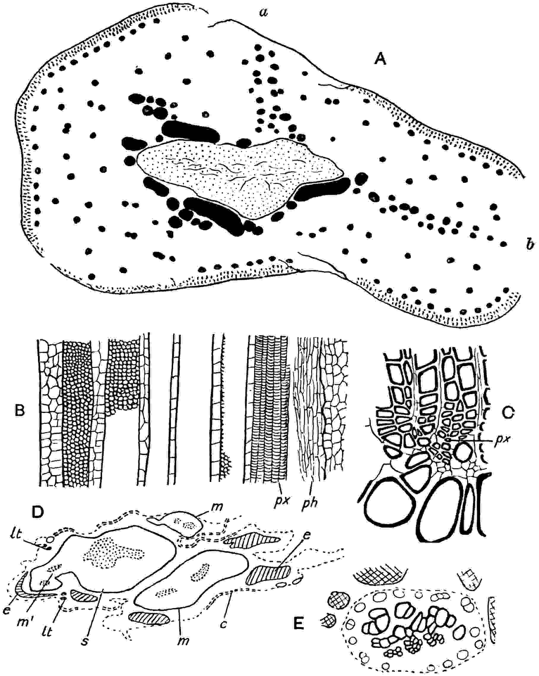
Fig. 439.
Sutcliffia insignia Scott. A, B, E, forma α. (After Scott.) C, D, forma β. (After de Fraine.)
A. Transverse section of stem; a, b, double rows of leaf-trace bundles. The larger black masses are the meristeles; the smaller patches represent bundles derived from the meristeles.
B. Longitudinal section of a radially symmetrical bundle from a leaf-base.
C. The junction between the primary and secondary xylem.
D. Vascular system in transverse section: m, m′, meristeles; s, main stele; e, extrafascicular strands; lt, leaf-traces; c, secondary cortex.
E. Vascular bundle from petiole showing a ring of large sieve-tubes surrounding the xylem, and portions of stereome strands.
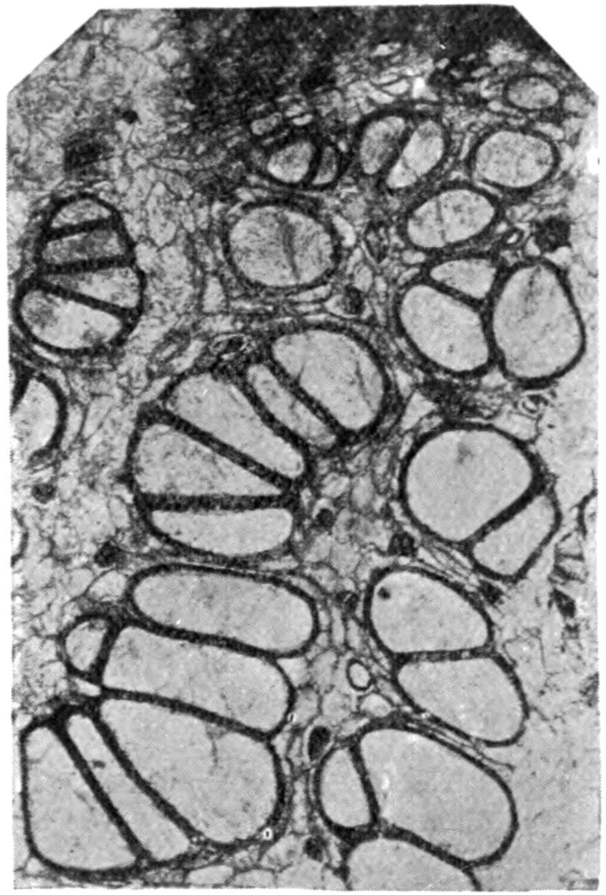
Fig. 440.
Sutcliffia insignis.
Part of the primary xylem showing on the upper edge the beginning of secondary growth. (Cambridge Botany School, 560.)
A conspicuous feature of the stem of forma α is the occurrence of two double rows of vascular strands stretching across the cortex (fig. 439, A, a, b). These are interpreted by Scott as downward continuations in the stem of the inner surface of leaf-bases. The outer cortex of the stem and leaf-bases has hypodermal strands of stereome which remain separate or rarely anastomose, and form a superficial zone exactly like that of some species of Medullosa. The leaf-trace bundles may be radially symmetrical or unilateral in the arrangement of the xylem which is in all cases completely surrounded by phloem. Fig. 439, B, shows part of a longitudinal section of a large leaf-trace bundle: spiral protoxylem elements (px) abut on the phloem (ph) and are succeeded to the left by narrow scalariform and large reticulately pitted tracheids. In the larger and radially constructed traces there are several protoxylem-strands distributed over the surface of the xylem, while in the smaller unilateral traces there may be one or two protoxylem strands. A characteristic feature of the xylem of the leaf-traces is the admixture of parenchyma with the tracheids (fig. 439, B, E) and another noteworthy character is the occurrence of large thin-walled tubes in the phloem described by Scott as sieve-tubes and compared with the large sieve-tubes in Marattiaceous leaf-bundles. Immediately internal to the hypoderm is a row of leaf-bundles (fig. 439, A) each of which is accompanied by stereome strands.
The petioles, which reach a diameter of 12 cm., contain numerous, occasionally anastomosing, concentric bundles. Nothing is known of the fronds as a whole beyond the fact that they are spirally disposed and had decurrent bases of large dimensions in proportion to the stem.
Sutcliffia insignis, forma β.
This form, described by Miss de Fraine as Sutcliffia insignis[412], is represented by a stem rather smaller than the type-specimen described by Scott, though it is probably an older example of the same species. It is distinguished by a greater development of secondary xylem and phloem both on the main stele and the meristeles; it differs also in the absence of the greater part of the cortex and leaf-bases which have been cut off as the result of the formation of a deep-seated periderm (fig. 439, D, C). The meristeles are smaller and fewer than in the larger form and are distinguished by some other peculiarities. At m′ in fig. 439, D, a meristele is seen attached to the main stele. In Sutcliffia insignis forma α the main stele is enclosed by an irregular network of subsidiary steles or meristeles and these form the points of departure of the leaf-traces, but the meristeles are not completely used up in the process of conversion into leaf-traces. In Sutcliffia insignis forma β the meristeles agree in structure with the main stele except in the smaller amount of secondary tissue: they do not, however, form a network as in forma α but occur as strands parallel to the central stele, ‘giving off leaf-traces and ultimately dividing up into smaller strands, often unequal in size, the primary wood of the meristeles being entirely used up in the production of radially symmetrical or unilateral bundles.’ In this respect, as Miss de Fraine points out, forma β agrees more closely than forma α with Medullosa anglica and supports Scott’s view that Sutcliffia is a primitive type of Medullosan stem. The leaves were given off at fairly long intervals as in some species of Medullosa: the leaf-traces are exarch and similar to those of the type-species. The most striking feature of the new stem is the presence of a vascular network (fig. 439, D, e) which encloses both the main steles and the meristeles; it consists of extrafascicular strands composed of normally orientated bands of secondary xylem and phloem often assuming a fan-like arrangement and occasionally almost concentric or inversely orientated. These strands are always accompanied by short, usually reticulate, tracheids on the inner margin of the xylem: similar isodiametric tracheids also occur in the pericyclic region. The extrafascicular strands are believed to be secondary structures phylogenetically independent of the meristeles and main stele, comparable with the successive cylinders or arcs of secondary xylem and phloem in some recent Cycads and in some species of Medullosa[413].
The stele is exarch and roughly triangular; except in the broader zone of secondary tissue it agrees with the protostele of forma α. The secondary xylem (fig. 439, C) possesses numerous medullary rays 3–4 cells broad and of considerable depth: the secondary phloem is characterised by the presence of thick-walled elements, presumably sieve-tubes, like those of Medullosa Leuckarti and M. anglica.
The ground-tissue is rich in secretory tissue and the stem-surface, from which the leaf-bases have been detached, is limited by a wide zone of secondary tissue produced by a phellogen.
Sutcliffia Williamsoni
(Seward).
1876. Myelopteris (pars) Williamson, Phil. Trans. R. Soc. Vol. 166, Pl. ii. figs. 7, 8; Pl. iv. fig. 17.
1893. Rachiopteris Williamsoni Seward, Ann. Bot. Vol. vii. p. 1.
1894. Rachiopteris Williamsoni Seward, ibid. Vol. viii. p. 207, Pl. xiii.
1906. Sutcliffia Williamsoni Scott, Trans. Linn. Soc. Vol. vii. pt iv. p. 62.
In an account of Myelopteris (= Myeloxylon) published in 1876 Williamson included some sections of petioles from the Lower Coal Measures which I afterwards with his concurrence transferred to the genus Rachiopteris as R. Williamsoni. In Rachiopteris Williamsoni the vascular bundles are concentric and not collateral, and are further distinguished from those of Myeloxylon by the association of parenchyma with the tracheids. In the arrangement of the bundles and in the structure of the ground-tissue the petioles of Rachiopteris Williamsoni agree with those of Myeloxylon. An outstanding feature of the vascular strands of the former is the occurrence at fairly regular intervals in the peripheral part of the phloem of comparatively large tubes described by me as secretory canals on the ground that evidence was furnished of their development by the schizogenous separation of cells to form a central canal. The canals were compared with the large sieve-tubes of the Marattiaceae, but with the qualification that ‘their mature form and their manner of development are strongly suggestive of small secretory canals.’ Nothing was known as to the stem which bore these petioles until Scott’s discovery of Sutcliffia with vascular bundles in the cortex and leaf-bases of the same type as those of Rachiopteris Williamsoni. As Scott says, there are a few distinguishing features which suggest that R. Williamsoni is not specifically identical with the petioles of Sutcliffia insignis, though the agreement is such as to justify the substitution of Sutcliffia for Rachiopteris. The tubular elements in the phloem which I considered to be secretory canals are regarded by Scott and Miss de Fraine as sieve-tubes.
Miss de Fraine gives an excellent summary of our knowledge of the genus and discusses, in the light of the additional facts furnished by the second stem, the position of Sutcliffia in a phylogenetic series. The new form strengthens the comparison instituted by Scott between Sutcliffia and Medullosa and gives support to a close connexion between the Medulloseae and the Cycadaceae. Scott lays stress on the fact that the Medulloseae, except Sutcliffia, are polystelic, while the Cycads, at least the adult stems, are monostelic. The views of Worsdell and other botanists who believe that the Medulloseae and the Cycads are intimately related are discussed on another page: in reference to these views Miss de Fraine adds a caveat as to the danger of attaching excessive importance to evidence based on seedling anatomy when we are concerned with broad phylogenetic questions. The stele of a cycadean stem may be derived from a protostelic type such as that of Sutcliffia by the gradual disappearance of the internal tracheids: in Sutcliffia, as in some Cycads and species of Medullosa, extrafascicular strands and cylinders are a characteristic feature and these may well have arisen independently of the central stele in response to physiological requirements. From such a type as Sutcliffia evolution may have proceeded along two lines; in one direction new types were produced in which increasing complexity, as represented by a multiplication of steles, was an outstanding feature. These forms, illustrated by Medullosa anglica and other more elaborate species, proved inefficient and were unproductive. Along another line the protostelic condition was maintained though in some cases extrafascicular strands or cylinders and cortical steles were superadded: it was this line that led to the recent Cycads.
General considerations suggested by the anatomical features of Medullosa.
A comparison of the stems described under the generic name Medullosa reveals a considerable range in the grosser anatomical features superadded to certain fundamental characters denoting a common origin[414]. If additional data were available giving us a fuller knowledge of individual plants differences between species would be more clearly defined and would provide adequate grounds for the institution of new genera for some of the types now included in the comprehensive genus Medullosa. A proposal by Dr Lotsy[415] to adopt the names Pecopteromedullosa and Neuropteromedullosa rests primarily on the relatively unimportant difference between the fronds associated with certain Medullosan stems and, as Scott[416] points out, this two-fold division if applied to such a species as Medullosa Leuckarti would result in its separation from species which anatomically are clearly of the same generic type. In Medullosa anglica, one of the oldest and simplest types, there are three steles of equal importance, and each of them is practically identical with the single stele of Heterangium. Each stele—as indeed all Medullosan steles—consists of a strand of primary xylem enclosed by secondary xylem and phloem, and it is obvious that the development of a constantly increasing cylinder of secondary conducting tissue about three centres would lead to serious mechanical difficulty: a stem constructed on the plan of Medullosa anglica or the smaller M. pusilla could not increase the thickness of its secondary vascular tissue beyond a certain point without detriment to its efficiency. In some types this difficulty is partially overcome by the production of complete concentric cylinders of centrifugally developed conducting tissue external to an inner system of concentric steles agreeing individually with those of Heterangium (fig. 415, B). Medullosa anglica, regarded from the point of view of the architectural efficiency of its vascular system, affords a much less promising point d’appui for further evolution than some of the forms described under Medullosa stellata in which the mechanical impasse is avoided by the adoption of the cycadean plan as represented by such genera as Cycas and Macrozamia. The English species Medullosa centrofilis (fig. 417) affords the first example of a characteristic Medullosan feature, namely the presence of a small concentric stele in the central region of the stem: this so-called star-ring differs not only in its smaller dimensions but in its more cylindrical form from the larger peripheral steles. In the later Permian species, e.g. Medullosa porosa and M. Solmsi, the single star-ring of the older M. centrofilis is replaced by a large number of precisely similar conducting strands. These star-rings are structurally comparable with the cortical steles of Cycas and, in position, with the medullary system of bundles in a Macrozamia; they are essentially cauline and take no part in the emission of leaf-traces. Medullosa Leuckarti (fig. 416, H) resembles in its vascular plan M. centrofilis, but in this larger stem there are several star-rings and the enlarged peripheral steles are more or less sinuous. In Medullosa Solmsi (fig. 416, E) the star-rings are still more numerous and the main vascular system consists of a double series of concentric steles, each agreeing with the larger peripheral steles of M. Leuckarti.
Some of the forms included in Medullosa stellata appear to be very different from M. anglica and M. Leuckarti (cf. fig. 416, F, A, H), but their similarity is apparent if we imagine Medullosa anglica with only one main stele (with the addition of star-rings) which is stretched tangentially until it becomes a long and narrow plate-ring and is then rolled into a hollow cylinder like that in fig. 416, D. A modification of the tubular type of stele is seen in Medullosa stellata var. corticata (fig. 416, G) in which the vascular cylinder is broken up into two or more curved plate-rings, a change superficially similar to that by which a dictyostele is produced from a solenostele, but in Medullosa it is not the overlapping of leaf-gaps that is the cause of the change. A striking feature in the stem of Medullosa stellata represented in fig. 416, D and F (especially F), is the inequality in breadth of the centrifugal and centripetal xylem: this inequality is an expression of the difficulty caused by the presence of an internal as well as an external addition of secondary conducting tissue. A limit is set to the production of secondary centripetal xylem by the space available for extension, whereas there is unlimited room for increase in the case of the centrifugal tissue. This tendency to a greater development of xylem and phloem on the outer side of the primary portion of the steles is illustrated also in M. Solmsi var. lignosa (fig. 416, L) where the outer of the two series of peripheral steles has a much larger proportion of centrifugal xylem. In M. anglica the secondary xylem on the inner side of the steles is not infrequently broader than the corresponding tissue on the abaxial side[417]; but this is exceptional in the genus. A further development of centrifugal conducting tissue without any corresponding development of centripetal tissue is effected in certain cases (fig. 416, K, L) by the addition of concentric cylinders of centrifugal xylem and phloem beyond the original concentric steles. The type illustrated by Medullosa Solmsi var. lignosa and M. stellata var. gigantea (fig. 416, L, K) at once suggests comparison with stems of Cycas, Macrozamia, and Encephalartos, the chief difference being the presence in Medullosa of an inner series of concentric steles and a central ground-tissue containing star-rings, though the latter may be regarded as corresponding to the medullary system of bundles in Macrozamia. In Macrozamia the central region of the stem is considered to be the pith of a monostelic stem, whereas in Medullosa the stem is polystelic. In recent cycadean stems it is not uncommon to find patches of inversely orientated xylem and phloem internal to one or more of the cylinders of centrifugal vascular tissue. These abnormal developments are considered by Worsdell[418] to be relics of the inner portions of concentric steles possessed by the Medullosan ancestors of recent Cycads. This interpretation affords a means of bringing into closer relationship the polystelic Medulloseae and the monostelic Cycadaceae, the apparent simplicity of the latter being the result of the progressive loss of centripetal xylem and phloem, the normal cycadean cylinder being therefore regarded as a one-sided remnant of a concentric Medullosan stele. In other words, the Cycads are descended from polystelic ancestors. As further evidence in support of this view Worsdell points to the occurrence of concentric steles in the cortex of Cycas and their occasional presence in the pith of other genera. Matte[419] has shown that in the seedling stem of Encephalartos Barteri (fig. 396, K) there are three concentric steles each similar to a normal Medullosan stele: at a higher level in the axis the steles become ‘unrolled’ and assume the form of one-sided cylinders of centrifugal xylem and phloem.
In the peduncles of some recent Cycads, e.g. Stangeria, there is a tendency towards a somewhat irregular orientation of the collateral bundles that constitute the vascular cylinder, and tracheids occasionally occur internal to the protoxylem of the individual bundles[420]. Worsdell regards these features as evidence of a Medullosan ancestry. If the sinuous plate-rings of a stem of Medullosa Leuckarti (fig. 416, H) were broken up into separate portions and wholly or in part deprived of the centripetal xylem, the result would be an arrangement of bundles comparable with that in a Stangeria peduncle[421]. The scattered centripetal tracheids discovered by Scott in Stangeria and other cycadean peduncles are interpreted by Scott and by Worsdell as relics of some ancestral centripetal xylem, but with this important difference in the point of view; Scott believes that they represent the almost completely aborted centripetal xylem of a single stele like that of Lyginopteris, while Worsdell sees in them fragmentary vestiges of the central primary xylem of two or more Medullosan steles. An abnormal seedling of Araucaria Bidwillii described by Shaw[422] exhibits features analogous to those in some cycadean seedlings: within the normal stele a cambium forms an inversely orientated vascular cylinder which at a lower level becomes continuous with the outer centrifugal tissue, the whole vascular system being eventually represented by two concentric steles. The polystelic stage is a development of a monostelic condition, and the inner or inverted portion of each of the two concentric steles is derived from an inversely orientated cylinder in the central region of the root. This abnormal root does not, however, supply an argument in favour of the derivation of a monostelic type of stem from one that was polystelic, but it shows a close relation between the two plans in one organ. The seedling is not altogether normal in form apart from structure and it is not improbable that the anatomical abnormality is connected with some pathological cause.
It has been suggested[423] that Worsdell attaches too much phylogenetic significance to the irregularities in the disposition and form of the vascular bundles in the peduncle of Stangeria, and the criticism that insufficient allowance is made for the possible reaction on structure of the special physiological requirements of reproductive shoots is well founded. Granting an overestimate of the arguments drawn from the occasional occurrence of concentric vascular strands, a considerable body of evidence remains in favour of Worsdell’s main contention.
Mrs Thoday[424] has drawn attention to certain features exhibited by the inflorescence-axes of Welwitschia, particularly the occurrence of concentric and inversely orientated bundles, similar to those characteristic of the seedling of Cycas siamensis[425] described by Matte, and to anatomical characters occasionally present in adult cycadean stems and normally represented in Medullosa. She is of opinion that the occurrence in Welwitschia of certain Medullosan features has a phylogenetic significance. The differences between the Gnetales, Medulloseae, and Cycadales are considerable, and it would seem unlikely that the anatomical resemblances described by Mrs Thoday are of great value as criteria of close relationship. The comparison of Lagenostoma with gnetalean seeds is alluded to elsewhere. To the statement that the presence of concentric and inversely orientated steles in Welwitschia are reminiscent of the polystelic Medulloseae, Mrs Thoday adds the qualifying remark that the occurrence of four concentric groups of vascular tissue in the hypocotyl of Welwitschia is not sufficient to justify the conclusion that the ancestral type was polystelic. This reservation accords with the contention of Scott and other botanists, that the occasional occurrence in cycadean seedlings and adult stems of anatomical features suggestive of polystely does not in itself furnish an adequate reason for doubting that the apparent monostely of Cycads is phylogenetically what it seems to be, namely, an indication of monostelic ancestry. This brings us to the question of a possible monostelic ancestor. It may be that the Upper Carboniferous genus Sutcliffia affords a clue to the problem of the origin of the polystelic type illustrated in various forms by Medullosa. The protostele of Sutcliffia bears a close resemblance to each of the three steles of Medullosa anglica; the fact that Sutcliffia is exarch and that Medullosa anglica has mesarch xylem is of secondary importance, particularly as exarchy is represented within the genus Medullosa. The extrafascicular strands of xylem and phloem and the accessory strands are points in which Sutcliffia and Medullosa anglica agree and, as Miss de Fraine[426] adds, the meristeles of Sutcliffia may be homologous with the leaf-trace strands of Medullosa. Scott[427] gave expression to the characters shared by these two types by describing Sutcliffia as the most primitive of the Medulloseae. It is suggested that the protostelic axis of Sutcliffia may be regarded as the starting-point of the monostelic Cycads, the central mass of tracheal tissue being replaced by a parenchymatous pith, while the extrafascicular and accessory strands arose independently of the central stele in response to increased physiological demands consequent on the increase in size of the stem. From the same starting-point evolution may have progressed along another line through such a type as Medullosa anglica leading to the more complex Permian species of Medullosa. Chodat’s view[428] that the Medulloseae are Protocycadaceae, if we include Sutcliffia as well as Medullosa in the Medulloseae, is probably correct. There is clear evidence of a close bond of union between recent Cycads and the Medulloseae, and Sutcliffia offers a possible means of deriving complex polystelic types from a monostelic ancestor.
Worsdell’s opinion[429] that the stele of Lyginopteris affords evidence of derivation from a polystelic ancestor and is not homologous with the true monostele of Heterangium is opposed to the undoubted signs of intimate connexion exhibited by these genera. The Lyginopterideae are, as Scott[430] says, a less advanced group than the Medulloseae and, it may be added, they are more remote from the modern representatives of the Cycadales. The Lyginopterideae and the Medulloseae are probably offshoots of a common stock, but the Medulloseae occupy a position farther removed from the filicinean ancestry than Heterangium and Lyginopteris[431].
The relative meagreness of our knowledge of the reproductive organs of the Medulloseae gives precedence to anatomical data in phylogenetic considerations, but the evidence furnished by Trigonocarpus and other seeds that may fairly be assigned to Medullosan plants is in harmony with the conclusions based on vegetative characters with regard to a close affinity between the Medulloseae and Cycads.
The comparative examination of recent Cycads naturally suggested by any attempt to compare the group as a whole with Palaeozoic types leads to some apparently contradictory results. The habit of the megasporophyll of Cycas is usually quoted as a primitive attribute: the close resemblance in plan and in manner of occurrence on the stem between megasporophylls and foliage leaves recalls both Ferns and Medullosan fronds. On the other hand the production of eight body-cells in the pollen-tube of Microcycas[432] in place of the usual single cell may also be regarded as a primitive character. It is perhaps possible, as Miss Dorety[433] says, that the polyspermy may be a case of recurrence and not a direct inheritance. Microcycas differs from Cycas in having only one vascular cylinder, and if the presence of several concentric cylinders in Cycas be interpreted as an indication of a closer connexion with a Medullosan ancestry, the Microcycas type would represent a more advanced stage in evolution. Attempts to arrange plants according to a natural sequence are frequently frustrated by instances of unequal progress in the development of vegetative and reproductive organs; one or other set of members lags behind; some characters point to the retention of primitive traits while others indicate a marked progressive tendency. It is noteworthy that the Mesozoic Bennettitales are characterised by a greater simplicity of stem-structure than is the rule in recent Cycads, and both in their vegetative features and in the structure of the seeds they are further removed from the Medullosan type.
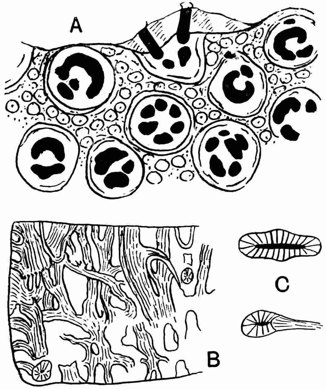
Fig. 441.
Steloxylon Ludwigii.
A, surface-view (longitudinal) of stem showing leaf-bases. (After Solms-Laubach.) B, longitudinal section showing anastomosing vascular strands. C, steles, the lower one showing the emission of a leaf-trace. (B, C, after Schenk.)
III. A. STELOXYLEAE.
Steloxylon. Solms-Laubach.
Steloxylon Ludwigii (Goeppert and Stenzel). The genus is founded on a piece of stem from Siberia, possibly of Permian age though not improbably older, which was originally described as Medullosa Ludwigii[434]. It is characterised by numerous cylindrical and band-like vascular strands forming an irregular anastomosing system (fig. 441, B) and by crowded spiral leaf-scars on the exposed face. The appearance presented by the transverse section figured by these authors, while suggesting comparison with Medullosa, reveals a distinctive character, namely the absence of a definite peripheral system of vascular rings such as forms a striking feature of the continental Medulloseae. A more complete description was afterwards published by Schenk[435] who recognised more fully the peculiar features and hinted at the possibility that the species might more appropriately be regarded as a member of a distinct group. Solms-Laubach[436] went a step further and instituted the generic name Steloxylon, and in a later publication gave a fuller account of the anatomical characters. The complete stem must have reached a diameter of approximately 13 cm. The homogeneous ground-tissue forms a matrix enclosing an anastomosing vascular system of cylindrical or oval steles (fig. 441, C). Each strand consists of a band of secondary xylem tracheids with one or several rows of circular or oval bordered pits on the radial walls and narrow medullary rays usually 1–2 cells broad and 1–4 cells deep, though occasionally deeper. No phloem is preserved. The tissue in the centre of each stele is very imperfectly preserved, but it is clear that the secondary xylem enclosed a central region (‘partial pith’) like that in the steles of a Medullosa, doubtless consisting of primary xylem and conjunctive parenchyma.
The stem is covered with leaf-bases of oval or circular section and between them are small organs, probably multicellular hairs (fig. 441, A). A leaf-base consists of an outer zone of strengthening tissue and a parenchymatous ground-tissue traversed by two or more small vascular strands which assume various forms. These petiolar strands are simply portions of the main vascular system which bend outwards at the periphery of the anastomosing network. The more noteworthy features in which Steloxylon differs from Medullosa, particularly such species as M. stellata and M. Leuckarti, are (i) the crowded and comparatively small leaf-bases in place of the massive decurrent petioles of Medullosa; (ii) the supply of the leaves by compact branches of the stelar network instead of the bundles detached as leaf-traces from a stem-stele of Medullosa (the origin of a leaf-trace in Steloxylon is shown in fig. 441, C); (iii) the absence of a peripheral system of vascular plate-rings and the irregular distribution of cylindrical and plate-steles in the ground-tissue. Nothing is known of the reproductive organs or leaves beyond the structure of the attached leaf-bases. The opinion expressed by P. Bertrand[437] that the fossil described by Stenzel as Asterochlaena (Clepsydropsis) kirgisica is the petiole of Steloxylon was abandoned after the additional facts published by Solms-Laubach.
As regards the affinities of Steloxylon: the structure of the steles agrees closely with that of the star- and plate-rings of a Medullosa, while the pitting of the tracheids is more like that in Medullosa than Cladoxylon. In the tendency to a more radial than tangential disposition of the band-like steles Steloxylon recalls Cladoxylon rather than Medullosa, but in Cladoxylon the vascular system does not form an irregular network as in Steloxylon. The information as to the structure of the primary xylem is very meagre, but it points to a closer connexion with Medullosa than with Cladoxylon. On the whole Steloxylon may perhaps be defined as a genus allied to the Medulloseae in the anatomical features of the stem more closely than to other genera, but sufficiently distinct to be excluded from the Medulloseae as at present understood[438].