Chapter VII
Reading edition
THALLOPHYTA.
The divisions of the plant kingdom dealt with in the following chapters of Volume I. are taken in their natural sequence, beginning with the lowest and passing gradually to the highest groups. The list of the classes and families included in Chapters VII.–XI. is given in the table of contents preceding Chapter I.
Thallophytes are of the simplest type, but they exhibit a very wide range as regards both the structure and differentiation of the vegetative body and the methods of reproduction. In some cases the individual consists of a minute simple cell which multiplies by cell-division; in others the body or thallus is made up of a number of similar units, while in a great number of forms there is a well-marked physiological division of labour, as expressed both in the external division of the thallus into distinct organs corresponding in function to the root, stem, and leaves of the higher plants, and further in the high degree of histological differentiation of the tissues. In other thallophytes, again, the thallus is a coenocyte either unseptate or incompletely septate; that is, the individual consists of a single cell differing from a true plant-cell, in the stricter sense of the term, in possessing several nuclei, in other words, the thallus is divided up into compartments by transverse septa, but each division contains more than one nucleus. Such coenocytic plants may show well-marked external differentiation of the thallus into members or parts subserving different functions.
A similar wide range is covered by the methods of reproduction among thallophytes.
I. PERIDINIALES.
The organisms included under this head are of little importance from a palaeontological point of view, but a brief reference may be made to them as a section of the Thallophyta.
The Peridiniales include very small single-celled organisms which have often been described as occupying a position on the borderland between animals and plants, lying on the “shadowy boundary between animal and vegetable life.” The individuals are rarely naked, more frequently they are covered with a cellulose or mucilaginous investment which has frequently the form of two or more minute armour-like plates of a limiting membrane. The chromatophores are green, yellow, brown or colourless. Simple division is the usual method of reproduction, but spores have been described as occurring in some species. The motile forms are provided with cilia. The Peridiniaceae, a section of the Peridiniales, are regarded as nearly related to the Diatoms.
The Peridiniales play an important rôle in the Plankton flora of the sea and freshwater lakes, and have a world-wide distribution. In the narrative of the Challenger cruise they are described as occasionally filling the tow-nets with a yellow coloured slime[165]. Some genera, such as Ceratium, are found in enormous numbers off the British coast.
As an example of the occurrence of fossil representatives of the Peridiniaceae reference may be made to one of two species of Peridinium described by Ehrenberg in 1836. These were found in a siliceous rock described as Cretaceous in age from Delitzsch in Saxony. A comparison of Ehrenberg’s figures of the fossil species Peridinium pyrophorum Ehrenb.[166], with those of the recent species Peridinium divergens Ehrenb., as given by Schütt[167] and other writers, brings out clearly the very close resemblance if not identity of the two forms. Bütschli[168] in his account of the Dinoflagellata in Bronn’s Thier-Reich confirms Ehrenberg’s determination of Peridinium pyrophorum, and points out its striking agreement with the recent species.
II. COCCOSPHERES AND RHABDOSPHERES.
(Organisms of doubtful affinity.)
Our knowledge of these minute calcareous organisms is derived from Huxley’s description of coccoliths from the Atlantic in 1857, and from the accounts of Wallich, John Murray, and other writers. In the first volume of the narrative of the Challenger cruise[169] and in the volume on deep-sea deposits[170] these minute forms of life are figured and described. In the latter volume both genera are spoken of as extremely abundant in the surface waters of the tropical and temperate regions of the open ocean, and as forming an important constituent of the Globigerine ooze; they are said to occur entangled in the gelatinous substance of the Radiolarians, Diatoms, and Foraminifera, and are very common in the stomachs of Salps, Pteropods and other pelagic animals. Rhabdospheres are rare in regions where the temperature of the water sinks below 65° F.; the Coccospheres occur in tropical and temperate latitudes, and extend further north and south than the Rhabdospheres. As regards their botanical position, John Murray expresses the view that they are in all probability pelagic algae.
In the interesting memoir by Schütt on the Pflanzenleben der Hochsee[171] there occurs a short reference to the forms described in the Challenger Reports, but they were not obtained by the staff of the Hensen Plankton Expedition and Schütt’s remarks are not based therefore on personal observations. While admitting the existence of such bodies, he points out that Zoologists have referred Coccospheres and Rhabdospheres to the algae as organisms which cannot be included in any group of animals, and Schütt is unable to recognise a sufficient reason for referring them to this class of plants. It is suggested indeed that they may be purely inorganic structures.
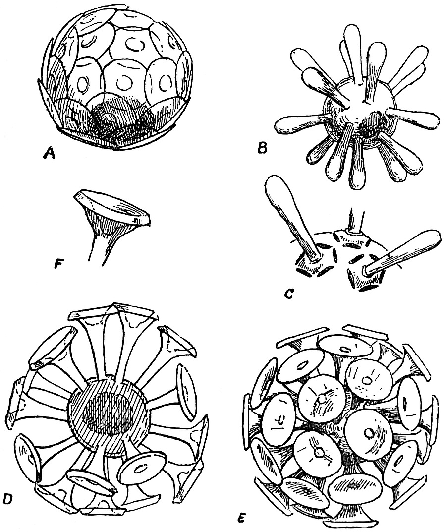
Fig.
25. (From Murray and Blackman).
A, Coccosphere × 1300. B, Rhabdosphere × 900. C, Portion of the same × 1300. D, Rhabdosphere of another type, in optical section × 1900. E, The same in surface view × 1900. F, End of one of the trumpet-shaped appendages of E.
The most recent account of these two genera is by Messrs G. Murray and Blackman a short notice in Nature for April 1, 1897[172]. Numerous examples of Coccospheres and Rhabdospheres were obtained by Capt. Milner of the R.M.S. Para during a voyage to Barbados by allowing the sea water to enter the feed-pipe of the boiler through a fine muslin net. All the forms described in the Challenger Reports were met with, and an examination of the material by means of extremely high objectives has confirmed the original account of the genera, and added some points to our previous knowledge.
Coccospheres (fig. 25 A). Spherical bodies of exceedingly small size, consisting of a central protoplasmic vesicle covered with overlapping circular calcareous scales, each of which is attached to the minute cell by a button-like projection. The scales are frequently found detached and are then spoken of as Coccoliths.
Rhabdospheres (fig. 25 B–F). Spherical bodies, extremely minute, consisting of a single cell, on the surface of which are embedded numerous calcareous plates bearing long blunt spines (fig. 25, C) or beautiful trumpet-like appendages (fig. 25, D–F). The detached plates of Rhabdospheres are known as Rhabdoliths.
In addition to the text-figures of Coccospheres and Rhabdospheres in the Challenger Reports, the same structures are shown in samples of globigerine ooze figured in Plate XI. of the Monograph on deep-sea deposits. In a recent number of Nature Messrs Dixon and Joly[173] have announced the discovery of Coccoliths and Coccospheres in the coastal waters off South County Dublin. They estimate that in one sample of water taken about three miles from the Irish coast there were 200 Coccoliths in each cubic centimetre of sea water.
The interest of these calcareous bodies from a palaeobotanical point of view lies in the fact that similar forms have been recognized in the Chalk and the Upper Lias. Sorby, in his memorable Address delivered before the Geological Society in 1879, refers to the abundance of Coccoliths in sections of chalk which he examined[174]. Rothpletz[175] has recently recorded the occurrence of numerous Coccoliths, 5–12 µ in diameter, associated with the skeleton of a horny sponge (Phymatoderma) of Liassic age.
The question of the nature of Coccospheres and Rhabdospheres cannot be regarded as definitely settled. It has been shown by J. Murray, and more recently by G. Murray and V. H. Blackman, that on the solution of the calcareous material by a weak acid there remains a small gelatinous body apparently protoplasmic in nature. We may at least express the opinion that Schütt’s suggestion as to their being inorganic must be ruled out of court. It would appear that they are extremely minute unicellular organisms characterised by a delicate calcareous armour consisting of numerous plates or scales. We know nothing as to their life-history, and cannot attempt to determine their affinities with any degree of certainty until further facts are before us. It is not improbable that they are algae of an extremely minute size, and the evidence so far obtained would lead us to regard them as complete individuals rather than the reproductive cells of some larger organism. Mr George Murray is of opinion that they are certainly algae, but he considers that they cannot be included in any existing family. It is conceivable that they may be minute eggs or reproductive cells of animals or plants, but on the whole the balance of probability would seem to be in favour of regarding them as autonomous organisms.
III. SCHIZOPHYTA.
SCHIZOPHYCEAE (Cyanophyceae).
SCHIZOMYCETES (Bacteria).
In this group are included small single-celled plants of an extremely low type of organisation, in which reproduction takes the form of multiplication by simple cell-division, or the formation of spores. The characteristic method of reproduction by division has given rise to the general term Fission-plants for this lowest sub-class in the vegetable kingdom. In many cases the members of this sub-class contain chlorophyll, and associated with it a blue-green colouring matter; such plants are classed together as the Blue-green algae, Cyanophyceae, or Schizophyceae. Others, again, are destitute of chlorophyll, and may be conveniently designated Schizomycetes or Fission-fungi. Seeing how close is the resemblance and relationship between the members of the sub-class, it has been the custom to include them as two parallel series under the general head, Schizophyta, rather than to incorporate them among the Algae and Fungi respectively.
A. SCHIZOPHYCEAE (Cyanophyceae or Blue-green Algae).
Chroococcaceae. Thallus of a single cell, the cells may be either free, or more usually joined together in colonies enveloped by a common gelatinous matrix, formed by the mucilaginous degeneration of the outer portion of the cell-walls. Reproduction by means of simple division or resting cells.
Nostocaceae. Thallus consists of simple or branched rows of cells in which special cells known as heterocysts often occur. Reproduction by means of germ-plants or hormogonia, or by resting cells specially modified to resist unfavourable conditions.
In both families the individuals are surrounded by a gelatinous envelope, which in some genera assumes the form of a conspicuous and comparatively resistant sheath. Marine, freshwater, and aerial forms are represented among recent genera. Several species occur as endophytes, living in the tissues or mucilage-containing spaces in the bodies of higher plants. In addition to the frequent occurrence of blue-green algae in freshwater streams and on damp surfaces, certain forms are particularly abundant in the open sea[176], and in lakes or meres[177] where they are the cause of what is known in some parts of the country as “the breaking of the meres” (“Fleurs d’eau”). From the narrative of the cruise of the Challenger, we learn that the Oscillariaceae are especially abundant in the surface waters of the ocean. The “sea sawdust” so named by Cook’s sailors[178], and the same floating scum collected by Darwin[179], affords an illustration of the abundance of some of these blue-green algae in the sea.
Another manner of occurrence of these plants has been recorded by different writers, which is of special importance from the point of view of fossil algae. On the shores of the Great Salt Lake, Utah, there are found numerous small oolitic calcareous bodies thrown up by the waves[180]. These are coated with the cells of Glœocapsa and Glœotheca, two genera of the Chroococcaceae. Sections of the grains reveal the presence of the same forms in the interior of the calcareous matrix, and it has been concluded, on good evidence that the algae are responsible for the deposition of the carbonate of lime of the oolitic grains. By extracting the carbonic acid which they require as a source of food, from the waters of the lake, the solvent power of the water is decreased and carbonate of lime is thrown down. In similar white grains from the Red Sea[181] there is a central nucleus in the form of a grain of sand, and cells of Chroococcaceae occur in the surrounding carbonate of lime as in the Salt Lake oolite. Prof. Cohn of Breslau in 1862 demonstrated the importance of low forms of plant life in the deposition of the Carlsbad “Sprudelstein[182].” On the bottom of Lough Belvedere, near Mullingar in Ireland[183], there occur numerous spherical calcareous pebbles, of all sizes up to that of a filbert. From a pond in Michigan (U.S.A.)[184] similar bodies have been obtained varying in diameter from one to three and a half inches. In the former pebbles a species of Schizothrix, one of the Nostocaceae occurs in abundance, in the form of chains of small cells enclosed in the characteristic and comparatively hard tubular sheath, and associated with Schizothrix fasciculata there have been found Nostoc cells and the siliceous frustules of Diatoms. In the Michigan nodules the same Schizothrix occurs, associated with Stigonema and Dichothrix, other genera of the Nostocaceae. One of the Michigan pebbles is shown in section in fig. 32 D.
OOLITIC STRUCTURE.
The connection between the well-known oolitic structure, characteristic of rocks of various ages in all parts of the world, and the presence of algal cells is of the greatest interest from a geological point of view. In recent years considerable attention has been paid to the structure of oolitic rocks, and in many instances there have been found in the calcareous grains tubular structures suggestive of simple cylindrical plants, which have probably been concerned in the deposition of the carbonate of lime of which the granules consist. In 1880 Messrs Nicholson and Etheridge[185] recorded the occurrence of such a tubular structure in calcareous nodules obtained from a rock of Ordovician age in the Girvan district of Scotland. These Authors considered the tubes to be those of some Rhizopod, and proposed to designate the fossil Girvanella.
Girvanella (fig. 26).
Messrs Nicholson and Etheridge defined the genus as follows:—
“Microscopic tubuli, with arenaceous or calcareous (?) walls, flexuous or contorted, circular in section, forming loosely compacted masses. The tubes, apparently simple cylinders, without perforations in their sides, and destitute of internal partitions or other structures of a similar kind.”
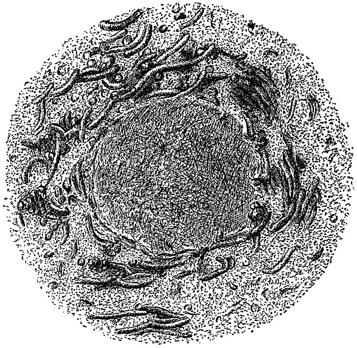
Fig. 26. Girvanella problematica, Eth. and Nich. Tubules of Girvanella lying in various positions and surrounding an inorganic ‘nucleus’ or centre. From a section of Wenlock limestone, May Hill. × 65
Since this diagnosis was published very many examples of similar tubular fossils have been described by several writers in rocks from widely separated geological horizons. The accompanying sketch (Fig. 26), drawn from a micro-photograph kindly lent to me by Mr Wethered of Cheltenham, who has made oolitic grains a special subject of careful investigation, affords a good example of the occurrence of such tubular structures in an oolitic grain of Silurian age from the Wenlock limestone of May Hill, Gloucestershire[186]. In the centre is a crystalline core or nucleus round which the tubules have grown, and presumably they had an important share in the deposition of the calcareous substance. The nature of Girvanella, and still more its exact position in the organic world, is quite uncertain; it is mentioned rather as à propos of the association of recent Cyanophyceae with oolitic structure, than as a well-defined genus of fossil algae.
In the typo description of the calcareous nodules from Michigan, Murray speaks of the Schizothrix filaments at the surface of the pebbles as fairly intact, while nearer the centre only sheaths were met with. It is conceivable that in some of the tubular structures referred to Girvanella we have the mineralised sheaths of a fossil Cyanophyceous genus[187]. The organic nature of these tubules has been a matter of dispute, but we may probably assume with safety that in some at least of the fossil oolitic grains there are distinct traces of some simple organism which was in all likelihood a plant. Some authors have suggested that Girvanella is a calcareous alga which should be included in the family Siphoneae[188]. As a matter of fact we must be content for the present to leave its precise nature as still sub judice, and while regarding it as probably an alga, we may venture to consider it more fittingly discussed under the Schizophyta than elsewhere.
Wethered[189] would go so far as to refer oolitic structure in general to an organic origin. While admitting that a Girvanella-like structure has been very frequently met with in oolitic rocks, it would be unwise to adopt so far-reaching a conclusion. It is at least premature to refer the formation of all oolitic structure to algal agency, and the evidence adduced is by no means convincing in every case. The discovery of Girvanella and allied forms in rocks from the Cambrian[190], Ordovician, Silurian, Carboniferous, Jurassic and other systems is a striking fact, and lends support to the view that oolitic structure is in many cases intimately associated with the presence of a simple tubular organism. Among recent algae we find different genera, and representatives of different families, growing in such a manner and under such circumstances as are favourable to the formation of a ball-like mass of algal threads, which may or may not be encrusted with carbonate of lime. Similarly as regards oolitic grains of various sizes, and the occurrence in rocks of calcareous nodules, the tubular structure is not always of precisely the same type, and cannot always be included under the genus Girvanella.
Several observers have recorded the occurrence of low forms of plant-life in the waters of thermal springs. It has been already mentioned that Cohn described the occurrence of simple plants in the warm Carlsbad Springs, and fission-plants of various types have been discovered in the thermal waters of Iceland, the Azores[191], New Zealand, the Yellowstone Park, Japan, India, and numerous other places.
A few years ago Mr Weed, of the geological survey of the United States, published an interesting account of the formation of calcareous travertine and siliceous sinter in the Yellowstone Park district[192]. This author emphasizes the important rôle of certain forms of plants in the building up of the calcareous and siliceous material. Among other forms of frequent occurrence, Calothrix gypsophila and a species Leptothrix are mentioned, the former being a member of the Nostocaceae, allied to Rivularia, and the latter a genus of Schizomycetes. In many of the springs there are found masses of algal jelly like those previously described by Cohn in the Carlsbad waters. Sections of such dried jelly showed a number of interlaced filaments with glassy silica between them. Weed refers to the occurrence of small gritty particles in this mucilaginous material. These are calcareous oolitic granules which are eventually cemented together into a compact and firm mass of travertine by the continued deposition of carbonate of lime. The presence of the plant filaments is often difficult to recognise in the “leathery sheet of tough gelatinous material,” or in “the skeins of delicate white filaments” which make up the travertine deposits.
BORINGS IN SHELLS.
Under the head of Cyanophyceae, mention should be made of the recent genus Hyella[193], which occurs as a perforating or boring alga in the calcareous shells of molluscs. On dissolving the carbonate of lime of shells perforated by this alga, the latter is isolated and appears to consist of rows of small cells, with possibly some sporangia containing spores. Other boring algae have been recorded among the Chlorophyceae, and recently a member of the Rhodophyceae[194] has been found living in the substance of calcareous shells. Such examples are worthy of note in view of the not infrequent occurrence of fossil corals, shells and fish-scales, which have evidently been bored by an organism resembling in form and manner of occurrence these recent algal borers.
The occurrence of small ramifying tubes in recent and fossil corals, fish-scales, and bones was long ago pointed out by Quekett[195], Kölliker[196], Rose[197] and other writers[198]. These narrow tubular cavities have generally been attributed to the boring action of some parasitic organism, either a fungus or an alga. In 1876 Duncan published two important papers[199] dealing with the occurrence of such tubes in recent corals, as well as in the calcareous skeleton of Calceolina, Goniophyllum and other Palaeozoic, Mesozoic and Tertiary species of corals. This writer attributed the formation of the cavities in the case of the fossil species to the action of a fungus which he named Palaeachlya perforans, and considered as very nearly related to Achlya penetrans found in the “dense sclerenchyma” of recent corals. In fig. 27 A. is reproduced one of the drawings given by Rose[200] in his paper published in 1855; it shows a section of a fish-scale from the Kimeridge clay which has been attacked by a boring organism. Rose attributes the dichotomously branched canals to some “infusorial parasite.”
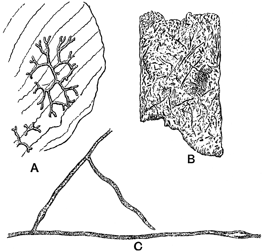
Fig. 27. A, Section of a fish-scale from the Kimeridge Clay, showing branched canals, made by a boring organism, × 85. B, Section of a Solen shell, penetrated in all directions by the boring thallus of Ostracoblabe (a fungus?), × 330. C, Piece of the thallus of Ostracoblabe isolated by decalcification, × 745. A, after Rose. B and C, after Bornet and Flahault.
In the important paper by MM. Bornet and Flahault on perforating algae a full description is given of various boring forms belonging to the Chlorophyceae and the Cyanophyceae[201]. The canals which these algae produce in calcareous shells and other hard substances are of the same type as those previously described in fossil corals, fish-scales and bones. In dealing with living perforating Thallophytes the colour and other cell-contents often enable us to distinguish between algae and fungi, but in fossil specimens such tests cannot be applied. The fossil tubular borings may or may not show traces of the transverse septa and reproductive cells; it is often the case that no trace of the organism has been left, but only the canals by which it penetrated the calcareous or bony skeleton. In some of the examples of Palaeachlya figured by Duncan there appear to be numerous spores in some of the sections, but it is generally a very difficult and often an impossible task to discriminate between the borings of fungi and algae in fossil material.
Fig. 27 B, which is copied from one of Bornet and Flahault’s drawings, represents a piece of Solen shell riddled with small canals made by the organism which has been named by the French authors Ostracoblabe implexa, and regarded by them as a fungus. Fig. 27 C represents a small piece of the vegetative body of Ostracoblabe obtained from a decalcified shell. In endeavouring to determine the organism which has produced borings in fossil corals or shells, it must be borne in mind that some forms of canals or passages may have been the work of perforating sponges, but these are larger in diameter than those made by algae or fungi. By some writers[202] the tubular cavities in shells have been referred to true algae, but others consider them to be of fungal origin.
As an example of a fossil alga referred to the Cyanophyceae, the genus Zonatrichites[203] may be quoted. Bornemann, who first described the specimens, points out the close resemblance in habit to some members of the recent Rivulariaceae.
Zonatrichites.
The author of the genus defines it as follows:—
“A calcareous alga, with radially arranged filaments, forming hemispherical or kidney-shaped layers, growing on or enclosing other bodies. Parallel or concentric zones are seen in cross-section, formed by the periodic growth of the alga, the older and dead layers serving as a foundation on which the young filaments grow in radially arranged groups.”
The nodules which are apparently formed by species of this genus occur in various sizes and shapes; Bornemann describes one hemispherical mass 8 cm. broad and 4 cm. thick. In some cases the organism has given rise to oolitic spherules, which in radial section exhibit the branched tubular cells spreading in fan-shaped groups from the centre of the oolitic grain. The section parallel to the surface of a nodule presents the appearance of a number of circular or elliptical tubes cut across transversely or more or less obliquely. The resemblance between the fossil and a specimen of the recent species Zonatrichia calcivora Braun, is certainly very close, but it is very difficult, in the absence of material exhibiting more detailed structure than is shown in the specimens described by Bornemann, to decide with any certainty the true position of the fossil. The figures do not enable us to recognise any trace of cells in the radiating tubes. It is possible that we have in Zonatrichites an example of a Cyanophyceous genus in which only the sheaths of the filaments have been preserved. In any case it is probable that this Mesozoic species affords another instance of a fossil alga which has been responsible for certain oolitic or other structures in limestone rocks.
The species described by Bornemann was obtained from a Breccia near Lissau in Silesia, of Keuper age.
M. Renault has recently described certain minute structures in a Palaeozoic coprolite to which he gives the name Gloioconis Borneti[204], and which he regards as a Permian gelatinous alga similar to the well-known recent genus Glœocapsa. The appearances revealed in a section of the coprolite are interpreted by this author as a collection of small colonies of a unicellular gelatinous alga in various stages of development. Renault’s figure shows a spherical group of faintly outlined and cloudy bodies, most of which include one or two small dark spots. The latter are regarded as the cells of the alga, and the surrounding cloudy substance is described as the gelatinous sheath. The absence of a nucleus in these extremely minute fossil cells (8–10 µ in diameter) is referred to as an argument in favour of referring the organism to the Cyanophyceae rather than to the Chlorophyceae. It is possible that the ill-defined structure described by Renault may be a petrified alga, but there is not sufficient evidence to warrant a decided opinion; the absence of nuclei can hardly be taken seriously in such a case as this as an argument in favour of the Cyanophyceae.
CYANOPHYCEAE.
Although our exact knowledge of fossil Cyanophyceae is extremely small, it is probable that such simple forms of plants existed in abundance during the past ages in the earth’s history. Several writers have expressed the opinion that the blue-green algae may be taken as the modern representatives of those earliest plants which first existed on an archaean land-surface. The living species possess the power of resisting unfavourable conditions in a marked degree, and are able to adapt themselves to very different surroundings. Their occurrence in hot springs proves them capable of living under conditions which are fatal to most plants, and suggests the possibility of their occurrence in the heated waters which probably constituted the medium in which vegetable life began. An interesting example of the growth of blue-green algae under unfavourable conditions was recorded in 1886 by Dr Treub[205] of the Buitenzorg Gardens, Java. In 1883 a considerable part of the island Krakatoa, situated in the Straits of Sunda, between Sumatra and Java, was entirely destroyed by a terrific volcanic explosion. What remained had been reduced to a lifeless mass of hot volcanic ashes. Three years later, Treub visited the island, and found that several plants had already established themselves on the volcanic rocks. Various ferns and flowering plants were recorded in Treub’s description of this newly established flora. It seemed that the barren rocky surface had been prepared for the more highly organised plants by the action of certain forms of Cyanophyceae, which were able to live under conditions which would be fatal to more complex types.
In the petrified tissues of fossil plants there are occasionally found small spherical vesicles, with delicate limiting membranes, in the cavities of parenchymatous cells or in the elements of vascular tissue. Some of these spherical inclusions have been described as possibly simple forms of endophytic algae[206], such as we are now familiar with in species of the Cyanophyceae and other algae. So far, however, no recorded instance of such fossil endophytic algae is entirely satisfactory. Some of the cells figured by Williamson as possibly algae, endophytic in the tissues of Coal-Measure plants, are no doubt thin-walled vesicles which formed part of a highly vacuolated cell-contents. Examples of such vesicles in living and fossil cells are shown in fig. 42. The fact that the contents of living plant tissues have been erroneously described as endophytic organisms, should serve as a warning against describing fossil endophytes without the test of good evidence to support them.
The description of a fossil Nostoc by the late Prof. Heer[207] from the Tertiary rocks of Switzerland cannot be accepted as a trustworthy example of a fossil plant, much less of a genus of recent algae. The application of recent generic names to fossils which are possibly not even organic must do more harm than good.
B. SCHIZOMYCETES (Bacteria).
It is impossible to draw a sharp line between the two subdivisions of the Schizophyta. The so-called Fission-Fungi or Bacteria differ from the Schizophyceae or Fission-Algae in the cell-contents being either colourless, blood-red or green, but never blue-green. We may regard the Bacteria, generally, as the lowest forms of plants; they are extremely simple organisms which have been derived from some primitive types which possessed the power of independent existence and contained chlorophyll—that important substance which enables a plant to obtain its carbon first-hand from the carbon dioxide of the atmosphere.
Bacteria may be briefly described as single-celled plants, and as de Bary suggested comparable in shape to a billiard ball, a lead pencil or a corkscrew[208]. A single spherical or cylindrical cell measures about 1 µ in diameter[209]. They occur either singly or in filaments, or as masses of various shapes consisting of numberless bacterial cells. The nature and manner of life of Bacteria, and their extraordinary power of successfully resisting the most unfavourable conditions, render it probable that they constitute an extremely ancient group of organisms.
The wonderful perfection of preservation of many fossil plants enables us to investigate the contents of petrified cells and to examine in minutest detail the histology of extinct plants. To those who are familiar with the possibilities of microscopical research as applied to silicified and calcified fossil tissues, it is by no means incredible that evidence has been detected of the existence of Bacteria as far back in the history of the earth as the Carboniferous and Devonian periods.
Were there no trustworthy records of the occurrence of Bacteria in Palaeozoic times, it would still be a natural supposition that these ubiquitous organisms must have been abundantly represented. It has been suggested as a probable conclusion that some forms of Bacteria, which produced chemical changes in the soil necessary for the nutrition of plants, must have existed contemporaneously with the oldest vegetation[210].
The paper-coal of Toula, which in some places reaches a thickness of 20 cm., is a plant-bed of exceptional interest. It differs from ordinary coal in being made up of numberless thin brown-papery sheets associated with a darker coloured substance largely composed of ulmic acid. Prof. Zeiller[211], in an interesting account of the papery layers, has shown that they consist of the cuticles of a Lepidodendroid plant, Bothrodendron. An examination of a piece of one of the sheets at once reveals the existence of a regular network of which the walls of the meshes are the outlines of the epidermal cells, the meshes being bridged across by a thin light brown membrane which represents the layer of cuticularised cell-wall of each epidermal cell. At regular intervals and disposed in a spiral arrangement, we find small gaps in the papery cuticle which mark the position of the Bothrodendron leaves. These Palaeozoic cuticles are not petrified; they are only slightly altered, and have retained the power of swelling in water, being able to take up stains like recent tissues. It may reasonably be assumed that the persistent cuticles owe their preservation to a greater power of resistance to destructive agents than was possessed by the other tissues of the plant. It is by no means unlikely, as Renault[212] has recently suggested, that as the Bothrodendron stem-fragments lay in the swamps or marshes the tissues were gradually eaten away by Bacteria, but the cuticles successfully resisted the attacks of the bacterial saprophytes. The same observer has described what he regards as the actual organism which effected this wholesale destruction, under the name Micrococcus Zeilleri. He finds, after treating the cuticles with ammonia to remove the ulmic acid, that there occur numerous minute spherical bodies, each surrounded by a thin envelope, either singly or in groups on the surface of the cuticular membrane. These vary in size from ·5µ to 1µ in diameter. I have not been able to detect any satisfactory proof of such Micrococci in specimens of the paper-coal which were treated according to Renault’s method, but it is extremely probable that this unusual method of preservation of stem-cuticles is the result of selective bacterial action.
Renault believes that some of the minute spherulitic structures which are seen in sections of decayed tissues of Palaeozoic plants owe their origin, in part, to the ravages of bacteria. The disorganisation of parenchymatous cells gives rise to a gelatinous substance in which needle-like crystals of silica may be deposited, from a siliceous solution, in a matrix which has resulted from bacterial activity. In some of the sections of tissues figured by Renault[213] the outlines of a few cells are still indicated by fragments of the partially decayed wall, while in other cells the walls have been completely destroyed by Bacteria of which some are preserved in the centre of the cell-area, forming a kind of nucleus to the siliceous spherulites.
BACILLI.
In addition to the Micrococcus described by Renault from the Toula paper-coal, there are a host of other forms which have been minutely diagnosed and figured by Profs. Renault and Bertrand[214]. These authors have discovered what they believe to be well-defined species of Micrococcus and Bacillus ranging in age from Devonian to Jurassic. The material which has afforded the somewhat startling results of their investigations consists partly of the coprolites of reptiles and fishes, and of silicified and calcified plant tissues.
Bacillus Permicus. Ren. and Bert.[215] (Fig. 28 B.)
This Bacillus, which was discovered in sections of a Permian coprolite from Central France, has the form of cylindrical rods 12–14µ in length, and 1·3–1·5µ broad, rounded at each end. The rods occur either singly or occasionally, two or three individuals are joined end to end. Fig. 28 B represents a piece of one of Renault and Bertrand’s sections; the small rods are clearly seen lying in various directions in the homogeneous matrix of the coprolite. Each individual is said to be surrounded by an extremely minute empty space ·4µ in width, originally occupied by the Bacillus membrane, the central rod representing the mineralised cell-contents. In this example the petrifying substance was probably derived from the phosphate of calcium of bones which were attacked by Bacteria. I am indebted to Prof. Renault for an opportunity of examining specimens of this and other fossil Bacteria, and in this particular case there is undoubtedly strong evidence in favour of the author’s determination.
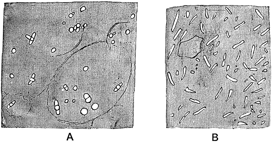
Fig. 28. A, Bacillus Tieghemi Ren. and Micrococcus Guignardi Ren. B, Bacillus Permicus Ren. (After Renault.)
Renault has given the name Bacillus Tieghemi to certain minute rods 6–10µ, in length, and 2·2–3·8µ broad, often containing a dark coloured spherical spore-like body 2µ in diameter, which have been found in the tissues of a Coal-Measure plant.
The name Micrococcus Guignardi has been applied to more or less spherical bodies 2·2µ in diameter, also met with in silicified plants.
A portion of one of Renault’s figures is reproduced in Fig. 28 A. The faint and broken lines mark the position of the middle lamellae of parenchymatous cells from the pith of a Calamite. The tissue has been almost completely destroyed, but the more resistant middle lamellae have been partially preserved. The short and broad rods represent what Renault terms Bacillus Tieghemi; the small circle in the middle of some of these being referred to as a spore, and in one specimen shown in the figure, the second rod at right angles to the first is described as a small daughter-Bacillus formed by the germination of the central spore.
The isolated circles in the figure are referred to Micrococcus.
FOSSIL BACTERIA.
It is unnecessary to give an account of the numerous examples of Micrococci and Bacilli described by Renault from Devonian, Carboniferous, Permian and Jurassic rocks. We may, however, in a few words consider the general question of the existence and possible determination of fossil Bacteria.
In 1877 Prof. Van Tieghem[218] of Paris drew attention to the method of operation and plan of attack of Bacillus amylobacter as a destructive agent in the decay of plant débris in water. He was able to follow the gradual disorganisation of the tissues and the various steps in the ‘butyric fermentation’ effected by this Bacterium. Similarly the same author[219] was able to detect the action of an allied organism in some silicified tissues from the Carboniferous nodules of Grand-Croix, a well-known locality for petrified plants near Saint-Étienne. He recognised also the traces of the Bacillus itself in the partially destroyed plant tissues. The Palaeozoic Bacteria made use of some cellulose-dissolving ferment of which the action is clearly demonstrated in sections of silicified tissues. Many of the phenomena described by Renault and Bertrand as due to similar Bacterial action, afford additional evidence that the gradual disorganisation of vegetable tissues was effected in precisely the same manner as at the present day.
In some cases we have I believe trustworthy examples of the Bacteria themselves, both in coprolites and plant-tissues, but it is more than probable that some of the recorded examples are not of any scientific value. The examination of petrified tissues under the higher powers of a microscope often reveals the existence of numerous spherical particles and rod-like bodies which agree in shape with Micrococci or Bacilli. Minute crystals of mineral substances may occur in the siliceous or calcareous matrix of a petrified plant which simulate minute organic forms. Vogelsang[220] in his important work die Krystalliten has thrown considerable light on the ontogeny of crystals, and the minute globulites and other forms of incipient crystallisation might well be mistaken for Bacterial cells. Granting, however, that we have satisfactory evidence, both direct and indirect, that some forms of Bacteria lived in the decaying tissues of Palaeozoic plants, and in the intestines of reptiles and other animals, we cannot safely proceed to specific diagnoses and determinations[221].
Renault has pointed out that fossil Bacteria may often be more readily detected than living forms owing to the presence of a brown ulmic substance which results from the carbonisation of the protoplasm. He is forced to admit, however, that such diagnostic characters as are obtained by Bacteriologists by means of cultures cannot be utilised when we are dealing with fossil examples! We are told that “Partout où nous avons cherché des Bacteriaceés, nous en avons rencontré.”[222] This indeed is the danger; an extended examination of fossil sections under an immersion-lens must almost inevitably lead to the discovery of minute bodies of a more or less spherical form which might be Micrococci. To measure, and name such bodies as definite species of Micrococci is, I believe, but wasted energy and an attempt to compass the impossible.
Specialists tell us that the accurate determination of species of recent Bacteria is practically hopeless: may we not reasonably conclude that the attempt to specifically diagnose fossil forms is absolutely hopeless? “The imagination of man is naturally sublime, delighted with whatever is remote and extraordinary—”, but it is to be deplored if the fascination of fossil bacteriology is allowed to warp sound scientific sense.
IV. ALGAE.
DIATOMACEAE. (Diatoms.)
CHLOROPHYCEAE. (Green algae.)
RHODOPHYCEAE. (Red algae.)
PHAEOPHYCEAE. (Brown algae.)
The presence of chlorophyll is one common characteristic of the numerous plants included in the Algae. The generally adopted classification rests in part on an artificial distinction, namely the prevailing colour of the plant.
It must be definitely admitted, at the outset, that palaeobotany has so far afforded extremely little trustworthy information as to the past history of algae. Were we to measure the importance of the geological history of these plants by the number of recorded fossil species, we should arrive at a totally wrong and misleading estimate. By far the greater number of the supposed fossil algae have no claim to be regarded as authentic records of this class of Thallophytes. It has been justly said that palaeontologists have been in the habit of referring to algae such impressions or markings on rocks as cannot well be included in any other group. “A fossil alga,” has often been the dernier ressort of the doubtful student.
LARGE SEAWEEDS.
Before discussing our knowledge, or rather lack of knowledge, of fossil algae at greater length, it will be well to briefly consider the manner of occurrence and botanical nature of existing forms. In the sea and in fresh water, as well as in damp places and even in situations subject to periods of drought, algae occur in abundance in all parts of the world. We find them attaining full development and reproducing themselves at a temperature of −1° C. in the Arctic Seas, and again living in enormous numbers in the waters of thermal springs. Around the coast-line of land areas, and on the floor of shallow seas algae exhibit a remarkable wealth of form and luxuriance of growth. As regards habit and structure, there is every gradation from algae in which the whole individual consists of a thin-walled unseptate vesicle, to those in which the thallus attains a length unsurpassed by any other plant, and of which the anatomical features clearly express a well-marked physiological division of labour such as occurs in the highest plants.
The large and leathery seaweeds which flourish in the extreme northern and southern seas are plants which it is reasonable to suppose might well have left traces of their existence in ancient sediments. Sir Joseph Hooker, in his account of the Antarctic flora[223], investigated during Sir James Ross’s voyage in H.M. ships Erebus and Terror, has given an exceedingly interesting description of the gigantic brown seaweeds of southern latitudes. The trunks are described as usually 5–10 feet long, and as thick as a human thigh, dividing towards the summit into numerous pendulous branches which are again broken up into sprays with linear ‘leaves.’ Hooker records how a captain of a brig employed his crew for two bitterly cold days in collecting Lessonia stems which had been washed up on the beach, thinking they were trunks of trees fit for burning. On our own coasts we are familiar with the common Laminaria, the large brown seaweed with long and strap-shaped or digitate fronds which grows on the rocks below low-tide level. The frond passes downwards into a thick and tough stipe firmly attached to the ground by special holdfasts. A transverse section of the stalk of a fairly old plant presents an appearance not unlike that of a section of a woody plant. In the centre there is a well-defined axial region or pith consisting of thick walled, long and narrow tubes pursuing a generally vertical though irregular course, and embedded in a matrix of gelatinous substance derived from the mucilaginous degeneration of the outer portions of the cell-walls. The greater part of such a section consists, however, of regularly disposed rows of cells which have obviously been formed by the activity of a zone of dividing or meristematic elements. The occurrence of distinct concentric rings in this secondary tissue clearly points to some periodicity of growth which is expressed by the alternation of narrow and broader cells. In the Antarctic genus Lessonia, the stem reaches a girth equal to that of a man’s thigh, and in structure it agrees closely with the smaller stem of Laminaria. In these large algal stems, the cells are not lignified as in woody plants, and in longitudinal section they have for the most part the form of somewhat elongated parenchyma, differing widely in appearance from the tracheids or vessels of woody plants. At the periphery of the Laminaria stem, represented in fig. 29, there occur numerous and comparatively large mucilage ducts.
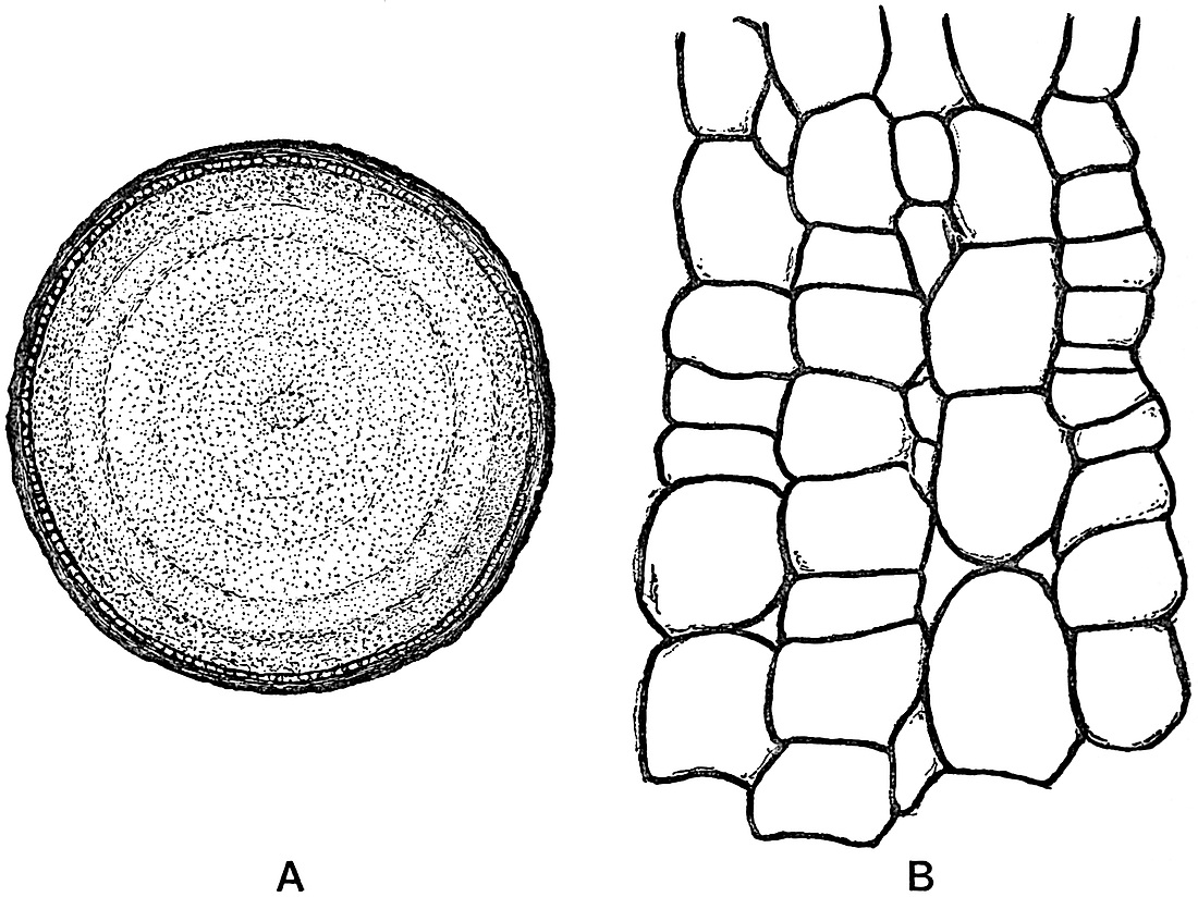
Fig. 29. A, Transverse section of the stipe of a Laminaria, slightly enlarged. B, A small piece of the tissue between the central ‘pith’ and ‘cortex’ showing the radially disposed secondary elements more highly magnified.
In certain algae of different families the thallus is encrusted with carbonate of lime, and is thus rendered much more resistant. The Diatoms, on the other hand, possess still more durable siliceous tests which are particularly well adapted to resist the solvent action of water and other agents of destruction. It is these calcareous and siliceous forms which supply the greater part of the trustworthy data furnished by fossil algae.
SCARCITY OF FOSSIL ALGAE.
It remains to consider some of the causes to which we may attribute the scarcity of fossil algae, and the possible sources of error which beset any attempt to describe or assign names to impressions and casts simulating algal forms.
In the first place, the delicate nature of algal cells is a serious obstacle to fossilisation. Even in plants in which the woody stems have been preserved by a siliceous or calcareous solution, we frequently find the more delicate cells represented by a mass of crystalline matter without any trace of the cell-walls being preserved. In such plants as algae, where the cell-walls are not lignified, but consist of cellulose or some special form of cellulose, which readily breaks down into a mucilaginous product, the tissues have but a small chance of withstanding the wear and tear of fossilisation.
The danger of relying on external form as a means of recognition is especially patent in the case of those numerous markings or impressions frequently met with on rocks, and which resemble in outline the thallus of recent algae. Among animals, such as certain Polyzoa, the flat branching body of various algae is closely simulated, and in other plants, such as the frondose liverworts, the same thalloid and branched form of body is again met with. Some of the much dissected Aphlebia leaves of ferns (e.g. Rhacophyllum species) bear a striking resemblance to fossil algae; and numerous other examples might be quoted. In palaeobotanical literature we find a host of names, such as Chondrites, Fucoides[224], Caulerpites and others applied to indefinite and indistinct surface markings which happen to resemble in shape certain of the better known genera of recent seaweeds.
The close parallelism in outward form displayed by different genera and families of algae is in itself sufficient argument against the use of recent generic names for fossils of which the algal nature is often more than doubtful. Were external form to be accepted as a trustworthy guide, in the absence of internal structure and reproductive organs, such a genus as Caulerpa[225] would afford material for numerous generic designations. A comparison of the different species of this Siphoneous green alga brings out very clearly the exceedingly protean nature of this interesting genus, and serves as one instance among many of the small taxonomic value which can be attached to external configuration. Caulerpa pusilla Mart. and Her., C. taxifolia (Vahl.), C. plumaris Forsk., C. abies-marina J. Ag., C. ericifolia (Turn.), C. hypnoides (R. Br.), C. cactoides (Turn.), C. scalpelli formis (R. Br.), and others clearly illustrate the almost endless variety of form exhibited by the species of a single genus of algae. We constantly find in the several classes of plants a repetition of the same form either in the whole or in the separate members of the vegetative body, and but a slight acquaintance with plant types should lead us to use the test of external resemblance with the greatest possible caution. To emphasize this danger may seem merely the needless reiteration of a self-evident fact, but there is, perhaps, no source of error which has been more responsible for the creation of numerous worthless species among fossil plants.
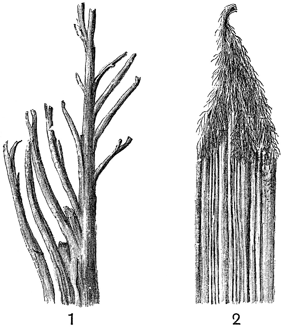
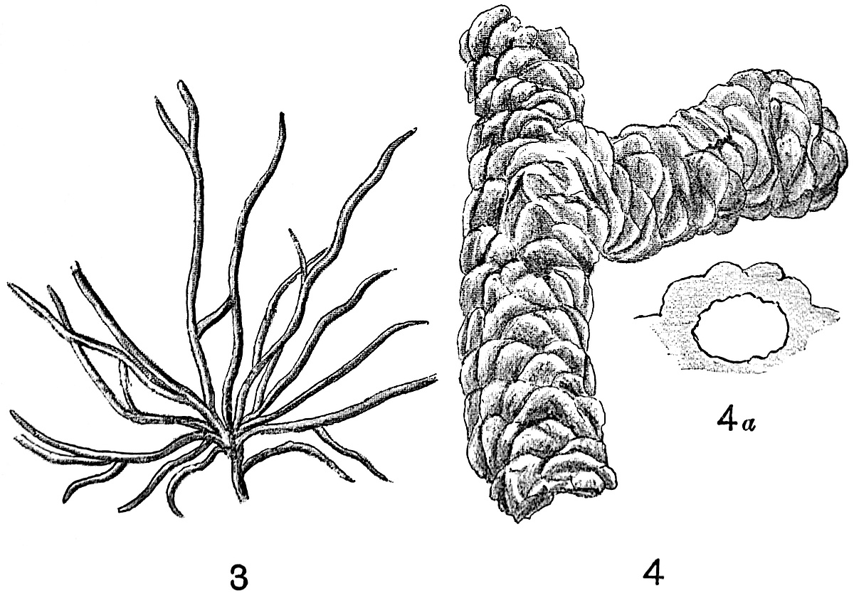
Fig. 30. 1. Rill-mark (after Williamson). 2. Trail made by a seaweed dragged along a soft plaster of Paris surface (after Nathorst). 3. Tracks made by Goniada maculata, a Polychaet (after Nathorst). 4. Burrow of an insect. 4a. Section of the gallery (after Zeiller).
There is, however, another category of impressions and casts of common occurrence in sedimentary rocks which requires a brief notice. Very many of the fossil algae described in text-books and palaeobotanical memoirs have been shown to be of animal origin, and to be merely the casts of tracks and burrows. A few examples will best serve to illustrate the identity of many of the fossils referred to algae with animal trails and with impressions produced by inorganic agency.
Dr Nathorst of Stockholm has done more than any other worker to demonstrate the true nature of many of the species of Chondrites, Cruziana, Spirophyton, Eophyton, and numerous other genera. In 1867 there were discovered in certain Cambrian beds of Vestrogothia, long convex and furrowed structures in sandstone rocks which were described as the remains of some comparatively highly organised plant, and described under the generic name Eophyton[226]. By many authors these fossils have been referred to algae, but Nathorst has shown that the frond of an alga trailed along the surface of soft plaster of Paris produces a finely furrowed groove (fig. 30, 2) which would afford a cast similar to that of Eophyton. The same author has also adduced good reasons for believing that the Eophytons of Cambrian rocks may represent the trails made by the tentacles of a Medusa having a habit similar to that of Polydonia frondosa Ag. Impressions of Medusae have been described by Nathorst from the beds in which Eophyton occurs; and the specimens in the Stockholm Museum afford a remarkable instance of the rare preservation of a soft-bodied organism[227]. By allowing various animals to crawl over a soft-prepared surface it is possible to obtain moulds and casts which suggest in a striking manner the branched thallus of an alga. The tracks of the Polychaet, Goniada maculata Örstd.[228], one of the Glyceridae, are always branched and very algal-like in form (fig. 30, 3). Many of the so-called fossil algae are undoubtedly mere tracks or trails of this type. In the fossil-plant gallery of the British Museum there are several specimens of small branched casts, clearly marked as whitish fossils on a dark grey rock of Lower Eocene age from Bognor; these were described by Mantell and Brongniart[229] as an alga, but there is little doubt of their being of the same category as the track shown in fig. 30, 3.
FOSSILS SIMULATING ALGAE.
The well-known half-relief casts met with in Cambrian, Silurian and Carboniferous rocks, and known as Cruziana or Bilobites, are probably casts of the tracks of Crustaceans. The impression left by a King-Crab (Limulus) as it walks over a soft surface affords an example of this form of cast. It has been suggested that some of the Bilobites may be the casts of an organism like Balanoglossus[230], a worm-like animal supposed by some to have vertebrate affinities. The resemblance between some of the lower Palaeozoic Bilobites and the external features of a Balanoglossus is very striking, and such a comparison is worth considering in view of the fact that soft-bodied animals have occasionally left distinct impressions on ancient sediments.
The literature on the subject of fossil algae versus inorganic and animal markings is too extensive and too wearisome to consider in a short summary; the student will find a sufficient amount of such controversial writing—with references to more—in the works quoted below[231].
In the Stockholm Museum of Palaeobotany there is an exceedingly interesting collection of plaster casts obtained by Dr Nathorst in his experiments on the manufacture of fossil ‘algae,’ which afford convincing proof of the value and correctness of his general conclusions.
The pressure of the hand on a soft moist surface produces a raised pattern like a branched and delicate thallus. The well-known Oldhamia antiqua Forbes and Oldhamia radiata Forbes[232], from the Cambrian rocks of Ireland may, in part at least, owe their origin to mechanical causes, and we have no sufficient evidence for including them among the select class of true fossil algae. Sollas[233] has shown that the structure known as Oldhamia radiata is not merely superficial but that it extends across the cleavage-planes. Oldhamia is recorded from Lower Palaeozoic rocks in the Pyrenees[234] by Barrois, who agrees with Salter, Göppert and others in classing the fossil among the algae. The photograph accompanying Barrois’ description does not, however, add further evidence in favour of accepting Oldhamia as a genus of fossil algae.
The burrows made by Gryllotalpa vulgaris Latr., the Mole-cricket, have been shown by Zeiller to bear a close resemblance to a branch of a conifer in half-relief (fig. 30, 4), or to such a supposed algal genus as Phymatoderma[235].
In fig. 30, 1, we have what might well be described as a fossil alga. This is merely a cast of a miniature river-system such as one frequently sees cut out by the small rills of water flowing over a gently-sloping sandy beach. A cast figured and described by Newberry as an alga, Dendrophycus triassicus[236], from the Trias of the Connecticut Valley, is practically identical with the rill-marks shown in fig. 30, 1. The cracks produced in drying and contracting sediment may form moulds in which casts are subsequently produced by the deposition of an overlying layer of sand, and such casts have been erroneously referred to algal impressions[237]. Dawson[238] has figured two good examples of Carboniferous rill-marks from Nova Scotia in his paper on Palaeozoic burrows and tracks of invertebrate animals.
RECOGNITION OF FOSSIL ALGAE.
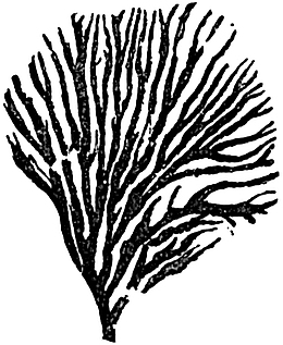
Fig. 31. Chondrites verisimilis Salt. Wenlock limestone, Dudley. From a specimen in the British Museum (V. 2550). Slightly reduced.
The specimen represented in fig. 31 affords an example of a fairly well-known fossil from the Wenlock limestone, originally described by Salter as Chondrites verisimilis Salt, from Dudley[239]. He regarded it as an alga, and the graphitic impression agrees closely in form with the thallus of some small seaweeds. A closer examination of the fossil reveals a curious and characteristic irregular wrinkling on the graphite surface, which suggests an organism of more chitinous and firmer material than that of an alga.
A similar and probably an identical fossil is described and figured by Lapworth[240] in an appendix to a paper by Walter Keeping on the geology of Central Wales, under the name of Odontocaulis Keepingi Lap. and regarded as a dendroid graptolite. In any case we have no satisfactory grounds for including these fossils in the plant-kingdom.
How then are we to recognise the traces of ancient algae? There is no golden rule, and we must admit the difficulty of separating real fossil algae from markings made by animal or mechanical agency. The presence of a carbonaceous film is occasionally a help, but its occurrence is no sure test of plant origin, nor is its absence a fatal objection to an organic origin. While being fully alive to the small value of external resemblance, and to the numerous agents which have been shown to be capable of producing appearances indistinguishable from plant impressions, we must not go too far in a purely negative direction.
An important contribution to the subject of fossil algae has lately appeared by Prof. Rothpletz[241]. He deals more particularly with the much discussed Flysch[242] Fucoids of Tertiary age, and while refusing to accept certain examples as fossil algae, he brings forward weighty arguments in favour of including several other forms among the algae. He is of opinion that most of the main divisions of the algae are represented among the Flysch Fucoids, but considers that the Phaeophyceae are the most numerous.
Rothpletz’s work is chiefly interesting as illustrating the application of microscopic examination and chemical analysis to the determination of fossil algae. Although he makes out a good case in favour of restoring many of the Tertiary fossils to the plant kingdom, the material at his disposal does not admit of satisfactory botanical diagnosis.
No doubt some of the fossils from the Silurian and Cambrian rocks are true algae, and Nathorst has pointed out that such a species as Hall’s Sphenothallus angustifolius[243] may well be an alga. Additional examples might be quoted from Bornemann and other writers, but in view of the attempts which are sometimes made to trace the development of more recent plants to more than doubtful Lower Palaeozoic Algae, one must agree with Nathorst’s opinion,—“Je crois que l’on rend un bien mauvais service à la théorie de l’évolution, en essayant de baser l’arbre généalogique des algues fossiles sur des corps aussi douteux que les Bilobites, Crossochorda, Eophyton, etc.[244]”
There are many carbonaceous impressions on rocks of different ages which it is reasonable to refer to algal origin, and although such are of little or no botanical value, it may be a convenience to refer to them under a definite term. The comprehensive generic name Algites[245] has been suggested as a convenient designation for impressions or casts which are probably those of algae.
SUPPOSED FOSSIL ALGAE.
Some of the fossils described by Mr Kidston from British Carboniferous rocks as probably algae present an undoubted algal appearance, and might be placed in the genus Algites; but in some cases—e.g. Chondrites plumosa[246] Kidst. from the Calciferous Sandstone of Eskdale, one feels much more doubtful; in this particular instance the impressions suggest the fine roots of a water-plant.
The statement is occasionally made that the numerous fossil algae and the absence of higher plants in the older strata justify the description of the oldest rocks as belonging to the ‘age of algae.’ Such an assertion rests on an unsound basis, and is rather the expression of what might be expected than what has been proved to be the case. The oldest plants with which we are at all closely acquainted are of such a type as to forcibly suggest that in the lowest fossiliferous rocks we are still very far from the sediments of that age which witnessed the dawn of plant life.
Many of the obscure markings on rock surfaces which have been referred to existing genera of algae or described as new genera, are much too doubtful to be included even under such a comprehensive name as Algites. Space does not admit of further reference to determinations of this type which abound in palaeontological literature.
It would be very difficult to produce satisfactory evidence for the algal nature of many of the supposed fossil algae from Cambrian rocks[247]; there has been a special tendency to recognise algal remains in the oldest fossiliferous strata, due in part no doubt to the fallacy that in that period nothing higher than Thallophytes is likely to have existed. The so-called Phycodes referred to by Credner[248] as characteristic of the Cambrian rocks of the Fichtelgebirge (“Phycoden-Schiefer”) is probably of inorganic origin, and comparable to the genus Vexillum of Saporta[249] and other writers, which Solms-Laubach has described as being formed every day in the soft mud of our ponds where local currents are checked by branches and other obstacles[250]. There are several good specimens of Phycodes in the Bergakademie of Berlin and in the Leipzig Museum which, I believe, clearly demonstrate the absence of all satisfactory evidence of an algal origin.
We may next pass to a short description of a few representative types of algae, which may reasonably be classed under definite families, and accepted as evidence possessing some botanical value.
A. DIATOMACEAE (Bacillariaceae).
This family occupies a somewhat isolated position among the algae, and is best considered as a distinct subdivision rather than as a family of the Phaeophyceae or Brown algae, with which it possesses as a common characteristic a brown-colouring matter.
Single-celled plants consisting of a simple protoplasmic body containing a nucleus and brown colouring matter (diatomin) associated with the chlorophyll. The cell-wall is in the form of two halves, known as valves, which fit into one another like the two portions of a pill-box. The cell-wall contains a large amount of silica, and the siliceous cases of the diatoms are commonly spoken of as the valves of the individual, or the frustules. Diatoms exhibit a characteristic creeping movement, and are reproduced by division, also by the development of spores in various forms[251].
The recent members of the family have an exceedingly wide distribution, occurring both in freshwater and in the sea. Owing to the lightness of the frustules, they are frequently carried along in the air, and atmospheric dust falling on ships at sea has been found to contain large numbers of diatoms[252]. The siliceous valves are abundant in guano deposits, and they have been found also in association with volcanic material. Diatomaceous deposits are now being formed in the Yellowstone Park district; “they cover many square miles in the vicinity of active or extinct hot spring vents of the park, and are often three feet, four feet, and sometimes five to six feet thick[253].” The gradual accumulation of the siliceous tests on the floor of a fresh-water lake results in the formation of a sediment consisting in part of pure silica. Such deposits, often spoken of as kieselguhr or diatomite, and used as a polishing material, occur in many parts of Britain, marking the sites of dried-up pools or lakes. At the northern end of the island of Skye there occurs an unusually pure deposit of diatomite overlain by peat and turf, and extending over an area of fifty-eight square miles. Many of the individuals in this deposit were in all probability carried into the lake by running water, while others lived in the lake and after death their tests contributed to the siliceous deposit[254]. The late Dr Ehrenberg published numerous papers on diatomaceous deposits in different parts of the world, and in his great work, Zur Mikrogeologie[255], he gave numerous and beautifully executed illustrations of such siliceous accumulations. In many of the samples he figures one sees fragments of plant tissues, spores of conifers and ferns, associated with the diatom tests. The occurrence of the pollen grains of coniferous trees in lacustrine and marine deposits is not surprising in view of their abundance in Lake Constance and other lakes. It is stated that the pollen of conifers in the Norwegian fiords plays an important part in the nourishment of the Rhizopod Saccamina[256].
DIATOMACEOUS OOZE.
In the waters of the ocean diatoms are of frequent occurrence, and very widely distributed. Sir Joseph Hooker records the existence of masses of diatomaceous ooze over a wide area in Antarctic regions[257]. Along the shores of the Victoria Barrier, a perpendicular wall of ice, between one and two hundred feet above sea-level, the soundings were found to be invariably charged with diatom remains, and from the base of the ice-wall there appeared to be in process of formation a bank of these tests stretching north for a distance of 200 miles. The more extended researches conducted during the cruise of the Challenger have clearly proved the enormous accumulations of diatoms now being formed on the ocean-bed[258]. South of latitude 45° S. there is now being built up a vast deposit which may be eventually upraised as a fairly pure siliceous rock. From extreme northern latitudes Nansen has recently recorded the occurrence of these lowly organised plants. He writes,—“I found a whole world of diatoms and other microscopical organisms, both vegetable and animal, living in the fresh-water pools on the Polar drift-ice, and constantly travelling from Siberia to the east coast of Greenland[259].” In warmer latitudes diatoms abound in the surface waters, but there they are associated with numerous other forms of the Plankton vegetation. The waters of the Amazon carry with them into the sea large numbers of fresh-water forms, which are floated out to sea and finally added to the rock-building material which is constantly accumulating on the ocean floor[260]. No definite results have so far been obtained as to the geographical and bathymetrical distribution of marine diatoms.
The enormous number of recent species precludes any attempt to give a description of the better-known forms. It is more important for us to realize how common and widely distributed are the living genera. The hard and almost indestructible valves have been frequently found in a fossil condition, often forming thick and extensive masses of siliceous rock. From diatom-beds now forming in lakes and on the ocean-bed we pass to deposits such as those in Skye and elsewhere, which mark the site of recently dried-up sheets of water, and so to older rocks of Tertiary age formed under similar conditions. Among the many examples of diatomaceous deposits of Tertiary and Cretaceous age mention should be made of those of Berlin, Königsberg, Bilin in Bohemia, and Richmond in Virginia. The diatoms in the beds of Berlin are regarded as fresh-water, and those of Richmond as marine. It has been pointed out by Pfitzer that it is a comparatively easy matter to distinguish between fresh-water and marine forms of diatoms. The diatomaceous rocks of Bilin are known as polishing slates; they attain a thickness of 50 feet. In these, as in many other cases, the deposit has become cemented together as a hard flinty or glassy rock, in which the cementing material was formed by the solution of some of the diatom tests[261]. In many cases in which calcareous and siliceous rocks reveal no direct evidence of organic origin it is probable that they were originally formed by the accumulation of plants of which the structure has been completely obliterated by secondary causes. The genus Gallionella plays an important part in the composition of the Bilin beds. Occasionally impressions of leaves and other organic remains are found associated with the diatoms in the siliceous rocks. In the British Museum (Botanical department) a large block of white powdery rock is exhibited as an example of a diatomaceous deposit of Tertiary age from Australia. It is described as being largely made up of the tests of fresh-water diatoms, such as Navicula, Gomphonema, Cymbella, Synedra, and others.
FOSSIL DIATOMS.
The abundance of Diatoms in Cretaceous rocks of the Paris basin has recently been recorded by Cayeux[262]; it would seem that these algae had already assumed an important rôle as rock-builders in pre-Tertiary times. Cayeux points out that the silica of these Cretaceous diatomaceous frustules has often been replaced by carbonate of calcium.
In addition to the occurrence of Diatoms in the various diatomaceous deposits, their siliceous tests may occasionally be recognised in argillaceous or other sediments. Shrubsole and Kitton[263] have described several species of Diatoms from the London Clay of Lower Eocene age. In many localities in the London basin the clay obtained from well-sinkings presented the appearance of being dusted with sulphur-like particles of a dark bronze or golden colour which glistened in the sunlight. These yellow bodies have been found to be diatomaceous frustules in which the silica has been replaced by iron pyrites. The genus Coscinodiscus is one of the commonest forms recorded from the London Clay[264].
Without further considering individual examples of diatomaceous rocks we may briefly notice the general facts of the geological history of the family. As Ehrenberg pointed out several years ago, the Tertiary and Cretaceous species of diatoms show a very marked resemblance to living forms. In many cases the species are identical, and the fossil deposits as a whole seem to differ in no special respect from those now being built up.
With the exception of two species of Liassic Diatoms, no trustworthy examples of the Diatomaceae have been found below the Cretaceous series. The oldest known Diatoms were discovered by Rothpletz[265] among the fibres of an Upper Lias sponge from Boll in Württemberg. They occur as small thimble-shaped siliceous tests with coccoliths and foraminifera in the horny skeleton of Phymatoderma, a genus formerly regarded as an alga. Rothpletz describes two species which he includes in the genus Pyxidicula, P. bollensis and P. liasica. This generic name of Ehrenberg is used by Schütt[266] as a subgenus of Stephanopyxis.
Seeing how great a resemblance there is between the recent and Cretaceous species, and how many examples there are of Tertiary diatom deposits, it is not a little surprising that the past history of these plants has not been traced to earlier periods. In 1876 Castracane[267], an Italian diatomist, gave an account of certain species of diatoms said to have been found in a block of coal from Liverpool obtained from the English Coal-Measures. The species were found to be identical with recent forms. It is generally agreed that these specimens cannot have been from the coal itself, but that they must have been living forms which had come to be associated with the coal. The late Prof. Williamson spent many years examining thin sections and other preparations of coal from various parts of the world, but he never found a trace of any fossil diatom. There is no apparent reason why diatoms should not be found in Pre-Cretaceous rocks, and the microscopic investigation of old sediments may well lead to their discovery. Prof. Bertrand of Lille, who has devoted himself for some time past to a detailed microscopical examination of coal, informs me that he has so far failed to discover any trace of Palaeozoic diatomaceous tests.
BACTRYLLIUM.
The genus Bactryllium is often quoted in text-books as a probable example of a Triassic diatom. It was first described by Heer[268] from the Trias of Switzerland and North Italy, also from the neighbourhood of Heidelberg, and regarded as an extinct member of the Diatomaceae. Heer defined the genus as follows:
“Small bodies, with parallel sides, rounded at either end, the surface traversed by one or two longitudinal grooves.”
(fig. 32, C.) Several species have been figured by Heer from beds of Muschelkalk, Keuper and Rhaetic age. He describes the wall as thick and firm (fig. 32, C. ii.) and probably composed of silica, with a hollow interior. The specimen shown in fig. 32, C. was found in the Rhaetic beds, and named by Heer Bactryllium deplanatum; it has a length of 4·5 mm.; the surface is transversely striated and traversed by a single longitudinal groove. Stefani[269] has given reasons in favour of removing Bactryllium from the plant to the animal kingdom; he points out that the specimens are too large for diatoms, and moreover that they are asymmetrical in form and possessed a calcareous and not a siliceous shell. He would place the fossil among the Pteropods, comparing it with such genera as Cuvierina and Hyalaea. In view of Stefani’s opinion we cannot attach any importance to this supposed diatom, especially as it has generally been regarded as at best but an unsatisfactory genus.
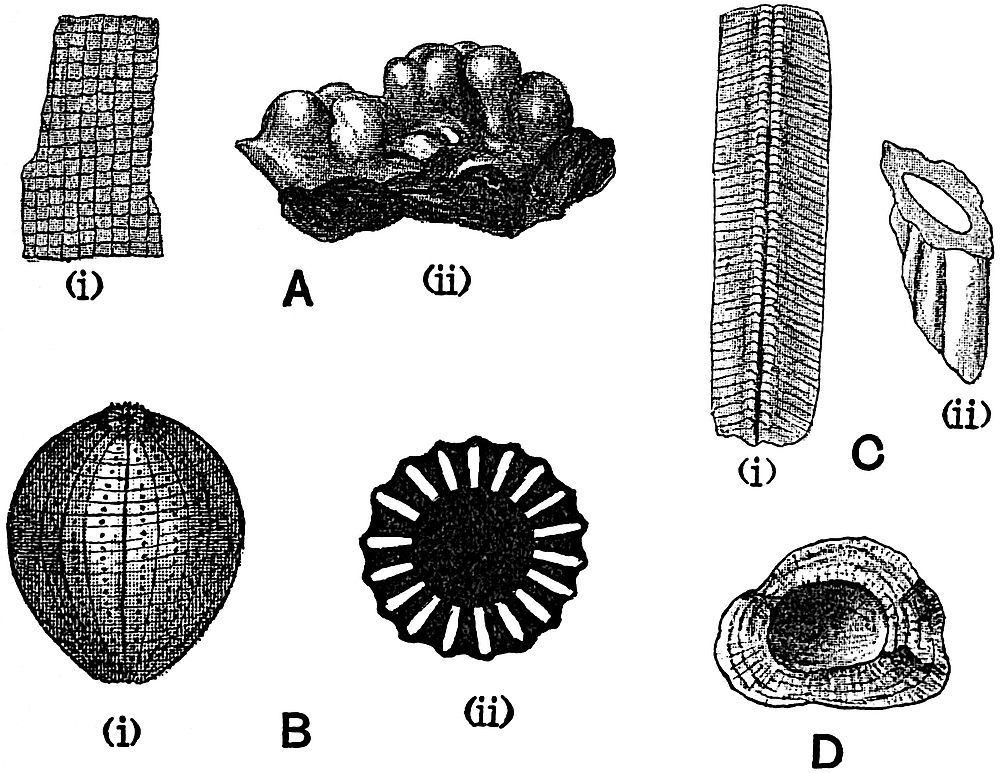
Fig. 32. A, Lithothamnion mamillosum Gümb. (i) In section, (ii) surface view [after Gümbel. (i) × 320, (ii) nat. size. B, Sycidium melo Sandb. (i) Surface view, (ii) transverse section (after Deecke). C, Bactryllium deplanatum Heer. (i) Surface view, (ii) section, showing the thick wall and hollow interior (after Heer). D, Calcareous pebble from a lake in Michigan. Rather less than nat. size (after Murray).
B. CHLOROPHYCEAE (Green Algae).
Thallus unseptate, having the form of a vesicle or a variously branched coenocyte, which may or may not be encrusted with carbonate of lime, or of filaments composed of cells containing a single nucleus, or of cells in which more than one nucleus occurs; in other instances consisting of a plate of cells or a cell-mass. Asexual reproduction by zoospores and other reproductive cells; sexual reproduction by means of the conjugation of similar gametes or by the fertilisation of a typical egg-cell by a motile spermatozoid.
This family of algae is represented at the present day by numerous and widely distributed marine and fresh-water genera, as well as by genera growing in moist air or as endophytes in the tissues of higher plants[270].
Seeing how very few fossil forms have been described which have any claim to inclusion in this subdivision of the Algae, it is unnecessary to enumerate or define the various families of the Chlorophyceae. It is true that many species have been figured as examples of different genera of green algae, but few of these possess any scientific value. There is, however, one division of the Chlorophyceae, the Siphoneae, which must be treated at some length on account of its importance from a palaeobotanical and geological point of view.
a. Siphoneae.
Thallus consisting of simple or branched cells very rarely divided by septa, and containing many nuclei. In certain genera the branches form a pseudoparenchymatous tissue by their repeated branching, and as a result of the intimate felting together of the branched cells. Reproduction is effected either by the conjugation of similar gametes or by the fertilisation of an egg-cell.
Vaucheria and Botrydium are two well-known British genera of this order, but most of the recent representatives live in tropical and subtropical seas. The most striking characteristic feature of this division of the Chlorophyceae is the fact that the thallus of a siphoneous alga consists of an unseptate coenocyte; the plant may be extremely small and simple, or it may reach a length of several inches, but in all cases the body does not consist of more than one cell or coenocyte.
From a palaeontological standpoint the Siphoneae are of exceptional interest. It is impossible to do more than refer to a few of the living and fossil genera. There are numerous fossil representatives already known, and there can be little doubt that further research would be productive of valuable results.
As examples of the order, a few genera may be described belonging to the three families Caulerpaceae, Codiaceae, and Dasycladaceae.
α. Caulerpaceae.
Thallus unseptate, showing an extraordinary variation in the external differentiation of the plant-body. Reproduction is effected by means of detached portions of the parent plant.
The genus Caulerpa, represented by a few species in the Mediterranean and by many tropical forms, has already been alluded to as a striking example of a plant which appears under a great many different forms[271]. As a recent writer has said, “Nature seems to have shown in this genus the utmost possibilities of the siphoneous thallus[272],” Fragments of coniferous twigs, the tracks and burrows of various animals and other objects have been described by several authors as fossil species of Caulerpa. As an illustration of the identification of a very doubtful fossil as a species of Caulerpites, reference may be made to such a form as C. cactoides Göpp.[273] from Silurian and Cambrian rocks. There are several examples of this fossil in the Brussels Museum which probably owe their origin to some burrowing animal, and may be compared with Zeiller’s figures of the tunnels made by the mole-cricket (fig. 30, 4)[274].
Mr Murray, of the British Museum, has recently described what he regards as a trustworthy example of a fossil Caulerpa from the Kimeridge Clay near Weymouth[275]. Specimens of the fossil were first figured in a book on the geology of the Dorset coast as casts of an equisetaceous plant[276].
To this fossil Murray has assigned the name Caulerpa Carruthersi, and given to it a scientific diagnosis. The best specimens have the form of a slender central axis, giving off at fairly regular intervals whorls of short and somewhat clavate branches; they bear a superficial resemblance to such a recent species as Caulerpa cactoides Ag. An examination of several examples of this fossil leads me to express the opinion that there is not sufficient reason for assigning to them the name of a recent genus of algae[277]. To use the generic name of a recent plant without following the common custom of adding on the termination “ites” (i.e. Caulerpites) is as a general rule to be avoided in dealing with fossil forms; and there are, I believe, no satisfactory grounds for referring to these fossils as trustworthy examples of a Mesozoic alga.
In the present case I am disposed to regard the Caulerpa-like casts as of animal rather than plant origin. The clavate branches have the form of very deep moulds in the hard brown rock which have been filled in with blue mud. It is hardly conceivable that the branches of a soft watery plant such as Caulerpa could leave more than a faint impression on an old sea-floor. The specimens occur in different positions in the matrix of the rock and they are not confined to the lines of bedding; in none of the examples is there any trace of carbonaceous matter in association with the deep moulds. On the whole, then, this Kimeridge fossil cannot, I believe, be accepted as an authentic example of a Mesozoic Caulerpa.
It is not improbable that some of the supposed fossil algae may be casts of egg-cases or spawn-clusters of animals. In Ellis’ Natural History of the Corallines[278] there is a drawing representing a number of disc-like ovaries attached to a tough ligament, and referred to the mollusc Buccinum, which bears a certain resemblance to the Weymouth fossil. A similar body is figured by Fuchs[279] in an important memoir on supposed fossil algae.
It is not suggested that the Caulerpa Carruthersi of Murray should be regarded as the cast of some molluscan egg-case attached to a slender axis, but it is important to bear in mind the possibility of matching such extremely doubtful fossils with other organic bodies than the thallus of a Caulerpa. In an example of an egg-case in the Cambridge Zoological Museum, referred to a species of Pyrula, there is a hard, long and slender axis, bearing a series of semicircular chambers divided into radial compartments. The whole is hard and horny and might well be preserved as a fossil.
β. Codiaceae.
The members of this Order present a considerable diversity of form as regards the shape of the plant-body; the thallus of some species is encrusted with carbonate of lime. The order is widely distributed in tropical and temperate seas.
Among the recent genera Penicillus and Codium may be chosen as important types from the point of view of fossil representatives.
Codium.
The thallus of Codium consists of a spongy mass of tubular cell-branches which are differentiated into two fairly distinct regions, an outer peripheral layer in which the branches have long club-shaped terminations, and an inner region consisting of loosely interwoven filaments.
Codium Bursa L. and C. tomentosum Huds. are two well-known British species, the former presents the appearance of a spongy ball of cells, and in the latter the thallus is divided up into dichotomously forked branches[280]. In this genus the thallus is not encrusted with carbonate of lime, at least in recent species.
Sphaerocodium. Fig. 37, D.
Rothpletz[281] instituted this genus for certain small spherical or oval bodies varying from 1 mm. to 2 cm. in diameter, which have been found on crinoid stems or shell fragments of Triassic age. Each spherical body consists of dichotomously branched single-celled filaments, between 50 and 100µ in breadth, and from 300–500µ in height. The tubular cavities occasionally swell out into spherical spaces which are regarded by Rothpletz as sporangia.
There is not sufficient evidence that Sphaerocodium Bornemanni Roth. has been correctly referred to the Codiaceae. The sporangia-like swellings described by the author of the species are not by any means conclusive as characters of important taxonomic value. Figure 37, D, illustrates the general structure of the fossil as seen in a transverse section of one of the calcareous grains.
Like Girvanella, which has been referred by some writers to the Siphoneae, Sphaerocodium occurs in the form of oolitic grains. In the Triassic Raibler and St Cassian beds of the Tyrol, as well as in rocks of Rhaetic age in the Eastern Alps, it makes up large masses of limestone. Rothpletz compares the structure of this genus with that of the recent alga Codium adhaerens Ag., but it is wiser to regard such tubular structures as Girvanella, Siphonema[282] and Sphaerocodium as closely allied organisms, which are probably algae, but too imperfectly known to be referred to any particular family.
Penicillus.
The recent genus Penicillus is one of those algae formerly included among animals. Fig. 33, O, has been copied from a drawing of a species of Penicillus given by Lamouroux[283] under the generic name of Nesea in his treatise on the genera of Polyps published in 1821. He describes the genus as a brush-like Polyp with a simple stem.
The thallus consists of a stout stem terminating in a brush-like tuft of fine dichotomously-branched filaments. The apical branches are divided by regular constrictions into short oval or rod-like segments which may be encrusted with carbonate of lime. A few of the segments from the terminal tuft of a recent Penicillus are shown in fig. 35, E. Each of these calcareous segments has the form of an oval shell perforated at each end, and the wall is pierced by numerous fine canals. Penicillus is represented by about 10 recent species, which with one exception live in tropical seas.
The recognition of Penicillus, or a very similar type, in a fossil condition is due to Munier-Chalmas[284]. This keen observer has rendered great service to palaeobotany by directing attention to the calcareous algae in the Paris basin beds, and by proving that many of the fossils from these Tertiary deposits have been erroneously included by previous writers among the Foraminifera[285]. It is greatly to be desired that Prof. Munier-Chalmas may soon publish a monograph on the fossil Siphoneous forms of which he possesses a unique collection.
Ovulites. Figs. 33, K, L, and 35, F.
In his Natural History of Invertebrate Animals, Lamarck described some small oval bodies from the Calcaire Grossier (Eocene) of the Paris basin under the name of Ovulites. He defined them as follows:—“Polypier pierreux, libre, ovuliforme ou cylindracé, creux intérieurement, souvent percé aux deux bouts. Pores très petits, régulièrement disposés à la surface[286].”
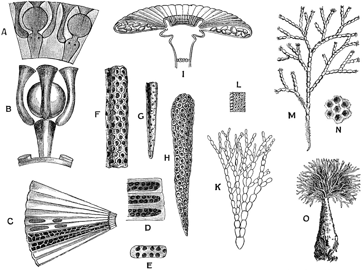
Fig. 33. A and B, Cymopolia barbata (L.); A, transverse section of the calcareous cylinder. B, verticillate branches and sporangium after removal of the calcareous matrix (A and B after Munier-Chalmas). C and D, Acicularia Andrussowi Solms (C, after Andrussowi; D, after Solms). E, Acicularia Miocenica Reuss; section of a spicula (after Reuss). F and G, Acicularia sp. (after Carpenter), F × 40; G × 20. H, Acicularia Schencki (Möb.) (after Solms). I, Acetabularia Mediterranea Lamx.; section of the cap (after Falkenberg). K and L, Ovulites margaritula (Lam.) (after Munier-Chalmas); K slightly enlarged; L, a piece of the thallus more highly magnified. M, Cymopolia barbata (L.) (after Ellis, nat. size). N, C. barbata (L.); the surface of the thallus; magnified. O, Penicillus pyramidalis (Lamx.) (after Lamouroux, nat. size).
The specimens are referred to two species, Ovulites margaritula and O. elongata.
By some subsequent writers[287] these calcareous fossils, like miniature birds’ eggs with a hole at either end, were included among the Zoophytes. Carpenter and others afterwards referred Ovulites to the Foraminifera, and compared the genus with Lagena[288]. The single specimens of Ovulites have a length of 2–6 mm. At each end there is usually a fairly large and somewhat irregular hole (fig. 35, F), and in some rarer cases there may be two apertures at the broader end of an Ovulite. A good example of Ovulites margaritula with two pores at the broader end is figured by Michelin[289]. The surface of the shell when seen under a low magnifying power appears to be covered over with regularly arranged circular pores, which are the external openings of fine canals (fig. 33, L).
In 1878 Munier-Chalmas expressed the opinion, which was supported by strong evidence, that Ovulites should be referred to the siphoneous algae[290]. He regarded it as generically identical with Penicillus (Coralliodendron, Kützing). It has already been pointed out that in Penicillus the apical tuft of filaments is partially calcareous (fig. 33, O)[291]. The individual calcareous segments agree almost exactly with the fossil Ovulites. As a rule the Ovulites occur as separate egg- or rod-like bodies, but Munier-Chalmas informs me that occasionally two or three have been found joined end to end in their natural position. The terminal holes in the fossil specimens represent the apertures left after the detachment of the calcareous segments from the uncalcified filaments of the alga. The segments with two holes at the broader end were no doubt situated at the base of dichotomising branches as shown in fig. 33, K. The restoration of Ovulites, shown in fig. 33, K, bears a striking resemblance to the figure of an Australian Penicillus given by Harvey in his Phycologia Australica[292].
It is probable that these Eocene forms agreed closely in habit with the recent species of Penicillus. The portions preserved as fossils are segments of the filaments which probably formed a terminal brush of fine branches supported on a stem. The retention of the original generic name Ovulites is on the whole better than the inclusion of the fossil species in the recent genus. The Tertiary species lived in warm seas of the Lower and Middle Eocene of England, Belgium, France and Italy.
Halimeda.
An example of an Eocene species of Halimeda has been recorded by Fuchs from Greifenstein under the name of Halimeda Saportae[293]. The impression has the form of a branched plant consisting of wedge-shaped or oval segments, and there is a close resemblance to the thallus of a recent Halimeda, e.g. H. gracilis Harv. It is not improbable that Fuchs’ determination is correct, but without more definite evidence than is afforded by a mere impression it is a little rash to make use of the recent generic name.
γ. Dasycladaceae.
In this family of Siphoneae are included a number of genera represented by species living in tropical and subtropical seas.
The thallus consists of an elongated axial cell fixed to the substratum by basal rhizoids, and bearing whorls of lateral appendages of limited growth which may be either simple or branched. Many of the lateral branches bear sporangia or spores. The thallus is in many species encrusted with carbonate of lime.
The two genera Acetabularia and Cymopolia may be briefly described as recent types which are represented by trustworthy fossil forms.
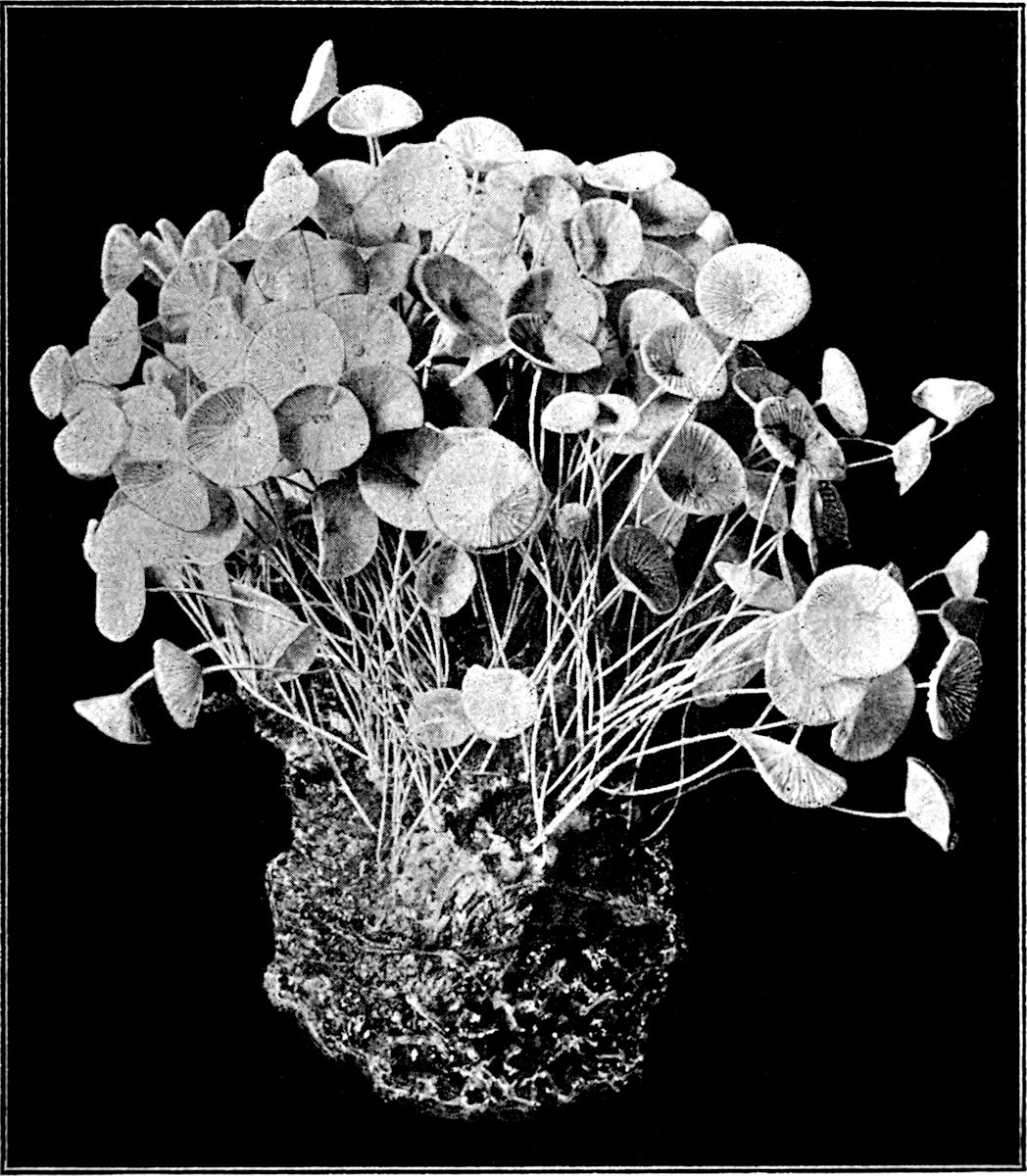
Fig. 34. Acetabularia mediterranea Lamx. From a specimen in the Cambridge Botanical Museum (nat. size).
Acetabularia. Figs. 33, I, and 34.
With the exception of A. mediterranea Lamx. (fig. 34) the few living species of this genus are confined to tropical seas.
The habit of Acetabularia is well illustrated by the photograph of a cluster of plants of A. mediterranea Lamx.[294] reproduced in fig. 34. The thallus consists of a delicate stalk attached to the substratum by a tuft of basal holdfasts, and expanded distally into a small circular disc 10–12 mm. in diameter and more or less concave above. This terminal cap is made up of a number of laterally fused appendages given off from the upper part of the stalk in the form of a crowded whorl. The whole thallus resembles a small and long-stalked calcareous fungus. In each radially elongated compartment of the fertile cap (fig. 33, I) there are several sporangia (gametangia) developed; these eventually open and produce numerous ciliated gametes which give rise to zygospores by conjugation. Fig. 33, I, represents the cap of an Acetabularia in radial section and surface-view; the two radial compartments seen in section contain the elliptical gametangia; the circular markings at the base of the figure are scars of sterile deciduous branches.
The whole plant is unicellular, each chamber in the disc being in open communication with the stem of the plant.
Acicularia. Fig. 33, C–H.
In a recent monograph on the Acetabularieae, Solms-Laubach[295] has described a new type of these algae which is of special importance from the point of view of the past history of the family. Möbius described an example of Acetabularia in 1889 under the name A. Schencki; this species has since been placed in D’Archiac’s genus Acicularia[296]. Acicularia Schencki[297] bears a close resemblance as regards external form to Acetabularia mediterranea. In the latter species the walls of the terminal disc compartments are calcified, and the cavity of each of the laterally fused members contains numerous free spores; in Acicularia, the cavity of each disc-ray is occupied by a calcareous substance in the form of a spicule containing numerous cavities in each of which is a single sporangium. A single spicule is seen in fig. 33, H, showing the spherical pockets in which the sporangia were originally situated. This species, Acicularia Schencki, has been recorded from Martinique, Guadeloupe, Brazil, and a few other places.
The genus Acicularia was founded by D’Archiac for certain minute calcareous spicules found in the Eocene sands (Calcaire Grossier) of the Paris basin. D’Archiac describes one species, Acicularia pavantina, which he defines as follows:—“Polypier aciculaire, élargi, et légèrement comprimé à sa partie supérieure, qui est échancrée au milieu. Surface couverte de petits pores simples, nombreux, disposés irrégulièrement[298].” The same species is figured also in Michelin’s Iconographie Zoophytologique, and described as an organism of which the exact zoological position is uncertain[299]. After these fossils had been placed in various divisions of the animal kingdom, Carpenter[300] described several specimens as portions of foraminifera. Finally, Munier-Chalmas removed Acicularia to the plant kingdom, and “with rare divination” placed the genus among the Acetabularieae. The history of our knowledge of the true nature of Acicularia is of unusual interest. Some of the specimens of this genus figured in Carpenter’s monograph have the form of imperfect long and narrow bodies tapering to a point at one end and broad at the other (fig. 33, F and G); they are joined together laterally and pitted with numerous small cavities. From the resemblance of such specimens to a fragment of the terminal fertile disc of the recent Acetabularias, Munier-Chalmas referred the fossils to this type of algae. In the living species which were then known the radiating chambers of the disc contained loose sporangia, without any calcareous matrix filling the cavity of the chambers. In the fossil Acicularias, on the other hand, the manner of preservation of the pitted calcareous spicules pointed to the occurrence of sporangia embedded in cavities in a calcareous matrix. Subsequent to Munier-Chalmas’ somewhat daring conclusions as to the relation of Acicularia to Acetabularia, Solms-Laubach found that the species originally described by Möbius as Acetabularia Schencki from Guadeloupe presented exactly those characters in which the fossil specimens differ from Acetabularia. The genus Acicularia formerly restricted to fossil species is now applied also to this single living species Acicularia Schencki.
The genus is thus defined by Solms-Laubach:—
“Discus fertilis terminalis e radiis inter se conjunctis formatus, coronis et inferiore et superiore praeditis, sporae massa mucosa calce incrustata coalitae, pro radio spiculam solidam cuneatam formantes[301].”
As Solms-Laubach points out in his recent monograph, Munier-Chalmas’ conjecture, “which had little to support it in the fossil material, has been more recently proved true in the most brilliant fashion by the discovery of a living species of this genus.”
•••••
1. Acicularia Andrussowi Solms[302]. Fig. 33, C and D. This species was first described by Andrussow[303] as Acetabularia miocenica from the Crimea. It occurs in Miocene rocks south of Sevastopol, and, with Ostrea and Pecten, forms masses of white limestone.
In each sporangial ray of the disc the cavity contains a calcareous spicula bearing spore cavities in four rows. “Round each spore-cavity there is a circular zone which stands out, when viewed in reflected light, through its white colour against the central mass of the spicule, though a sharp contour is not visible[304].” Fig. 33, C, is taken from a somewhat diagrammatic sketch by Andrussow; it shows ten of the fertile rays of the disc. The thick walls of the chambers are seen in the two lowest rays, and in the next two rays the spore-cavities are represented. A more accurate drawing, from Solms-Laubach’s memoir, is reproduced in fig. 33, D. The calcareous spicule with numerous spore-cavities shown in fig. 33, H, is from a fertile ray of the recent species Acicularia Schencki. This corresponds to the spore-containing calcareous matrix in each ray of the disc of Acicularia Andrussowi Solms. The spicule copied in fig. 33, F from one of Carpenter’s drawings[305] of an Eocene specimen bears the closest resemblance to the recent spicule of fig. 33, H, and emphasizes the very close relationship between the fossil forms and the single rare tropical species.
2. Acicularia miocenica Reuss. Another Tertiary species has been described under this name by Reuss[306] from the Miocene of the Vienna district, from the Leithakalk of Moravia and elsewhere. It agrees very closely with the recent species A. Schencki. A section of one of the spicules of this species is shown in fig. 33, E; the dark patches represent the pockets in the calcareous spicule which were originally occupied by sporangia and spores.
Cymopolia. Fig. 33, A, B, M and N.
The genus Cymopolia is at present represented by two species, C. barbata (L.) and C. mexicana, Ag., living in the Gulf of Mexico and off the Canary Islands.
Cymopolia and Acetabularia, with several other calcareous algae, are figured by Ellis and other writers as members of the animal kingdom. Ellis speaks of the species of Cymopolia which he figures as the Rosary Bead-Coralline of Jamaica.
Fig. 33, M, has been drawn from a figure published by Ellis in his Natural History of the Corallines published in 1755[307]. The thallus has the form of a repeatedly forked body, of which the branches are divided into cylindrical joints thickly encrusted with carbonate of lime, but constricted and uncalcified at the limits of each segment. A tuft of hairs is given off from the terminal segment of each branch. The axis of each branch of the thallus is occupied by a cylindrical and unseptate cell which gives off crowded whorls of lateral branches. In the lower part of fig. 33, M, the calcareous investment has been removed, and the branches are seen as fine hair-like appendages of the central cell. The branches given off from the constricted portions of the axis are unbranched simple appendages, but the others terminate in bladder-like swellings, each of which bears an apical sporangium. The sporangia are surrounded and enclosed by the swollen tips of four to six branches which spring from the summit of the sporangial branch. Fig. 33, A, represents part of a transverse section through the calcareous outer portion of a branch of Cymopolia; the darker portions or cavities in the calcareous matrix were originally occupied by the lateral branches and sporangia[308].
In Fig. 33, B, the sporangial branch with the terminal sporangium and three of the investing branches are more clearly shown, the surrounding calcareous investment and the thallus having been removed by the action of an acid.
In a transverse section of a branch from which the organic matter had been removed, and only the calcareous matrix left, one would see a central circular cavity surrounded by a thick calcareous wall perforated by radially disposed canals and containing globular cavities; the canals and cavities being occupied in the living plant by branches and sporangia respectively.
The two circular cavities shown in the figure mark the position of the sporangia which are borne on branches with somewhat swollen tips. From the summit the left-hand sporangial branch shown in fig. 33, A, three of the secondary branches are represented by channels in the calcareous matrix; the two black dots on the face of the sporangiophore being the scars of the remaining two secondary branches.
By the lateral contact of the swollen ends of the ultimate branches enclosing the sporangia the whole surface of the thallus, when examined with a lens, presents a pitted appearance. Each pit or circular depression (fig. 33, N) marks the position of the swollen tip of a branch.