Chapter VII · (continued 2)
Reading edition
This form of thallus represents a type which is met with in several members of the Dasycladaceae. It would carry us beyond the limits of a short account to describe additional recent genera which throw light on the numerous fossil species. For further information as to the recent members of the family, the student should refer to Murray’s Seaweeds[309], and for a more detailed memoir on the group to Wille’s recent contribution to the Pflanzenfamilien[310] of Engler and Prantl. Among the various special contributions to our knowledge of the Dasycladaceae, those by Munier-Chalmas[311], Cramer[312], Solms-Laubach[313], and Church[314], may be mentioned.
PALAEOZOIC SIPHONEAE.
The publication of a short preliminary note by Prof. Munier-Chalmas in the Comptes Rendus for 1877 was the means of calling attention to the exceptional importance of the calcareous Siphoneae as algae possessing an interesting past history, of which satisfactory records had been preserved in rocks of various ages. Decaisne had pointed out in 1842 that certain marine organisms previously regarded as animals should be transferred to the plant kingdom. Such seaweeds as Halimeda, Udotea, Penicillus and others were thus assigned to their correct position. Many fossil algae belonging to this group continued to be dealt with as Foraminifera until Munier-Chalmas demonstrated their true affinities. In Gümbel’s monograph on the so-called Nullipores found in limestone rocks, published in 1871[315], several examples of siphoneous algae are included among the fossil Protozoa.
In recent years there have been several additions to an already long list of fossil Siphoneae. In addition to the numerous and well-preserved specimens, representing a large number of generic and specific forms, which have been collected from the Eocene of the Paris basin, there is plenty of evidence of the abundance of the members of the Dasycladaceae in the Triassic seas. In the Triassic limestones of the Tyrol, as well as in other regions, the calcareous bodies of siphoneous algae have played no inconsiderable part as agents of rock-building[316]. Genera have been recorded from Silurian and other Palaeozoic horizons, and there is no doubt that the Verticillate Siphoneae of to-day are the remnants of an extremely ancient family, which in former periods was represented by a much more widely distributed and more varied assemblage of species. There is probably no more promising field of work in the domain of fossil algae than the further investigation of the numerous forms included in Munier-Chalmas’ class of Siphoneae Verticillatae. A brief description of a few genera from different geological horizons must suffice to draw attention to the character of the data for a phylogenetic history of this group.
The fossil examples of the genus Cymopolia (Polytrypa) were originally described by Defrance[317] in the Dictionnaire des Sciences Naturelles as small polyps under the generic name Polytrypa.
In the Eocene sands of the Paris basin there have been found numerous specimens of short, calcareous tubes which Munier-Chalmas has shewn are no doubt the isolated segments of an alga practically identical with the recent Cymopolia. A section[318] through one of the fossil segments presents precisely the same features as those which are represented in fig. 33, A. The habit of the Eocene alga and its minute structure were apparently almost identical with those of the recent species, Cymopolia barbata. The two drawings of Cymopolia reproduced in fig. 33, A and B, have been copied from Munier-Chalmas’ note in the Comptes Rendus[319]; the corresponding figures given by this author of the Eocene species (Cymopolia elongata Deb.) are practically identical with figs. A and B, and show no points of real difference. The segments of the thallus of the fossil species, as figured by Defrance[320], appear to be rather longer than those of the recent species. The calcareous investment of the axial cell of the thallus was traversed by regular verticils of branches or ‘leaves’; the central branch of each whorl terminates in an oval sporangial cavity, exactly as in fig. 33, A and B; and from the top of this branch there is given off a ring of slender prolongations which terminate on the surface of the calcareous tube as regularly disposed depressions, which were no doubt originally occupied by their swollen distal ends as in the recent species.
Vermiporella.
This generic name was proposed by Stolley for certain branched and curved tubes found in Silurian boulders from the North German drift[321]. The tubes have a diameter of ·5–1 mm., and are perforated by radial canals which probably mark the position of verticils of branches given off at right angles to the central axis. The surface of the tubes is divided into regular hexagonal areas.
The resemblance of these Silurian fossils to Diplopora and other genera favours their inclusion in the Verticillate Siphoneae.
Sycidium. Fig. 32, B.
The fossils included in this genus were first described by Sandberger from the middle Devonian rocks of the Eifel, and referred by him to the animal kingdom. More recently Deecke has suggested the removal of the genus to the calcareous Siphoneae, and such a view appears perfectly reasonable, although without more data it is not possible to speak with absolute certainty.
Sycidium melo. (Sandb.) Fig. 32, B. The specimen represented in fig. 32, B (i), (ii), drawn from Deecke’s figures[322], has the form of a small oval calcareous body, 1 mm. in transverse diameter and 1–1·3 mm. in longitudinal diameter. It is pointed at one end and flattened at the other. At the flatter end there is a circular depression, continued into a funnel-shaped cavity, and on the walls of this cavity there are 18–20 radially disposed ribs, which extend over the surface of the whole body. A series of transverse ribs intersects the vertical ribs at right angles. The calcareous wall is perforated by numerous whorls of circular pores, and the internal cavity is a simple undivided space. Each of these oval bodies (fig. 33, B) is probably the segment of a thallus, and the perforations in the wall may have been originally occupied by lateral prolongations from the unseptate axial cell of the thallus. Sycidium bears a fairly close resemblance to the Tertiary Ovulites.
Diplopora. Fig. 35, A and B.
This genus of algae is characteristic of Triassic rocks, and is especially abundant in Muschelkalk and Lower Keuper limestones of the Alps, Silesia, and elsewhere. The thallus, or rather the calcareous portion of the thallus, has the form of a thick-walled tube, with a diameter of about 4 mm., and occasionally reaching a length of 50 mm. At one end the tube has a rounded and closed termination, and the wall is pierced throughout its whole length by regular whorls of fine canals. Diplopora agrees with Cymopolia in its main features.
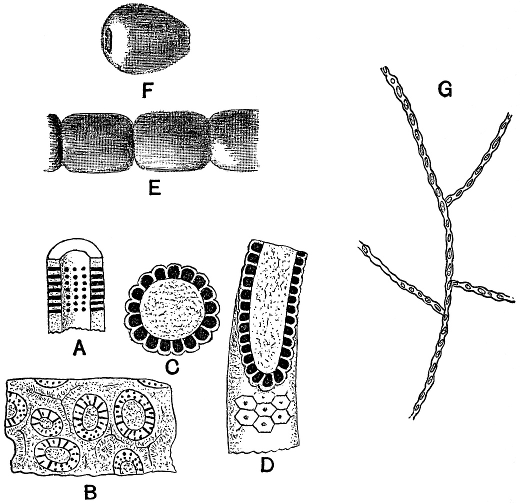
Fig. 35. A, B, Diplopora. × 2. C, D, Gyroporella (after Benecke. × 4). E, Calcareous segments of Penicillus, from a specimen in the British Museum. × 5. F, a single segment of Ovulites margaritula Lam. × 4. G, Confervites chantransioides Born. (after Bornemann. × 150).
Fig. 35, A, affords a diagrammatic view of a Diplopora tube, and shews the arrangement of the numerous whorls of canals. In fig. 35, B, a piece of limestone is represented containing several Diploporas cut across transversely and more or less obliquely. In an obliquely transverse section of a tube perforated by horizontal canals the cavities of the canals necessarily appear as holes or discontinuous canals in the substance of the calcareous wall. The manner of occurrence of the specimens points to the abundance of this genus in the Triassic seas, and suggests that the calcareous tubes of Diplopora may have been important factors in the building up of limestone sediments[323]. In many instances no doubt the carbonate of lime of the thallus has been dissolved and recrystallised, and the original form completely obliterated. As in the rocks built up largely of calcareous Florideae (p. 185) which have lost their structure, it is a legitimate inference that some of the limestone rocks which shew no trace of organic structure may have been in part derived from the calcareous incrustation of various algal genera.
Gyroporella. Fig. 35, C and D.
In this genus from the Alpine Trias the structure of the calcareous tube is very similar to that in Diplopora, but in Gyroporella the canals form less distinct whorls and are closed externally by a small plate, as seen in figs. 35, C and D.
As Solms-Laubach has pointed out, the branch-systems of Diplopora, Gyroporella and other older genera are much simpler than in the Tertiary genera Dactylopora and others[324].
A species of Gyroporella, G. bellerophontis, has recently been described by Rothpletz[325] from Permian rocks in the Southern Tyrol. The thallus is tubular in form and has a diameter of ·5–1 mm.
Dactylopora.
The genus Dactylopora was founded by Lamarck[326] on some fossil specimens from the Calcaire Grossier and included among the Zoophytes. D’Orbigny afterwards included it among the Foraminifera, and the structure of the calcareous body has been described by Carpenter[327] and other writers on the Foraminifera. In a specimen of Dactylopora cylindracea Lam. from the Paris basin, for which I am indebted to Munier-Chalmas, the tubular thallus measures 4 mm. in diameter; at the complete end it is closed and bluntly rounded. The wall of the tube is perforated by numerous canals, and contains oval cavities which were no doubt originally occupied by sporangia. The shape of the specimens is similar to that of Diplopora, but the canals and cavities present a characteristic and more complex appearance, when seen in a transverse section of the wall, than in the older genus Diplopora. Gümbel has given a detailed account of this Tertiary genus in his memoir on Die sogenannten Nulliporen[328]; he distinguishes between Dactyloporella and Gyroporella by the existence of cavities in the calcareous wall of the tube in the former genus, and by their absence in the latter. The oval cavities in a Dactyloporella were originally occupied by sporangia; in Diplopora and Gyroporella the sporangia were probably borne externally and on an uncalcified portion of the thallus.
•••••
In addition to the few examples of fossil species described above there are numerous others of considerable interest, which illustrate the great wealth of form among the Tertiary and other representatives of the Verticillate Siphoneae.
Reference has already been made to Vermiporella as an example of a Silurian genus. Other genera have been described by Stolley from Silurian boulders in the North-German drift under the names Palaeoporella, Dasyporella and Rhabdoporella[329]; the latter genus is compared with the Triassic Diplopora, and the two preceding with the recent Bornetella.
Schlüter has transferred a supposed Devonian Foraminiferal genus, Coelotrochium[330], to the list of Palaeozoic Siphoneae. Munier-Chalmas regards some of the fossils described by Saporta under the name of Goniolina[331], and classed among the inflorescences of pro-angiospermous plants, as examples of Jurassic Siphoneae. The shape and surface-features of some of the examples of Goniolina suggest a comparison with Echinoid spines, but the resemblance which many of the forms in the Sorbonne collection present to large calcareous Siphoneae is still more striking. A comparison of Saporta’s fig. 5, Pl. xxxiii. and fig. 4, Pl. xxxii. in volume iv. of the Flore Jurassique, with the figures given by Solms-Laubach[332] and Cramer[333] of species of Bornetella brings out a close similarity between Goniolina and recent algae; the chief difference being the greater size of the fossil forms. The possibility of confounding Echinoid spines with calcareous Siphoneae is illustrated by Rothpletz[334], who has expressed the opinion that Gümbel’s Haploporella fasciculata is not an alga but the spine of a sea-urchin.
Among Cretaceous forms, in addition to Goniolina, which passes upwards from Jurassic rocks, Triploporella[335] and other genera have been recorded.
Uteria[336] is an interesting type of Tertiary genera; it occurs in the form of barrel-shaped rings, which are probably the detached segments of a form in which the central axial cell was encrusted with carbonate of lime, but the sporangia and the whorls of branches differed from those of Cymopolia in being without a calcareous investment.
b. Confervoideae.
Without attempting to describe at length the fossil forms referred to this division of the Chlorophyceae, there is one fossil which deserves a passing notice. Brongniart in 1828[337] instituted the generic term Confervites for filamentous fossils resembling recent species of confervoid algae. Numerous fossils have been referred to this genus by different authors, but they are for the most part valueless and need not be further considered. In 1887 Bornemann described some new forms which he referred to this genus from the Cambrian rocks of Sardinia. He describes the red marble of San Pietra, near Masne, as being in places full of the delicate remains of algae having the form of branched filaments, and appearing in sections of the rock as white lines on a dark crystalline matrix. In fig. 35, G, one of these Sardinian specimens is represented. This form is named Confervites Chantransioides[338]; the thallus consists of branched cell-filaments, having a breadth of 6–7µ, and composed of ovate cells. It is possible that this is a fragment of a Cambrian alga, but the figures and descriptions do not afford by any means convincing evidence. From post-Tertiary beds various genera, such as Vaucheria and others, have been recorded, but they possess but little botanical value.
C. INCERTAE SEDIS.
Fossils in Boghead ‘Coal’ referred by some authors to the Chlorophyceae.
During the last few years much has been written by two French authors, Dr Renault and Prof. Bertrand, on the subject of the so-called Boghead of France, Scotland, and other countries. They hold the view that the formation of the extensive beds of this carbonaceous material was due to the accumulation and preservation of enormous numbers of minute algae which lived in Permo-Carboniferous lakes.
In an article contributed to Science-Progress in 1895 I ventured to express doubts as to the correctness of the conclusions of MM. Renault and Bertrand[339]. Since then Prof. Bertrand has very kindly demonstrated to me many of his microscopic preparations of various Bogheads, and I am indebted to Prof. Bayley Balfour of Edinburgh for an opportunity of examining a series of sections of the Scotch Boghead. The examination of these specimens has convinced me of the difficulties of the problems which many investigators have tried to solve, but it has by no means led me to entirely adopt the views expressed by MM. Bertrand and Renault.
BOGHEAD.
The Boghead or Torbanite of Scotland was rendered famous by a protracted lawsuit tried in Edinburgh from July 29th to August 4th, 1853. A lease had been granted by Mr and Mrs Gillespie, of Torbanehill, in Fifeshire, to Messrs James Russell and Son, coal-masters of Falkirk, of “the whole coal, ironstone, iron-ore, limestone, and fire-clay (but not to comprehend copper, or any other minerals whatsoever, except those specified) with lands of Torbanehill[340].” After the Boghead had been worked for two years the Gillespies challenged the right of Messrs Russell, and argued that the valuable mineral Torbanite was not included among the substances named in the agreement. The defendants maintained that it was a coal, known as gas-, cannel- or parrot-coal. A verdict was given for the defendants. Some of the scientific experts who gave evidence at the trial considered that the Boghead afforded indications of organic structure, while others regarded it as essentially mineral in origin.
The Torbanite or Boghead is a close-grained brown rock, of peculiar toughness and having a subconchoidal fracture. It contains about 65% carbon, with some hydrogen, oxygen, sulphur, and mineral substances. A thin section examined under the microscope presents the appearance of a dark and amorphous matrix, containing numerous oval, spherical and irregularly shaped bright orange-yellow patches. Fig. 36, 1 shows the manner of occurrence of the yellow bodies in a piece of Scotch Boghead, as seen in a slightly magnified horizontal section. Under a higher power the light patches in the figure reveal traces of a faint radial striation, which in some cases suggests the occurrence of a number of oval or polygonal cells.
The Autun Boghead possesses practically the same structure. The yellow bodies are often sufficiently abundant to impart a bright yellow colour to a thin section. If the section is vertical the coloured bodies are seen to be arranged in more or less regular layers parallel to the plane of bedding.
The Kerosene shale of New South Wales agrees closely with the Scotch and French Boghead; it is approximately of the same geological age, and is largely made up of orange or yellow bodies similar to those of the European Boghead, but much more clearly preserved.
The nature and manner of formation of the various forms of coal should be dealt with in a later chapter devoted to the subject of plants as rock-builders, but in view of the recent statements as to the algal nature of these bituminous deposits it may not be out of place to state briefly the main conclusions of the French authors.
MM. Renault and Bertrand regard each of the yellow bodies in the European and Australian Boghead as the thallus of an alga. To the form which is most abundant in the Kerosene shale they have given the generic name of Reinschia, while that in the Scotch and French Boghead is named Pila.
Reinschia. Fig. 36, 3.
A section of a piece of Kerosene shale at right angles to the bedding appears to be made up of fairly regular layers of flattened elliptical sacs of an orange or yellow colour. Each sac or thallus is about 300µ in length and 150µ broad (fig. 36, 3). A single row of cells constitutes the wall surrounding the central globular cavity. The cells are more or less pyriform in shape, and the cell-cavities are filled with a dark substance, described by Renault and Bertrand as protoplasm, and the cell-walls are fairly thick. In some of the larger specimens there are often found a few smaller sacs enclosed in the cavity of the partially disorganised mother-thallus. In the larger specimens the wall is usually invaginated in several places, giving the whole thallus a lobed or brain-like appearance. The supposed alga, which makes up ⁹⁄₁₀ths of the contents of a block of Kerosene shale, is named Reinschia Australis; it is regarded by the authors of the species as nearly related to the Hydrodictyaceae or Volvocineae.
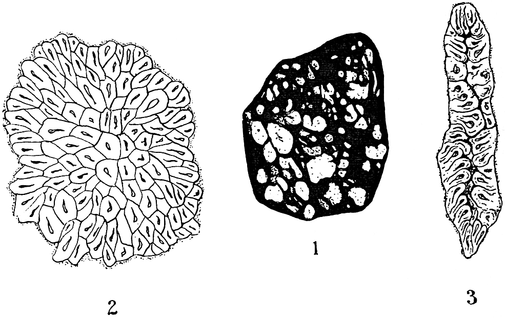
Fig. 36, 1. Section of a piece of Scotch Torbanite. Slightly enlarged. 2. Pila bibractensis from the Autun Boghead, × 282 (after Bertrand). 3. Reinschia Australis, from the Kerosene shale of New South Wales, × 592 (after Bertrand).
In the Kerosene shale from certain localities in New South Wales Bertrand recognises a second form of thallus, which he refers to the genus Pila, characteristic of the European Bogheads.
Pila. Fig. 36, 2.
The “thallus” characteristic of the Scotch Boghead has been named Pila scotica, and that of the Autun Boghead, Pila bibractensis.
In the latter form, which has been studied in more detail by MM. Renault and Bertrand, the thallus consists of about 6–700 cells, and is irregularly ellipsoidal in form, from ·189–·225mm. in length, and ·136–·160mm. broad. The surface-cells are radially disposed and pyramidal in shape, the internal cells are polygonal in outline and less regularly arranged (fig. 36, 2). The Pila thalli make up ¾ths of the mass in an average sample of the Autun Boghead. The Autun Boghead often contains siliceous nodules, and sections of these occasionally include cells of a Pila in which the protoplasmic contents and nuclei have been described by the French authors. The evidence for the existence of these supposed nuclei is, however, not entirely satisfactory; sections of silicified thalli which were shown to me by Prof. Bertrand did not satisfy me as to the minute histological details recognised by Bertrand and Renault.
The species of Pila are compared with the recent genus Celastrum, and regarded as most nearly allied to the Chroococcaceae or Pleurococcaceae among recent algae. Prof. Bornet[341] has suggested Gomphosphaeria as a genus which presents a resemblance to the Autun Pila.
In addition to the Bogheads of Autun, Torbanehill, and New South Wales, there are similar Palaeozoic deposits in Russia, America, and various other parts of the world. Full details of the structure of Boghead and the supposed algae referred to Reinschia, Pila, and other genera will be found in the writings of Bertrand and Renault[342].
The Kerosene shale of New South Wales affords the most striking and well-preserved examples of the cellular orange and yellow bodies referred to as the globular thalli of algae. It is almost impossible to conceive a purely inorganic material assuming such forms as those which occur in the Australian Boghead. On the other hand, it is hardly less easy to understand the possibility of such explanations as have been suggested of the organic origin of these characteristic bodies.
The ground-mass or matrix of the Boghead is referred to a brown ulmic precipitate thrown down on the floor of a Permian or Carboniferous lake, probably under the action of calcareous water. In this material there accumulated countless thalli of minute gelatinous algae, which probably at certain seasons completely covered the surface of the waters, as the fleurs d’eau in many of our fresh-water lakes. In addition to the thalli of Reinschia and Pila the Bogheads contain a few remains of various plant fragments, pollen-grains, and pieces of wood. Fish-scales and the coprolites of reptiles and fishes occur in some of the beds. On a piece of Kerosene shale in the Woodwardian Museum, Cambridge, there are two well-preserved graphitic impressions of the tongue-shaped fronds of Glossopteris Browniana, Brongn. There can be little doubt that the beds of Boghead were deposited under water as members of a regular sequence of sedimentary strata. The yellow bodies which form so great a part of the beds are practically all of the same type. Reinschia and Pila cannot always be distinguished, and it would seem that there are no adequate grounds for instituting two distinct genera and referring them to different families of recent algae.
Stated briefly, my conclusion is that the algae of the French authors may be definite organic bodies, but it is unwise to attempt to determine their affinities within such narrow limits as have been referred to in the above résumé. The structure of the bituminous deposits is worthy of careful study, and it is by no means impossible that further research might lead us to accept the view of the earlier investigators, that the brightly coloured organic-like bodies may be inorganic in origin.
D. RHODOPHYCEAE. (Florideae. Red Algae.)
The thallus of the members of this group assumes various forms, and consists of branched cell-filaments of a more or less complex structure. Cells of the thallus contain a red colouring matter in addition to the green chlorophyll. The reproduction is asexual and sexual; the formation of asexual reproductive cells (tetraspores) in groups of four in sporangia is a characteristic method of reproduction. Sexual reproduction is effected by means of distinct male and female cells.
With the exception of a few fresh-water genera all the red algae are marine. The Rhodophyceae, like the Cyanophyceae and Chlorophyceae, include a shell-boring form which has been found in the common razor-shell[343]. Several genera live as endophytes in the tissues of other algae. The recent species of this section of algae are characteristic of temperate and tropical seas. One subdivision of the red algae, the Corallinaceae, is extremely important from a geological point of view and must be dealt with in some detail.
Corallinaceae.
The thallus is usually encrusted with carbonate of lime; it is of a branched cylindrical form in the well-known Corallina officinalis, Linn. of the British coasts, of an encrusting and foliaceous type, in the genus Lithophyllum, and of a more coral-like form in the genus Lithothamnion. The reproductive organs occur in conceptacles, having the form of small depressed cavities in the thallus, or projecting as warty swellings above the surface of the plant. Asexual reproduction is by means of tetraspores formed in conceptacles resembling those containing the sexual cells. The Corallinaceae may be subdivided into the two families Melobesieae and Corallineae[344].
Melobesieae. Thallus encrusting, leaf- or coral-like; unsegmented. (Melobesia, Lithophyllum, Lithothamnion.)
Corallineae. Cylindrical filamentous and segmented thallus. (Amphiroa and Corallina.)
The genus Corallina is the best known British representative of the Corallinaceae. With other members of the group it was long regarded as a coralline animal, and it is only comparatively recently that the plant-nature of these forms has been generally admitted. Lithophyllum, Lithothamnion, Melobesia, and other genera of the Corallinaceae and some of the Siphoneae play a very important part in the building and cementing of coral-reefs. The pink or rose-coloured calcareous thallus of some of these calcareous algae or Nullipores imparts to coral-reefs a characteristic appearance. In some cases, indeed, the coral-reefs are very largely composed of algae. Saville Kent[345] describes the Corallines or Nullipores of the Australian Barrier-reef as furnishing a considerable quota towards the composition of the coral rock. Mr Stanley Gardiner, who accompanied the coral-boring expedition to the island of Funafuti, has kindly allowed me to quote the following extract from his notes, which affords an interesting example of the importance of calcareous algae as reef-building organisms. “It is quite a misnomer to speak of the outer edge of a reef like this (Rotuma Island) as being formed of coral. It would be far better to call it a Nullipore reef, as it is completely encrusted by these algae, while outside in the perfectly clear water, 10 to 15 fathoms in depth, the bottom has a most brilliant appearance from masses of red, white and pink Nullipores, with only a stray coral here and there.”
Agassiz[346] has given an account of the occurrence of immense masses of Nullipores (Udotea, Halimeda etc.) in the Florida reefs; his description is illustrated by good figures of these algae.
In the Mediterranean there are true Nullipore reefs, which are interesting geologically as well as botanically. Walther[347] has described one of these limestone-banks in the Gulf of Naples which occurs about 1 kilometre from the coast and 30 metres below the surface of the water. Every dredging, he says, brings up numberless masses of Lithothamnion fasciculatum (Lamarck), and L. crassum (Phil.). Between the branches of the algae, gasteropods and other animals become completely enclosed by the growing plants, while diatoms, foraminifera, and other forms of life are abundant. Water percolating through the mass gradually destroys the structure of the algal thalli, and in places reduces the whole bank to a compact structureless limestone.
The same author[348] has also called attention to the importance of Lithophyllum as a constructive element in the coral-reefs off the Sinai peninsula.
Lithothamnion a typical genus of the Corallinaceae may be briefly described.
Lithothamnion. Fig. 37.
Philippi[349] was the first writer to describe this and other genera as plants. He gave the following definition of Lithothamnion:
“Stirps calcarea rigida, e ramis cylindricis vel compressiusculis dichotoma ramosis constans.”
The thallus of Lithothamnion grows attached to the face of a rock or other foundation, and forms a hard, stony mass, assuming various coralline shapes. The exposed face may have the form of numerous short branches or of an irregular warty surface.
In section (fig. 37, A.) the lower part of the thallus is seen to be made up of rows of cells radiating out from a central point, and the upper portion consists of vertical and horizontal rows of cells. The whole body is divided up into a large number of small cells by anticlinal and periclinal walls, and possesses an evident cellular as distinct from a tubular structure. Conceptacles containing reproductive organs are either sunk in the thallus or project above the surface. The two types of structure in a single thallus are shown in fig. 37, A, also a conceptacle containing tetraspores.
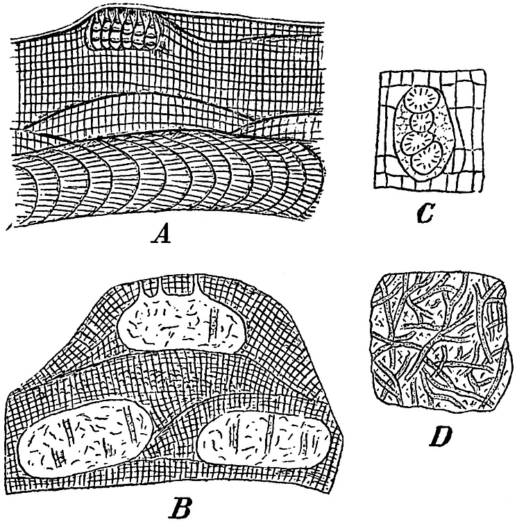
Fig. 37, A. Section of a recent Lithothamnion (after Rosanoff[350], × 200). B. Section of Lithothamnion suganum, Roth (after Rothpletz[351], × 100). C. A conceptacle with tetraspores from a Tertiary Lithothamnion (after Früh[352], × 300). D. Sphaerocodium Bornemanni Roth, (after Rothpletz, × 150).
In the closely allied Lithophyllum the thallus is encrusting, and in section it presents the same appearance as the lower part of a Lithothamnion thallus.
Species of Lithothamnion occur in the Mediterranean Sea, and are abundant in the arctic regions[353], while on the British coasts the genus is represented by four species[354]. Some large specimens of Lithothamnion and Lithophyllum are exhibited in one of the show-cases in the botanical department of the British Museum. For the best figures and descriptions of recent species reference should be made to the works of Hauck, Rosanoff, Rosenvinge, Kjellman and Solms-Laubach[355].
It is to be expected that such calcareous algae as Lithothamnion should be widely represented by fossil forms. In addition to the botanical importance of the data furnished by the fossil species as to the past history of the Corallinaceae, there is much of geological interest to be learnt from a study of the manner of occurrence of both the fossil and recent representatives. As agents of rock-building the coralline algae are especially important. The late Prof. Unger[356] in 1858 gave an account of the so-called Leithakalk of the Tertiary Vienna basin, and recognised the importance of fossil algae as rock-forming organisms. The Miocene Leithakalk, which is widely used in Vienna as a building stone[357], consists in part of limestone rocks consisting to a large extent of Lithothamnion.
Since the publication of Unger’s work several writers have described numerous fossil species of Lithothamnion from various geological horizons. A few examples will suffice to illustrate the range and structure of this and other genera of the Corallinaceae. In dealing with the fossil species it is often impossible to make use of those characters which are of primary importance in the recognition of recent species. The fossil thallus is usually too intimately associated with the surrounding rock to admit of any use being made of external form as a diagnostic feature. The size and form of the cells must be taken as the chief basis on which to determine specific differences. In the absence of conceptacles or reproductive organs it is not always easy to distinguish calcareous algae from fossil Hydrozoa or Bryozoa. In many instances, however, apart from the nature and size of the elements composing the thallus, the conceptacles afford a valuable aid to identification. An example of a fossil conceptacle containing tetraspores is shown in fig. 37, C; it is from a Tertiary species of Lithothamnion, described by Früh from Montévraz in Switzerland.
•••••
1. Lithothamnion mamillosum Gümb. Fig. 32, A (i) and (ii). (p. 155.) This species was first recorded by Gümbel[358] from the Upper Cretaceous (Danian) rocks of Petersbergs, near Maëstricht, on the Belgian frontier. It was originally described as a Bryozoan. The thallus has the form of an encrusting calcareous structure bearing on its upper surface thick nodular branches, as shown in fig. 32, A (ii); in section, A (i), the thallus consists of a regular series of rectangular cells.
The specific name mamillosum has also been given to a recent species by Hauck[359], but probably in ignorance of the existence of Gümbel’s Cretaceous species.
•••••
2. Lithothamnion suganum Roth. Fig. 37, B. The section of this form given in fig. 37, B shows three oval conceptacles filled with crystalline material. The two lower conceptacles originally communicated with the surface of the thallus, but as in recent species the deeper portions of the algal body became covered over by additions to the surface, forming merely dead foundations for new and overlying living tissues.
The cells of the thallus have a breadth of 7–9µ, and a length of 9–12µ.
The specimen was obtained from a Lithothamnion bank, probably of Upper Oligocene age, in Val Sugana[360], in the Austrian Tyrol.
Numerous other species of Jurassic, Cretaceous and Tertiary age might be quoted, but the above may suffice to illustrate the general characters and mode of occurrence of the genus. It is important that the student should become familiar with the Lithothamnion and Lithophyllum types of thallus, in view of their frequent occurrence in crystalline limestone rocks and in such comparatively recent deposits as those of upraised coral-reefs. The coral-rock of Barbados and other West-Indian islands affords a good illustration of the manner of occurrence of fossil coralline algae in association with corals and other organisms[361].
In the fossil species of Lithothamnion hitherto recorded there do not appear to be any important features in which they differ from recent forms; the geological history of the genus so far as it is known, favours the view that the generic characters are of considerable antiquity.
Solenopora. Fig. 38.
Mr A. Brown[362], of Aberdeen, has recently brought forward good evidence for including various calcareous fossils, described by several authors under different names and referred to various genera of fossil animals, in the genus Solenopora, which he places among the coralline algae.
Species of this genus have been described from England, Scotland, Esthonia, Russia, and other countries. The geological range of Solenopora appears to be from Ordovician to Jurassic rocks; in some cases it is an important constituent of beds of limestone.
•••••
Solenopora compacta (Billings). Fig. 38. This species was originally described by Billings as Stromatopora compacta, and afterwards defined by Nicholson and Etheridge. The thallus forms sub-spheroidal masses, from the size of a hemp-seed to that of an orange. The external surface is lobulate; the fractured surface has a porcellaneous and sometimes a fibrous appearance, and is usually white or light brown in colour. In vertical section (fig. 38, B) the cells are elongated and arranged in a radiating and parallel fashion; they often occur in concentric layers. The cells have a diameter of about ¹⁄₁₇ mm. and possess distinctly undulating walls, as seen in a tangential section (fig. 38, A). Brown describes certain larger cells in the thallus (fig. 38, A) as sporangia[363], but it is difficult to recognise any distinct sporangial cavities in the drawing. The example figured is from the Trenton limestone of Canada; a variety of the same species has been recorded from the Ordovician rocks of Girvan in Ayrshire. There appear to be good reasons for accepting Brown’s conclusion that Solenopora belongs to the Corallinaceae rather than to the Hydrozoa, among which it was originally included. After comparing Solenopora with recent genera of Florideae, Brown concludes that “the forms of the cells and cell-walls, the method of increase, and the arrangement of the tissue cells in the various species of Solenopora bear strong evidence of relationship between that genus and the calcareous algae[364].”
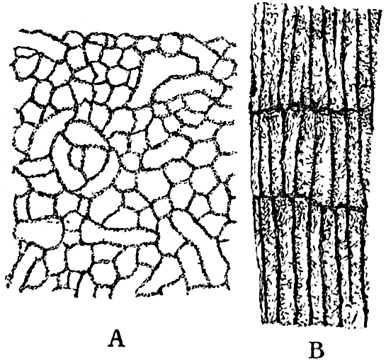
Fig. 38. Solenopora compacta (Billings). A. Tangential section. × 100. B. Vertical section. × 50. (After Brown.)
The importance of the calcareous Rhodophyceae has been frequently emphasised by recent researches, and our knowledge of the rock-building forms is already fairly extensive. We possess evidence of the existence of species of different genera in Ordovician seas, as well as in those of the Silurian, Triassic, Jurassic, and more recent periods. It is reasonable to prophesy that further researches into the structure of ancient limestones will considerably extend our knowledge of the geological and botanical history of the Corallinaceae.
Numerous fossils have been described as examples of other genera[365] of Rhodophyceae than those included in the Corallinaceae, but these possess little or no scientific value and need not be considered.
E. PHAEOPHYCEAE (Brown Algae).
Olive-brown algae, thallus often leathery in texture, composed of cell-filaments or parenchymatous tissue, in some cases exhibiting a considerable degree of internal differentiation. The sexual reproductive organs may be either in the form of passive egg-cells and motile antherozoids or of motile cells showing no external sexual difference.
With one or two exceptions all the genera are marine. They have a wide distribution at the present day, and are especially characteristic of far northern and extreme southern latitudes. The gigantic forms Lessonia, Macrocystis and others already alluded to, belong to this group; also the genus Sargassum, of which the numberless floating plants constitute the characteristic vegetation of the Sargasso Sea.
Palaeobotanical literature is full of descriptions of supposed fossil representatives of the brown algae, but only a few of the recorded species possess more than a very doubtful value; most of them are worthless as trustworthy botanical records. Many of the numerous impressions referred to as species of Fucoides and other genera present a superficial resemblance to the thallus of the common Bladder-wrack and other brown seaweeds. Such similarity of form, however, in the case of flat and branched algal-like fossils is of no scientific value. In many instances the impressions are probably those of an alga, but they are of no botanical interest. The flat and forked type of thallus of Fucus, Chondrus crispus (L.) and other members of the Phaeophyceae is met with also among the red and green algae, to say nothing of its occurrence in the group of thalloid Liverworts, or of the almost identical form of various members of the animal kingdom. The variety of form of the thallus in one species is well illustrated by the common Chondrus crispus (L.). This alga was described by Turner[366] in his classic work on the Fuci under the name of Fucus crispus as “a marine Proteus.” It affords an interesting example of the different appearance presented by the same species under different conditions, and at the same time it furnishes another proof of the futility of relying on imperfectly preserved external features as taxonomic characters of primary importance.
An example of a supposed Jurassic Fucus is shown in fig. 49, and briefly described in the Chapter dealing with fossil Bryophytes.
Several species of Flysch Algae have recently been referred by Rothpletz[367] to the Phaeophyceae under the provisional generic name Phycopsis, but they are of no special botanical interest.
The extremely interesting genus Nematophycus has lately been assigned by a Canadian author[368] to a position in the Phaeophyceae. Although the particular points on which he chiefly relies are not perhaps thoroughly established, there are certain considerations which lead us to include Nematophycus as a doubtful member of the present group of algae.
Nematophycus.
The stem attains a diameter of between 2 and 3 feet in the largest specimens; it is made up either of comparatively wide and loosely arranged tubes pursuing a slightly irregular vertical course accompanied by a plexus of much narrower tubes, or of tubes varying in diameter but not divisible into two distinct types. Rings of growth occur in some forms but not in others. Radially elongated or isodiametric spaces occur in the stem tissues in which the tubes are less abundant.
Reproductive organs unknown, with the possible exception of some very doubtful bodies described as spores.
In 1856 Sir William Dawson proposed the generic name Prototaxites for some large silicified trunks discovered in the Lower and Middle Devonian rocks of Canada. A few years later the same writer[369] published a detailed account of the new fossils and arrived at the conclusion that the Devonian stem showed definite points of affinity with the recent genus Taxus, and the generic name suggests that he regarded it as the type of Coniferous trees belonging to the sub-family Taxineae. The reasons for this determination were afterwards shown by Carruthers to be erroneous. Dawson thought he recognised pits and spiral thickenings in the walls of the tubular elements, as well as pointed ends in some of the latter. The spiral markings were in reality small hyphal tubes passing obliquely across the face of the wider tubes, and the apparent ends of the supposed tracheids were deceptive appearances due to the fact that the tubes had in some cases been cut through in an oblique direction. In 1870 Carruthers[370] expressed the opinion that Dawson’s Prototaxites was a “colossal fossil seaweed” and not a coniferous plant. The same author[371] in 1872 published a full and able account of the genus, and conclusively proved that Prototaxites could not be accepted as a Phanerogam; he brought forward almost convincing evidence in favour of including the genus among the algae. The name Prototaxites was now changed for that of Nematophycus. Carruthers compares the rings of growth in the fossil stems with those in the large Antarctic Lessonia stems, but he regards the histological characters as pointing to the Siphoneae as the most likely group of recent algae in which to include the Palaeozoic genus.
We may pass over various notes and additional contributions by Dawson, who did not admit the corrections to his original descriptions which Carruthers’ work supplied. In 1889 an important memoir appeared by Penhallow[372] of Montreal in which he confirmed Carruthers’ decision as to the algal nature of Prototaxites; he contributed some new facts to the previous account by Carruthers, and expressed himself in favour of regarding the fossil plant as a near ally of the recent Laminariae. The next addition to our botanical knowledge of this genus was made by Barber[373] who described a new specific type of Nematophycus—N. Storriei—found by Storrie in beds of Wenlock limestone age near Cardiff. Solms-Laubach[374], in a recent memoir on Devonian plants, recorded the occurrence of another species of this genus in Middle Devonian rocks near Gräfrath on the Lower Rhine. Lastly Penhallow[375], in describing a new species, lays stress on the resemblance of some of the tubular elements in the stem to the sieve-hyphae of the recent seaweeds Macrocystis and Laminaria. He concludes that the new facts he records make it clear that Nematophycus “is an alga, and of an alliance with the Laminarias.” The recent evidence brought forward by Penhallow is not entirely satisfactory; the drawings and descriptions of the supposed trumpet-shaped sieve-hyphae are not conclusive. On the whole it is probably the better course to speak of Nematophycus as a possible ally of the brown algae rather than as an extinct type of the Siphoneae, but until our knowledge is more complete it is practically impossible to decide the exact position of this Siluro-Devonian genus.
Solms-Laubach[376] has suggested that the generic name Nematophyton, used by Penhallow in preference to Carruthers’ term Nematophycus, is the more suitable as being a neutral designation and not one which assumes a definite botanical position. In view of the nature of the evidence in favour of the algal affinities of the fossil, the reasons for discarding Carruthers’ original name are hardly sufficient.
Before discussing more fully the distribution and botanical position of Nematophycus we may describe at length one of the best known species, and give a short account of some other forms.
•••••
1. Nematophycus Logani (Daws.). Fig. 39, A–E. The stem possesses well marked concentric rings of growth due to a periodic difference in size of the large tubular elements. The tissues consist of two distinct kinds of tubular elements, the larger tubes loosely arranged and pursuing a fairly regular longitudinal course, and having a diameter of 13–35µ; the smaller tubes, with a diameter of 5–6µ, ramify in different directions and form a loose plexus among the larger and more regularly disposed elements. Branching occurs in both kinds of tubes; septa have been recognised only in the smaller tubes. Irregular and discontinuous radial spaces traverse the stem tissues, having a superficial resemblance in their manner of occurrence to the medullary rays of the higher plants.
The best specimens of this species were obtained by Sir William Dawson from the Devonian Sandstones of Gaspé in New Brunswick. The largest stems had a diameter of 3 feet and reached a length of several feet[377]; in some examples Dawson found lateral appendages attached to the stem which he described as “spreading roots.” Externally the specimens were occasionally covered with a layer of friable coal, and internally the tissues were found to be more or less perfectly preserved by the infiltration of a siliceous solution. Most of the examples of Nematophycus from Britain and Germany are much smaller and less perfectly preserved than those from Canada. The Peter Redpath Museum, Montreal, contains several very large blocks of Nematophycus, in many of which one sees the concentric rings of growth clearly etched out by weathering agents in a cross section of a large stem.
In fig. 39, A, a sketch is given of a thin transverse section of a stem, drawn natural size. The lines of growth are clearly seen, and as in coniferous stems the breadth of the concentric zones varies considerably. The short lines traversing the tissues in a radial direction represent the medullary-ray-like spaces referred to in the specific diagnosis. A transverse section examined under a low-power objective presents the appearance of a number of thick-walled and comparatively wide tubes loosely arranged; they may be in contact or separated from one another. If the microscope be carefully focussed through the thickness of the section the transversely-cut tubes appear to move laterally, producing a curiously dazzling effect if the objective is raised or lowered rapidly. This lateral movement is due to the undulating vertical course of the tubes. Under a higher power the lighter-coloured matrix in which the tubes are embedded shows a number of very much smaller and thinner-walled hyphal elements; some of these are cut across transversely, others more or less obliquely and others again longitudinally. These smaller tubes constitute an irregular plexus surrounding and ramifying between the larger elements. The diameter of the larger tubes decreases for a certain distance in a radial direction as seen in a transverse section, and this change in size gives rise to the appearance of concentric lines indicating periodic changes in growth.
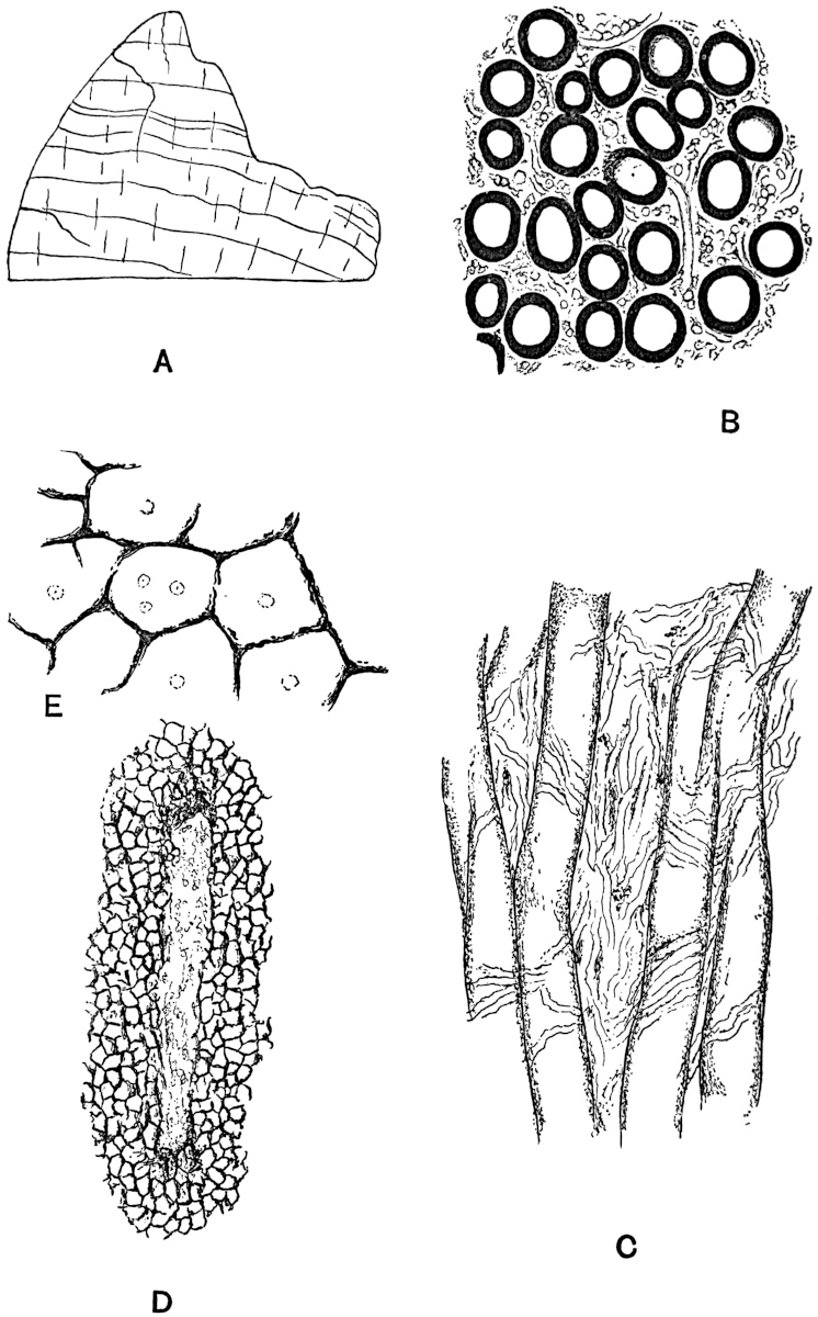
Fig. 39. Nematophycus Logani (Daws.). A. Part of a transverse section from a specimen in the British Museum. (Nat. size.) B. Transverse section from specimens in Mr Barber’s possession. C. Longitudinal section. (B and C × 160.) D. Transverse section showing a radial space. E. Transverse section; a few ‘cells’ more highly magnified. D and E from a specimen in the British Museum.
The radial spaces are characterised by the partial absence of the larger tubes, and as seen in longitudinal sections these spaces constitute regions in which the smaller tubes branch very freely. Fig. 39, B, represents a small piece of a transverse section seen under a fairly high power. In fig. 39, C, the tubes are seen in longitudinal section. The larger elements are unseptate and not very regular in their vertical course through the stem; the smaller elements are seen as fine tubes lying between and across the larger tubes. In the sections I have examined no undoubted transverse septa were detected in any of the tubular elements.
The question as to the possible connection between the larger and smaller elements is one which is not as yet satisfactorily disposed of. Penhallow[378] regards the finer hyphal elements as branches of the larger tubes, but Barber[379], who has carefully examined good material of Nematophycus Logani, was unable to detect any organic connection between the two. My own observations are in accord with those of Barber. Further details and numerous figures of this species of Nematophycus will be found in the memoirs of Carruthers, Penhallow and Barber.
•••••
Some specimens of silicified Nematophycus stems afford particularly instructive examples of the state of preservation or method of mineralisation as a source of error in histological work. The sketches reproduced in fig. 39, D and E, were made from a section of a large specimen of Nematophycus in the British Museum. In fig. D we have one of the radial spaces containing some indistinct small elements, the tissue surrounding the space appears to consist of polygonal cells suggesting ordinary parenchymatous tissue. In fig. E a few of these ‘cells’ are seen more clearly, they have black and ragged walls, and often contain very small and faint circles of which the precise nature is uncertain. The true interpretation of this form of structure was first supplied by Penhallow[380]. The black network simulating parenchymatous tissue consists of the substance of Nematophycus tubes which has been completely redistributed during fossilisation and collected along fairly regular lines, as seen in figs. D and E. The original structure has been almost completely destroyed, and the material composing the walls of the large tubes has finally been rearranged as a network, interrupted here and there by the characteristic radial spaces which remain as evidence of the original Nematophycus characters. It is possible in some cases to trace every gradation from sections exhibiting the normal structure through those having the appearance shown in figs. D and E to others in which the structure is completely lost. Penhallow describes this method of fossilisation in N. crassus (Daws.); an examination of several specimens in the National Collection leads me to entirely confirm his general conclusions, and also to the opinion that N. Logani shows exactly the same manner of mineralisation as N. crassus. The chief point of interest as regards this method of preservation lies in the fact that a fossil described by Dawson[381] as Celluloxylon primaevum, and referred to as a probable conifer, is undoubtedly a badly preserved Nematophycus. Penhallow examined Dawson’s specimens and obtained convincing evidence of their identity with certain forms of highly altered Nematophycus stems.
•••••
2. Nematophycus Storriei Barber. Fig. 40. The specimens on which Barber[382] founded this species were obtained by Mr Storrie from the Tymawr quarry near Cardiff, in beds of Wenlock age. The fragmentary nature of the material is largely compensated for by the excellence of the preservation. We may briefly define the species as follows:
The stem consists of separate interlacing undivided and usually unbranched tubes of varying diameter. Spaces more or less isodiametric in dimensions are scattered through the tissue. The spaces constitute regions in which the tubular elements branch freely.
The main distinguishing features of this British species are (i) the absence of two distinct and well-defined forms of tubular elements. The main part of the stem consists of thick walled tubes similar to those of N. Logani, but the spaces between them are occupied by thinner-walled and smaller tubes varying considerably in diameter; (ii) the form of the spaces which are not radially elongated as in N. Logani.
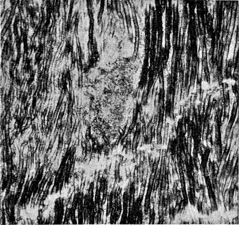
Fig. 40. Nematophycus Storriei Barb. Longitudinal section, from a photograph by Mr C. A. Barber. × 45.
Fig. 40 shows the undulating course of the tubes as seen in a longitudinal section; the black colour of some of the elements is due to the fact that the surface of the wall is seen, while in the lighter-coloured portions of the tubes the wall has been cut through. The lighter patch about the middle of the figure shows the form of one of the spaces in which the tubes are freely branched.
In addition to the two species already described six others have been recorded, but with these we need not concern ourselves in detail. One of these species, N. Hicksi, was found by Dr Hicks[383] in the Denbighshire grits quarry of Pen-y-Glog near Corwen in North Wales. The position of these beds has recently been determined by Mr Lake[384] as corresponding to that of the Wenlock limestone. This species and N. Storriei are both Silurian examples of the genus. It is possible, as Barber has suggested, that the specimens described under these two names should be referred to one species. The specimens found by Hicks were small and imperfectly preserved fragments; Etheridge has given a full description of their structure, and Barber has subsequently examined the material. The preservation is not such as will admit of any very precise specific diagnosis; the fragments are correctly referred to Nematophycus, but their specific characters cannot be clearly determined.
Solms-Laubach[385] has described some fragments of another species of Nematophycus from the Devonian rocks of the Lower Rhine. His specimens are chiefly interesting as extending the geographical range of the genus, and as affording examples of a curious method of preservation. The specimens obtained were small fragments, flattened and very dark brown in colour. The tubular elements consisted of an external membrane of black coal, enclosing a central core of dark red iron-oxide. On burning the fragment on a piece of platinum foil the coal composing the wall of the tubes was removed and the deep-red casts of the tube-cavities remained[386]. The investigation of the structural characters of this imperfect material was conducted by reflected light. Under certain conditions, when it is impossible to obtain thin sections for examination by transmitted light, it is possible to accomplish much, as shown by Solms-Laubach’s work, by means of observation with direct light.
The last species to be noticed is Nematophycus Ortoni recently described by Penhallow. There are no concentric rings of growth, no radial spaces and no smaller hyphae in the tissues of this type of stem. In longitudinal section, the tubes show occasional local expansions of the lumen which Penhallow compares with the ‘trumpet-hyphae’ of some recent brown algae. No actual sieve-plates or transverse walls have been detected, but the general appearance of the tubes is considered to afford distinct evidence of the original existence of such walls. The figures accompanying the description do not carry conviction as to the correctness of the reference of the tubes to imperfectly preserved sieve-hyphae.
The following list, taken, with a few alterations, from Penhallow’s memoir[387], shows the geographical and geological range of the species of Nematophycus hitherto recorded.
Nematophycus Logani (Daws.) Lower Devonian of Gaspé.
Silurian [Wenlock] of England.
Silurian of New Brunswick.
N. Hicksi (Eth.) Silurian. (Wenlock) of N. Wales.
N. crassus (Daws.)[388] Middle Devonian of Gaspé and New York.
N. laxus (Daws.) Lower Devonian of Gaspé.
N. tenuis (Daws.) Lower Devonian of Gaspé.
N. Storriei (Barb.) Silurian (Wenlock) of Wales (Cardiff).
N. dechenianus (Pied.) Upper Devonian of Germany (Gräfrath).
N. Ortoni (Pen.) Upper Erian of Ohio.
In summing up our information as to the structure of Nematophycus we find there are certain points not definitely settled, and which are of considerable importance. The few recorded instances of spore-like bodies by Penhallow and Barber are not satisfactory; we are still ignorant of the nature of the reproductive organs. Such instances of lateral appendages as have been referred to do not throw much light on the habit of the plant. So far as we know at present the stem of Nematophycus was not differentiated internally into a cortical and central region. It may be that the specimens have been only partially preserved, and the coaly layer which occasionally surrounds a stem may represent a carbonised cortex which has never been petrified. The large and loosely arranged tubes constitute the chief characteristic feature of the genus; in some cases (N. Logani) there is an accompanying plexus of smaller hyphae, in others (N. Storriei) there is no definite division of the tissue into two sets of tubes of uniform size, and in N. Ortoni the tubular elements are all of the large type.
Penhallow has recognised the branching of large tubes in N. Logani and N. crassus giving rise to the small hyphal elements. In most specimens, however, no such mode of origin of the smaller tubes can be detected. The spaces which interrupt the homogeneity of the tissues in some forms have been described as branching depots, on account of the frequent occurrence in these areas of much branched hyphae. The function of these spaces (fig. 39, D, and fig. 40) may be connected with aeration of the stem-tissues.
As Carruthers first pointed out the unseptate nature of the elements and the occurrence of large and small tubes forming a comparatively lax tissue suggested affinities with such recent genera as Penicillus, Halimeda, Udotea and other members of the Siphoneae. In those fossil stems which possess tubes of two distinct sizes, we cannot as a rule trace any organic connection between the two sets of tubular elements. Transverse septa have been detected in the tubes of some specimens of N. Logani. These considerations and the large size and habit of growth of the stem leave one sceptical as to the wisdom of assigning the fossil genus to the Siphoneae. On the other hand, apart from the doubtful sieve-hyphae of Penhallow, the manner of growth of the plant, the concentric rings, marked by a decrease in the diameter of the tubes, the lax arrangement and irregular course of the elements, afford points of agreement with some recent Phaeophyceae. The stem of a Laminaria (fig. 29) or of a Lessonia are the most obvious structures with which to compare Nematophycus. The medullary region of a Laminaria or Fucus and of other genera presents a certain resemblance to the tissues of the fossil stems. On the whole we may be content to leave Nematophycus for the present as probably an extinct type of alga, more closely allied to the large members of the Phaeophyceae than to any other recent seaweeds.
Pachytheca.
(A fossil of uncertain affinity.)
There is another fossil occasionally associated with Nematophycus and referred by many writers to the Algae, which calls for a brief notice. Pachytheca is too doubtful a genus to justify a detailed treatment in the present work. Although, as I have elsewhere suggested[389], we are hardly in a position to speak with any degree of certainty as to its affinity, it is not improbable that it may eventually be shown to be an alga.
Without attempting a full diagnosis of the genus, we may briefly refer to its most striking characters.
Pachytheca usually occurs in the form of small spherical bodies, about ·5 cm. in diameter, in Old Red Sandstone or Silurian rocks. In section a single sphere is found to consist of two well marked regions; in the centre, of a number of ramifying and irregularly placed narrow tubes, and in the peripheral or cortical region, of numerous regular and radially disposed simple or forked septate tubes. The tubular elements of the two regions are in organic connection.
The name was proposed by Sir Joseph Hooker for some specimens found by Dr Strickland[390] in the Ludlow bone-bed (Silurian) of Woolhope and May-Hill. Examples were subsequently recorded from the Wenlock limestone of Malvern and from Silurian and Old Red Sandstone rocks of other districts. Hicks[391] found Pachytheca in the Pen-y-Glog grits of Corwen in association with Nematophycus, and the two fossils have been found together elsewhere. This association led to the suggestion that Pachytheca might be the sporangium of Nematophycus, and Dawson[392], in conformity with his belief in the coniferous character of the latter plant, referred to Pachytheca as a true seed.
The best sections of this fossil have been prepared with remarkable skill by Mr Storrie of Cardiff; they were carefully examined and described by Barber in two memoirs[393] published in the Annals of Botany, the account being illustrated by several well executed drawings and microphotographs.
Among other difficulties to contend against in the interpretation of Pachytheca there is that of mineralisation. The preservation is such as to render the discrimination of original structure as distinct from structural features of secondary origin, consequent on a particular manner of crystallisation of the siliceous material, a matter of considerable difficulty.
Suggestions as to the nature of Pachytheca have been particularly numerous; it has been referred to most classes of plants and relegated by some writers to the animal kingdom. The most recent addition to our knowledge of this problematic fossil was the discovery of a specimen by Mr Storrie in which the Pachytheca sphere rested in a small cup, like an acorn fruit in its cupule. This specimen was figured and described by Mr George Murray[394] in 1895; he expresses the opinion that the discovery makes the taxonomic position of the genus still more obscure. Solms-Laubach briefly refers to Pachytheca in connection with Nematophycus, and regards its precise nature almost as much an unsolved riddle now as it was when first discovered. For a fuller account of this fossil reference must be made to the contributions of Hooker[395], Barber[396] and others. The literature is quoted by Barber and more recently by Solms-Laubach[397]. There are several specimens and microscopic sections of Pachytheca in the geological and botanical departments of the British Museum. The genus has been recorded from Shropshire, North Wales, Malvern, Herefordshire, Perthshire and other British localities, as well as from Canada; it occurs in both Silurian and Old Red Sandstone rocks.
Algites.
A generic name for those fossils which in all probability belong to the class Algae, but which, by reason of the absence of reproductive organs, internal structure, or characters of a trustworthy nature in the determination of affinity, cannot be referred with any degree of certainty to a particular recent genus or family.
This term was suggested in 1894[398] as a provisional and comprehensive designation under which might be included such impressions or casts as might reasonably be referred to Algae. The practice of applying to alga-like fossils names suggestive of a definite alliance with recent genera is as a rule unsound. It would simplify nomenclature, and avoid the multiplication of generic names, if the term Algites were applied to such algal fossils from rocks of various ages as afford no trustworthy data by which their family or generic affinity can be established.
V. MYXOMYCETES (MYCETOZOA).
This class of organisms affords an interesting example of the impossibility of maintaining a hard and fast line between the animal and plant kingdom. Zoologists and Botanists usually include the Myxomycetes[399] in the text-books of their respective subjects, and the name Animal-fungi which has been applied to these organisms expresses their dual relationship. They constitute one of three groups which we may include in that intermediate zone or ‘buffer-state’ between the two kingdoms. From a palaeobotanical point of view the Myxomycetes are of little interest, but a very brief reference may be made to them rather for the sake of avoiding unnecessary incompleteness in our classification than from their importance as possible fossils.
They are organisms without chlorophyll, consisting of a naked mass of protoplasm, known as the plasmodium, which may attain a size of several inches. Such plasmodia creep over the surface of decaying organic substrata, and in forming their asexual reproductive cells they are converted into somewhat complex fruits containing spores. The spores produce motile swarm-cells, which eventually coalesce together to form a new plasmodium.
A few examples of fossil Myxomycetes have been recorded from the Palaeozoic and more recent formations, but none of them are entirely beyond suspicion. We may mention three examples of fossils referred to this group, but only one of these is entitled to serious consideration.
Myxomycetes Mangini Ren.[400] It is not uncommon to find distinct traces of original or secondary cell-contents in well preserved petrified plant-tissues. There is often a difficulty, however, in distinguishing between the true cell-contents and the cells of some parasitic or saprophytic intruder. In some petrified corky tissue in a silicified nodule from the Permo-Carboniferous beds of Autun, Renault has recently discovered what he believes to be traces of a Myxomycetous plasmodium. The cork-cells would be without protoplasmic contents of their own, and their cavities contain a number of fine strands stretching from the cell-walls in different directions and uniting in places as irregular or more or less spherical masses. The drawings given by Renault of these irregular reticulated structures with scattered patches of what may possibly be petrified plasmodial protoplasm bear a striking resemblance to the plasmodium of a Myxomycete. A figure of the capillitium of a species of Leocarpus figured by Schröter[401] in his account of the Myxomycetes in Engler and Prantl’s work is very similar to that of Renault’s ‘plasmodium.’
It is by no means inconceivable that the Myxomycetes Mangini may be correctly referred to this group, but the wisdom of assigning a name to such structures may well be questioned.
•••••
The other two examples call for little notice. Messrs Cash and Hick[402] in a paper on fossil fungi from the Coal-Measures refer to some small spherical bodies as possibly the spores of a Myxomycete. They might be referred equally well to numerous other organisms.
Göppert and Menge[403] in their monograph on plants in the Baltic Tertiary Amber, express the opinion that an ill-defined tangle of threads which they figure may be a Myxomycete.
It would serve no useful purpose to quote other instances of possible representatives of fossil Mycetozoa; but the consideration of the above examples may serve to emphasize the desirability of refraining from converting a possibility into an apparently recognised fact by the application of definite generic and specific names.
VI. FUNGI.
The most striking difference between the fungi and algae is the absence of chlorophyll in the former, and the consequent inability of fungi to manufacture their organic compounds from inorganic material. Fungi live therefore either as parasites or saprophytes, and as the same species may pass part of its life in a living host to occur at another stage of its development as a saprophyte, it is impossible to distinguish definitely between parasitic and saprophytic forms. The vegetative body of a fungus, that is the portion which is concerned with providing nourishment and preparing the plastic food-substance for the reproductive organs, is known as the mycelium. It consists either of a single and branched tubular cell known as a hypha, or of several hyphae or thread-like elements (filamentous fungi). The hyphal filaments may be closely packed together and form a felted mass of compact tissue, which in cross section closely simulates the parenchyma of the higher plants. This pseudoparenchymatous form of thallus is particularly well illustrated by the so-called sclerotia; these are sharply defined and often tuberous masses of hyphal tissue covered by a firm rind and containing supplies of food in the inner hyphae. They are able to remain in a quiescent state for some time, and to resist unfavourable conditions until germination and the formation of a new individual take place. The reproductive structures assume various forms; in some of the simpler fungi (Phycomycetes) sexual organs occur, as in the parallel group of Siphoneae among the algae, but in the higher fungi the reproduction is usually entirely asexual. An interesting case has recently been recorded among the more highly differentiated fungi in which distinct sexuality has been established[404]. In addition to the reproductive organs, such as oogonia and antheridia, the asexual cells or spores are borne either in special sporangia, or they occur as exposed conidia on supporting hyphae or conidiophores. Thick-walled and resistant resting-spores of various forms are also met with.
Without going into further details we may very briefly refer to the larger subdivisions of this group of Thallophytes.
PHYCOMYCETES. ZYGOMYCETES, OOMYCETES, including Chytridiaceae, &c. Mycelium usually consisting of a single cell. Reproduction by means of conidia, and in many cases also by the conjugation of two similar hyphae or by the fertilisation of an egg-cell contained in an oogonium.
MESOMYCETES, including the Sub-classes HEMIASCI and HEMIBASIDII. Intermediate between the Phycomycetes and the higher fungi. Multicellular hyphae. No sexual organs.
MYCOMYCETES. including the Sub-classes ASCOMYCETES and BASIDIOMYCETES. Septate vegetative mycelium. No sexual reproduction—as a general rule. Asexual conidia and other forms of spores. In the Ascomycetes the spores are found in characteristic club-shaped cases or asci; in the Basidiomycetes the spores are borne on special branches from swollen cells known as basidia. The sporophore or spore-bearing body in this group may attain a considerable size (e.g. Agaricus, Polyporus, &c.) and exhibit a distinct internal differentiation.
Before describing a few examples of fossil fungi, it is important to consider the general question of their manner of occurrence and determination. Considering the small size and delicate nature of most fungi, it is not surprising that we have but few satisfactory records of well-defined fossil forms. The large leathery sporophores of Polyporus and other genera of the Basidiomycetes, which are familiar objects as yellow or brown brackets projecting from the trunks of diseased forest trees, have been found in a fairly perfect condition in the Cambridgeshire peat-beds, and examples have been described also by continental writers[405]. As a general rule, however, we have to depend on the chance mineralisation or petrifaction of the hyphae of a fungus-mycelium which has invaded the living or dead tissues of some higher plant. In the literature on fossil plants there are numerous recorded species of fungi founded on dark coloured spots and blotches on the impression of a leaf. Most of such records are worthless; the external features being usually too imperfect to allow of accurate identification. The occurrence of recent fungi as discolourations on leaves is exceedingly common, and the characteristic perithecia or compact and more or less spherical cases enclosing a group of sporangia in certain Ascomycetous species, might be readily preserved in a fossil condition.
Ascomycetes.
Some examples of possible Ascomycetous fungi have been recently recorded by Potonié from leaves and other portions of plants of Permian age. There is a distinct superficial resemblance between the specimens he figures and the fructifications of recent Ascomycetes, but in the absence of internal structure, it would be rash to do more than suggest the probable nature of the markings he describes. For one of the fungus-like impressions Potonié proposes the generic name Rosellinites; he compares certain irregularly shaped projections on a piece of Permian wood with the perithecia of Rosellinia, a member of the Sphaeriaceae, and describes them as Rosellinites Beyschlagii Pot.[406] Various other records of similar Ascomycetes-like fossils may be found in palaeobotanical literature[407], but it is unnecessary to examine these in detail. Unless we are able to determine the nature of the supposed fungus by microscopical methods our identifications cannot in most cases be of any great value.
An example of the perithecia of a fungus (Rosellinia congregata [Beck])[408] has been recorded from the Oligocene of Saxony, which would appear to rest on a more satisfactory basis than is often the case. In this particular instance the small projections on a piece of fossil coniferous stem present a form which naturally suggests a fungus perithecium. In cases where the black spots on a fossil stem or leaf possess a definite form and structure, it is perfectly legitimate to refer them to a group of fungi; but in very many instances the forms referred to such genera as Sphaerites and others are of little or no value. Many forms of scale-insects and galls on leaves present an obvious superficial resemblance to epiphyllous fungi, and might readily be mistaken for the fructifications of certain Ascomycetous species. As examples of scale-insects simulating fungi, reference may be made to such genera of the Coccineae as Aspidiotus, Diaspis, Lecanium, Coccus, and others. The female insects lying on the surface of a leaf, if preserved as a fossil impression, might easily be mistaken for perithecia[409].
Another pitfall in fossil mycology may be illustrated by a description of a supposed fungus, Sclerotites Salisburiae[410], Mass. on a Tertiary Ginkgo leaf. The figure given by Massalongo represents a Ginkgo leaf with well marked veins, the lamina between the veins being traversed by short discontinuous and longitudinally-running lines; the latter are referred to as the fungus. In a recent Ginkgo leaf one may easily detect with the naked eye a number of short lines between and parallel to the veins, which if examined in section are found to be secretory canals. There can be little doubt that Sclerotites Salisburiae owes its existence to the preservation of these canals.
The list of fossil fungi given by Meschinelli in Saccardo’s Sylloge Fungorum[411] includes certain species which are of no botanical value, and should have no place in any list which claims to be authentic.
Among the numerous examples of fossil ‘fungi’ which have no claim to be classed with plants, there are some which are in all probability the galleries of wood-eating animals. The radiating grooves frequently found on the inner face of the bark of a pine tree made by species of the beetle Bostrychus might be mistaken for the impressions of the firm strands of mycelial tissue of some Basidiomycetous fungus.
In some notes on fossil fungi by J. F. James[412] contributed to the American Journal of Mycology in 1893, it is pointed out that a supposed fungus described by Lesquereux from the Lower Coal-Measures as Rhizomorpha Sigillariae[413], bears a strong likeness to some insect-burrows, such as those of Bostrychus.
“A new fungus from the Coal-Measures” described by Herzer in 1893[414] may probably be referred to animal agency. In any case there is no evidence as to the fungoid nature of the object represented in the figure accompanying Herzer’s description.
Basidiomycetes.
More trustworthy evidence of fossil fungi is afforded by the marks of disease in petrified tissue and by the presence of true mycelia. In examining closely the calcareous and siliceous plant-tissues from the Coal-Measures and other geological horizons, one occasionally sees fine thread-like hyphae ramifying through the cells or tracheal cavities; in many cases the hyphae bear no reproductive organs and cannot as a rule be referred to a particular type of fungus. If the hyphal filaments are unseptate, they most likely belong to some Phycomycetous species; or if they are obviously septate the Mesomycetes or the Mycomycetes are the more probable groups. Occasionally there may be found indications of the characteristic clamp-connections in the septate filaments; a small semicircular branch, which is given off from a mycelium immediately above a transverse wall, bends round to fuse with the filament just below the septum, thus serving as a small loop-line connecting the cell-cavity above and below a cross wall. Such clamp-connections are usually confined to the hyphae of Basidiomycetes and thus serve as a useful aid in identification. A good example of a clamp-connection in a fossil mycelium is figured by Conwentz[415] in his monograph on the Baltic amber-trees of Oligocene age. The stout and thick type of hypha found in some fossil woods agrees closely with that of Polyporus, Agaricus melleus and other well-known recent Basidiomycetes.
In a section of a piece of lignified coniferous wood recently brought by Col. Feilden from Kolguev island[416], the brown and stout hyphae of a fungus are clearly seen as distinct dark lines traversing the tracheal tissue. The occurrence of septa and the large diameter of the mycelial branches at once suggest a comparison with such recent forms as Agaricus melleus, Polyporus and other Basidiomycetes. The age of the Kolguev wood is not known with any certainty.
The vesicular swellings such as those represented in fig. 41, A, B, D and E, may easily be misinterpreted. Such spherical expansions in a mycelium, either terminal or intercalary, may be sporangia, oogonia or large resting-spores, or non-fungal cell-contents, and it is usually impossible in the absence of the contents to determine their precise nature. Hartig[417] and others have drawn attention to the occurrence of such bladder-like swellings in the mycelia of recent fungi, which have nothing to do with reproductive purposes; under certain conditions the hyphae of a fungus growing in the cavity of a cell or trachea may form such vesicles, and these, as in fig. 42, D, m may completely fill up the cavity of a large tracheid.
Some good examples of bladder-like swellings, such as occur in the mycelium of Agaricus melleus and other recent fungi, have been figured by Conwentz[418] in fossil wood of Tertiary age from Karlsdorf. The swellings in this fossil fungus might easily be mistaken for oogonia or sporangia; especially as they are few in number and spherical in form.
A similar appearance is presented by a mass of tyloses in the cavity of an old vessel or tracheid; and vesicular cell-contents, as in the cells of fig. 41, A, 2–5, may closely simulate a number of thin-walled fungal spores or sporangia.
A good example of such a vesicular tissue, in addition to that already quoted, is afforded by a specimen of an Eocene fern, Osmundites Dowkeri Carr.[419] described by Carruthers in 1870. The ground-tissue cells contain traces of distinct fungal hyphae (fig. 41, B), and in many of the parenchymatous elements the cavity is completely filled with spherical vesicles; in other cases one finds hyphae in the centre of the cell while vesicles line the wall, as shewn in fig. 41, B. Carruthers refers to these bladders as starch grains, and this may be their true nature; their appearance and abundant occurrence in the parenchyma certainly suggest vesicular cell-contents rather than fungal cells. I could detect no proof of any connection between the hyphae and bladders, and the absence of the latter in the cavities of the tracheids, fig. 41, C, favoured the view of their being either starch-grains or other vacuolated contents similar to that in the cells of the Portland Cycad (fig. 41, A) referred to on p. 88.
PATHOLOGY OF FOSSIL TISSUES.
The vacuolated cell-contents partially filling the cells in fig. 41, D, present a striking resemblance to the contents of the cells 2–5 in fig. 41, A. In fig. D the frothy and contracted substance might be easily mistaken for a parasitic or saprophytic fungus, but this resemblance is entirely misleading. It is by no means uncommon to find the cells of recent plants occupied by such vacuolated contents, especially in diseased tissues in which a pathological effect produces an appearance which has more than once misled the most practised observers.
In the important work recently published by Renault on the Permo-Carboniferous flora of Autun, there is a small spore-like body described as a teleutospore, and classed with the Puccineae[420]. We have as yet no satisfactory evidence of the existence of this section of Fungi in Palaeozoic times, and Renault’s description of Teleutospora Milloti from Autun might be seriously misleading if accepted without reference to his figure. The fragment he describes cannot be accepted as sufficient evidence for the existence of a Palaeozoic Puccinia.
The same author refers another Palaeozoic fungus to the Mucorineae under the name of Mucor Combrensis[421]; this identification is based on a mycelium having a resemblance to the branched thallus of Mucor, but in the absence of reproductive organs such resemblance is hardly adequate as a means of recognition.
The occurrence of hyphal cells in calcareous shells and corals has already been alluded to.[422] In addition to the examples referred to above, there is one which has been described by Etheridge[423] from a Permo-Carboniferous coral. This observer records the occurrence of tubular cavities in the calices of Stenopora crinita Lonsd., and attributes their origin to a fungus which he names Palaeoperone endophytica; he mentions one case in which a tube contains fine spherical spore-like bodies which he compares with the spores of a Saprolegnia. As pointed out above (p. 128), it is almost impossible to decide how far these tubes in shells and corals should be attributed to fungi, and how far to algae.
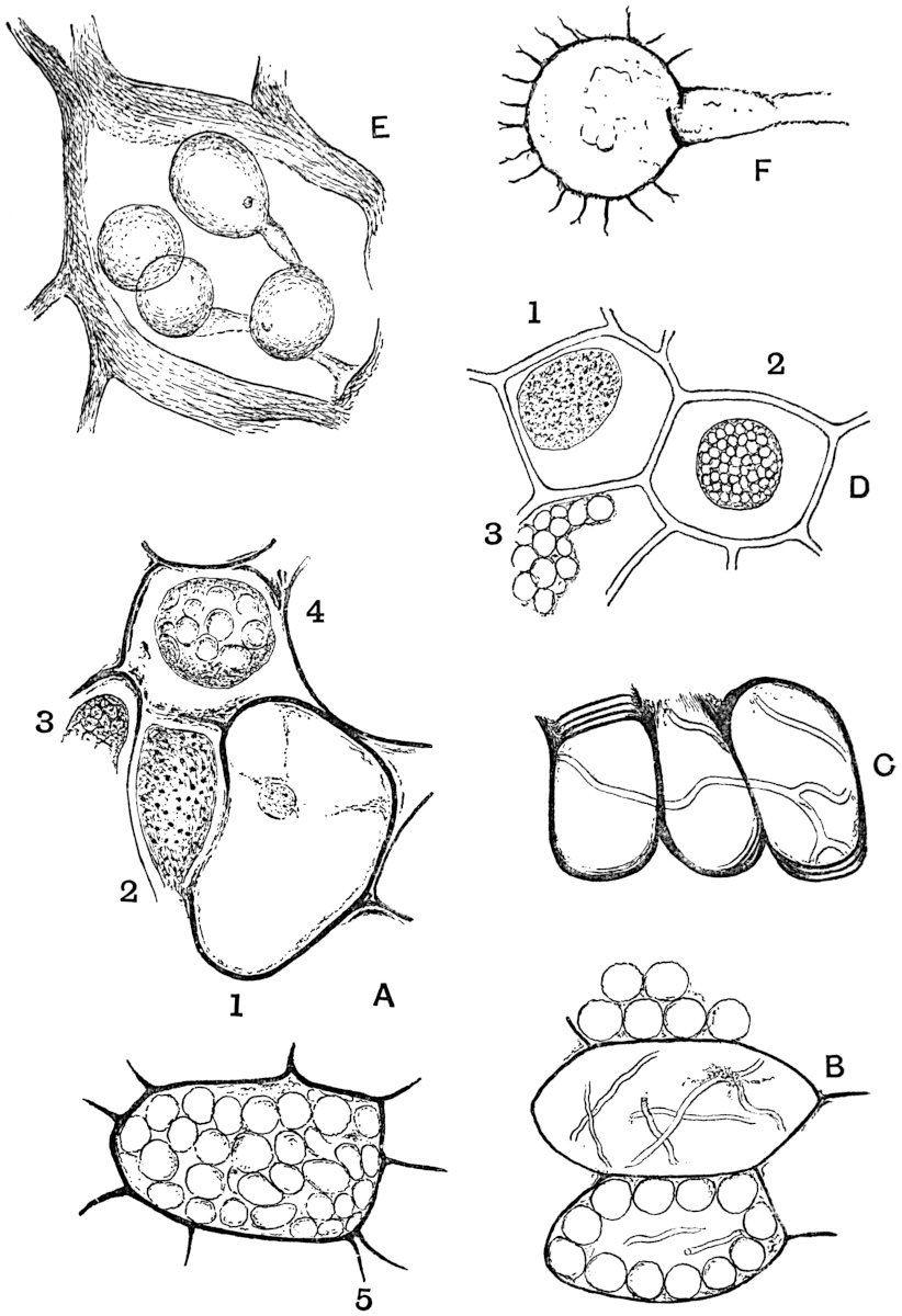
Fig. 41. A. Cells of Cycadeoidea gigantea Sew. × 355. B and C. Parenchymatous cells and scalariform tracheids of Osmundites Dowkeri Carr. × 230. D. Epidermal cells of Memecylon (Melastomaceae) with vacuolated contents. E. Peronosporites antiquarius Smith, (No. 1923 in the Williamson collection). × 230. F. Zygosporites. × 230. (A, B, C and E drawn from specimens in the British Museum; D from a drawing by Prof. Marshall Ward; F from a specimen in the Botanical Laboratory Collection, Cambridge.)
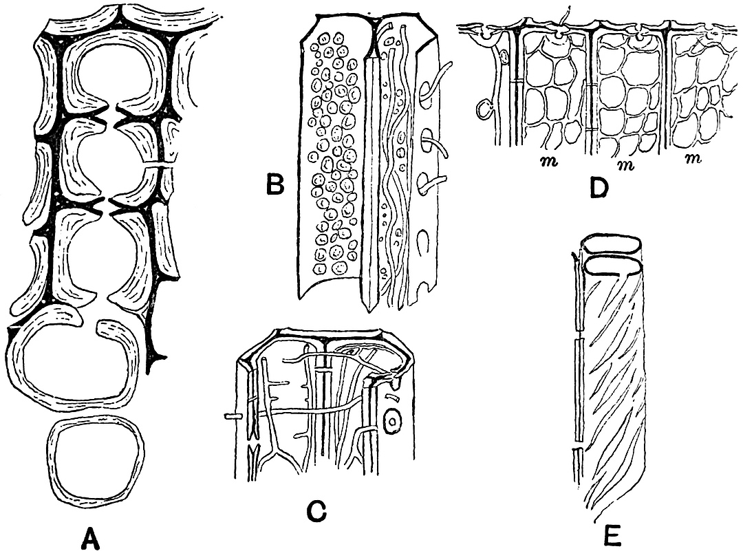
Fig. 42. A, B, C. Tracheids of coniferous wood attacked by Trametes radiciperda Hart. (Polyporus annosus Fr.) D and E. Tracheids attacked by Agaricus melleus Vahl. A, × 650, B–E, × 360. (After Hartig.)
Passing from the direct evidence obtained from the presence of fungal hyphae in petrified tissues, we must draw attention to the indirect evidence of fungal action afforded by many fossil plants. It is important to be familiar with at least the more striking effects of fungal ravages in recent wood in order that we may escape some of the mistakes to which pathological phenomena may lead us in the case of fossils[424].
The gradual dissociation of the elements in a piece of fossil wood owing to the destruction of the middle lamellae, the occurrence of various forms of slit-like apertures in the walls of tracheids (fig. 42, E) and the production of a system of fine parallel striation on the walls of a vessel are among the results produced by parasitic and saprophytic fungi. With the help of a ferment secreted by its hyphae, a fungus is able to eat away either the thickening cell layers or the middle lamellae or both, and if, as in fig. 42, A, only the middle lamellae are left one might easily regard such tissue in a fossil condition as consisting of delicate thin-walled elements. The oblique striae on the walls of a tracheid may often be due to the action of a ferment which has dissolved the membrane in such a manner as to etch out a system of spiral lines, probably as a consequence of the original structure of the tracheids. In distinguishing between the woods of Conifers the presence of spiral thickening layers in the wood element is an important diagnostic character, and it is necessary to guard against the confusion of purely secondary structures, due to fungal action, with original features which may be of value in determining the generic affinity of a piece of fossil wood.
Oochytrium Lepidodendri, Ren. Fig. 43, 1.
Under this name Renault has recently described a filamentous fungus endophytic in the cavities of the scalariform tracheids of a Lepidodendron[425]. The mycelium has the form of slender branched hyphae with transverse septa. Numerous ovoid and more or less spherical sporangia occur as terminal swellings of the mycelial threads. The long axis of the ovoid forms measures 12–15 µ, and the shorter axis 9–10 µ; the contents may be seen as a slightly contracted mass in the sporangial cavity. In some of the sporangia one sees a short apical prolongation in the form of a small elongated papilla, as shown in fig. 43, 1. Renault refers this fungus to the Chytridineae, and compares it with Cladochytrium, Woronina, Olpidium, and other recent genera.
In the immediate neighbourhood of two of the sporangia shown in the uppermost tracheid of fig. 43, 1, there are seen a few minute dark dots which are described as spores petrified in the act of escaping from a lateral pore. This interpretation strikes one as lacking in scientific caution.
The sporangia of Hyphochytrium infestans[426], as figured by Fischer in Rabenhorst’s work bear a close resemblance to those of the fossil. It would seem very probable that Renault’s species may be reasonably referred to the Chytridineae, as he proposes.
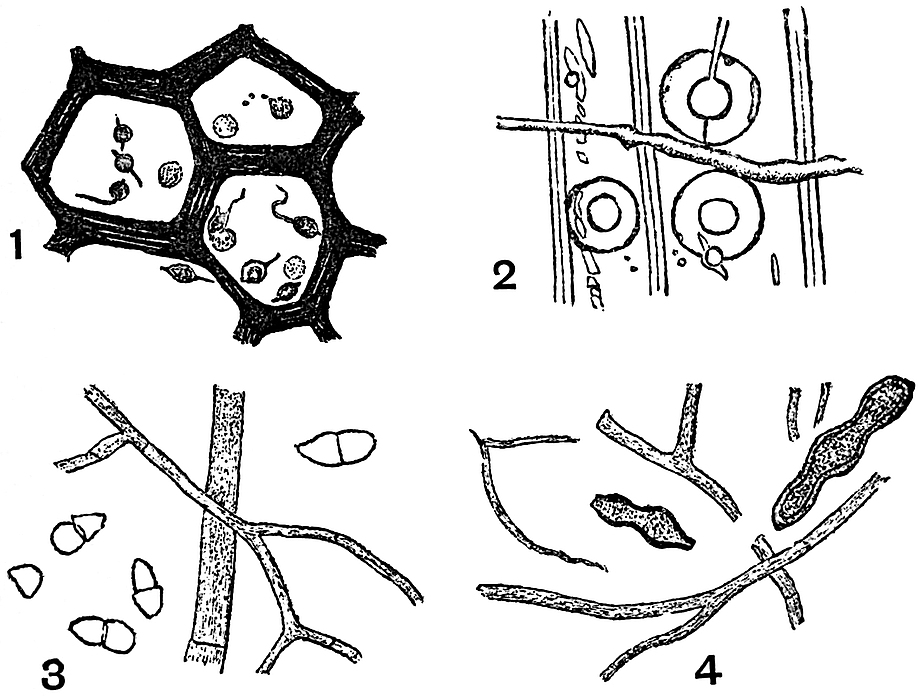
Fig. 43. 1. Oochytrium Lepidodendri, Ren. (After Renault.) 2. Polyporus vaporarius Fr. var. succinea. (After Conwentz.) 3. Cladosporites bipartitus Fel. (After Felix.) 4. Haplographites cateniger Fel. (After Felix.)
Peronosporites antiquarius W. Smith. Fig. 41, E.
In an address to the Geologists’ Association delivered by Mr Carruthers in 1876 a brief reference, accompanied by a small-scale drawing, is made to the discovery of a fungus in the scalariform tracheids of a Lepidodendron from the English Coal-Measures[427]. In the following year Worthington Smith published a fuller account of the fungus, and proposed for it the above name[428], which he chose on the ground of a close similarity between the mycelium and reproductive organs of the fossil form and recent members of the Peronosporeae. In Smith’s description the mycelium is described as bearing spherical swellings containing zoospores. These spherical organs are fairly abundant and not infrequently met with in sections of petrified plant-tissues from the English Coal-Measures; they may be oogonia or sporangia, or in some cases mere vesicular expansions of a purely vegetative hypha. No confirmation has been given to the supposed spores referred to by Smith. Prof. Williamson and others have carefully examined the specimens, but they have failed to detect any trace of reproductive cells enclosed in the spherical sacs[429]. The mycelium does not appear to show any satisfactory evidence of its being septate as figured by Smith.
The example shown in fig. 41 E has been drawn from one of the Williamson specimens: it illustrates the form and manner of occurrence of the characteristic swellings. It is probable that some at least of the vesicles are either sporangia or oogonia, but we cannot speak with absolute confidence as to their precise nature. The general habit and structure of the fungus favour its inclusion in the class of Phycomycetes. The occurrence of several of the vesicles close together on short hyphal branches, as shown in Williamson’s figures, suggests the spherical swellings on vegetative hyphae, but it is impossible to speak with absolute confidence. There is a close resemblance between this English form and one recently described by Renault as Palaeomyces gracilis Ren.[430]; the two fossils should probably be placed in the same genus.
The examples referred to below and originally recorded by Cash and Hick no doubt belong to the same type as Smith’s Peronosporites.
The sketches reproduced in fig. 44 have been drawn from specimens originally described by Cash and Hick in 1878[431]. The sections were cut from a calcareous nodule from the Halifax Coal-Measures containing fragments of various plants and among others a piece of cortical tissue, probably of a Lepidodendron or Stigmaria. In a transverse section of this tissue one sees under a moderately high power that the cells have become partially separated from one another by the destruction of the middle lamellae (fig. 44 A). The cell-cavities and the spaces between the isolated cells contain numerous fine fungal hyphae, which here and there terminate in spherical swellings. One such swelling is shown under a low power in fig. 44 A, in the middle uppermost cell, and more highly magnified in fig. 44 B. In fig. C there are two such swellings (the larger one having a diameter of ·003 mm.) in contact, but the connection does not appear to be organic. The cell-walls of the infected tissue present a ragged and untidy appearance, and in places (e.g. fig. 44 D) the membrane has been pierced by some of the mycelial branches.
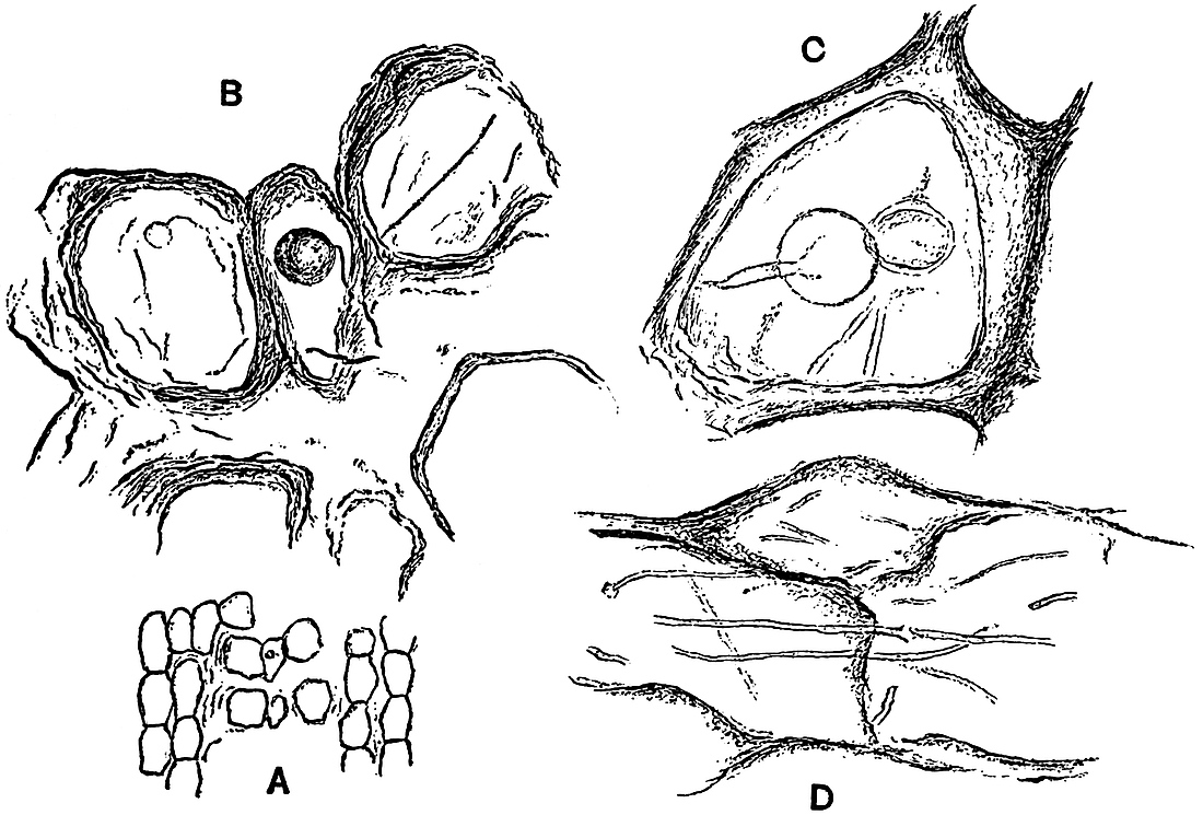
Fig. 44. Cells with fungal hyphae. A. A piece of disorganised tissue, showing the separation of the cells. B. Part of A more highly magnified. C. A single cell containing two swollen hyphae. D. Partially destroyed cell-membranes pierced by fungal hyphae. (Drawn from sections in the Edinburgh Botanical Museum, originally described by Cash and Hick.)
This fungus bears a close resemblance to Peronosporites antiquarius, but it is impossible to determine its precise botanical position without further data. In Cash and Hick’s paper in which the above fungus is briefly dealt with, some small spore-like bodies are figured which the authors speak of as possibly a Myxomycetous fungus[432]. There is however no sound reason for such a supposition.
As examples of Ascomycetous fungi found in silicified wood of Tertiary age, two species may be quoted from Felix.
Cladosporites bipartitus Felix[433], fig. 43, 3.
The mycelium and conidia of this form were discovered in some Eocene silicified wood from Perekeschkul near Baku, on the shores of the Caspian. The conidia are elliptical or pyriform in shape and divided by a transverse septum into two cells. No traces were found of any special conidiophores. The mycelium consists of septate branched hyphae, rendered conspicuous by a brown colouration. Felix compares the fossil with the recent genera Cephalothecium and Cladosporium.
Haplographites cateniger Felix[434], fig. 43, 4.
The conidia of this form were found to be fairly abundant in the silicified tissue investigated by Felix; they occur usually in chains of 2 to 6 conidia having an ovoid or flask-shaped form, with a thick membrane (fig. 43, 4). The mycelium consists of branched hyphae divided into long cylindrical cells by transverse septa; occasional instances were found of an H-shaped fusion between lateral branches of parallel hyphae.
Felix compares this species with examples of the genera Haptographium and Dematium of the family Sphaeriaceae; it was found in the woody tissue of a dicotyledonous stem from Perekeschkul.
Zygosporites sp.
The object represented in fig. 41 F consists of a stalked spherical sac bearing a number of radiating arms which are divided distally into delicate terminations. We find similar bodies figured by Williamson[435] in his IXth and Xth Memoirs on the Coal-Measure plants; he includes some of them under the generic term Zygosporites, and compares them with the zygospores of the freshwater algae Desmideae. Hitherto these spore-like fossils have only been recorded as isolated spheres, but in the example shown in fig. 41 F there is a distinct tubular and thin-walled stalk attached to the Zygosporites. The specimen was found in the partially disorganised cortical tissue of a Lyginodendron stem from the English Coal-Measures. It is difficult to decide as to the precise nature of the fossil, but the presence of the hyphal stalk points to a fungus rather than an alga as the most probable type of plant with which to connect it. It may possibly be a sporangium of a fungus comparable with the common mould Mucor, or it may be a zygospore formed by the conjugation of two hyphae of which only one has been preserved.
POLYPORUS.
For an example of a fossil representative of the Basidiomycetes we may turn to the excellent monograph by Conwentz on the Baltic amber trees, and quote one of the forms which he has described.
Polyporus vaporarius Fr. f. succinea[436], fig. 43, 2.
In several preparations of the wood preserved by petrifaction in amber Conwentz found distinct indications of the ravages of a fungus, which suggested the presence of the recent species Polyporus vaporarius Fr. With the help of the indirect evidence afforded by the pathological effects as seen in the tissues of the host-plant, and the direct evidence of the fungal mycelium Conwentz was led to this identification.
The mycelium is brown in colour, in part thick-walled, and in part with thin walls, transversely septate and not much branched. In the portion of one of Conwentz’ figures reproduced in fig. 43, 2, the rents and holes in the tracheid walls are clearly shown; they afford the indirect evidence of fungal attacks, and are of the same nature as those shown in fig. 42, B, C and E.
•••••
Enough has been said to call attention to the paucity of exact data on which to generalise as to the geological history of fungi. The types selected for description or passing allusion have not been chosen in each case because of their special intrinsic value, but rather as convenient examples by which to illustrate authentic records or to serve as warnings against possible sources of error.
It would seem that we have fairly good and conclusive evidence of the existence in Permo-Carboniferous times of Phycomycetous fungi, but it is not until we pass to post-Palaeozoic or even Tertiary plants that we discover satisfactory representatives of the higher fungi or Mycomycetes. If special attention were paid to the investigation of fossil fungi, it is quite possible that our knowledge of the past history of the group might be considerably extended. It is essential that the greatest caution should be exercised in the identification of forms and in their reference to definite families; otherwise our lists of fossil species will serve to mislead, and to emphasize the untrustworthy character of palaeobotanical data. Unless we feel satisfied as to the position of a fossil fungus it is unwise to use a generic term suggestive of a definite family or recent genus. Such a name as Renault has used in one instance, Palaeomyces, might be employed as a useful and comprehensive designation.
VII. CHAROPHYTA.
CHARACEÆ. NITELLEÆ.
It has been the general custom to include the Characeæ or Stoneworts among the Chlorophyceae (green algae), of which they form a distinctly isolated family. On the whole, it would seem better to follow the course lately adopted by Migula[437] and allow the Characeæ to rank as a family of a distinct group, Charophyta. While agreeing in many respects with plants higher in the scale than Thallophytes, the Stoneworts do not sufficiently resemble the Bryophyta to be included in that group.
The Charophyta are plants containing chlorophyll, living in fresh and brackish water; the stem is jointed, and bears at the nodes whorls of leaves, on which are borne the reproductive organs. The antheridia are spherical in shape and of complex structure, containing numerous biciliate antherozoids. The oogonia are oval in form and contain a single large egg-cell. The Chara-plant is developed from a protonema formed from the germinating oospore. Vegetative reproduction is effected by means of bulbils, accessory shoots, etc.
The Nitelleæ have not been recognised in a fossil condition. The absence or feeble development of a calcareous incrustation renders the genera of this family less likely to be preserved than such a genus as Chara.
Chareae.
Leaves and stems with or without a cortical investment. Fruit with a five-celled corona. The envelope of the ‘fruit’ and other parts of the plant are frequently encrusted with carbonate of lime.
In the genus Chara, the best known member of the family, the plant as a whole resembles in its general habit and external differentiation of parts the higher plants. The stem consists of long internodes separated by short nodes bearing whorls of leaves. Each internode consists of a long cylindrical cell, which becomes enclosed by a cortical sheath composed of rows of cells which have grown upwards and downwards from the peripheral nodal cells. The cortical cells are usually spirally twisted and impart to the stem a characteristic appearance; they are divided by transverse walls into numerous cells some of which occasionally grow out into short processes (fig. 45 c). The leaves repeat on a smaller scale the structural features of the stem, but possess a limited growth, whereas the stem has an unlimited power of growth by means of a large hemispherical apical cell. Branches arise in the axils of the leaves. The plants are either monoecious or dioecious. The oogonium is elliptical in shape, and is borne on a short stalk-cell, it contains a single oosphere. The wall of the oogonium is formed of five spirally twisted cells which have grown over it from the five peripheral cells of a leaf-node. The tips of the investing cells project at the apex in the form of a terminal crown or corona (fig. 45, E, c). The antheridia have a complex structure, and produce a very large number of motile antherozoids.
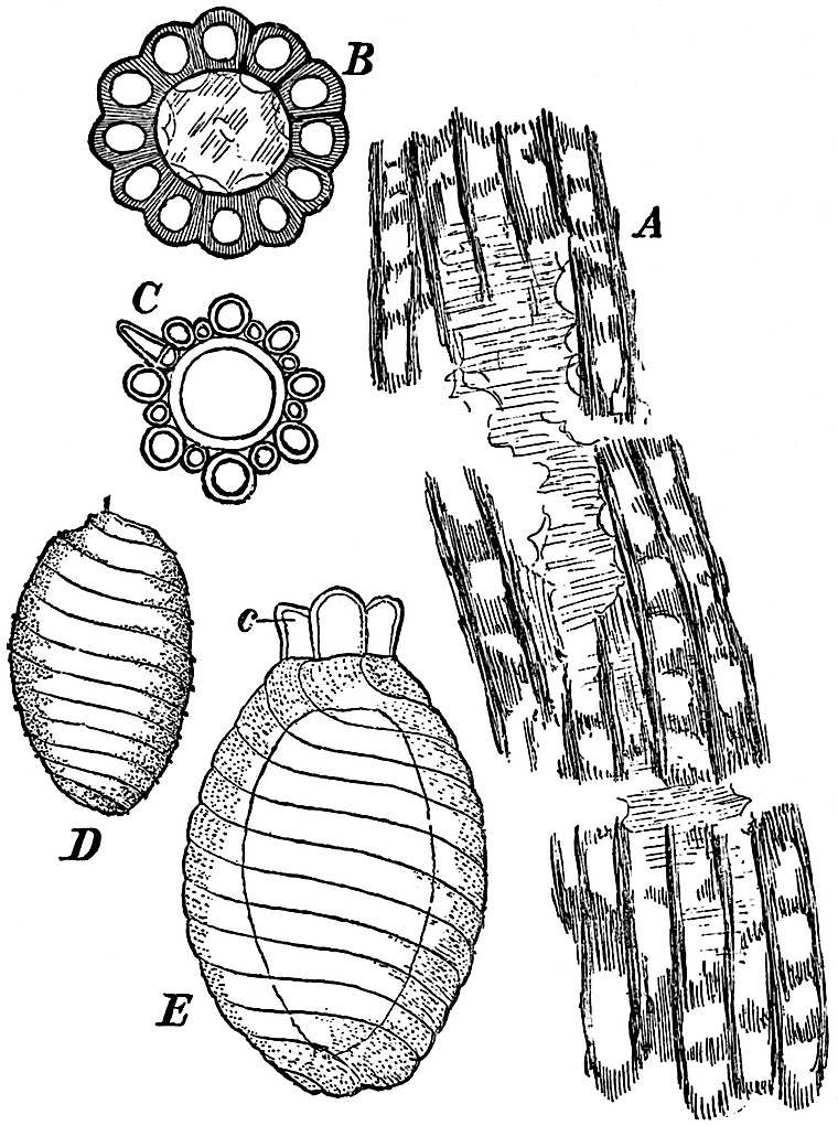
Fig. 45. A and B. Chara Knowltoni Sew. From a section in the British Museum. C. Stem of Chara foetida A. Br. in transverse section (after Migula. × 18). D. Interior of oogonium of C. foetida. E. Oogonium of C. foetida (D and E after Migula. × 50).
After fertilisation, the egg-cell becomes surrounded by a membrane, at first colourless, but afterwards yellow or brown. The inner cell-walls of the cells surrounding the oospore become thicker and darker in colour; the outer walls remain thin and eventually fall away. The lateral walls may or may not become thickened. In most of the Chareae a calcareous deposit is formed between the hard shell and the outer walls of the cells enveloping the oospore. This calcareous shell is developed subsequently to the thickening and hardening of the inner walls of the fruit-case. The cells of the corona and stalk do not become calcareous. In the fossil Charas, it is this calcareous shell that is preserved. In the members of the Chareae the stems are usually encrusted with carbonate of lime, and thus have a much better chance of preservation than the slightly calcareous Nitelleæ.
Chara.
The generic characters have already been described in the brief account of the family Chareae.
The generic name was proposed by Vaillant in 1719[438], and adopted by Linnaeus, who classed the Stoneworts with aquatic phanerogams. As long ago as 1623[439] a figure of Chara was published by Caspar Bauhin as a form of Equisetum. The generic name Chara has usually been applied to recent and fossil species alike. The existing species have a wide distribution; Chara foetida, A. Br., a common British form, occurs in practically all parts of the world. Stems and calcareous ‘fruit-cases’ occur fairly commonly in a fossil state, and differ but little from recent species, at least as regards essential features.
It is difficult to say at what geological horizon the Stoneworts are first represented. The first certain traces of Chara occur in Jurassic rocks, but certain spirally marked subspherical bodies have been recorded from Devonian and Carboniferous strata, which closely resemble Chara oogonia, and may be Palaeozoic representatives of the genus.