Chapter X
Reading edition
I. EQUISETALES (continued).
(CALAMARIEAE.)
In order to minimise repetition and digression the following account of the Calamarieae is divided into sections, under each of which a certain part of the subject is more particularly dealt with. After a brief sketch of the history of our knowledge of Calamites, and a short description of the characteristics of the genus, the morphological features are more fully considered. A description of the most striking features of the better known Calamitean types is followed by a short discussion on the question of nomenclature and classification, and reference is made to the manner of occurrence of Calamites and to some of the possible sources of error in identification.
D. Calamites.
I. Historical Sketch.
In the following account of the Calamarieae the generic name Calamites is used in a somewhat comprehensive sense. As previous writers have pointed out, it is probable that under this generic name there may be included more than one type of plant worthy of generic designation. Owing to the various opinions which have been held by different authors, as to the relationship and botanical position of plants now generally included in the Calamarieae, there has been no little confusion in nomenclature. Facts as to the nature of the genus Calamites have occasionally to be selected from writings containing many speculative and erroneous views, but the data at our disposal enable us to give a fairly complete account of the morphology of this Palaeozoic plant.
In the earliest works on fossil plants we find several figures of Calamites, which are in most cases described as those of fossil reeds or grasses. The Herbarium diluvianum of Scheuchzer[580] contains a figure of a Calamitean cast which is described as probably a reed. Another specimen is figured by Volkmann[581] in his Silesia subterranea and compared with a piece of sugar-cane. A similar flattened cast in the old Woodwardian collection at Cambridge is described by Woodward[582] as “part of a broad long flat leaf, appearing to be of some Iris, or rather an Aloe, but ’tis striated without.” Schulze[583], one of the earlier German writers, figured a Calamitean branch bearing verticils of leaves, and described the fossil as probably the impression of an Equisetaceous plant. It has been pointed out by another German writer that the Equisetaceous character of Calamites was recognised by laymen many years before specialists shared this view.
One of the most interesting and important of all the older records of Calamites is that published by Suckow[584] in 1784. Suckow is usually quoted as the author of the generic name Calamites; he does not attempt any diagnosis of the plant, but merely speaks of the specimens he is describing as “Calamiten.” The examples figured in this classic paper are characteristic casts from the Coal-Measures of Western Germany. Suckow describes them as ribbed stems, which were found in an oblique position in the strata and termed by the workmen Jupiter’s nails (“Nägel”). Previous writers had regarded the fossils as casts of reeds, but Suckow correctly points out that the ribbed character is hardly consistent with the view that the casts are those of reeds or grasses. He goes on to say that the material filling up the hollow pith of a reed would not have impressed upon it a number of ribs and grooves such as occur on the Calamites. He considers it more probable that the casts are those of some well-developed tree, probably a foreign plant. Equisetum giganteum L. is mentioned as a species with which Calamites may be compared, although the stem of the Palaeozoic genus was much larger than that of the recent Horse-tail. The tree of which the Calamites are the casts must, he adds, have possessed a ribbed stem, and the bark must also have been marked by vertical ribs and grooves on its inner face. It is clear, therefore, that Suckow inclined to the view that Calamites should be regarded as an internal cast of a woody plant. Such an interpretation of the fossils was generally accepted by palaeobotanists only a comparatively few years ago, and the first suggestion of this view is usually attributed to Germar, Dawes, and other authors who wrote more than fifty years later than Suckow.
One of the earliest notices of Calamites in the present century is by Steinhauer[585], who published a memoir in the Transactions of the American Philosophical Society in 1818 on Fossil reliquia of unknown vegetables in the Carboniferous rocks. He gives some good figures of Calamitean casts under the generic name of Phytolithus, one of those general terms often used by the older writers on fossils. Among English authors, Martin[586] may be mentioned as figuring casts of Calamites, which he describes as probably grass stems. By far the best of the earlier figures are those by Artis[587] in his Antediluvian Phytology. This writer does not discuss the botanical nature of the specimens beyond a brief reference to the views of earlier authors. Adolphe Brongniart[588], writing in 1822, expresses the opinion that the Calamites are related to the genus Equisetum, and refers to M. de Candolle as having first suggested this view. In a later work Brongniart[589] includes species of Calamites as figured by Suckow, Schlotheim, Sternberg and Artis in the family Equisetaceae. Lindley and Hutton[590] give several figures of Calamites in their Fossil flora, but do not commit themselves to an Equisetaceous affinity.
An important advance was made in 1835 by Cotta[591], a German writer, who gave a short account of the internal structure of some Calamite stems, which he referred to a new genus Calamitea. The British Museum collection includes some silicified fragments of the stems figured and described by Cotta in his Dendrolithen. Some of the specimens described by this author as examples of Calamitea have since been recognised as members of another family.
PETZHOLDT AND UNGER.
In 1840 Unger[592] published a note on the structure and affinities of Calamites, and expressed his belief in the close relationship of the Palaeozoic plant and recent Horse-tails.
An important contribution to our knowledge of Calamites was supplied by Petzholdt[593] in 1841. His main contention was the Equisetaceous character of this Palaeozoic genus. The external resemblance between Calamite casts and Equisetum stems had long been recognised, but after Cotta’s account of the internal structure it was believed that the apparent relation between Equisetum and Calamites was not confirmed by the facts of anatomy. Petzholdt based his conclusions on certain partially preserved Permian stems from Plauenscher Grund, near Dresden. Although his account of the fossils is not accurate his general conclusions are correct. The specimens described by Petzholdt differ from the common Calamite casts in having some carbonised remnants of cortical and woody tissue. A transverse section of one of the Plauenscher Grund fossils is shown in fig. 70. The irregular black patches were described by Petzholdt as portions of cortical tissue, while he regarded the spaces as marking the position of canals like the vallecular canals in an Equisetum. Our more complete knowledge of the structure of a Calamite stem enables us to correlate the patches in which no tissue has been preserved with the broad medullary rays, which separated the wedge-shaped groups of xylem elements; the latter being more resistant were converted into a black coaly substance, while the cells of the medullary rays left little or no trace in the sandstone matrix. The thin black line, which forms the limit of the drawing in fig. 70, external to the carbonised wood, no doubt marks the limit of the cortex, and the appendage indicated in the lower part of the figure may possibly be an adventitious root. It is interesting to note that Unger[594] in 1844 expressed the opinion, which we now know to be correct, that the coaly mass in the specimens described by Petzholdt represented the wood, and that there was no proof of the existence of canals in the cortex as Petzholdt believed.
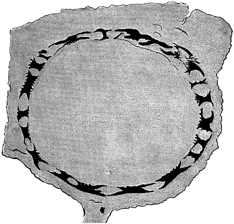
Fig. 70. Transverse section of a Calamite stem, showing carbonised remnants of secondary wood. From a specimen (no. 40934), presented to the British Museum by Dr Petzholdt from Plauenscher Grund, Dresden. ½ nat. size.
Turning to Brongniart’s later work[595] we find an important proposal which led to no little controversy. While retaining the genus Calamites for such specimens as possess a thin bark and a ribbed external surface, showing occasional branch-scars at the nodes, and having such characters as warrant their inclusion in the Equisetaceae, he proposes a second generic name for other specimens which had hitherto been included in Calamites. The fossils assigned to his new genus Calamodendron are described as having a thick woody stem, and as differing from Equisetum in their arborescent nature. Brongniart’s genus Calamodendron is made to include the plants for which Cotta instituted the name Calamitea, and it is placed among the Gymnosperms. This distinction between the Vascular Cryptogam Calamites and the supposed Gymnosperm Calamodendron is based on the presence of secondary wood in the latter type of stem. The prominence formerly assigned to the power of secondary thickening possessed by a plant as a taxonomic feature, is now known to have been the result of imperfect knowledge. The occurrence of a cambium layer and the ability of a plant to increase in girth by the activity of a definite meristem, is a feature which some recent Vascular Cryptogams[596] share with the higher plants; and in former ages many of the Pteridophytes possessed this method of growth in a striking degree.
Although Brongniart’s distinction between Calamites and Calamodendron has not been borne out by subsequent researches, the latter term is still used as a convenient designation for a special type of Calamitean structure. One of the earliest accounts of the anatomy of Calamodendron stems is by Mougeot[597], who published figures and descriptions of two species, Calamodendron striatum and C. bistriatum.
Some years later Göppert[598], who was one of the greatest of the older palaeobotanists, instituted another genus, Arthropitys[599], for certain specimens of silicified stems from the Permian rocks of Chemnitz in Saxony, which Cotta had previously placed in his genus Calamitea under the name of Calamitea bistriata[600]. Göppert rightly decided that the plants so named by Cotta differed in important histological characters from other species of Calamitea. The generic name Arthropitys has been widely adopted for a type of Calamitean stem characterised by definite structural features. The great majority of the petrified Calamite stems found in the English Coal-Measures belong to Göppert’s Arthropitys.
WILLIAMSON.
The next proposal to be noticed is one by Williamson[601] in 1868; he instituted the generic name Calamopitys for a few examples of English stems, which differed in the structure of the wood and primary medullary rays from previously recorded types. We have thus four names which all stand for generic types of Calamitean stems. Of these Calamodendron and Arthropitys are still used as convenient designations for stems with well-defined anatomical characters. The genus Calamitea is no longer in use, and Williamson’s name Calamopitys had previously been made use of by Unger[602] for plants which do not belong to the Calamarieae. As it is convenient to have some term to apply to such stems as those which Williamson made the type of Calamopitys, the name Arthrodendron is suggested by my friend Dr Scott[603] as a substitute for Williamson’s genus.
The twofold division of the Calamites instituted by Brongniart has already been alluded to, and for many years it was generally agreed that both Pteridophytes and Gymnosperms were represented among the Palaeozoic fossils known as Calamites. The work of Prof. Williamson was largely instrumental in proving the unsound basis for this artificial separation; he insisted on the inclusion of all Calamites in the Vascular Cryptogams, irrespective of the presence or absence of secondary wood. By degrees the adherents of Brongniart’s views acknowledged the force of the English botanist’s contention. It is one of the many signs of the value of Williamson’s work that there is now almost complete accord among palaeobotanical writers as to the affinities of Calamitean plants.
In the following account of the Calamites, the generic name Calamites is used in a wide sense as including stems possessing different types of internal structure; when it is possible to recognise any of these structural types the terms Calamodendron, Arthropitys or Arthrodendron are used as subgenera. The reasons for this nomenclature are discussed in a later part of the Chapter.
Genus Calamites, Suckow, 1714 This term was originally applied to the common pith-casts of Calamitean stems, without reference to internal structure.
Subgenera Calamodendron, Brongniart, 1849 These names have primarily reference to internal structure.
Arthropitys Göppert, 1864
Arthrodendron Scott 1897
(= Calamopitys Williamson, 1871
II. Description of the anatomy of Calamites.
a. Stems. b. Leaves. c. Roots. d. Cones.
No fossils are better known to collectors of Coal-Measure plants than the casts and impressions of the numerous species of Calamites. In sandstone quarries of Upper Carboniferous rocks there are frequently found cylindrical or somewhat flattened fossils, varying from one to several inches in diameter, marked on the surface by longitudinal ridges and grooves, and at more or less regular intervals by regular transverse constrictions. Similar specimens are still more abundant as flattened casts in the blocks of shale found on the rubbish heaps of collieries. The sandstone casts are often separated from the surrounding rock by a loose brown or black crumbling material, and the specimens in the shale are frequently covered by a thin layer of coal.
Most of the earlier writers regarded such specimens as the impressions of the ribbed stems of plants similar to or identical with reeds or grasses. Suckow, and afterwards Dawes and others, expressed the opinion that the ordinary Calamite cast represented a hardened mass of sand or marl, which had filled up the pith of a stem either originally fistular or rendered hollow by decay. The investigation of the internal structure confirmed this view, and proved that the surface-features of a Calamite stem do not represent the external markings of the original plant, but the form of the inner face of the cylinder of wood. The ribs represent the medullary rays of the original stem or branch, and the intervening grooves mark the position of the strands of xylem which are arranged in a ring round a large hollow pith[604].
With this brief preliminary account we may pass to a detailed description of the anatomical characters of Calamites.
The genus Calamites may be briefly defined as follows:—
Arborescent plants reaching a height of several meters, and having a diameter of proportional size. In habit of growth the Calamites bore a close resemblance to Equisetum; an underground rhizome giving off lateral branches and erect aerial shoots bearing branches, either in whorls from regularly recurring branch-bearing nodes, or two or three from each node; and in some cases the stems bore occasional branches from widely separated nodes. The leaves were disposed in whorls either as star-shaped verticils on slender foliage shoots, or in the form of a circle of long narrow leaves on the node of a thicker branch. Adventitious roots were developed from the nodal regions of underground and aerial stems. The cones had the form of long and narrow strobili consisting of a central axis bearing whorls of sterile and fertile appendages; the latter in the form of sporangiophores bearing groups of sporangia. The strobili were heterosporous in some cases, isosporous in others. The stems had a large hollow pith bridged across by a transverse diaphragm at the nodes in the centre of the single stele; the latter consisted of a ring of collateral bundles separated from one another by primary medullary rays. Each group of xylem was composed of spiral, annular, scalariform and occasionally reticulate tracheids, the position of the protoxylem being marked by a longitudinal carinal canal. The shoots and roots grew in thickness by means of a regular cambium layer. The cortex consisted of parenchymatous and sclerenchymatous cells, with scattered secretory sacs. The increase in girth of the central cylinder was often accompanied by a considerable development of cortical periderm. The roots differed from the shoots in having no carinal canals, and in the possession of a solid pith and centripetally developed primary xylem groups alternating with strands of phloem.
The above incomplete diagnosis includes only some of the more important structural features of the genus. Thanks to the researches begun by the late Mr Binney of Manchester and considerably extended by Carruthers, Williamson and later investigators, we are now in a position to give a fairly complete account of Calamites. The type of stem most frequently met with in a petrified condition in the English rocks is that to which Göppert applied the name Arthropitys, and it is this subgenus that forms the subject of the following description. Our knowledge of Calamitean anatomy is based on the examination of numerous fragments of petrified twigs and other portions of different specific types of the genus. It is seldom possible to differentiate specifically between the isolated fragments of stems and branches which are met with in calcareous or siliceous nodules. As so frequently happens in fossil-plant material, large specimens showing good surface features and broken fragments with well-preserved internal structure have to be dealt with separately.
YOUNG STEM.
a. Stems.
A transverse section of a young twig, such as is represented in fig. 71, illustrates the chief characteristics of the primary structure of a young branch of Calamites. The figure has been drawn from a section originally described by Hick[605] in 1894. A very young Calamite twig bears an exceedingly close resemblance to the stem of a recent Equisetum. The axial region of the stem may be occupied by parenchymatous cells, or the absence of cells in the centre may indicate the beginning of the gradual formation of the hollow pith, which is one of the characteristics of Calamites. The student of petrified Palaeozoic plants must constantly be on his guard against the possible misinterpretation of Stigmarian ‘rootlets,’ which are frequently found in intimate association with fossil tissues. The intrusion of these rootlets is admirably illustrated by a section of a Calamite stem in the Williamson Collection (No. 1558) in which the hollow pith, 2 cm. broad, contains more than a dozen Stigmarian appendages.
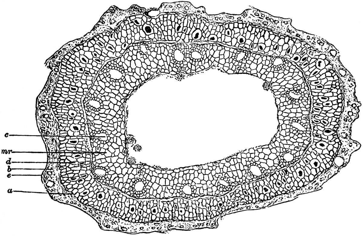
Fig. 71. Transverse section of a young Calamite stem. c, carinal canals; mr, primary medullary rays; a, b, and d, cortex; e, epidermis. From a section in the Manchester Museum, Owens College, × 60.
In the figured specimen of a Calamite twig (fig. 71) there is a clearly marked differentiation into a cortical region and a large stele or central cylinder. The pith-cells are already partially disorganised, but there still remain a few fairly large parenchymatous cells internal to the ring of vascular bundles. The few irregular projections into the cavity of the large pith consist of small fragments of cells, which may be the result of fungal action. Mycelia of fungi are occasionally met with in the tissues of older Calamite stems.
The position of the primary xylem groups is shown by the conspicuous and regularly placed canals, c; these have been formed in precisely the same manner as the corresponding spaces in an Equisetum stem, and they are spoken of in both genera as the carinal canals. Each canal owes its origin to the disorganization and tearing apart of the protoxylem elements and the surrounding cells. This may be occasionally seen in examples of very young Calamites; the canals of a young twig often contain apparently isolated rings which are coils of elongated spiral threads. Fig. 72, B represents the canal of a twig, cut in an oblique direction, in which the remains of spiral tracheids are distinctly seen. In the stem of fig. 71 the development has not advanced far enough to enable us to clearly define the exact limits of each xylem strand. The smaller elements bordering the canals constitute the primary xylem, they are fairly distinct on the outer margin of some of the canals seen in the section. Between the small patches of primary xylem the outward extensions of the parenchyma of the pith constitute the primary medullary rays, mr. The distinct line encircling the canals and primary xylem has been described by Hick as marking the position of the endodermis, but it may possibly owe its existence to the tearing of the tissues along the line where cambial activity is just beginning. This layer of delicate dividing cells would constitute a natural line of weakness. External to this line we have a zone of tissue a, d, containing here and there larger cells with black contents, which are no doubt secretory sacs. It is impossible to distinguish with certainty any definite phloem groups, but in other specimens these have been recognised immediately external to each primary xylem group; the bundles were typically collateral in structure. Towards the periphery of the twig the preservation is much less perfect; the outer portion of the inner cortex, d, consists of rather smaller and thicker-walled cells, but this is succeeded by an ill-defined zone containing a few scattered cells, b, which have been more perfectly preserved. The twig is too young to show any secondary tissue in the cortex; but the tangential walls in some of the cortical cells afford evidence of meristematic activity, which probably represents the beginning of cork-formation. The limiting line, e, possibly represents the cuticularised outer walls of an epidermal layer. The irregularly wavy character of the surface of the specimen is probably the result of shrinking, and does not indicate original surface features.
VASCULAR SYSTEM.
In examining sections of calcareous nodules from the coal seams one meets with numerous fragments of small Calamitean twigs with little or no secondary wood; in some of these there is a small number of carinal canals, in others the canals are much more abundant. The former probably represent the smaller ramifications of a plant, and the latter may be regarded as the young stages of branches capable of developing into stout woody shoots[606]. Longitudinal sections of small branches teach us that the xylem elements next the carinal canals are either spiral or reticulate in character, the older tracheids being for the most part of the scalariform type, with bordered pits on the radial walls. This and other histological characters are admirably shown in the illustrations accompanying Williamson and Scott’s memoir on Calamites. The student should treat the account of the anatomy of Calamites given in these pages as introductory to the much more complete description by these authors. They thus describe the course of the vascular bundles in a Calamitean branch:—
“The bundle-system of Calamites bears a general resemblance to that of Equisetum. A single leaf-trace enters the stem from each leaf, and passes vertically downwards to the next node. In the simplest cases the bundle here forks, its two branches attaching themselves to the alternating bundles which enter the stem at this node. In other cases both the forks attach themselves to the same bundle, so that, in this case, there is no regular alternation. In other cases, again, the bundle runs past one node without forking, and ultimately forms a junction with the traces of the second node below its starting-point. These variations may all occur in the same specimen. The xylem at the node usually forms a continuous ring, for where the regular dichotomous forks of the bundles are absent their place is usually taken by anastomoses[607].”
As in Equisetum, the xylem at the nodes possesses certain characteristic features which distinguish it from the internodal strands. It has already been pointed out that the xylem of Equisetum increases in breadth at the nodes (p. 251, fig. 55, 4); the same is true of Calamites. In fig. 72, C, we have part of a radial section of a Calamite twig in which the broad mass of short nodal tracheids is clearly shown; this nodal wood forms a prominent projection towards the pith. In the lower part of the section the remains of some spiral protoxylem tracheids are seen in a carinal canal.
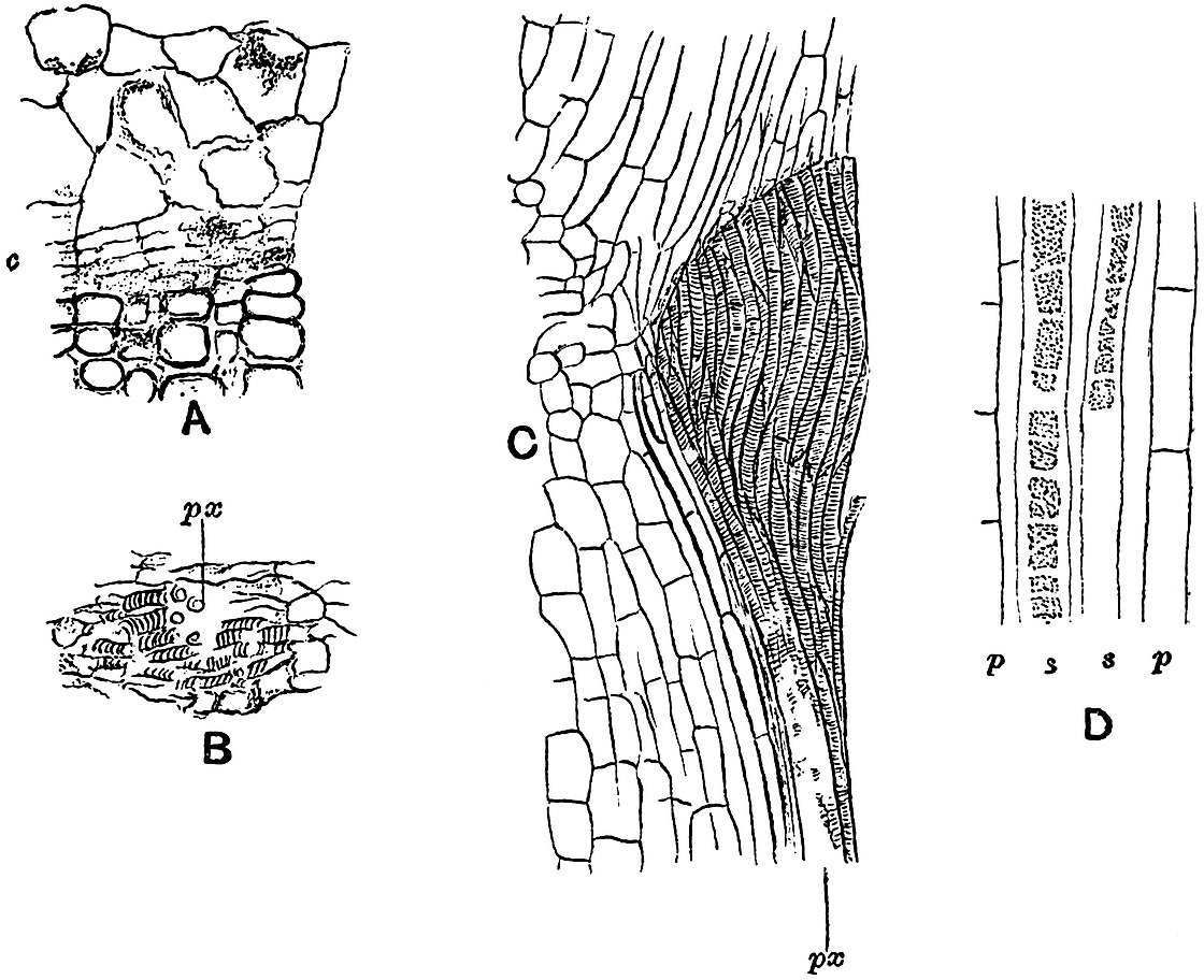
Fig.
72.
A. External xylem elements and cambium, c, with imperfect phloem, × 100.
B. Carinal canal containing protoxylem, px. × 65.
C. Radial longitudinal section through nodal xylem, px. × 35.
D. Phloem elements; s, sieve-tubes; p, p, parenchymatous cells.
(A–C. After Williamson and Scott. D. After Renault.)
The tracheids of the nodal wood are often reticularly pitted, and so differ in appearance from the ordinary scalariform elements.
It is rare to find the phloem clearly preserved, but in specimens where it has been possible to examine this portion of the vascular bundles, it is found to consist of elongated cambiform cells and sieve-tubes. An unusually perfect specimen has been described by Renault[608] in which the phloem elements are preserved in silica. Fig. 72, D, is copied from one of Renault’s drawings, the sieve-tubes, s, s, show several distinct sieve-plates on the lateral walls of the tubes, reminding one to some extent of the sieve-tubes in a Bracken Fern. The cells, p, p, associated with the sieve-tubes are square-ended elongated parenchymatous elements. Another characteristic feature illustrated by longitudinal sections is the nodal diaphragm; except in the smallest branches the interior of each internode is hollow, and the ring of vascular bundles is separated from the pith-cavity by a band of parenchymatous tissue. At each node this parenchyma extends across the central cavity in the form of a nodal diaphragm, as in the stem of Equisetum.
By far the greater number of the petrified fragments of Calamites afford proof of cambial activity, and possess obvious secondary tissues. In exceptionally perfect specimens the xylem tracheids are found to be succeeded externally by a few flattened thin-walled cells which are in a meristematic condition (fig. 72, A, c); these constitute the cambium zone, and it is the secondary structure that results from the activity of the meristematic cells that we have now to consider.
SECONDARY THICKENING.
In petrified examples of branches in which the secondary thickening has reached a fairly advanced stage, the wood is usually the outermost tissue preserved, the more external tissues having been detached along the line of cambium cells. It is only in a few cases that we are able to examine all the tissues of older examples.
The specimen represented in fig. 73 illustrates very clearly the extension of the hollow pith up to the inner surface of the vascular ring; the disorganisation of the pith-cells which had already begun in the twig of fig. 71 has here advanced much further. The bluntly rounded projections represent the prominent primary xylem strands, each of which is traversed by the characteristic carinal canal. Alternating with the wedge-shaped groups of secondary xylem, x, we have the broad principal medullary rays, mr, which become slightly narrower towards the outside. The inner face of each of these wide rays has a concave form, due to the less resistent nature of the medullary-ray cells as compared with the stronger xylem. The regularly sinuous form of the inner face of the vascular cylinder enables one to realise how the Calamite-casts (figs. 82, 99, and 101) have come to have the regular ridges and grooves on their surface. The broad ridges on the cast mark the position of the wide medullary rays, while the grooves correspond to the more prominent ends of the vascular strands. The tissues external to the wood have not been preserved in the example shown in fig. 73. Some silicified specimens described by Stur[609] from Bohemia and now in the Museum of the Austrian Geological Survey, Vienna, admirably illustrate the connection between the surface features of a Calamite cast and the anatomy of the stem.
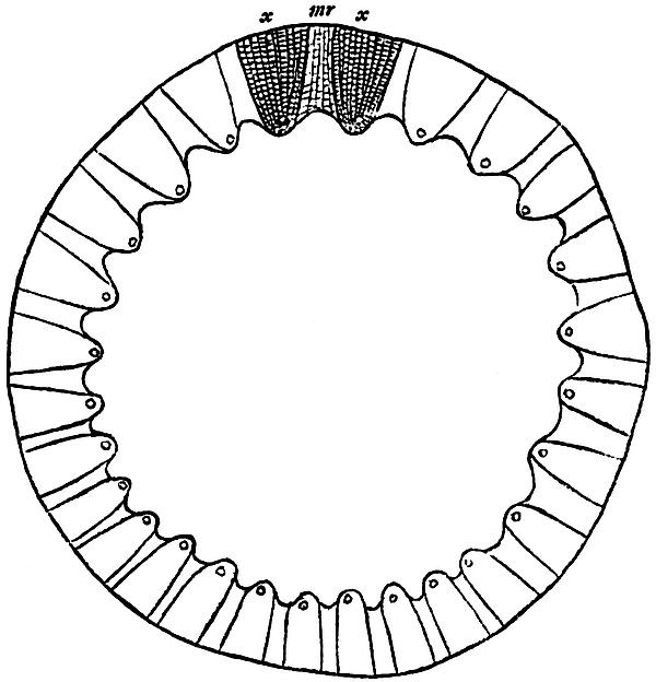
Fig. 73. Transverse section of a Calamite stem. mr, medullary ray. After Williamson. x, x, xylem. (No. 1933 A.A. in the Williamson Collection.)
ARTHROPITYS.
In the large section of a calcareous nodule diagrammatically shown in fig. 17 II. (p. 85) the secondary wood of a slightly flattened Calamite is the most prominent plant fragment. The pith-cavity has been almost obliterated by the lateral compression of the woody cylinder, but the presence of the carinal canals along the inner edge of the wood may still be readily recognised. The appearance presented by a transverse section of the secondary wood of a Calamite is that of regular radial series of rather small rectangular tracheids, with occasional secondary medullary rays consisting of narrow and radially elongated parenchymatous cells. The principal rays[610] in the Arthropitys type of a Calamite stem are often found to gradually decrease in breadth as they pass into the secondary wood, until in the outer portion of the wood the primary medullary rays are practically obliterated by the formation of interfascicular xylem.
In fig. 74, A, we have a portion of a single xylem group of a thick woody stem. The stem from which the figure has been drawn was originally described by Binney[611] as Calamodendron commune; we now recognise it as a typical example of the subgenus Arthropitys. The specific term communis was used by Ettingshausen[612] in 1855 in a comprehensive sense to include more than twenty species of the genus Calamites, but since Binney’s use of the term it has come to be associated with a definite type of Arthropitys stem, in which the primary medullary rays decrease rapidly in breadth towards the periphery of the wood. The wood of Binney’s stem[613] measures 2·5 cm. across, but the pith-cavity has been crushed to the limits of a narrow band represented in the figure by the shaded portion. The strand of cells, s, in the pith is a portion of a Stigmarian appendage (“rootlet”), which penetrated into the hollow stem of the Calamite and became petrified by the same agency to which the preservation of the stem is due. These intruded Stigmarian appendages are of constant occurrence in the calcareous nodules; their intimate association with the tissues of other plants is often a serious source of error in the identification of petrified tissues. The inner portion of one of the xylem groups is shown in fig. 74, A. External to the carinal canal, the xylem tracheids are disposed in regular series and associated with numerous narrow secondary medullary rays. The width of the xylem wedge increases gradually as we pass outwards, this is due to the formation of interfascicular xylem, which in the more peripheral portion of the stem extends across the primary medullary rays. The few primary medullary-ray cells shown in the drawing illustrate the characteristic tangentially elongated form and large size of the parenchymatous elements. Williamson and Scott have pointed out that the tangentially elongated form of the medullary-ray cells is the result of active growth, and not merely the expression of the tangential stretching of the stem consequent on secondary thickening.
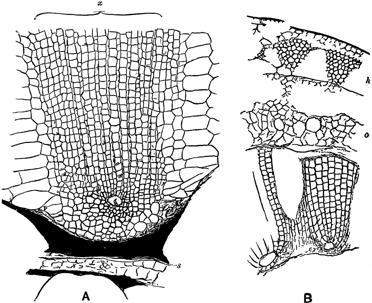
Fig.
74.
Transverse section of part of a Calamite stem. [Calamites (Arthropitys) communis (Binney).] s, Stigmarian appendage. x, xylem. From a specimen in the Binney Collection, Cambridge, × 50.
Transverse section of a stem. h, hypodermal tissue; c, inner cortex. From a specimen in the Williamson Collection (no. 62). × 35.
A glance at the complete transverse section of the stem,—of which a small portion is shown in fig. 74 A,—suggests the existence of annual rings in the wood, but this appearance of rings is merely the result of compression. The secondary wood of a Calamite does not exhibit any regular zones of growth comparable with the annual rings of our forest trees.
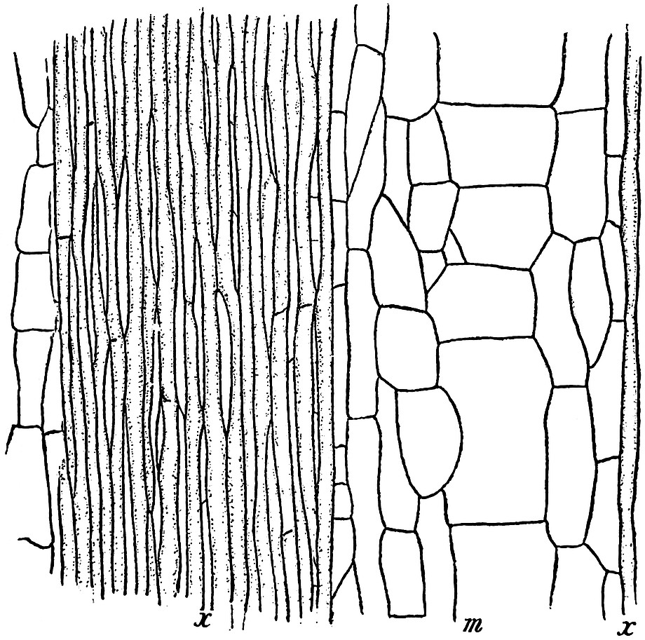
Fig. 75. Longitudinal tangential section near the inner edge of the wood of the Calamite of fig. 74. x, x, secondary xylem and medullary rays; m, principal medullary ray. From a section in the Binney Collection, × 50.
Before passing to other examples of Calamitean stems, reference may be made to the sections shown in figs. 75 and 76, which illustrate some further points in the structure of Binney’s stems. In fig. 75 the xylem tracheids are shown at x, and between them the secondary medullary rays present the appearance of long and narrow parenchymatous cells; as the section is tangential the characteristic scalariform character of the tracheids is not shown, the ladder-like bordered pits being confined to the radial walls of the tracheal elements. The much greater length than breadth of the cells which form the rays associated with the xylem tracheids, is a characteristic feature in Calamitean stems. The breadth of the principal ray, m, shows that the section has passed through the wood a short distance from the pith; in a tangential section cut further into the wood the breadth of the principal rays would be considerably reduced. The large medullary-ray tissue consists of square-walled parenchymatous cells. The more highly magnified section, in fig. 76, shows a central group of parenchyma containing a few transversely cut tracheids, but the two kinds of elements are not clearly differentiated in the figure; this group of cells is an outgoing leaf-trace which is enclosed by the strongly curved tracheids of the stem. The section is taken from the node of a stem where several leaf-trace bundles are passing out to a whorl of leaves; the few cells intercalated between the tracheids belong to the parenchyma of the secondary medullary rays.
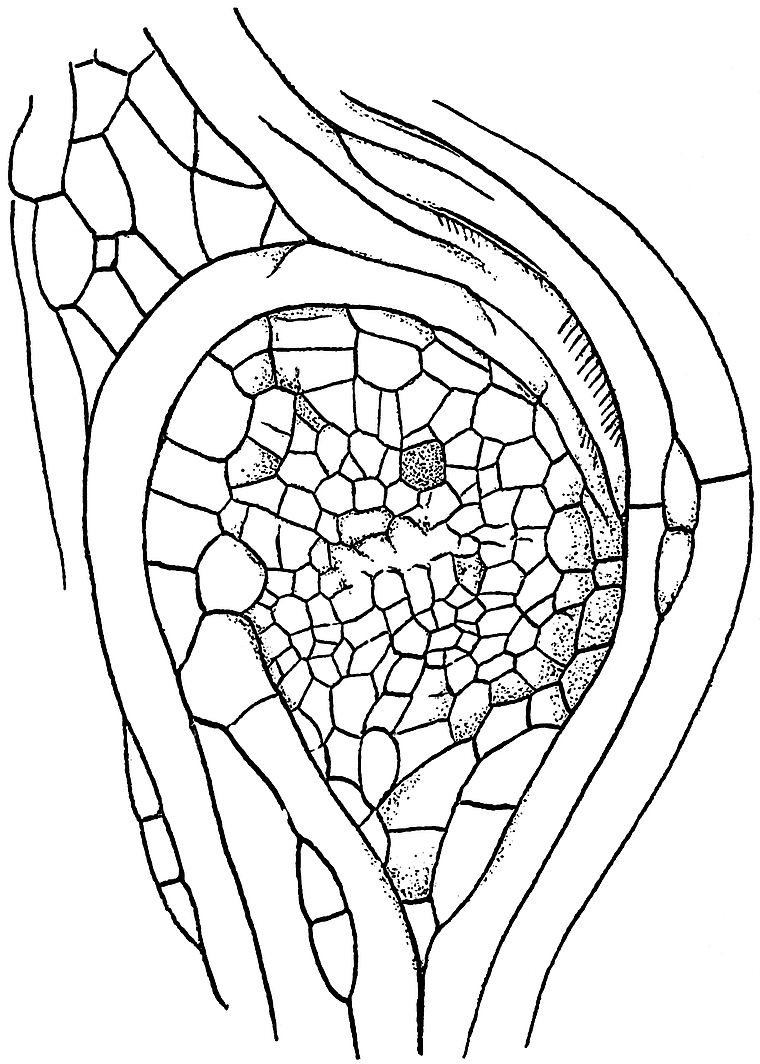
Fig. 76. Longitudinal tangential section of the same Calamite as that of figs. 74 and 75, showing a leaf-trace and curved tracheids at a node. From a section in the Binney Collection, × 100.
ARTHROPITYS. SURFACE FEATURES.
In the small portion of a stem represented in fig. 74 B, the cortical tissues have been partially preserved; at the inner edge, next the hollow pith, there are two xylem groups, each with a carinal canal, and between them is part of a broad “principal” medullary ray[614]. The cambium has not been preserved, but beyond this region we have some of the large cells, c, of the inner cortex; these are followed by a few remnants of a smaller-celled tissue, and external to this part of the cortex there is a series of triangular groups, h, consisting of small thick-walled cells alternating with spaces which were originally occupied by more delicate parenchyma. The darker groups constitute hypodermal strands of mechanical tissue or stereome which lent support to the stem. The surface of a stem possessing such supporting strands would probably assume a longitudinally wrinkled or grooved appearance on drying; the intervening parenchyma, contracting and yielding more readily, would tend to produce shallow grooves alternating with the ridges above the stereome strands.
The complete section of the stem of which a small portion is shown in fig. 74 B, is figured by Williamson[615] in his 12th memoir on Coal-Measure plants. The section was obtained from Ashton-under-Lyne in Lancashire; it illustrates very clearly a method of preservation which is occasionally met with among petrified plants. The walls of the various tissue elements are black in colour and somewhat ragged, and the general appearance of the section is similar to that of a section of a charred piece of stem. It is possible that the Calamite twig was reduced to charcoal before petrifaction by a lightning flash or some other cause.
It is often said that the surface of a Calamite stem was probably marked by regular ridges and grooves similar to those of the pith-cast, and that such external features are connected with the arrangement of the tissues in the vascular cylinder. The indication of grooves and ridges on the bark of fossil Calamites is no doubt the result of the existence in the hypoderm of firm strands alternating with strands of less resistant cells. It is very common to find Calamite pith-casts covered with a layer of coal presenting a ribbed surface, but this is simply due to the moulding of the coaly film on an internal pith-cast. The broad grooves on such a specimen as that of fig. 77 are, on the other hand, probably an indication of the existence of hypoderm bands similar to those in fig. 74 B, h. The specimen from which fig. 77 is drawn shows many interesting features. The figure given by Grand’Eury, of which fig. 77 is a copy, is somewhat idealised, but the various surfaces can be made out in the fossil. The surface of the coaly envelope surrounding the pith-cast, a, is distinctly grooved, but the depressions have nothing to do with the surface features of the wood or the pith-cast; they are no doubt due to the occurrence of alternating bands of thick- and thin-walled tissue in the hypodermal region of the cortex; the peripheral strands of bast cells would stand out as prominent ribs as the stem tissue contracted during fossilisation. At b (fig. 77) we have a view of the wood in which the position of the principal rays is indicated by fine longitudinal lines at regular intervals; the oval projections just below the nodal line are probably the casts of infranodal canals (cf. p. 324). At a the characteristic pith-cast is seen with a small branch-scar on the node. The scar on the middle node, N 2, is probably that of a root, and a root R is still attached to the node, N 3.
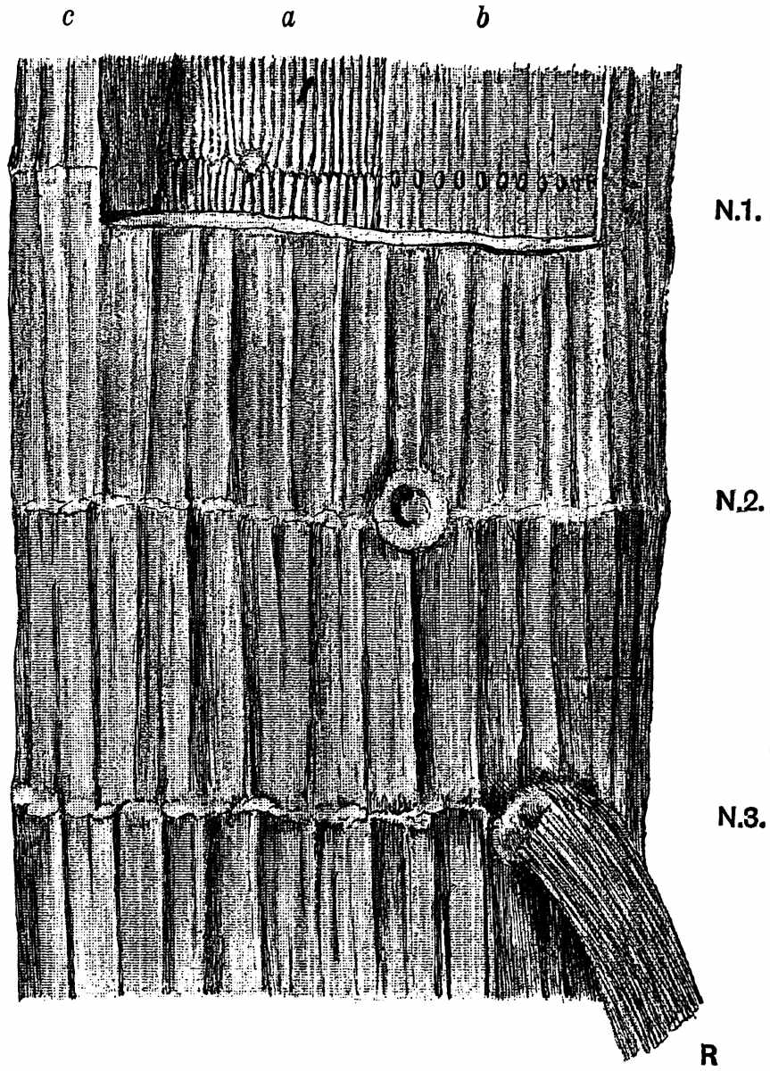
Fig. 77. Portion of a Calamite stem, showing the surface of the bark, c; the wood, b; the surface of the pith-cast, a. N.1-N.3. Nodes. R. Root. (After Grand’Eury. Partially restored from a specimen in the École des Mines, Paris.) ¾ nat. size.
PERIDERM IN STEMS.
An interesting feature observed in some specimens of older Calamite branches is the development of periderm or cork. This is illustrated on a large scale by a unique specimen originally described by Williamson in 1878[616]. Figs. 78 and 79 represent transverse and longitudinal sections of this stem. This unusually large petrified stem was found in the Coal-Measures of Oldham, in Lancashire. In the slightly reduced drawing, fig. 78, the large and somewhat flattened pith, p, 4·2 cm. in diameter, is shown towards the bottom of the figure. Surrounding this we have 58 or 59 wedge-shaped projecting xylem groups and broad medullary rays; the latter soon become indistinguishable as they are traced radially through the thick mass of secondary wood, 5 cm. wide, composed of scalariform tracheids and secondary medullary rays (fig. 78, 3). The secondary wood presents the features characteristic of Calamites (Arthropitys) communis (Binney). External to the wood there is a broken-up mass, about 5·5 cm. wide composed of regularly arranged (fig. 78, 2) and rather thick-walled cells; this consists of periderm, a secondary tissue, which has been developed by a cork-cambium during the increase in girth of the plant. The more delicate cortical tissues have not been preserved, and the more resistant portion of the bark has been broken up into small pieces of corky tissue, among which are seen numerous Stigmarian appendages, pieces of sporangia and other plant fragments. These associated structures cannot of course be shown in the small-scale drawing of the figure.
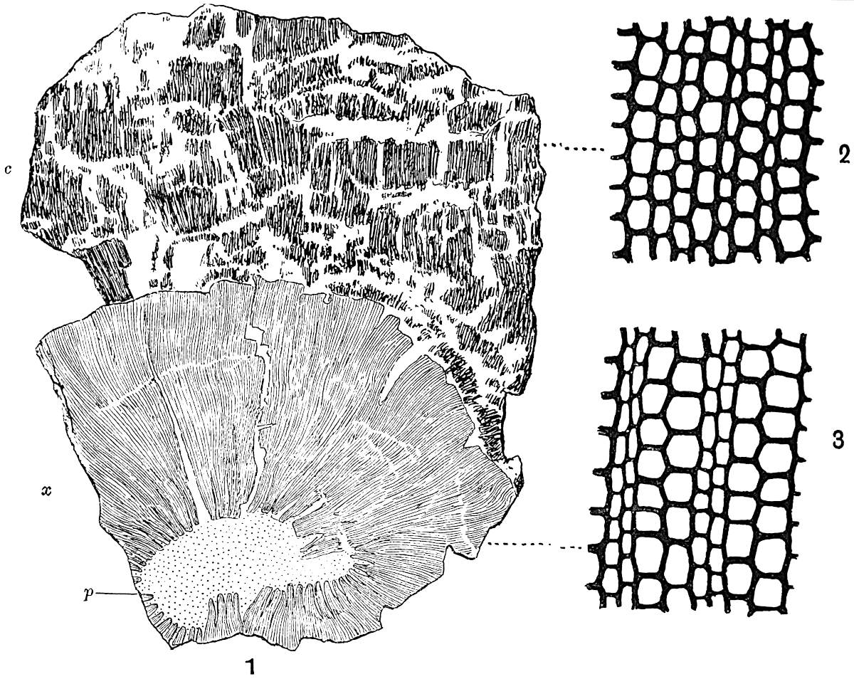
Fig.
78.
Transverse section of a thick Calamite stem. p, pith; x, secondary wood; c, bark. (⅔ nat. size.)
Periderm cells of bark.
Xylem and medullary rays. (2 and 3, × 80.)
From a specimen in the Williamson Collection (no. 79).
In the radial longitudinal section (fig. 79) we see the pith with the projecting wood and the remains of a diaphragm at the node. The mottled or watered appearance of the wood is due to numerous medullary rays which sweep across the tracheids. The periderm elements, as seen in longitudinal section, are fibrous in form.
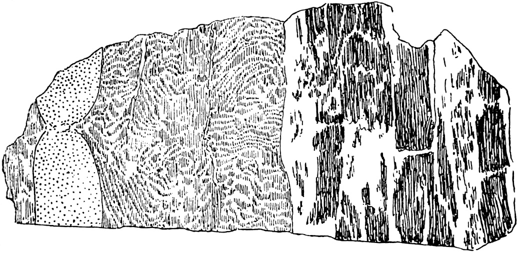
Fig. 79. Longitudinal section of the specimen stem in fig. 78. From a specimen in the Williamson Collection, British Museum (no. 80). ⅔ nat. size.
CALLUS WOOD.
The development of cork in a younger Calamite stem is clearly shown in a specimen described by Williamson and Scott in their Memoir of 1894. In a transverse section of the stem several large cells of the inner cortex are seen to be in process of division by tangential walls, and giving rise to radially arranged periderm tissue[617].
The section diagrammatically sketched in fig. 80 is that of a Calamite twig in which the wood appears to have been injured, and the wound has been almost covered over by the formation of callus wood. The young trees in a Palaeozoic forest might easily be injured by some of the large amphibians, which were the highest representatives of animal life during the Carboniferous period, just as our forest trees are often barked by deer, rabbits, and other animals. Fissures might also be formed by the expansion of the bark under the heating influence of the sun’s rays[618]. Such a specimen as that of fig. 80 gives an air of living reality to the petrified fragments of the Coal period trees. It is well known how a wound on the branch of a forest tree becomes gradually overgrown by the activity of the cambium giving rise to a thick callus, which gradually closes over the wounded surface in the form of two lips of wood which finally meet over the middle of the scar. The two lips of callus are clearly shown in the fossil branch arching over the tear in the wood just beyond the ring of carinal canals. The tissue external to the wood represents the imperfectly preserved cortex. A section which was cut parallel to that of fig. 80 shows a continuous band of wood beyond the wound, and the latter has the form of a small triangular gap; this section appears to have passed across the wound where it was narrower and has already been closed over by the callus. The formation of a rather different kind of callus wood has been described by Renault[619] and by Williamson and Scott[620], in stems where aborted or deciduous branches have been overgrown and sealed up by cambial activity.
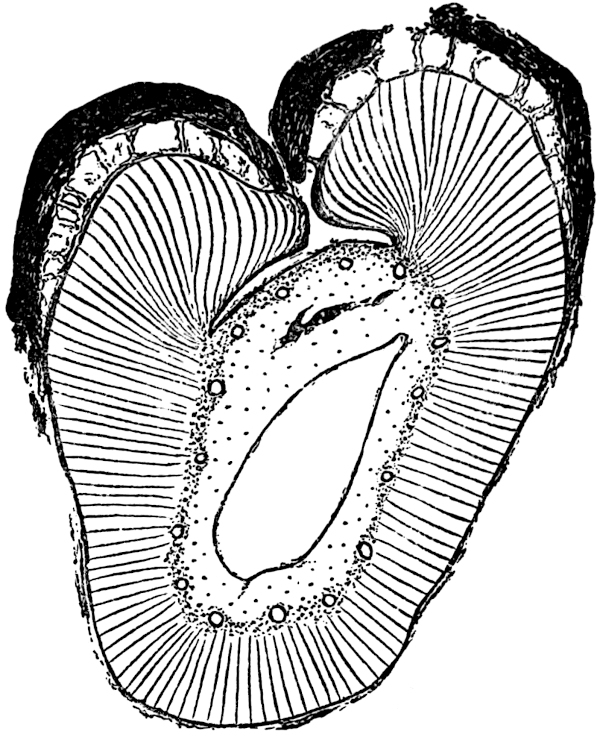
Fig. 80. Diagrammatic sketch of a transverse section of a Calamite twig, showing callus wood. From a specimen in the Cambridge Botanical Laboratory Collection. × ca. 10.
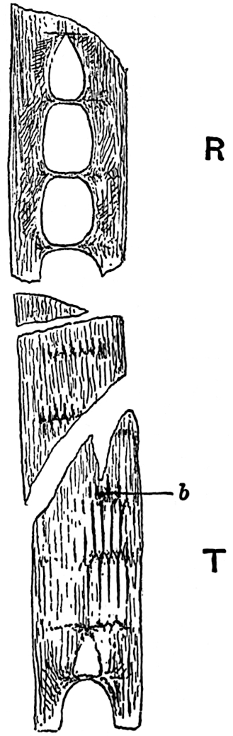
Fig. 81. Calamites. Longitudinal section (R, radial; T, tangential) of a small branch. b, position of a lateral branch. From a specimen (no. 1937) in the Williamson Collection. Slightly enlarged.
Some of the features to be noticed in longitudinal sections of Calamite stems have already been described, at least as regards younger branches. The specimen shown in fig. 81 illustrates the general appearance of a stem as seen in tangential and radial section. In the lower portion, T, the course of the vascular bundles is shown by the black lines which represent the xylem tracheids, bifurcating and usually alternating at each node. Between the xylem strands are the broad principal medullary rays. At b a branch has been cut through on its passage out from the parent stem, just above the nodal line. In tangential sections of Calamite stems one frequently sees both branches and leaf-trace bundles (fig. 83, A), passing horizontally through the wood and enclosed by strongly curved and twisted tracheids. In the upper part of the figure (81, R), the section has passed through the centre of the stem, and the wood is seen in radial view; each node is bridged across by a diaphragm of parenchymatous cells capable of giving rise to a surface layer of periderm[621].
An outgoing branch, as seen in a tangential section of a stem, consists of a parenchymatous pith surrounded by a ring of vascular bundles, in which the characteristic carinal canals have not yet been formed, but if the section has cut the branch further from its base, there may be seen a circle of irregular gaps marking the position of the carinal canals. Such gaps are often occupied by thin parenchyma, and contain protoxylem elements. The outgoing branches, as seen in a tangential section of a Calamite stem, are seen to be connected with the wood of the parent stem by curved and sinuous tracheids, which give to the stem-wood a curiously characteristic appearance[622], as if the xylem elements had been pushed aside and contorted by the pressure of the outgoing member. A tangential section through a Pine stem[623] in the region of a lateral branch presents precisely the same features as in Calamites. The branches are given off from the stem immediately above a node and usually between two outgoing leaf-trace bundles.
RHIZOME OF CALAMITES.
Specimens of pith-casts occasionally present the appearance of a curved and rapidly tapered ram’s horn, and the narrow end of such a cast is sometimes found in contact with the node of another cast. This juxtaposition of casts is shown unusually well in fig. 82. In some of the published restorations of Calamites the plant is represented as having thick branches attached to the main stem by little more than a point. Williamson[624] clearly explained this apparently unusual and indeed physically impossible method of branching, by means of sections of petrified stems. The branches seen in fig. 82 are of course pith-casts, and in the living plant the pith of each branch was surrounded by a mass of secondary wood developed from as many primary groups of xylem as there are grooves on the surface of the cast, each of the grooves on an internode corresponding to the projecting edge of a xylem group. At the junction of one branch with another the pith was much narrower and the enclosing wood thicker, so that the tapered ends of the cast merely show the continuity by a narrow union between the pith-cavities of different branches. Most probably the casts of fig. 82 are those of a branched rhizome which grew underground, giving off aerial shoots and adventitious roots. There is a fairly close resemblance between the Calamite casts of fig. 82 and a stout branching rhizome of a Bamboo, e.g. Bambusa arundinacea Willd.; it is not surprising that the earlier writers looked upon the Calamite as a reed-like plant.
Before leaving the consideration of stem structures there is another feature to which attention must be drawn. On the casts shown in fig. 82 there is a circle of small oval scars situated just below the nodes, these are clearly shown at c, c, c. Each of the scars is in reality a slight projection from the upper end of an internodal ridge. As the ridges correspond to the broad inner faces of medullary rays, the small projection at the upper end of each ridge is a cast of a depression or canal which existed in the medullary tissue of the living plant. There have been various suggestions as to the meaning of these oval projections; several writers have referred to them as the points of attachments of roots or other appendages, but Williamson proved them to be the casts of canal-like gaps which traversed the upper ends of principal medullary rays in a horizontal direction. In a tangential section of a Calamite stem the summit of each primary medullary ray often contains a group of smaller elements which are in process of disorganisation, and in some cases these cells give place to an oval and somewhat irregular canal. In the diagrammatic tangential section represented in fig. 83, A the upper end of each ray is perforated by a large oval space, which has been formed as the result of the breaking down of a horizontal band of cells. Williamson designated these spaces infranodal canals. While proving that they had nothing to do with the attachment of lateral members, he suggested that they might be concerned with secretion; but their physiological significance is still a matter of speculation. The casts of infranodal canals are especially large and conspicuous in the subgenus Arthrodendron, a form of Calamite characterised by certain histological features to be referred to later. Williamson[625] originally regarded the presence of infranodal canals as one of the distinguishing features of Arthrodendron, but they occur also in the casts of the commoner type Arthropitys. As a rule we have only the cast of the inner ends of the infranodal canals preserved as slight projections like those in fig. 83, A; but in one exceptionally interesting pith-cast described by Williamson, these casts of the infranodal canals have been preserved as slender spoke-like columns radiating from the upper ends of the ridges of the infranodal region of a pith-cast.
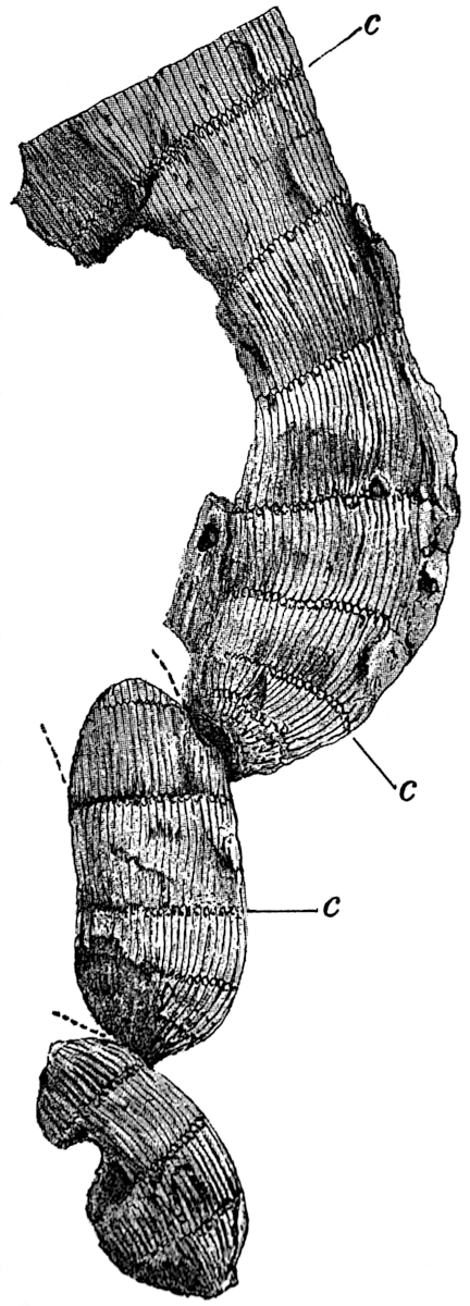
Fig. 82. Branched rhizome of Calamites. ½ nat. size. C, C, nodes showing casts of infranodal canals. From a specimen in the Manchester Museum, Owens College.
This specimen, which was figured by Williamson[626] in two of his papers, and by Lyell[627] in the fifth edition of his Elementary Geology, is historically interesting as being one of the first important plants obtained by Williamson early in the fifties, when he began his researches into the structure of Carboniferous plants. A joiner, who was employed by Williamson to make a piece of machinery for grinding fossils, brought a number of sandstone fragments as an offering to his employer, whom he found to be interested in stones. The specimens “were in the main the merest rubbish, but amongst them,” writes Williamson, “I detected a fragment which was equally elegant and remarkable.... In later days, when the specimen so oddly and accidentally obtained, came to be intelligently studied, its history became clear enough, and the priceless fragment is now one of the most precious gems in my cabinet[628].”
Comparison of three types of structure met with in Calamitean stems,—Arthropitys, Arthrodendron, and Calamodendron.
ARTHROPITYS.
The anatomical features which have so far been described as characteristic of Calamites represent the common type met with in the English Coal-Measures. The same type occurs also in France, Germany and elsewhere. It is that form of stem known as Arthropitys, a sub-genus of Calamites.
Arthropitys may be briefly diagnosed as follows,—confining our attention to the structure of the stem: A ring of collateral bundles surrounds a large hollow pith, each primary xylem strand terminates internally in a more or less bluntly rounded apex traversed by a longitudinally carinal canal. The principal medullary rays consist of large-celled parenchyma, of which the individual elements are usually tangentially elongated as seen in transverse section, and four or five times longer than broad as seen in a tangential longitudinal section. The secondary xylem consists of scalariform and reticulately pitted tracheids; the interfascicular xylem may be formed completely across each primary ray at an early stage in the growth of the stem[629], or it may be developed more gradually so as to leave a tapering principal ray of parenchyma between each primary xylem bundle. In the latter case the principal rays present the characteristic appearance shown in figs. 71, 74, A, 75 and 78, a type of stem which we may refer to as Calamites (Arthropitys) communis. In the former case the stem presents the appearance shown in fig. 83, D[630]. A third variety of Arthropitys stem is one which was originally named by Göppert Arthropitys bistriata; in this form the principal rays retain their individuality as bands of parenchyma throughout the whole thickness of the wood[631]. Such stems as those of figs. 73 and 74, B, may be young examples of Arthropitys communis or possibly of A. bistriata. The narrow secondary medullary rays of Arthropitys usually consist of a single row of cells which are three to five times higher than broad, as seen in tangential longitudinal section. Infranodal canals occur in some examples of Arthropitys.
ARTHRODENDRON.
In the subgenus Arthrodendron, a type of stem first recognised by Williamson and named by him Calamopitys[632], the principal medullary rays consist of prosenchymatous cells (i.e. elongated pointed elements) and not parenchyma. These elongated elements are not pitted like tracheids, and they are shorter and broader than the xylem elements. In some examples of this subgenus the primary rays are bridged across at an early stage by the formation of secondary interfascicular xylem, and in others they persist as bands of ray tissue, as in Arthropitys. Other characteristics of Arthrodendron are the abundance of reticulated instead of scalariform tracheids in the secondary wood, and the large size of the infranodal canals.
Fig. 83, D represents part of a transverse section of Arthrodendron; in this stem the rays have been occupied by interfascicular xylem at a very early stage of the secondary growth. The section from which fig. 83, D is drawn was described by Williamson in 1871; the complete section shows about 80 carinal canals and primary xylem groups. The prosenchymatous form of the principal medullary rays is seen in fig 83, C, and the reticulate pitting on the radial wall of a tracheid is shown in fig. 83, B. Fig. 83, A illustrates the large infranodal canals as seen in a tangential section of a stem. The same section shows also the course of the vascular bundles characteristic of Calamites as of Equisetum, and the position of outgoing leaf-traces is represented by unshaded areas in the black vascular strands.
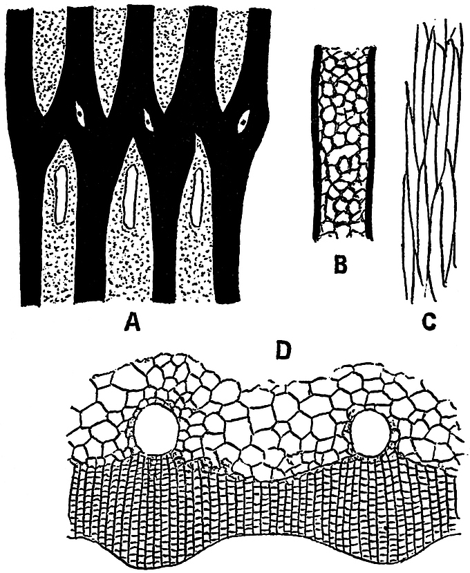
Fig.
83.
Calamites
(
Arthrodendron
).
Tangential section (diagrammatic) showing the course of the vascular strands, also leaf-traces and infranodal canals.
Radial face of a tracheid.
Prosenchymatous elements of a principal medullary ray.
Transverse section of the wood. (After Williamson.) No. 36 in the Williamson Collection.
The subgenus Arthrodendron is very rarely met with, and our information as to this type is far from complete[633].
The third subgenus Calamodendron has not been discovered in English rocks, and our knowledge of this type is derived from French and German silicified specimens[634]. There is the same large hollow pith surrounded by a ring of collateral bundles with carinal canals, as in the two preceding subgenera. The tracheids are scalariform and reticulate, and the secondary medullary rays consist of rows of parenchymatous cells which are longer than broad, as in Arthropitys and Arthrodendron.
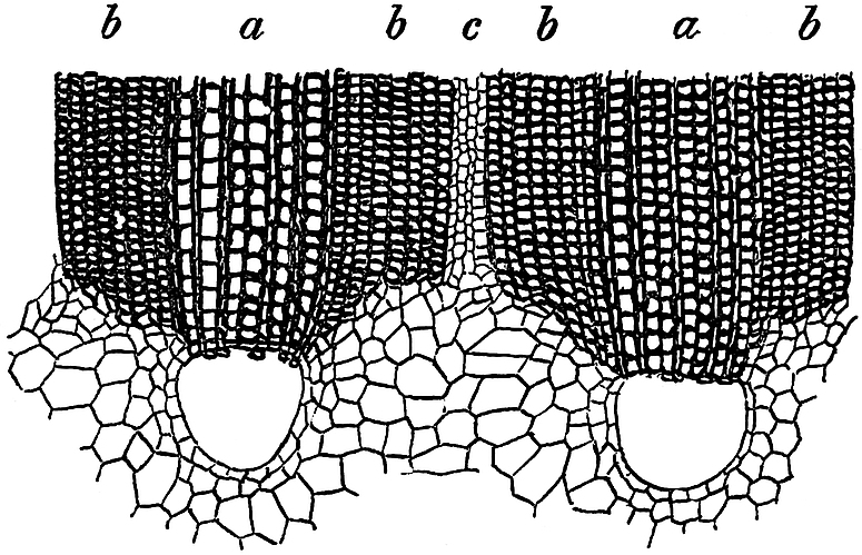
Fig. 84. Calamites (Calamodendron) intermedium, Ren. Transverse section through two vascular bundles. a, a, xylem tracheids, b, b, bands of prosenchyma, c, medullary ray. (After Renault.)
The most characteristic feature of Calamodendron is the occurrence of several rows of radially disposed thick-walled prosenchymatous elements (fig. 84, b) on either flank of each wedge-shaped group of xylem. Each principal ray is thus nearly filled up by bands of fibrous cells on the sides of adjacent xylem groups, but the centre of each principal ray is occupied by a narrow band of parenchyma (fig. 84, c). The relative breadth of the xylem and prosenchymatous bands has been made use of by Renault as a specific character in Calamodendron stems. Fig. 84 is copied from a drawing recently published by this French author of a new species of Calamodendron, C. intermedium[635]. In this case the bands of fibrous cells, b, are slightly broader, as seen in a transverse section of the stem, than the bands of xylem tracheids, a. The narrow band, c, consists of four rows of the parenchymatous tissue of a medullary ray. At the inner end of each group of tracheids there is a large carinal canal.
The question of the recognition of the pith-casts of stems possessing the structure of any of the three subgenera of Calamites is referred to in a later section of this chapter.
b. Leaves
Leaves of Calamites and Calamitean foliage-shoots, including an account of (α) Calamocladus (Asterophyllites) and (β) Annularia.
Our knowledge of the structure and manner of occurrence of Calamite leaves is very incomplete. There are numerous foliage-shoots among the fossils of the Coal-Measures which are no doubt Calamitean, but as they are nearly always found apart from the main branches and stems, it is generally impossible to do more than speak of them as probably the leaf-bearing branches of a Calamite. The familiar fossils known as Asterophyllites, and in recent years often referred to the genus Calamocladus, are no doubt Calamitean shoots; but they are usually found as isolated fragments, and it is seldom that we are able to refer them to definite forms of Calamites. Another common Coal-Measure genus, Annularia, is also Calamitean, and at least some of the species are no doubt leafy shoots of Calamites. Although it is generally accepted that the fossils referred to as Asterophyllites or Calamocladus are portions of Calamites, and not distinct plants, it is convenient, and indeed necessary, to retain such a term as Calamocladus as a means of recording foliage-shoots, which may possess both a botanical and a geological value.
Some of the Calamite casts, especially those referred to the subgenus Calamitina, are occasionally found with leaves attached to the nodes. In some stems the leaves are arranged in a close verticil, and each leaf has a narrow linear form and is traversed by a single median vein. Figures of Calamite stems with verticils of long and narrow leaves may be found in Lindley and Hutton[636], and in the writings of many other authors[637]. In the specimen shown in fig. 85 the leaves are preserved apart from the stem, but from their close association with a Calamite cast, and from the proofs afforded by other specimens, it is quite certain they formed part of a whorl of leaves attached to the node of a true Calamite, and a stem having that particular type known as Calamitina[638] (figs. 99, 100). It is probable that in some Calamites, and especially in younger shoots, the leaves had the form of narrow sheaths split up into linear segments. This question has already been referred to in dealing with certain Palaeozoic fossils referred to Equisetites[639].
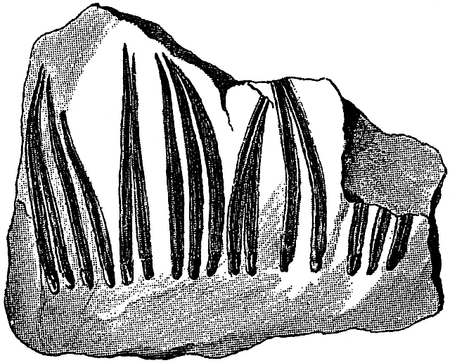
Fig. 85. Linear leaves of a Calamite (Calamitina). After Weiss, slightly reduced.
A few years ago the late Thomas Hick[640], of Manchester, described the structure of some leaves which he believed to be those of a Calamite. He found them attached to a slender axis which possessed the characteristics of a young Calamite branch. There can be little doubt that his specimens are true Calamite leaves. The sketches of fig. 86 have been made from the sections originally described by Hick. Fig. 86, 1 shows a leaf in transverse section; on the outside there is a well-defined epidermal layer with a limiting cuticle. Internal to this we have radially elongated parenchymatous cells forming a loose or spongy tissue, the cells being often separated by fairly large spaces (fig. 86, 5), especially in the region of the blunt lateral wings of the leaf. Some of these cells contain a single dark dot, which in all probability is the mineralised nucleus. These pallisade-like cells probably contained chlorophyll and constituted the assimilating tissue of the leaf. In the centre there is a circular strand of cells limited by a layer of larger cells with black contents, enclosing an inner group of small-celled parenchyma and traversed by a few spiral or scalariform tracheids constituting the single median vein. It is hardly possible to recognise any phloem elements in the small vascular bundle; there appear to be a few narrow tracheids surrounded by larger parenchymatous elements (fig. 86, 2). At one point in the epidermis of fig. 86, 1, there appears to be a stoma, but the details are not very clearly shown (fig. 86, 4); the two cells, s, s, bordering the small aperture are probably guard-cells.
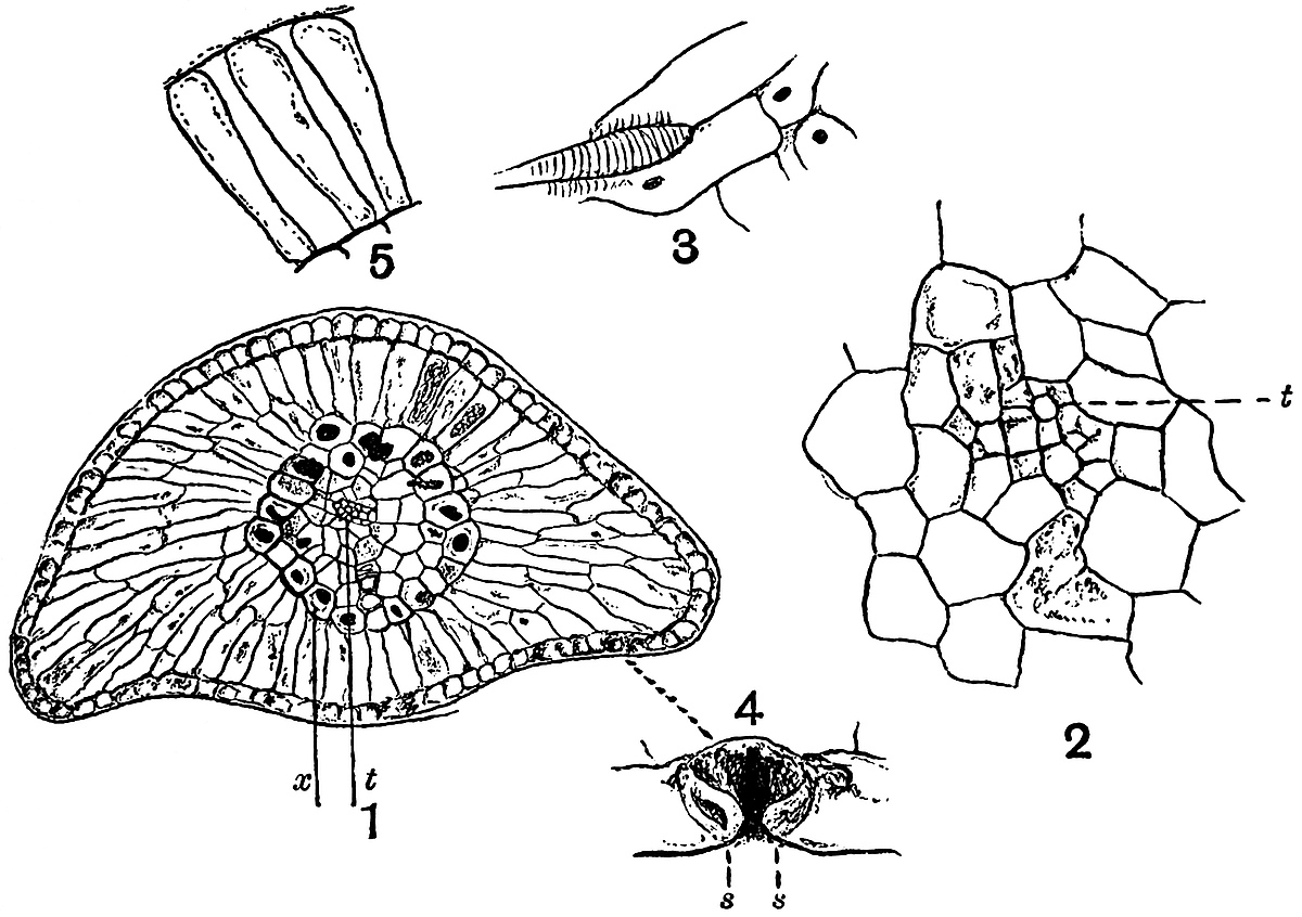
Fig.
86. A leaf of
Calamites
.
Transverse section; t, vascular bundle; x, sheath of cells. × 35.
Vascular bundle consisting of a few small tracheids, t.
A tracheid and a few parenchymatous cells, the latter with nuclei.
A stoma; s, s, guard-cells.
Pallisade cells and intercellular spaces.
From a section in the Manchester Museum, Owens College.
The nature of the assimilating tissue, the comparatively thick band of thin-walled cells with intercellular spaces, and the exposed position of the stomata suggest that the plant lived in a fairly damp climate; at least there is nothing to indicate any adaptation to a dry climate.
In the Binney collection of plants in the Woodwardian Museum, Cambridge, there is a species of a very small shoot bearing three or four verticils of leaves which possess the same structure as those of fig. 86. We may probably regard such twigs as the slender terminal branches of Calamitean shoots.
α. Calamocladus (Asterophyllites).
The generic name Asterophyllites was proposed by Brongniart[641] in 1822 for a fossil previously named by Schlotheim[642] Casuarinites, and afterwards transferred to Sternberg’s genus Annularia. In 1828 Brongniart[643] gave the following diagnosis of the fossils which he included under the genus Asterophyllites:—“Stems rarely simple, usually branched, with opposite branches, which are always disposed in the same plane; leaves flat, more or less linear, pointed, traversed by a simple median vein, free to the base.” Lindley and Hutton described examples of Brongniart’s genus as species of Hippurites[644], and other authors adopted different names for specimens afterwards referred to Asterophyllites.
At a later date Ettingshausen[645] and other writers expressed the view that the fossils which Brongniart regarded as a distinct genus were the foliage-shoots of Calamites, and Ettingshausen went so far as to include them in that genus. In view of the generally expressed opinion as to the Calamitean nature of Asterophyllites, Schimper[646] proposed the convenient generic name Calamocladus for “rami et ramuli foliosi” of Calamites. Some recent authors have adopted this genus, but others prefer to retain Asterophyllites. In a recent important monograph by Grand’Eury[647] Calamitean foliage-shoots are included under the two names, Asterophyllites and Calamocladus; the latter type of foliage-shoots he associates with the stems of the subgenus Calamodendron, and the former he connects with those Calamitean stems which belong to the subgenus Arthropitys.
It is an almost hopeless task to attempt to connect the various forms of foliage-shoots with their respective stems, and to determine what particular anatomical features characterised the plants bearing these various forms of shoots. We may adopt Schimper’s generic name Calamocladus in the same sense as Asterophyllites, but as including such other foliage-shoots as we have reason to believe belonged to Calamites. Those leaf-bearing branches which conform to the type known as Annularia are however not included in Calamocladus, as we cannot definitely assert that these foliage-shoots belong in all cases to Calamitean stems. Grand’Eury’s use of Calamocladus in a more restricted sense is inadvisable as leading to confusion, seeing that this name was originally defined in a more comprehensive manner as including Calamitean leaf-bearing branches generally. We may define Calamocladus as follows:—
Branched or simple articulated branches bearing whorls of uni-nerved linear leaves at the nodes; the leaves may be either free to the base or fused basally into a cup-like sheath (e.g. Grand’Eury’s Calamocladus). The several acicular linear leaves or segments which are given off from the nodes spread out radially in an open manner in all directions; they may be either almost at right angles to the axis or inclined at different angles. Each segment is traversed by a single vein and terminates in an acuminate apex.
As a typical example of a Calamitean foliage-shoot the species Calamocladus equisetiformis (Schloth.) may be briefly described. The synonymy of the commoner species of fossil plants is a constant source of confusion and difficulty; in order to illustrate the necessity of careful comparison of specimens and published illustrations, it may be helpful to quote a few synonyms of the species more particularly dealt with. The exhaustive lists drawn up by Kidston in his Catalogue of Palaeozoic plants in the British Museum will be found extremely useful by those concerned with a systematic study of the older plants.
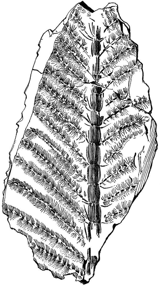
Fig. 87. Calamocladus equisetiformis (Schloth.). From a specimen in the British Museum (McMurtrie Collection, no. V. 2963). ca. ⅓ nat. size.
Calamocladus equisetiformis (Schloth.). Fig. 87.
1809. Phytolithus, Martin[648].
1820. Casuarinites equisetiformis, Schlotheim[649].
1825. Bornia equisetiformis, Sternberg[650].
1828. Asterophyllites equisetiformis, Brongniart[651].
1836. Hippurites longifolia, Lindley and Hutton[652].
1855. Calamites equisetiformis, Ettingshausen[653].
1869. Calamocladus equisetiformis, Schimper[654].
1869. Annularia calamitoides, Schimper[654].
The above synonyms do not exhaust the list[655], but they suffice to illustrate the necessity of a careful comparison in drawing up tables of species, in connection with geographical distribution or for other purposes.
Calamocladus equisetiformis may be briefly defined as follows:—A central axis possessing a hollow pith of Calamitean character, divided externally into well-marked slightly constricted nodes and internodes; from the nodes long narrow and free leaves are borne in whorls; from the axils of some of the leaves lateral branches are given off inclined at a fairly wide angle to the main axis, and bearing crowded verticils of spreading acicular leaves.
The unusually good specimen, 38·5 cm. long, shown on a much reduced scale in fig. 87, illustrates the characteristic habit of this form of Calamocladus. It is from the Radstock coal-field of Somersetshire, one of the best English localities for Coal-Measure plants. An exceedingly good collection of Radstock plants has recently been presented to the British Museum by Mr J. McMurtrie; it includes many fine specimens of Calamites. A small example—probably of this species—from Coalbrook Dale, near Dudley, in Shropshire, and now in the British Museum, illustrates very well the appearance of a young and partially expanded Calamitean foliage-shoot. The central axis, 6·5 cm. in length, includes about 15 internodes, and terminates in a bud covered by several small leaves. Lateral branches are given off at a wide angle, and small unexpanded buds occur in the axils of several of the leaves.
As an example of the leaf-bearing branches which Grand’Eury has recently described as Calamocladus, using the genus in a more restricted sense than is adopted in the present chapter, reference may be made to the fragment shown in fig. 68, A. The foliage-shoots of this type bore verticils of linear leaves, coherent basally in the form of a cup, at the ends of branches and not in a succession of whorls on each branch. The association of reproductive organs, in the form of long and narrow strobili, with Calamocladus is referred to in the sequel.
The specimens described by Grand’Eury are in the École des Mines Museum, Paris; some of the shoots which are well preserved bear a resemblance in habit of growth to the genus Archaeocalamites.
β. Annularia.
In 1820 this generic name was applied by Sternberg[656] to some specimens of branches bearing verticils of linear leaves. In 1828 Brongniart[657] thus defined the genus Annularia:—“Slender stem, articulated, with opposite branches arising above the leaves. Leaves verticillate, flat, frequently obtuse, traversed by a single vein, fused basally and of unequal length.”
In the works of earlier writers we find frequent illustrations of specimens of Annularia, which are compared with Asters and other recent flowering plants. Lehmann[658] contributed a paper to the Royal Academy of Berlin in 1756, in which he referred to certain fossil plants as probable examples of flowers, among them being a specimen of Annularia. He refers to the occurrence of fossil ferns and other plants, and asks why we do not find flowers of the rose or tulip; his object being “not to acquire vain glory, but to give occasion for others to look into the matter more clearly.”
The general habit of the fossils which are now included under Annularia agrees closely with that of Calamocladus. There is the same spreading form and a similar foliage in the two genera, but in Annularia the members of a whorl are always fused into a basal sheath, and the segments are not of equal length. We may thus summarise the characteristic features of the genus:—
Opposite branches are given off in one plane from the nodes of a main axis; the leaves are in the form of narrow sheaths divided into numerous and unequal linear or narrow lanceolate segments, each with a median vein. The segments in each whorl appear to be spread out in one plane very oblique to the axis of a branch, instead of spreading radially in all directions; the lateral segments are usually longer than the upper and lower members of a whorl. The vegetative branches possess the same type of structure as Calamites.
A comparison of Annularia and Phyllotheca has already been made in Chapter IX. (p. 282). Potonié[659] has recently given a detailed account of Annularian leaves; he compares them with those of Equisetum, and describes the occurrence on the lamina of each leaf-segment of a broad central band or midrib, with a groove, probably containing stomata, on either side. He shows that in well-preserved specimens of Annularia, it is possible to recognise certain minute surface-features, such as the presence of hairs and stomata, which enable one to detect a close resemblance between the leaves of Calamite stems and those of Annularian shoots.
It is not always easy to distinguish between Annularia and Calamocladus; the collar-like basal sheath in the leaves of the former is a characteristic feature, but that cannot always be recognised. On the other hand, the leaves of Calamocladus may sometimes be flattened out on the surface of the rock and simulate the deeply cut sheaths of Annularia. It is difficult to decide how far the manner of occurrence of Annularian leaves in one plane, which is commonly insisted on as a generic character, is an original feature, or how far it is the result of compression in fossilisation. Probably the leaves of a living Annularia were spread out at right angles to the axis, as in the ‘verticils’ of such a plant as Galium.
Dawson[660] has described some fossils from the Devonian rocks of Canada as species of Asterophyllites; the figures bear a closer resemblance to the genus Annularia. The same author figures some irregularly whorled impressions as Protannularia, which appear to be identical with a fossil described by Nicholson[661] from the Skiddaw slates (Ordovician) of Cumberland as Buthotrephis radiata, but the specimens are too imperfect to admit of accurate determination.
Annularia stellata (Schloth.). Fig. 88.
1820. Casuarinites stellatus, Schlotheim[662].
1826. Bornia stellata, Sternberg[663].
1828. Annularia longifolia, Brongniart[664].
1834. Asterophyllites equisetiformis, Lindley and Hutton[665].
1868. Asterophyllites longifolius, Binney[666].
1887. Annularia Geinitzi, Stur[667].
1887. Annularia westphalica, Stur.
This species was figured by Scheuchzer[668] in his Herbarium Diluvianum, and compared by him with a species of Galium (Bedstraw). Brongniart first made use of the generic name Annularia for this common Coal-Measure species, which may be defined as follows:—
Stem reaching a diameter of about 6–8 cm., with internodes 6–12 cm. in length, the surface either smooth or faintly ribbed. Primary branches given off in opposite pairs from the nodes, the lateral branches giving off smaller branches disposed in the same manner. The smaller branches bear verticils of leaves at each node; both leaves and ultimate branches being in one plane. The leaves are narrow, lanceolate-spathulate in form, broadest about the middle, 1–5 cm. in length and 1–3 mm. broad, hairy on the upper surface[669]; each leaf is traversed by a single vein.
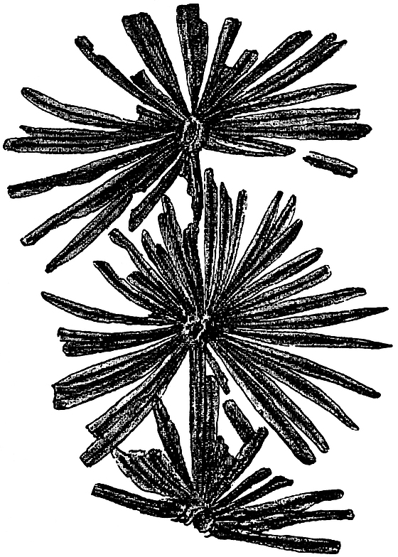
Fig. 88. Branch of Annularia stellata (Schloth.). ⅘ nat. size. From a specimen in the Collection of Mr R. Kidston. Upper Coal-Measures, Radstock.
Each whorl contains 16–32 segments, which are connected basally into a collar or narrow sheath; the lateral segments are usually longer than the upper and lower. The branches are about 6–20 mm. broad, with finely ribbed internodes 3–7 cm. long, bearing verticils of leaves; the ultimate branches arise in pairs in the axils of the lateral segments of the verticils.
The strobili are of the Calamostachys[670] type and are borne on the main branches or possibly on the stem; they have a long and narrow form and are attached in verticils at the nodes. Each strobilus consists of a central axis bearing alternate whorls of linear lanceolate sterile bracts and sporangiophores, about half as numerous as the sterile bracts; each sporangiophore bears four ovoid sporangia.
The anatomical structure of a specimen referred to Annularia stellata has been described by Renault[671]. The cortex consists of parenchyma traversed by lacunae and limited peripherally by a denser hypodermal tissue. In the stele Renault describes 14 xylem strands, each with a large carinal canal. The pith was apparently large and hollow. The same author describes an Annularia strobilus in which the lower sporangiophores bear macrosporangia, and the upper microsporangia.
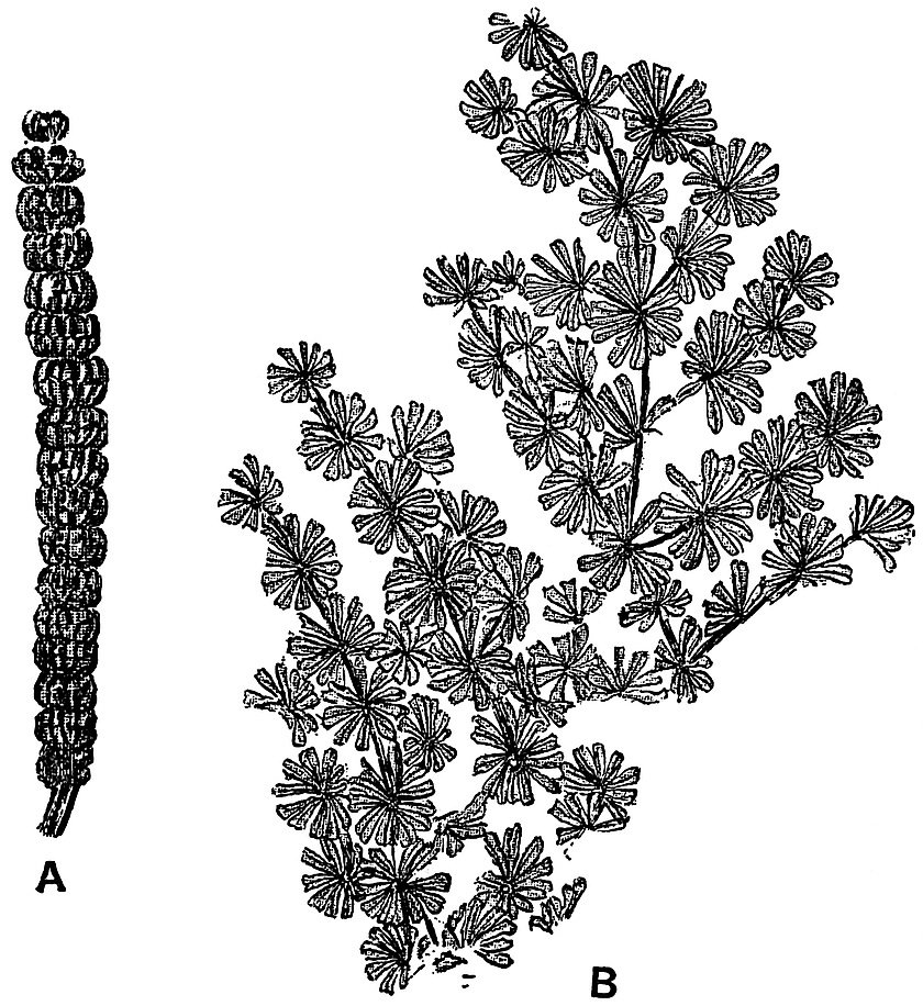
Fig. 89. Annularia sphenophylloides (Zenk.). A. Strobilus (Stachannularia calathifera, Weiss). ⅔ nat. size. B. Vegetative shoot. ⅘ nat. size. From specimens in the Collection of Mr R. Kidston. Upper Coal-Measures, Radstock.
The references in the footnote should be consulted for figures of this species of Annularia; it is from the examination of such specimens as are referred to in the note that the above diagnosis has been compiled[672].
Annularia sphenophylloides (Zenk.). Fig. 89.
1833. Galium sphenophylloides, Zenker[673].
1865. Annularia brevifolia, Heer[674], Strobilus.
1876. Calamostachys (Stachannularia) calathifera, Weiss[675].
Principal branches 8–12 mm. wide, with internodes 8–10 cm. in length, giving off two opposite branches at the nodes; from the secondary branches arise smaller branches in opposite pairs. The leaf-verticils and branches are all in one plane. Each verticil consists of 12–18 spathulate segments, 3–10 mm. long, cuneiform at the base and broader above, with an acuminate tip; the lateral segments are slightly longer than the upper and lower members of a whorl.
The small and crowded leaf-whorls give to this species a characteristic appearance, which readily distinguishes it from the larger-leaved forms such as Annularia stellata. A fossil figured by Lhwyd[676] in 1699 as Rubeola mineralis is no doubt an example of Annularia sphenophylloides.
Annularian branches are occasionally found with cones given off from the axils of some of the leaf-whorls. An interesting specimen, which is now in the Leipzig Museum, was described by Sterzel in 1882[677], showing cones attached to a vegetative shoot of Annularia sphenophylloides. The long and narrow strobili—2·5 cm. long and about 6 mm. broad—appear very large in proportion to the size of the vegetative branches. A fertile shoot consists of a central axis bearing whorls of bracts alternating with sporangiophores, to each of which are attached four sporangia. The specimen in fig. 89, A, does not show the details clearly; each transverse constriction represents the attachment of a whorl of linear bracts; the whole cone appears to consist of a series of short broad segments. The divisions in the lower half of each segment mark the position of the sterile bracts, while those of the upper half represent the outlines of the upper sporangia of each whorl of sporangiophores, the lower sporangia being hidden by the ring of linear bracts[678]. On some portions of the specimen of fig. 89, A, it is possible to recognise the outlines of cells on the coaly surface-film; these probably belong to the sporangium wall. This type of cone is included under the genus Calamostachys, a name applied to Calamitean strobili with certain morphological characters, as described on p. 351.
c. Roots.
In 1871 Williamson[679] described some sections of what he considered to be a distinct variety of a Calamite stem. The chief peculiarity which he noticed lay in the absence of carinal canals, and in the solid pith. Some years later the same observer[680] came to the conclusion that the specimens were probably those of a plant generically distinct from Calamites; he accordingly proposed a new name Astromyelon. Subsequently Cash and Hick[681] gave an account of some examples of apparently another form of plant, to which they gave the name Myriophylloides Williamsonis; and Williamson[682] suggested the term Helophyton as a more suitable generic designation. It was, however, demonstrated by Spencer[683] that the plant described by Cash and Hick was identical with Williamson’s Astromyelon. Williamson[684] then gave an account of several specimens of this type illustrating various stages in the growth and development of the Astromyelon ‘stems,’ which he compared with the rhizome of the recent genus Marsilia.
In 1885 Renault[685] published an account of Astromyelon in which he brought forward good evidence in favour of regarding it as a Calamitean root. The same author has recently given some excellent figures and a detailed description of certain specific types of these Calamite roots, and Williamson and Scott’s memoir on the roots of Calamites has rendered our knowledge of Astromyelon almost complete. Some of the finest specimens, in which the organic connection between typical Calamite stems and Astromyelon roots is clearly demonstrated, are in the Natural History Museum, Paris. There are several sections also from English material which show the connection between root and stem very clearly.
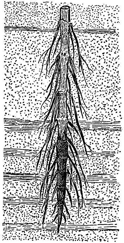
Fig. 90. Pith-cast of a Calamite stem, with roots; embedded in sandstone and shale. (After Grand’Eury.) Much reduced.
Casts of the hollow pith of Calamite rhizomes or aerial branches are occasionally found in which slender appendages are given off either singly or in tufts from the nodal regions. Many examples of such casts have been figured by Lindley and Hutton[686], Binney[687], Grand’Eury[688], Weiss[689], Stur, and other writers[690]. The large stem-cast of fig. 90 illustrates the manner of occurrence of long branched roots on the nodes of a Calamite growing in sandy or clay soil. The lower and more darkly shaded portion of the specimen is covered by a layer of coal representing the carbonised wood and cortex, which has been moulded on to the sandstone pith-cast. In fig. 77 (p. 316) a fairly thick root is seen, in organic connection with one of the nodes, N 3, and on N 2 there is a scar of another root.
There are certain external characters by which one may often recognise a Calamitean root. There is no division into nodes and internodes as in stems, and as the pith of the root was usually solid the parallel ribs and grooves of stem-casts are not present. In smaller flattened roots there may sometimes be seen a central or excentric black line representing the stele, and the surface of the root presents a curious wrinkled or shagreen texture, probably due to the shrinkage of the loose lacunar cortex. The occasional excentric position of the stele is no doubt due to the displacement of the vascular cylinder as a result of the rapid decay of the cortical tissues. In the Bergakademie of Berlin there are some unusually good examples of Calamite casts bearing well-preserved root-impressions; these include the original specimens figured by Weiss[691].
No doubt some of the roots figured by various writers under the names Pinnularia[692] and Hydatica[693] belong to Calamites, but it is often impossible to identify detached specimens with any certainty.
The section figured diagrammatically in fig. 91 A shows the characteristic single series of large lacunae, l, in the middle cortical region. In the centre there is a wide solid pith surrounded by a ring of vascular tissue, x. The appearance of the middle cortex is very like that of the stem of a water-plant such as Myriophyllum, the Water Milfoil; it shows that the Calamite roots grew either in water or swampy ground. In fig. 91 B, the root characters are clearly seen; the centre of the stele is occupied by large parenchymatous cells which are rather longer than broad in longitudinal view; at the periphery there are four protoxylem groups px, alternating with four groups of phloem, ph, the latter being situated a little further from the centre of the stele. The structure is therefore that of a typical tetrarch root. In the example represented in the figure secondary thickening has begun, and the cambial cells internal to each phloem group have given rise to a few radially disposed tracheids, x2. Beyond the phloem there are two layers of parenchyma representing, as regards position, a pericycle and an endodermis. In the ordinary pericycle and endodermis of the roots of most plants the cells of the two layers are on alternate radii, but in the Calamite root, as in Equisetum roots, the cells of these layers are placed on the same radii, as seen in the neighbourhood of x2 in the figure. This correspondence of the radial walls of the endodermal and pericyclic cells points to the development of both layers from one mother-layer, and suggests the ‘double endodermis’ or phloeoterma of Equisetum (p. 254). The cells in the outer of these two layers have slight thickenings on the radial walls recalling the usual character of endodermal cells. The phloeoterma is succeeded by a few layers of parenchyma, constituting the inner cortex, and beyond this we have the large lacunae separated from one another by slender trabeculae of cells. The outer cortex is limited by a well-defined layer of thick-walled cells, which may be spoken of as the epidermoidal[694] layer. Roots possessing this superficial layer of thicker cells have no doubt lost the original surface-layer which produced the absorptive root-hairs.
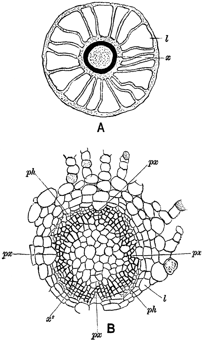
Fig. 91.
Diagrammatic sketch of a transverse section of a young root of Calamites. x, xylem; l, lacuna. After Hick.
Central cylinder (stele) of root, px, protoxylem; ph, phloem; x2, secondary xylem; l, phloeoterma. × 75. After Williamson and Scott.
The xylem elements have the form of spiral, reticulate and scalariform tracheids.
In roots or rootlets smaller than that shown in fig. 91 B, the primary xylem may extend to the centre of the stele, and form a continuous axial strand; in such examples the structure may be diarch, triarch or tetrarch. The origin of the cambium agrees with that in recent roots, the cells immediately external to the protoxylem tracheids become meristematic, as also those internal to the phloem. Another root-character is seen in the endogenous origin of lateral members. Good examples of branching roots are figured by Williamson[695] and by Williamson and Scott[696].
Older roots[697] are usually found in a decorticated condition. A transverse section of root in which secondary thickening has been active for some time presents on a superficial view a close resemblance to a stem of Calamites, but a careful comparison at once reveals important points of difference. The specimen diagrammatically sketched in fig. 92 illustrates very clearly the origin of a root from the node of a Calamite stem. The section has passed through a stem in a tangential direction, showing the characteristic arrangement of the vascular bundles x, and principal medullary rays m. The small leaf-traces, t, t, afford another feature characteristic of a Calamite stem. The portion of stem to the right of the figure has been slightly displaced, and between this piece and the root R, one of the ubiquitous Stigmarian appendages, s, has inserted itself. At R a fairly thick and decorticated root is seen in oblique transverse section; at the upper end the root tracheids are seen in direct continuity with the xylem of the stem. In the centre of the root is the large solid pith surrounded by twelve bluntly pointed xylem groups, composed in the main of radially disposed scalariform elements with narrow secondary medullary rays like those in a stem. Between each xylem group there is a broad medullary ray, which tapers rapidly towards the outside, and is soon obliterated by the formation of interfascicular secondary xylem. At R′ a portion of another root is seen in transverse section, and R″ the inner part of a single xylem group is shown more clearly. The solid pith and the absence of carinal canals are the two most obvious distinguishing features of the roots.
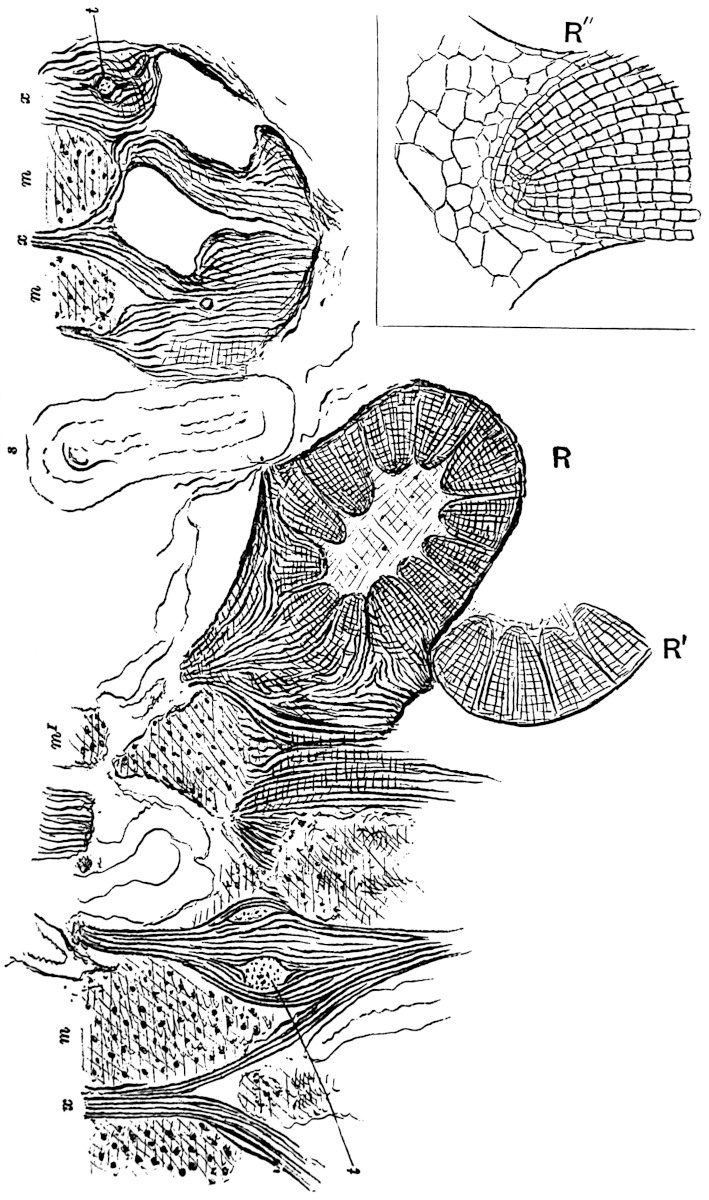
Fig. 92. Tangential section through a node of Calamites, showing a root in organic connection with the stem. R, R′. Root (Astromyelon) in transverse and oblique section, x, xylem; m, primary medullary ray; t, leaf-trace; s, Stigmarian appendage. R″, the inner portion of one of the xylem wedges of R′ more highly magnified. Sketched from a section in the Cambridge Botanical Laboratory Collection.
As Renault points out, roots of Calamites have been figured by some writers[698] as examples of stems, but it is usually comparatively easy to distinguish between roots and stems. On examining the xylem groups more closely, one notices that the apex of each is occupied by a triangular group of centripetally-developed primary tracheids, the narrow spiral protoxylem elements occupying the outwardly directed apex. The protoxylem apex is usually followed externally by a ray of one or two radially disposed series of parenchymatous cells. This ray is not distinguished in fig. 92 R″ from the rows of xylem tracheids. Each xylem group is thus formed partly of centripetal xylem and in part of secondary centrifugal xylem; the latter is associated with secondary medullary rays, as in stems, and contains a broader ray (fascicular ray of Williamson and Scott[699]) immediately opposite each protoxylem strand. In the roots of recent plants (e.g. Cucurbita, Phaseolus, &c.) a broad medullary ray is often found opposite the protoxylem, and such an arrangement is a perfectly normal structure in roots[700].
Renault has recently described several species of Calamite roots which he designates by specific names, some of them belonging to stems with the Arthropitys structure, and others to Calamodendron. Some of the roots figured by the French author have an axial strand of xylem with 7–15 projecting angles of protoxylem[701]. These he considers true roots, but the larger specimens with a wide pith he prefers to regard as stolons. In the latter he mentions the union of the primary centripetal with the secondary centrifugal wood as a distinguishing feature. It has been shown, however, that each group of secondary xylem includes a median ray of parenchyma, and that the whole structure is essentially that of a root, and not that of a modified stem or stolon. The organs described by Renault as true roots are probably rootlets, and as Williamson and Scott have demonstrated, there is every gradation between the smaller specimens with a solid xylem axis and those with a large central pith.
It is interesting to note that Renault’s figures of Calamodendron roots show the closest resemblance to those of the subgenus Arthropitys.
d. Cones.
The occurrence of fossil plants in the form of isolated fragments is a constant source of difficulty, and is well illustrated by the numerous examples of strobili which cannot be connected with their parent stems. We are, however, usually able to recognise Calamitean cones if the impressions or petrified specimens are fairly well preserved, but it is seldom possible to correlate particular types of cones with the corresponding species of foliage-shoots or stems. Palaeobotanical literature contains numerous illustrations and descriptions of long and narrow strobili designated by different generic terms such as Volkmannia, Brukmannia, Calamostachys, Macrostachya and others; many of these have since been recognised as the cones of Calamites, while some species of Volkmannia have been identified with Sphenophyllum stems. Before further considering the general question of Calamite cones, a few examples may be described in detail as types of fructification which are known to have been borne by Calamites. The examples selected are species of the two provisional genera Calamostachys and Palaeostachya.
The usual form of a Calamite cone is illustrated in fig. 93, which represents a fertile shoot bearing a few narrow linear leaves of the Calamocladus type; in the axils of some of these are borne the long strobili.
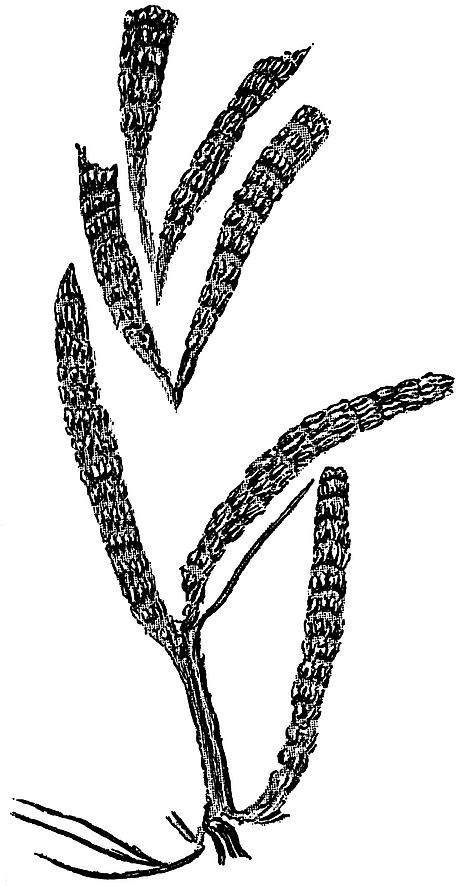
Fig. 93. Calamostachys sp. A fertile Calamitean shoot. From a specimen in the Geological Survey Museum, Jermyn Street, London. From the Upper Coal-Measures of Monmouthshire (No. 5539).
Calamostachys Binneyana (Carr.). Figs. 94 and 95.
In 1867 Carruthers[702] gave an account of the structural features of the species of cones named by him Volkmannia Ludwigi and V. Binneyi, the generic term having been originally used by Sternberg[703] for some impressions of Carboniferous strobili. Brongniart[704] in 1849 referred to the various forms of Volkmannia as cones of Asterophyllitean branches, and the latter he regarded as the foliage-shoots of a Calamite stem. In 1868 Binney[705] published a description, with several illustrations, of the cones named by Carruthers Volkmannia Binneyi, and referred to them as the fructification of that type of Calamite stem spoken of in a previous section of this chapter (p. 311) as Calamites (Arthropitys) communis (Binney). This cone is now usually spoken of as Calamostachys Binneyana; the specific name Binneyana being suggested by Schimper[706] in 1869 as more euphonious than that proposed by Carruthers. In recent years our knowledge of both C. Binneyana and C. Ludwigi has been considerably extended. We shall confine our attention in the following account to the former species[707]. Some excellent figures of the latter species may be found in Weiss’ Memoir[708] on Calamarieae.
One of the largest examples of Calamostachys Binneyana so far recorded has a length of 3–4 cm. and a maximum diameter of about 7·5 mm. The axis of the cone bears whorls of sterile leaves or bracts at equal distances; the linear bracts of each whorl are coherent basally as a disc or plate of tissue attached at right angles to the central axis of the cone. The periphery of each of these discs divides up into twelve linear segments, which curve upwards in a direction more or less parallel to the strobilus axis, and at right angles to the coherent portion of each whorl. The manner of occurrence of the whorls is shown in fig. 94, which has been sketched from a large section in the Williamson collection. The segments of the successive sterile verticils alternate with one another, so that in the surface-view of a cone the long and narrow free bracts appear spirally disposed. Midway between these alternating sterile verticils there is a series of fertile appendages, also given off in regular whorls. Each fertile whorl consists of about half as many members as the segments of a sterile whorl, and the members of the several fertile whorls are superposed and not alternate. Each member has the form of a stalk or sporangiophore given off at right angles from the cone axis; this is expanded distally into a peltate disc bearing four sporangia attached to its inner face. In fig. 94 we can only see the basal portions of the sporangiophores, which are shown in the upper part of the sketch as pointed projections, Sp, from the cone axis. Each sporangiophore is traversed by a vascular strand which sends off a branch to the base of a sporangium (fig. 95, A, t).
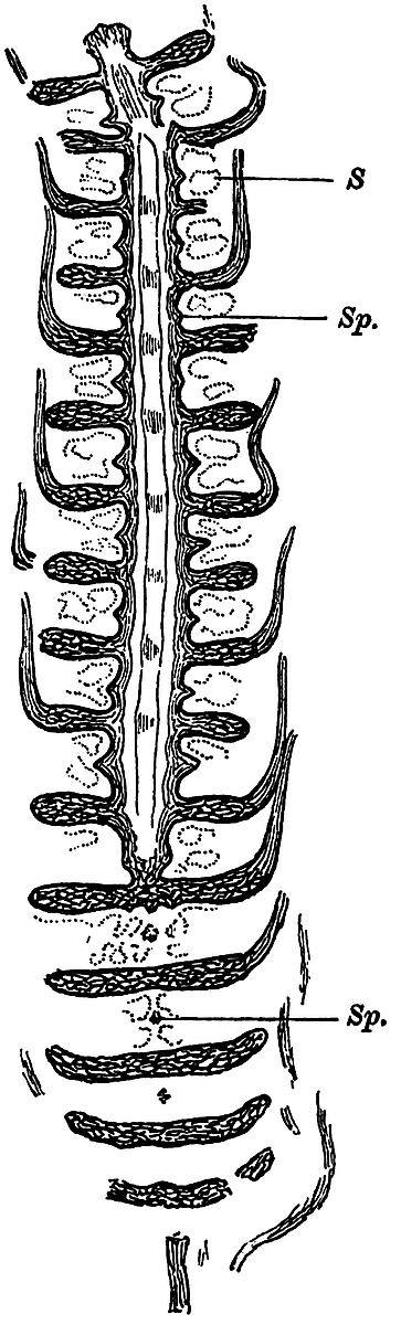
Fig. 94. Calamostachys Binneyana (Carr.) in longitudinal (radial and tangential) section. Sp, sporangiophores; S, sporangia. (From specimen no. 1022 in the Williamson Collection, British Museum.)
The axis of the cone is occupied by a single stele, usually triangular in section; the stele consists of a solid pith of elongated cells surrounded by six vascular bundles, two at each corner. A somewhat irregular gap marks the position of the protoxylem of each strand, and portions of spiral or annular tracheids may occasionally be seen in the cavity. These cavities, which may be spoken of as the carinal canals, disappear at the nodes, where there is a mass of short reticulately pitted tracheids, as in a Calamite stem. Vascular bundles pass upwards in an oblique direction from the central stele to supply the bracts, each of which is traversed by a single strand of tracheids. The coherent portion, or disc, of each sterile whorl consists of sclerenchymatous elements towards the upper surface, and of parenchyma below. The pedicel of the sporangiophores consists of fairly thick-walled cells traversed by a single vascular strand, and the peltate distal portions are made up of parenchymatous cells arranged in a palisade-like form at right angles to the free surface of the sporangiophores. The vascular strand of the pedicel forks into two halves just below the peltate head, and these branches again bifurcate to send a branch to each sporangium. The four sporangia of each sporangiophore are attached by a narrow band of tissue to the shield-shaped distal expansion (fig. 95, A).
In a tangential section of a cone, such as the lower portion of fig. 94 and in fig. 95, B, the sporangiophores present the appearance of narrow stalks (fig. 95, B, a) in the middle of a cluster of sporangia, and the latter appear more or less square in outline. The wall of a sporangium is made of a single layer of cells (fig. 95, B) which present a characteristic appearance in surface-view (fig. 95, C), the thin walls being crossed at right angles by small vertical plates. In the tangential section of the coherent sterile whorls (fig. 95, B, b and b) the vascular strands are occasionally seen in transverse section (fig. 95, B, t), as they pass outwards to the several free bracts.
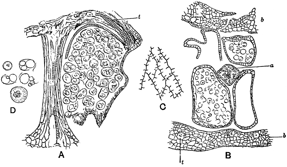
Fig. 95. Calamostachys Binneyana (Carr.).
A sporangiophore and one sporangium. t, vascular bundle. × 45.
Tangential section showing portions of two sterile discs, b, b; a sporangiophore, a, with its four sporangia, in two of which are seen the spores; t, vascular bundle. × 35.
Surface-view of cells of a sporangium wall. × 130.
Spores and remains of mother-cells. × 130.
(After Williamson and Scott.)
The spores in Calamostachys Binneyana are all of the same size, and no macrospores have ever been seen. In well preserved specimens tetrads of spores may be seen, still enclosed by the wall of the spore-mother-cell (fig. 95, A and D); and the torn remnants of the mother-cell sometimes simulate in appearance the elaters of an Equisetum spore. In surface-view a spore often shows clearly the three-rayed marking, which is a characteristic feature of daughter-cells formed in a tetrad from a mother-cell. The spores of a tetrad are in some cases of unequal size, some having developed more vigorously than others. This unequal growth and nourishment of spores is clearly shown in fig. 96, which represents a sporangium of a heterosporous Calamitean strobilus, C. Casheana. Williamson and Scott[709] have described striking examples of spores in different stages of abortion, and these authors draw attention to the importance of the phenomenon from the point of view of the origin of a heterosporous form of cone. The abortion of some of the members of a spore-tetrad and the consequent increased nutrition of the more favoured daughter-cells, might well be the starting-point of a process, which would ultimately lead to the production of well defined macrospores and microspores. The young microsporangia and macrosporangia of recent Vascular Cryptogams such as Selaginella, Salvinia and other heterosporous genera are identical in appearance[710]; it is not until the spore-producing tissue begins to differentiate into groups of spores, that the sporangia assume the form of macrosporangia and microsporangia. During the evolution of the various known types of pteridophytic plants heterospory gradually succeeded isospory, and this no doubt occurred several times and in different phyla of the plant kingdom. In the mature sporangia of some of the Calamitean strobili we have in the inequality of the spores in one sporangium an indication of the steps by which heterospory arose; and in the immature sporangia of some recent genera we are carried back to a stage still nearer the starting-point of the substitution of the heterosporous for the isosporous condition.
Calamostachys Casheana Will. Fig. 96.
To Williamson[711] again is largely due the information we possess as to the structure of this type of Calamitean strobilus. Its special interest lies in the occurrence of macrospores and microspores in the same cone.
The strobilus axis agrees in structure with that of C. Binneyana, but in C. Casheana a band of secondary xylem forms the peripheral portion of the triangular stele. Were any further proof needed of the now well-established fact that secondary growth in thickness is by no means unknown as an attribute of Vascular Cryptogams, the co-existence in the same cone of a cambium layer producing secondary wood and bark, and cryptogamic macrospores and microspores, affords conclusive evidence[712]. The dogma accepted by many writers for a considerable number of years that the power of secondary thickening is evidence against a cryptogamic affinity, has been responsible for no little confusion in palaeobotanical nomenclature.
On the axis of Calamostachys Casheana there are borne alternate whorls of fertile and sterile appendages similar to those in the homosporous C. Binneyana, but they are inclined more obliquely to the axis of the cone. Macrospores and microspores have been found in sporangia borne on the same sporangiophore.
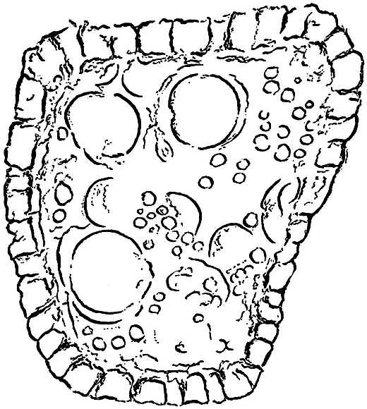
Fig. 96. Calamostachys Casheana Will. A sporangium with macrospores and abortive spores. × 65. (After Williamson and Scott.)
The spore-tetrads in the macrosporangia occasionally include aborted sister-cells like those noticed in C. Binneyana; this phenomenon is well illustrated by the unequally nourished spores in the sporangium of fig. 96, but no such starved spores have been found in the microsporangia. In this cone, then, heterospory has become firmly established, but the occurrence of undersized spores in a macrospore-tetrad leads us back to the probable lines of development of heterospory, which are seen in C. Binneyana at their starting-point.
In the two species of strobili which have been described, Calamostachys Binneyana and C. Casheana, the sporangiophores or sporophylls are given off at right angles to the axis, and midway between the sterile whorls. These are two of the most important distinguishing features of the Calamitean cones included under the generic term Calamostachys. In another form of cone, which also belongs to Calamitean stems, the sporangiophores arise in the axil of the sterile leaves, and are inclined obliquely to the axis of the cone. To this type the generic name Palaeostachya has been applied by the late Prof. Weiss[713] of Berlin. The portion of a cone shown in fig. 97 shows the arrangement of the sterile and fertile appendages characteristic of Palaeostachya.