VOLUME XIV SLICE V Indole to Insanity (continued 6)
Reading edition
The localities attacked by influenza in 1889-1890 appear in no case to have suffered severely for more than a month or six weeks. Thus in Europe and North America generally the visitation had come to an end in the first quarter of 1890. The earliest signs of an epidemic revival on a large scale occurred in March 1891, in the United States and the north of England. It was reported from Chicago and other large towns in the central states, whence it spread eastwards, reaching New York about the end of March. In England it began in the Yorkshire towns, particularly in Hull, and also independently in South Wales. In London influenza became epidemic for the second time about the end of April, and soon afterwards was widely distributed in England and Wales. The large towns in the north, together with London and Wales, suffered much more heavily in mortality than in the previous attack, but the south-west of England, Scotland and Ireland escaped with comparatively little sickness. The same may be said of the European continent generally, except parts of Russia, Scandinavia and perhaps the north of Germany. This second epidemic coincided with the spring and early summer; it had subsided in London by the end of June. The experience of Sheffield is interesting. In 1890 the attack, contrary to general experience, had been undecided, lingering and mild; in 1891 it was very sudden and extremely severe, the death-rate rising to 73.4 during the month of April, and subsiding with equal rapidity. During the third quarter of the year, while Europe was free, the antipodes had their second attack, which was more severe than the first. As in England, it reversed the previous order of things, beginning in the provinces and spreading thence to the capital towns. The last quarter of the year was signalized by another recrudescence in Europe, which reached its height during the winter. All parts, including Great Britain, were severely affected. In England those parts which had borne the brunt of the epidemic in the early part of the year escaped. In fact, these two revivals may be regarded as one, temporarily interrupted by the summer quarter.
The recrudescence at the end of 1891 lasted through mid-winter, and in many places, notably in London, it only reached its height in January 1892, subsiding slowly and irregularly in February and March. Brighton suffered with exceptional severity. The continent of Europe seems to have been similarly affected. In Italy the notifications of influenza were as follow: 1891—January to October, 0; November, 30; December, 6461; 1892—January, 84,543; February, 55,352; March, 28,046; April, 7962; May, 1468; June, 223. Other parts of the world affected were the West Indies, Tunis, Egypt, Sudan, Cape Town Teheran, Tongking and China. In August 1892 influenza was reported from Peru, and later in the year from various places in Europe.
A fourth recrudescence, but of a milder character, occurred in Great Britain in the spring of 1893, and a fifth in the following winter, but the year 1894 was freer from influenza than any since 1890. In 1895 another extensive epidemic took place. In 1896 influenza seemed to have spent its strength, but there was an increased prevalence of the disease in 1897, which was repeated on a larger scale in 1898, and again in 1899, when 12,417 deaths were recorded in England and Wales. This was the highest death-rate since 1892. After this the death-rate declined to half that amount and remained there with the slight upward variations until 1907, in which the total death-rate was 9257. The experience of other countries has been very similar; they have all been subjected to periodical revivals of epidemic influenza at irregular intervals and of varying intensity since its reappearance in 1889, but there has been a general though not a steady decline in its activity and potency. Its behaviour is, in short, quite in keeping with the experience of 1847-1860, though the later visitation appears to have been more violent and more fatal than the former. Its diffusion was also more rapid and probably more extensive.
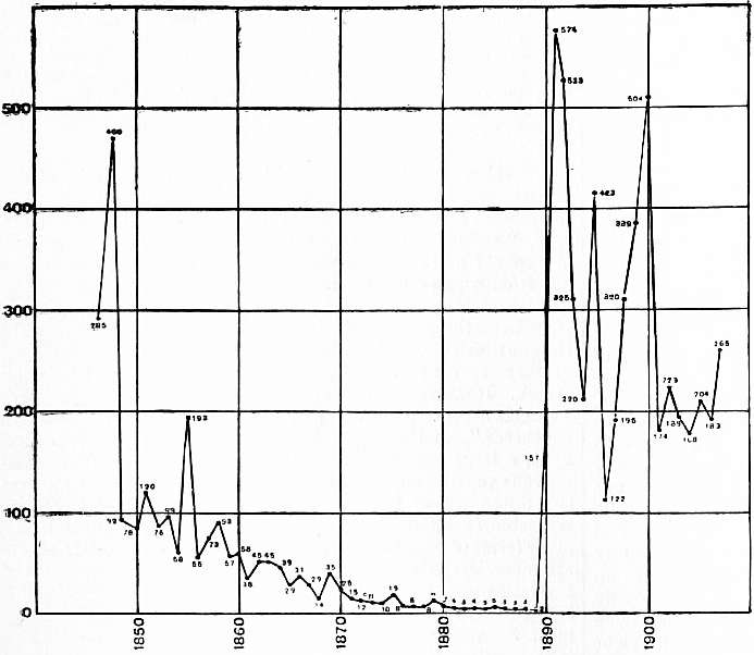
The foregoing general summary may be supplemented by some further details of the incidence in Great Britain. The number of deaths directly attributed to influenza, and the death-rates per million in each year in England and Wales, are as follow:—
Year. Deaths. Death-rates per million. Year. Deaths. Death-rates per million.
1890 4,523 157 1899 12,417 389
1891 16,686 574 1900 16,245 504
1892 15,737 534 1901 5,666 174
1893 9,669 325 1902 7,366 223
1894 6,625 220 1903 6,322 189
1895 12,880 424 1904 5,694 168
1896 3,753 122 1905 6,953 204
1897 6,088 196 1906 6,310 183
1898 10,405 331 1907 9,257 265
It is interesting to compare these figures with the corresponding ones for the previous visitation:—
Year. Deaths. Death-rates per million. Year. Deaths. Death-rates per million.
1847 4,881 285 1852 1,359 76
1848 7,963 460 1853 1,789 99
1849 1,611 92 1854 1,061 58
1850 1,380 78 1855 3,568 193
1851 2,152 120
The two sets of figures are not strictly comparable, because, during the first period, notification of the cause of death was not compulsory; but it seems clear that the later wave was much the more deadly. The average annual death-rate for the nine years is 320 in the one case against 162 in the other, or as nearly as possible double. In both epidemic periods the second year was far more fatal than the first, and in both a marked revival took place in the ninth year; in both also an intermediate recrudescence occurred, in the fifth year in one case, in the sixth in the other. The chief point of difference is the sudden and marked drop in 1849-1850, against a persistent high mortality in 1892-1893, especially in 1892, which was nearly as fatal as 1891.
To make the significance of these epidemic figures clear, it should be added that in the intervening period 1861-1889 the average annual death-rate from influenza was only fifteen, and in the ten years immediately preceding the 1890 outbreak it was only three. Moreover, in epidemic influenza, the mortality directly attributed to that disease is only a fraction of that actually caused by it. For instance, in January 1890 the deaths from influenza in London were 304, while the excess of deaths from respiratory diseases was 1454 and from all causes 1958 above the average.
We have seen above that the mortality was far greater in the second epidemic year than in the first, and this applies to all parts of England, and to rural as well as to urban communities, as the following table shows:—
Deaths from Influenza.
1890. 1891.
London 624 2302
24 Great Towns over 80,000 population 439 2417
35 Towns between 20,000 and 80,000 186 765
21 Towns between 10,000 and 20,000 46 196
60 Towns under 10,000 62 196
85 Rural Sanitary Districts 317 841
In spite of these figures, it appears that the 1890 attack, which was in general much more sudden in its onset than that of 1891, also caused a great deal more sickness. More people were “down with influenza,” though fewer died. For Instance, the number of persons treated at the Middlesex Hospital in the two months’ winter epidemic of 1890 was 1279; in the far more fatal three months’ spring epidemic of 1891 it was only 726. One explanation of this discrepancy between the incidence of sickness and mortality is that in the second attack, which was more protracted and more insidious, the stress of the disease fell more upon the lungs. Another is that its comparative mildness, combined with the time of year, in itself proved dangerous, because it tempted people to disregard the illness, whereas in the first epidemic they were too ill to resist. On the whole, rural districts showed a higher death-rate than towns, and small towns a higher one than large ones in both years. This is explained by the age distribution in such localities; influenza being particularly fatal to aged people, though no age is exempt. Certain counties were much more severely affected than others. The eastern counties, namely, Essex, Suffolk and Norfolk, together with Hampshire and one or two others, escaped lightly in both years; the western counties, namely, North and South Wales, with the adjoining counties of Monmouth, Hereford and Shropshire, suffered heavily in both years.
It will be convenient to discuss seriatim the various points of interest on which light has been thrown by the experience described above.
The bacteriology of influenza is discussed in the article on Parasitic Diseases. The disease is often called “Russian” influenza, and its origin in 1889 suggests that the name may have some foundation in fact. A writer, who saw the epidemic break out in Bokhara, is quoted by him to the following effect:—“The summer of 1888 was exceptionally hot and dry, and was followed by a bitterly cold winter and a rainy spring. The dried-up earth was full of cracks and holes from drought and subsequent frost, so that the spring rains formed ponds in these holes, inundated the new railway cuttings, and turned the country into a perfect marsh. When the hot weather set in the water gave off poisonous exhalations, rendering malaria general.” On account of the severe winter, the people were enfeebled from lack of nourishment, and when influenza broke out suddenly they died in large numbers. Europeans were very severely affected. Russians, hurrying home, carried the disease westwards, and caravans passing eastwards took it into Siberia. There is a striking similarity in the conditions described to those observed in connexion with outbreaks of other diseases, particularly typhoid fever and diphtheria, which have occurred on the supervention of heavy rain after a dry period, causing cracks and fissures in the earth. Assuming the existence of a living poison in the ground, we can easily understand that under certain conditions, such as an exceptionally dry season, it may develop exceptional properties and then be driven out by the subsequent rains, causing a violent outbreak of illness. Some such explanation is required to account for the periodical occurrence of epidemic and pandemic diffusions starting from an endemic centre. We may suppose that a micro-organism of peculiar robustness and virulence is bred and brought into activity by a combination of favourable conditions, and is then disseminated more or less widely according to its “staying power,” by human agency. Whether central Asia is an endemic centre for influenza or not there is no evidence, but the disease seems to be more often prevalent in the Russian Empire than elsewhere. Extensive outbreaks occurred there in 1886 and 1887, and it is certain that the 1889 wave was active in Siberia at an earlier date than in Europe, and that it moved eastwards. The hypothesis that it originated in China is unsupported by evidence. But whatever may be the truth with regard to origin, the dissemination of influenza by human agency must be held to be proved. This is the most important addition to our knowledge of the subject contributed by recent research. The upshot of the inquiry by Dr Parsons was to negative all theories of atmospheric influence, and to establish the conclusion that the disease was “propagated mainly, perhaps entirely, by human intercourse.”
He found that it prevailed independently of climate, season and weather; that it moved in a contrary direction to the prevailing winds; that it travelled along the lines of human intercourse, and not faster than human beings can travel; that in 1889 it travelled much faster than in previous epidemics, when the means of locomotion were very inferior; that it appeared first in capital towns, seaports and frontier towns, and only affected country districts later; that it never commenced suddenly with a large number of cases in a place previously free from disease, but that epidemic manifestations were generally preceded for some days or weeks by scattered cases; that conveyance of infection by individuals and its introduction into fresh places had been observed in many instances; that persons brought much into contact with others were generally the first to suffer; that persons brought together in large numbers in enclosed spaces suffered more in proportion than others, and that the rapidity and extent of the outbreak in institutions corresponded with the massing together of the inmates.
These conclusions, based upon the 1889-1890 epidemic, have been confirmed by subsequent experience, especially in regard to the complete independence of season and weather shown by influenza. It has appeared and disappeared at all seasons and in all weathers and only popular ignorance continues to ascribe its behaviour to atmospheric conditions. In Europe, however, it has prevailed more often in winter than in summer, which may be due to the greater susceptibility of persons in winter, or, more probably, to the fact that they congregate more in buildings and are less in the open air during that part of the year. No doubt is any longer entertained of its infectious character, though the degree of infectivity appears to vary considerably. Many cases have been recorded of individuals introducing it into houses, and of all or most of the other inmates then taking it from the first case. Difficulties in preventing the spread of infection are due to (1) the shortness of the period of incubation, (2) the disease being infectious in the earliest stages before the nature of the illness is recognized, (3) the milder varieties being equally infectious with the severe attacks, and the patient going to work and spreading the infection, (4) the diagnosis often being difficult, influenza being possibly confused with ordinary catarrhal attacks, typhoid fever and other diseases. Domestic animals seem to be free from any suspicion of being liable to human influenza. Sanitary conditions, other than overcrowding, do not appear to exercise any influence on the spread of influenza.
Influenza has been shown to be an acute specific fever having nothing whatever to do with a “bad cold.” There may be some inflammation of the respiratory passages, and then symptoms of catarrh are present, but that is not necessarily the case, and in some epidemics such symptoms are quite exceptional. This had been recognized by various writers before the 1889 visitation, but it had not been generally realized, as it has been since, and some medical authorities, who persisted in regarding influenza as essentially a “catarrhal” affection, were chiefly to blame for a widespread and tenacious popular fallacy.
Leichtenstern, in his masterly article in Nothnagel’s Handbuch, divides the disease as follows:—(1) Epidemic influenza vera caused by Pfeiffer’s bacillus; (2) Endemic-epidemic influenza vera, which occurs several years after a pandemic and is caused by the same bacillus; (3) Endemic influenza nostras or eatarrhal fever, called la grippe, and bearing the same relation to true influenza as cholera nostras does to Asiatic cholera.
The “period of incubation” is one to four days. Susceptibility varies greatly, but the conditions that influence it are matters of conjecture only. It appears that the inhabitants of Great Britain are less susceptible than those of many other countries. Dr Parsons gives the following list, showing the proportion of the population estimated to have been attacked in the 1889-1890 epidemic in different localities:—
Place. Per cent. Place. Per cent.
St Petersburg 50 Portugal 90
Berlin 33 Vienna 30-40
Nuremberg 67 Belgrade 33
Grand-Duchy of Hesse 25-30 Antwerp 33
Grand-Duchy, other Gaeta 50-77
Districts 50-75 Massachusetts 39
Heligoland 50 Peking 50
Budapest 50 St Louis (Mauritius) 67
In and about London he reckoned roughly from a number of returns that the proportion was about 12 1/2% among those employed out of doors and 25% among those in offices, &c. The proportion among the troops in the Home District was 9.3%. The General Post Office made the highest return with 33.6%, which is accounted for partly by the enormous number of persons massed together in the same room in more than one department, and partly by the facilities for obtaining medical advice, which would tend to bring very light cases, unnoticed elsewhere, upon the record. No public service was seriously disorganized in England by sickness in the same manner as on the continent of Europe. Some individuals appear to be totally immune; others take the disease over and over again, deriving no immunity, but apparently greater susceptibility from previous attacks.
The symptoms were thus described by Dr Bruce Low from observations made in St Thomas’s Hospital, London, in January 1890:—
The invasion is sudden; the patients can generally tell the time when they developed the disease; e.g. acute pains in the back and loins came on quite suddenly while they were at work or walking in the street, or in the case of a medical student, while playing cards, rendering him unable to continue the game. A workman wheeling a barrow had to put it down and leave it; and an omnibus driver was unable to pull up his horses. This sudden onset is often accompanied by vertigo and nausea, and sometimes actual vomiting of bilious matter. There are pains in the limbs and general sense of aching all over; frontal headache of special severity; pains in the eyeballs, increased by the slightest movement of the eyes; shivering; general feeling of misery and weakness, and great depression of spirits, many patients, both men and women, giving way to weeping; nervous restlessness; inability to sleep, and occasionally delirium. In some cases catarrhal symptoms develop, such as running at the eyes, which are sometimes injected on the second day; sneezing and sore throat; and epistaxis, swelling of the parotid and submaxillary glands, tonsilitis, and spitting of bright blood from the pharynx may occur. There is a hard, dry cough of a paroxysmal kind, worst at night. There is often tenderness of the spleen, which is almost always found enlarged, and this persists after the acute symptoms have passed. The temperature is high at the onset of the disease. In the first twenty-four hours its range is from 100° F. in mild cases to 105° in severe cases.
Dr J. S. Bristowe gave the following description of the illness during the same epidemic:—
The chief symptoms of influenza are, coldness along the back, with shivering, which may continue off and on for two or three days; severe pain in the head and eyes, often with tenderness in the eyes and pain in moving them; pains in the ears; pains in the small of the back; pains in the limbs, for the most part in the fleshy portions, but also in the bones and joints, and even in the fingers and toes; and febrile temperature, which may in the early period rise to 104° or 105° F. At the same time the patient feels excessively ill and prostrate, is apt to suffer from nausea or sickness and diarrhoea, and is for the most part restless, though often (and especially in the case of children and those advanced in age) drowsy.... In ordinary mild cases the above symptoms are the only important ones which present themselves, and the patient may recover in the course of three or four days. He may even have it so mildly that, although feeling very ill, he is able to go about his ordinary work. In some cases the patients have additionally some dryness or soreness of the throat, or some stiffness and discharge from the nose, which may be accompanied by slight bleeding. And in some cases, for the most part in the course of a few days, and at a time when the patient seems to be convalescent, he begins to suffer from wheezing in the chest, cough, and perhaps a little shortness of breath, and before long spits mucus in which are contained pellets streaked or tinged with blood.... Another complication is diarrhoea. Another is a roseolous spotty rash.... Influenza is by no means necessarily attended with the catarrhal symptoms which the general public have been taught to regard as its distinctive signs, and in a very large proportion of cases no catarrhal condition whatever becomes developed at any time.
Several writers have distinguished four main varieties of the disease—namely, (1) nervous, (2)gastro-intestinal, (3)respiratory, (4) febrile, a form chiefly found in children. Clifford Allbutt says, “Influenza simulates other diseases.” Many forms are of typhoid or comatose types. Cardiac attacks are common, not from organic disease but from the direct poisoning of the heart muscle by influenza.
Perhaps the most marked feature of influenza, and certainly the one which victims have learned to dread most, is the prolonged debility and nervous depression that frequently follow an attack. It was remarked by Nothnagel that “Influenza produces a specific nervous toxin which by its action on the cortex produces psychoses.” In the Paris epidemic of 1890 the suicides increased 25%, a large proportion of the excess being attributed to nervous prostration caused by the disease. Dr Rawes, medical superintendent of St Luke’s hospital, says that of insanities traceable to influenza melancholia is twice as frequent as all other forms of insanity put together. Other common after-effects are neuralgia, dyspepsia, insomnia, weakness or loss of the special senses, particularly taste and smell, abdominal pains, sore throat, rheumatism and muscular weakness. The feature most dangerous to life is the special liability of patients to inflammation of the lungs. This affection must be regarded as a complication rather than an integral part of the illness. The following diagram gives the annual death-rate per million in England and Wales, and is taken from an article by Dr Arthur Newsholme in The Practitioner (January 1907).
The deaths directly attributed to influenza are few in proportion to the number of cases. In the milder forms it offers hardly any danger to life if reasonable care be taken, but in the severer forms it is a fairly fatal disease. In eight London hospitals the case-mortality among in-patients in the 1890 outbreak was 34.5 per 1000; among all patients treated it was 1.6 per 1000. In the army it was rather less.
The infectious character of influenza having been determined, suggestions were made for its administrative control on the familiar lines of notification, isolation and disinfection, but this has not hitherto been found practicable. In March 1895, however, the Local Government Board issued a memorandum recommending the adoption of the following precautions wherever they can be carried out:—
1. The sick should be separated from the healthy. This is especially important in the case of first attacks in a locality or a household.
2. The sputa of the sick should, especially in the acute stage of the disease, be received into vessels containing disinfectants. Infected articles and rooms should be cleansed and disinfected.
3. When influenza threatens, unnecessary assemblages of persons should be avoided.
4. Buildings and rooms in which many people necessarily congregate should be efficiently aerated and cleansed during the intervals of occupation.
There is no routine treatment for influenza except bed. In all cases bed is advisable, because of the danger of lung complications, and in mild ones it is sufficient. Severer ones must be treated according to the symptoms. Quinine has been much used. Modern “anti-pyretic” drugs have also been extensively employed, and when applied with discretion they may be useful, but patients are not advised to prescribe them for themselves.
Sir Wm. Broadbent in a note on the prophylaxis of influenza recommends quinine in a dose of two grains every morning, and remarks: “I have had opportunities of obtaining extraordinary evidence of its protective power. In a large public school it was ordered to be taken every morning. Some of the boys in the school were home boarders, and it was found that while the boarders at the school took the quinine in the presence of a master every morning, there were scarcely any cases of influenza among them, although the home boarders suffered nearly as much as before.” He continues, “In a large girls’ school near London the same thing was ordered, and the girls and mistresses took their morning dose but the servants were forgotten. The result was that scarcely any girl or mistress suffered while the servants were all down with influenza.”
The liability to contract influenza, and the danger of an attack if contracted, are increased by depressing conditions, such as exposure to cold and to fatigue, whether mental or physical. Attention should, therefore, be paid to all measures tending to the maintenance of health. Persons who are attacked by influenza should at once seek rest, warmth and medical treatment, and they should bear in mind that the risk of relapse, with serious complications, constitutes a chief danger of the disease.
In addition to the ordinary text-books, see the series of articles by experts on different aspects in The Practitioner (London) for January 1907.
IN FORMÂ PAUPERIS (Latin, “in the character of pauper”), the legal phrase for a method of bringing or defending a case in court on the part of persons without means. By an English statute of 1495 (11 Hen. VII. c. 12), any poor person having cause of action was entitled to have a writ according to the nature of the case, without paying the fees thereon. The statute of 1495 was repealed by the Statute Law Revision and Civil Procedure Act 1883, but its provisions, as well as the chancery practice were incorporated into one code and embodied in the rules of the Supreme Court (O. xvi. rr. 22-31). Now any person may be admitted to sue as a pauper, on proof that he is not worth £25, his wearing apparel and the subject matter of the cause or matter excepted. He must lay his case before counsel for opinion, and counsel’s opinion thereon, with an affidavit of the party suing that the case contains a full and true statement of all the material facts to the best of his knowledge and belief, must be produced before the proper officers to whom the application is made. A person who desires to defend as a pauper must enter an appearance to a writ in the ordinary way and afterwards apply for an order to defend as a pauper. Where a person is admitted to sue or defend as a pauper, counsel and solicitor may be assigned to him, and such counsel and solicitor are not at liberty to refuse assistance unless there is some good reason for refusing. If any person admitted to sue or defend as a pauper agrees to pay fees to any person for the conduct of his business he will be dispaupered. Costs ordered to be paid to a pauper are taxed as in other cases. Appeals to the House of Lords in formâ pauperis were regulated by the Appeal (Formâ Pauperis) Act 1893, which gave the House of Lords power to refuse a petition for leave to sue.
INFORMATION (from Lat. informare, to give shape or form to, to represent, describe), the communication of knowledge; in English law, a proceeding on behalf of the crown against a subject otherwise than by indictment. A criminal information is a proceeding in the King’s bench by the attorney-general without the intervention of a grand jury. The attorney-general, or, in his absence, the solicitor-general, has a right ex officio to file a criminal information in respect of any indictments, but not for treason, felonies or misprision of treason. It is, however, seldom exercised, except in cases which might be described as “enormous misdemeanours,” such as those peculiarly tending to disturb or endanger the king’s government, e.g. seditions, obstructing the king’s officers in the execution of their duties, &c. In the form of the proceedings the attorney-general is said to “come into the court of our lord the king before the king himself at Westminster, and gives the court there to understand and be informed that, &c.” Then follows the statement of the offence as in an indictment. The information is filed in the crown office without the leave of the court. An information may also be filed at the instance of a private prosecutor for misdemeanours not affecting the government, but being peculiarly flagrant and pernicious. Thus criminal informations have been granted for bribing or attempting to bribe public functionaries, and for aggravated libels on public or private persons. Leave to file an information is obtained after an application to show cause, founded on a sworn statement of the material facts of the case.
Certain suits might also be filed in Chancery by way of information in the name of the attorney-general, but this species of information was superseded by Order 1, rule 1 of the Rules of the Supreme Court, 1883, under which they are instituted in the ordinary way. Informations in the Court of Exchequer in revenue cases, also filed by the attorney-general, are still resorted to (see A.-G. v. Williamson, 1889, 60 L.T. 930).
INFORMER, in a general sense, one who communicates information. The term is applied to a person who prosecutes in any of the courts of law those who break any law or penal statute. Such a person is called a common informer when he furnishes evidence on criminal trials or prosecutes for breaches of penal laws solely for the purpose of obtaining the penalty recovered, or a share of it. An action by a common informer is termed a popular or qui tam action, because it is brought by a person qui tam pro domino rege quam pro se ipso sequitur. A suit by an informer must be brought within a year of the offence, unless a specific time is prescribed by the statute. The term informer is also used of an accomplice in crime who turns what is called “king’s evidence” (see Accomplice). In Scotland, informer is the term applied to the party who, in criminal proceedings, sets the lord advocate in motion.
INFUSORIA, the name given by Bütschli (following O.F. Ledermüller, 1763) to a group of Protozoa. The name arose from the procedure adopted by the older microscopists to obtain animalcules. Infusions of most varied organic substances were prepared (hay and pepper being perhaps the favourite ones), the method of obtaining them including maceration and decoction, as well as infusion in the strict sense; they were then allowed to decompose in the air, so that various living beings developed therein. As classified by C. G. Ehrenberg in his monumental Infusionstierchen als volkommene Organismen, they included (1) Desmids, Diatoms and Schizomycetes, now regarded as essentially Plant Protista or Protophytes; (2) Sarcodina (excluding Foraminifera, as well as Radiolaria, which were only as yet known by their skeletons, and termed Polycystina), and (3) Rotifers, as well as (4) Flagellates and Infusoria in our present sense. F. Dujardin in his Histoire des zoophytes (1841) gave nearly as liberal an interpretation to the name; while C. T. Van Siebold (1845) narrowed it to its present limits save for the admission of several Flagellate families. O. Bütschli limited the group by removing the Flagellata, Dinoflagellata and Cystoflagellata (q.v.) under the name of “Mastigophora” proposed earlier by R. M. Diesing (1865). We now define it thus:—Protozoa bounded by a permanent plasmic pellicle and consequently of definite form, never using pseudopodia for locomotion or ingestion, provided (at least in the young state) with numerous cilia or organs derived from cilia and equipped with a double nuclear apparatus: the larger (mega-) nucleus usually dividing by constriction, and disappearing during conjugation: the smaller (micro-) nucleus (sometimes multiple) dividing by mitosis, and entering into conjugation and giving rise to the cycle of nuclei both large and small of the race succeeding conjugation.
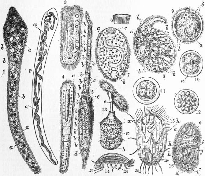
Fig. i. Ciliata.
1. Opalinopsis sepiolae, Foett.: a parasitic Holotrichous mouthless Ciliate from the liver of the Squid. a, branched meganucleus; b, vacuoles (non-contractile). 2. A similar specimen treated with picrocarmine, showing a remarkably branched and twisted meganucleus (a), in place of several nuclei. 3. Anoplophrya naidos, Duj.; a mouthless Holotrichous Ciliate parasitic in the worm Nais. a, the large axial meganucleus; b, contractile vacuoles. 4. Anoplophrya prolifera, C. and L.; from the intestine of Clitellio. Remarkable for the adhesion of incomplete fission-products in a metameric series. a, meganucleus. 5. Amphileptus gigas, C. and L. (Gymnostomaceae). b, contractile vacuoles; c, trichocysts (see fig. 2); d, meganucleus; e. pharynx. 6, 7. Prorodon niveus, Ehr. (Gymnostomaceae). a, meganucleus; b, contractile vacuole; c, pharynx with horny cuticular lining. 6. The fasciculate cuticle of the pharynx isolated. 8. Trachelius ovum, Ehr. (Gymnostomaceae); showing the reticulate arrangement of the endosarc, b, contractile vacuoles; c, the cuticle-lined pharynx. 9, 10, 11, 12. Icthyophthirius multifilius, Fouquet (Gymnostomaceae). Free individual and successive stages of division to form spores. a, meganucleus; b, contractile vacuoles. 13. Didinium nasutum, Müll. (Gymnostomaceae). The pharynx is everted and has seized a Paramecium as food. a, meganucleus; b, contractile vacuole; c, everted pharynx. 14. Euplotes charon, Müll. (Hypotrichaceae); lateral view of the animal when using its great cirrhi, x, as ambulatory organs. 15. Euplotes harpa, Stein (Hypotrichaceae); h, mouth; x, cirrhi. 16. Nyctotherus cordiformis, Stein (a Heterotriceae), parasitic in the intestine of the Frog; a, meganucleus; b, contractile vacuole; c, food particle; d, anus; e, heterotrichous band of membranelles; f, g, mouth; h, pharynx; i, small cilia.
Thus defined, the Infusoria fall into two groups:—(1) Ciliata, with cilia or organs derived from cilia throughout their lives, provided with a single permanent mouth (absent in the parasitic Opalinopsidae) flush with the body or at the base of an oral depression, and taking in food by active swallowing or by ciliary action: (2) Suctoria, rarely ciliated except in the young state, and taking in their food by suction through protrusible hollow tentacles, usually numerous.
The pellicle of the Infusoria is stronger and more permanent than in many Protozoa, and sometimes assumes the character of a mail of hard plates, closely fitting; but even in this case it undergoes solution soon after death. It is continuous with a firm ectosarc, highly differentiated in the Ciliata, and in both groups free from coarse movable granules. The endosarc is semifluid and rich in granules mostly “reserve” in nature, often showing proteid or fat reactions. One or more contractile vacuoles are present in some of the marine and all the freshwater species, and open to the surface by pores of permanent position: a system of canals in the deeper layers of the ectoplasm is sometimes connected with the vacucle. The body is often provided with not-living external formations “stalk” and “theca” (or “lorica”).
The character of the nuclear apparatus excludes two groups both parasitic and mouthless: (1) the Trichonymphidae, with a single nucleus of Leidy, parasitic in Insects, especially Termites; (2) the Opalinidae, with several (often numerous) uniform nuclei, parasitic in the gut of Batrachia, &c., and producing 1-nuclear zoospores which conjugate. Both these families we unite into a group of Pseudociliata, which may be referred to the Flagellata (q.v.). Lankester in the last edition of this Encyclopaedia called attention to the doubtful position of Opalina, and Delage and Hérouard placed Trichonymphidae among Flagellates.
The theca or shell is present in some pelagic species (fig. iii. 3, 5) and in many of the attached species, notably among the Peritricha (fig. iii. 21, 22, 25, 26) and Suctoria (fig. viii. 11); and is found in some free-swimming forms (fig. iii. 3, 5): it is usually chitinous, and forms a cup into which the animal, protruded when at its utmost elongation, can retract itself. In Metacineta mystacina it has several distinct slits (pylomes) for the passage of tufts of tentacles. In Stentor it is gelatinous; and in the Dictyocystids it is beautifully latticed.
The stalk is usually solid, and expanded at the base into a disk in Suctoria. In Peritrichaceae (fig. iii. 8-22, 25, 26), the only ciliate group with a stalk, it grows for some time after its formation, and on fission two new stalks continue the old one, so as to form a branched colony (fig. iii. 18). In Vorticella (fig. iii. 11, 12, 14, &c.) the stalk is hollow and elastic, and attached to it along a spiral is a prolongation of the ectosarc containing a bundle of myonemes, so that by the contractions of the bundle the stalk is pulled down into a corkscrew spiral, and on the relaxation of the muscle the elasticity of the hollow stalk straightens it out.
On fission the stalk may become branched, as the solid one of Epistylis and Opercularia (fig. iii. 20); and the myoneme also in the tubular stem of Zoothaminum; or the branch-myoneme for the one offspring may be inserted laterally on that for the other in Carchesium (fig. iii. 18). In several tubicolous Peritrichaceae there is some arrangement for closing their tubes. In Thuricola (fig. iii. 25-26) there is a valve which opens by the pressure of the animal on its protrusion, and closes automatically by elasticity on retraction. In Lagenophrys the animal adheres to the cup a little below the opening, so that its withdrawal closes the cup: at the adherent part the body mass is hardened, and so differentiated as to suggest the frame of the mouth of a purse. In Pyxicola (fig. iii. 21-22) the animal bears some way down the body a hardened shield (“operculum”) which closes the mouth of the shell on retraction.
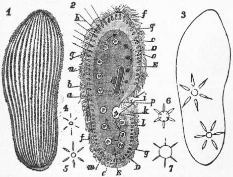
Fig. ii.
1, Surface view of Paramecium, showing the disposition of the cilia in longitudinal rows. 2, a, mega-; b, micronucleus; c, junction of ecto- and endosarc; D, pellicle; E, endosarc; f, cilia (much too numerous and crowded); g, trichocysts; g′, same with thread; h, discharged; i, pharynx, its undulating membrane not shown; k, food granules collecting into a bolus; l, m, n, o, food vacuoles, their contents being digested as they pass in the endosarc along the path indicated by the arrows. 3, Outline showing contractile vacuoles in commencing diastole, surrounded by five afferent canals. 4-7 Successive stages of diastole of contractile vacuole.
The cytoplasm of the Infusoria is very susceptible to injuries; and when cut or torn, unless the pellicle contracts rapidly to enclose the wounded surface, the substance of the body swells up, becoming frothy, with bubbles which rapidly enlarge and finally burst; the cell thus disintegrates, leaving only a few granules to mark where it was. This phenomenon, observed by Dujardin, is called “diffluence.” The contractile vacuole appears to be one of the means by which diffluence is avoided in cells with no strong wall to resist the absorption of water in excess; for after growing in size for some time, its walls contract suddenly, and its contents are expelled to the outside by a pore, which is, like the anus, usually invisible, but permanent in position. The contractile vacuole may be single or multiple; it may receive the contents of a canal, or of a system of canals, which only become visible at the moment of the contraction of the vacuole (fig. ii. 4-7), giving liquid time to accumulate in them, or when the vacuole is acting sluggishly or imperfectly, as in the approach of asphyxia (fig. ii. 3). Besides this function, since the system passes a large quantity of water from without through the substance of the cell, it must needs act as a means of respiration and excretion. In all Peritrichaceae it opens to the vestibule, and in some of them it discharges through an intervening reservoir, curiously recalling the arrangements in the Flagellate Euglenaceae.
The nuclear apparatus consists of two parts, the meganucleus, and the micronucleus or micronuclei (fig. iii. 17d, iv. 1). The meganucleus alone regarded and described as “the nucleus” by older observers is always single, subject to a few reservations. It is most frequently oval, and then is indented by the micronucleus; but it may be lobed, the lobes lying far apart and connected by a slender bridge or moniliform, or horseshoe-shaped (Peritrichaceae). It often contains darker inclusions, like nucleoles.
It has been shown, more especially by Gruber, that many Ciliata are multinucleate, and do not possess merely a single meganucleus and a micronucleus. In Oxytricha the nuclei are large and numerous (about forty), scattered through the protoplasm, whilst in other cases the nucleus is so finely divided as to appear like a powder diffused uniformly through the medullary protoplasm (Trachelocerca). Carmine staining, after treatment with absolute alcohol, has led to this remarkable discovery. The condition described by Foettinger in his Opalinopsis (fig. i. 1, 2) is an example of this pulverization of the nucleus. The condition of pulverization had led in some cases to a total failure to detect any nucleus in the living animal, and it was only by the use of reagents that the actual state of the case was revealed. Before fission, whatever be its habitual character, it condenses, becomes oval, and divides by constriction; and though it usually is then fibrillated, only in a few cases does it approach the typical mitotic condition. The micronucleus described by older writers as the “nucleolus” or “paranucleus” (“endoplastule” of Huxley), may be single or multiple. When the meganucleus is bilobed there are always two micronuclei, and at least one is found next to every enlargement of the moniliform meganucleus. In the fission of the Infusoria, every micronucleus divides by a true mitotic process, during which, however, its wall remains intact. From their relative sizes the meganucleus would appear to discharge during cell-life, exclusively, the functions of the nucleus in ordinary cells. Since in conjugation, however, the meganucleus degenerates and is in great part either digested or excreted as waste matter, while the new nuclear apparatus in both exconjugates arises, as we shall see, from a conjugation-nucleus of exclusively micronuclear origin, we infer that the micronucleus has for its function the carrying on of the nuclear functions of the race from one fission cycle to the next from which the meganucleus is excluded.
Fission is the ordinary mode of reproduction in the Infusoria, and is usually transverse, but oblique in Stentor, &., as in Flagellata, longitudinal in Peritrichaceae; in some cases it is always more or less unequal owing to the differentiation of the body, and consequently it must be followed by a regeneration of the missing organs in either daughter-cell. In some cases it becomes very uneven, affording every transition to budding, which process assumes especial importance in the Suctoria. Multiple fission (brood-formation or sporulation) is exceptional in Infusoria, and when it occurs the broods rarely exceed four or eight—another difference from Flagellata. The nuclear processes during conjugation suggest the phylogenetic loss of a process of multiple fission into active gametes. As noted, in fission the meganucleus divides by direct constriction; each micronucleus by a mode of mitosis. The process of fission is subject in its activity to the influences of nutrition and temperature, slackening as the food supply becomes inadequate or as the temperature recedes from the optimum for the process. Moreover, if the descendants of a single animal be raised, it is found that the rapidity of fission, other conditions being the same, varies periodically, undergoing periods of depression, which may be followed by either (1) spontaneous recovery, (2) recovery under stimulating food, (3) recovery through conjugation, or (4) the death of the cycle, which would have ensued if 2 or 3 had been omitted at an earlier stage, but which ultimately seems inevitable, even the induction of conjugation failing to restore it. These physiological conditions were first studied by E. Maupas, librarian to the city of Algiers, in his pioneering work in the later ’eighties, and have been confirmed and extended by later observers, among whom we may especially cite G. N. Calkins.
Syngamy, usually termed conjugation or “karyogamy,” is of exceptional character in the majority of this group—the Peritrichaceae alone evincing an approximation to the usual typical process of the permanent fusion of two cells (pairing-cells or gametes), cytoplasm to cytoplasm, nucleus to nucleus, to form a new cell (coupled cell, zygote).
This process was elucidated by E. Maupas in 1889, and his results, eagerly questioned and repeatedly tested, have been confirmed in every fact and in every generalization of importance.
Previously all that had been definitely made out was that under certain undetermined conditions a fit of pairing two and two occurred among the animals of the same species in a culture or in a locality in the open; that after a union prolonged over hours, and sometimes even days, the mates separated; that during the union the meganucleus underwent changes of a degenerative character; and that the micronucleus underwent repeated divisions, and that from the offspring of the micronuclei the new nuclear apparatus was evolved for each mate. Maupas discovered the biological conditions leading to conjugation: (1) the presence of individuals belonging to distinct stocks; (2) their belonging to a generation sufficiently removed from previous conjugation, but not too far removed therefrom; (3) a deficiency of food. He also showed that during conjugation a “migratory” nucleus, the offspring of the divisions of the micronucleus, passes from either mate to the other, while its sister nucleus remains “stationary”; and that reciprocal fusion of the migratory nucleus of the one mate with the stationary nucleus of the other takes place to form a zygote nucleus in either mate; and that from these zygote nuclei in each by division, at least two nuclei are formed, the one of which enlarges to form a meganucleus, while the other remains small as the first micronucleus of the new reorganized animal, which now separates as an “exconjugate” (fig. iv). Moreover, if pairing be prevented, or be not induced, the individuals produced by successive fissions become gradually weaker, their nuclear apparatus degenerates, and finally they cannot be induced under suitable conditions to pair normally, so that the cycle becomes extinct by senile decay. In Peritrichaceae the gametes are of unequal sizes (fig. iii. 11, 12), the smaller being formed by brood fissions (4 or 8); syngamy is here permanent, not temporary, the smaller (male) being absorbed into the body of the larger (female); and there are only two nuclei that pair. Thus we have a derived binary sexual process, comparable to that of ordinary bisexual organisms.
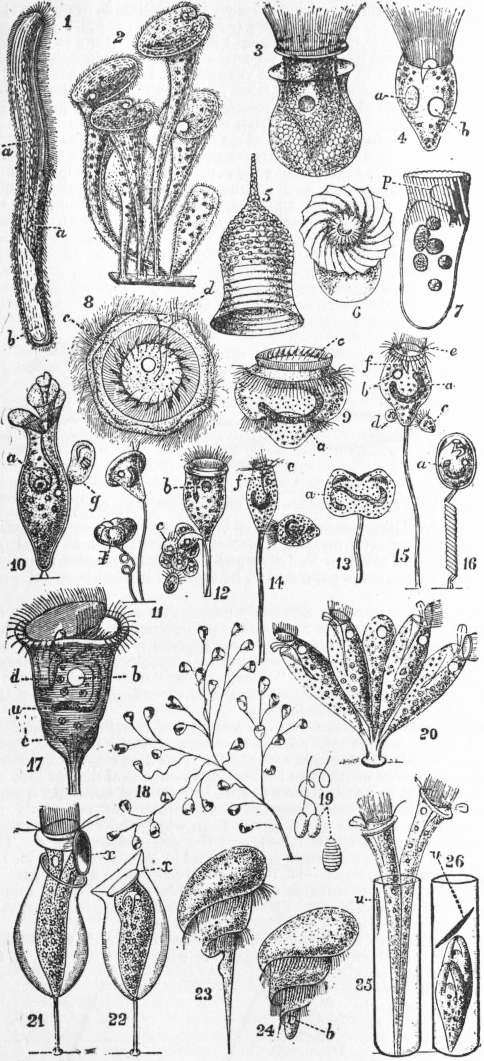
Fig. iii.— Ciliata: 1, 2, Heterotrichaceae; 3-7, 23-24, Oligotrichaceae; 8-22, 25, 26, Peritrichaceae.
1, Spirostomum ambiguum, Ehr.; on its left side oral groove and wreath of membranellae; a, moniliform meganucleus; b, position of contractile vacuole. 2, Group of Stentor polymorphus, O. F. Müller; the twisted end of the peristome indicating the position of the mouth. 3, Tintinnus lagenula, Cl. and L., in free shell. 4, Strombidium claparedii, S. Kent. 5, Shell of Codonella campanella, Haeck. 6, 7, Torquatella typica, Lank. (= Strombidium according to Bütschli); p, oral tube seen through peristomial wreath of apparently coalescent membranellae. 8. Basal, and 9, side (inverted) views of Trichodina pediculus, Ehr.; a, meganucleus; c, basal collar and ring of hooks; d, mouth; contractile vacuole and oral tube seen by transparency in 8. 10, Spirochona gammipara, Stein; a, meganucleus; g, bud. 11, 12, Vorticella microstoma, Ehr.; d, formation of a brood of 8 microgametes c by multiple fission; b, contr. vacuole. 13, Same sp. in binary fission; a, meganucleus. 14, V. nebulifera, Ehr.; bud swimming away by posterior wreath, peristome contracted; e, peristomial disk; f, oral tube. 15, V. microstoma; b, contr. vacuole; c, d, two microgametes seeking to conjugate. 16, V. nebulifera, contracted, with body encysted. 17, Same sp. enlarged; c, myonemes converging posteriorly to muscle of stalk; d, micronucleus. 18, Carchesium spectabile, Ehr.; (×50). 19, Nematocysts of Epistylis flavicans. Ehr. (after Greeff). 20, Opercularia stenostoma, St.; (×200); a small colony showing upstanding (“opercular”) peristomial disk, protruded oral undulating membranejand cilia in oral tube. 21, 22, Pyxicola affinis, S.K., with stalk and theca; x, chitinous disk, or true “operculum” closing theca in retracted state. 23, 24, Caenomorpha medusula, Perty, (×250), with spiral peristomial wreath. 25, 26, Thuricola valvata, Str. Wright, in sessile theca, with internal valve (v) to close tube, as in gastropod Clausilia; owing to recent fission two animals occupy one tube.
From Lankester’s Treatise on Zoology.
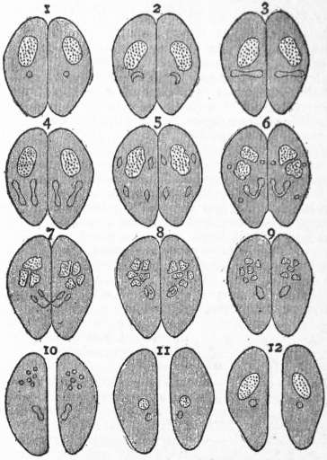
Fig. iv.—Diagrammatic Sketch of Changes during Conjugation in Ciliata. (From Hickson after Delage and Maupas.)
1, Two individuals at commencement of conjugation showing meganucleus (dotted) and micronucleus; successive stages of the disintegration of the meganucleus shown in all figures up to 9. 2, 3, First mitotic division of micronuclei. 4, 5, Second ditto. 6, One of the four nuclei resulting from the second division again dividing to form the pairing-nuclei in either mate, while the other 3 nuclei degenerate. 7, Migration of the migratory nuclei. 8, 9, Fusion of the incoming migratory with the stationary nucleus in either mate. 10, Fission of Zygote nucleus into two, the new mega- and micronucleus whose differentiation is shown in 11, 12. The vertical dotted line indicates the separation of the mates.
Ciliata.—The Ciliate Infusoria represent the highest type of Protozoa. They are distinctly animal in function, and the Gymnostomaceae are active predaceous beings preying on other Infusoria or Flagellates. Some possess shells (fig. iii. 3, 5, 21, 22, 25, 26), most have a distinct swallowing apparatus, and in Dysteria there is a complex jaw—or tooth-apparatus, which needs new investigation. In the active Ciliata we find locomotive organs of most varied kinds: tail-springs, cirrhi for crawling and darting, cilia and membranellae for continuous swimming in the open or gliding over surfaces or waltzing on the substratum (Trichodina, fig. iii. 8) or for eddying in wild turns through the water (Strombidium, Tintinnus, Halteria). Their forms offer a most interesting variety, and the flexibility of many adds to their easy grace of movement, especially where the front of the body is produced and elongated like the neck of a swan (Amphileptus, fig. iii. 5; Lacrymaria).
From Lankester’s Treatise on Zoology.
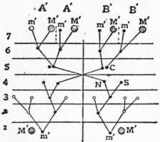
Fig. v.—Diagram 1 illustrating changes during conjugation of Colpidium colpoda. (From Hickson, after Maupas.)
M, Old meganucleus undergoing disintegration. m, Micronucleus. N, migratory, and S, Stationary pairing-nucleus. M′, M′, the new meganuclei, and m′, The new micronuclei in the products of the first fission of each of the exconjugates; the continuous vertical line indicates period of fusion, its cessation, separation; dotted lines indicate fission; the spaces lettered 1-7 successive stages in the process; the clear circles indicate functionless nuclei which degenerate.
The cytoplasm is very highly differentiated: especially the ectoplasm or ectosarc. This has always a distinct elastic “pellicle” or limiting layer, in a few cases hard, or even with local hardenings that affect the disposition of a coat of mail (Coleps) or a pair of valves (Dysteria); but is usually only marked into a rhomboidal network by intersecting depressions, with the cilia occupying the centres of the areas or meshes defined. The cytoplasm within is distinctly alveolated, and frequently contains tubular alveoli running along the length of the animal. Between these are dense fibrous thickenings, which from their double refraction, from their arrangement, and from their shortening in contracted animals are regarded as of muscular function and termed “myonemes.” Other threads running alongside of these, and not shortening but becoming wavy in the general contraction have been described in a few species as “neuronemes” and as possessing a nervous, conducting character. On this level, too, lie the dot-like granules at the bases of the cilia, which form definite groups in the case of such organs as are composed of fused cilia; in the deeper part of the ectoplasm the vacuoles or alveoli are more numerous, and reserve granules are also found; here too exist the canals, sometimes developed into a complex network, which open into the contractile vacuole.
The cilia themselves have a stiffer basal part, probably strengthened by an axial rod, and a distal flexible lash; when cilia are united by the outer plasmatic layer, they form (1) “Cirrhi,” stiff and either hook-like and pointed at the end, or brush-like, with a frayed apex; (2) membranelles, flattened organs composed of a number of cilia fused side by side, sometimes on a single row, sometimes on two rows approximated at either end so as to form a narrow oval, the membranelle thus being hollow; (3) the oral “undulating membrane,” merely a very elongated membranelle whose base may extend over a length nearly equal to the length of the animal; such membranes are present in the mouth oral depression and pharynx of all but Gymnostomaceae, and aid in ingestion; a second or third may be present, and behave like active lips; (4) in Peritrichaceae the cilia of the peristomial wreath are united below into a continuous undulating membrane, forming a spiral of more than one turn, and fray out distally into a fringe; (5) the dorsal cilia of Hypotrichaceae are slender and motionless, probably sensory.
Embedded in the ectosarc of many Ciliates are trichocysts, little elongated sacs at right angles to the surface, with a fine hair-like process projecting. On irritation these elongate into strong prominent threads, often with a more or less barb-like head, and may be ejected altogether from the body. Those over the surface of the body appear to be protective; but in the Gymnostomaceae specially strong ones surround the mouth. They can be injected into the prey pursued, and appear to have a distinctly poisonous effect on it. They are combined also into defensive batteries in the Gymnostome Loxophyllum. They are absent from most Heterotrichaceae and Hypotrichaceae, and from Peritrichaceae, except for a zone round the collar of the peristome.
The openings of the body are the mouth, absent in a few parasital species (Opalinopsis, fig. i. 1, 2), the anus and the pore of the contractile vacuole. The mouth is easily recognizable; in the most primitive forms of the Gymnostomaceae and some other groups, it is terminal, but it passes further and further back in more modified species, thereby defining a ventral, and correspondingly a dorsal surface; it usually lies on the left side. The anus is usually only visible during excretion, though its position is permanent; in a few genera it is always visible (e.g. Nyctotherus, fig. i. 16). The pore of the contractile vacuole might be described in the same terms.
The endoplasm has also an alveolar structure, and contains besides large food-vacuoles or digestive vacuoles, and shows movements of rotation within the ectoplasm, from which, however, it is not usually distinctly bounded. In Ophryoscolex and Didinium (fig. i. 13) a permanent cavity traverses it from mouth to anus.
From Calkins’ Protozoa, by permission of the Macmillan Company, N.Y.
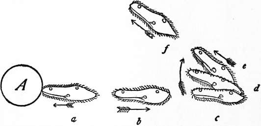
Fig. vi.—Diagrammatic view of behaviour of the motile reaction of Paramecium after meeting a mechanical obstruction at A. (From G. N. Calkins after H. S. Jennings.) For clearness and simplicity the normal motion is supposed to be straight instead of spiral.
Ingestion of food is of the same character in all the Hymenostomata. The ciliary current drives a powerful stream into the mouth, which impinges against the endosarc, carrying with it the food particles; these adhere and accumulate to form a pellet, which ultimately is pushed by an apparently sudden action into the substance of the endosarc which closes behind it (fig. ii. 2). In some of the Aspirotrichaceae accessory undulating membranes play the part of lips, and there is a closer approximation to true deglutition. The mouth is rarely terminal, more frequently at the bottom of a depression, the “vestibule,” which may be prolonged into a slender canal, sometimes called the “pharynx” or “oral tube,” ciliated as well as provided with a membrane, and extending deep down into the body in many Peritrichaceae.
In Spirostomaceae the “adoral wreath” of membranelles encloses more or less completely an anterior part of the body, the “peristome,” within which lies the vestibule. This area may be depressed, truncate, convex or produced into a short obconical disk or into one or more lobes, or finally form a funnel, or a twisted spiral like a paper cone. In most Peritrichaceae a collar-like rim surrounds the peristome, and marks out a gutter from which the vestibule opens; the peristome can be retracted, and the collar close over it. This rim forms a deep permanent spiral funnel in Spirochona (fig. iii. 10).
Movements of Ciliata.—H. S. Jennings has made a very detailed study of these movements, which resemble those of most minute free-swimming organisms. The following account applies practically to all active “Infusoria” in the widest sense.
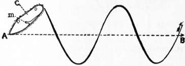
Fig. vii.—Diagram of a mode of progression of a Ciliate like Paramecium; m, mouth and pharynx; the straight line A, B, represents the axis of progression described by the posterior end, and the spiral line the curve described by the anterior end; the clear circles are the contractile vacuoles on the dorsal side.
The position of the free-swimming Infusoria, like that of Rotifers and other small swimming animals, is with the front end of the body inclined outward to the axis of advance, constantly changing its azimuth while preserving its angle constant or nearly so; if advance were ignored the body would thus rotate so as to trace out a cone, with the hinder end at the apex, and the front describing the base. On any irritation, (1) the motion is arrested, (2) the animal reverses its cilia and swims backwards, (3) it swerves outwards away from the axis so as to make a larger angle with it, and (4) then swims forwards along a new axis of progression, to which it is inclined at the same angle as to the previous axis (figs. vi., vii.). In this way it alters its axis of progression when it finds itself under conditions of stimulation. Thus a Paramecium coming into a region relatively too cold, too hot, or too poor in CO2 or in nutriment, alters its direction of swimming; in this way individuals come to assemble in crowds where food is abundant, or even where there is a slight excess of CO2. This reaction may lead to fatal results; if a solution of corrosive sublimate (Mercuric chloride) diffuses towards the hinder end of the animal faster than it progresses, the stimulus affecting the hinder end first, the axis of progression is altered so as to bring the animal after a few changes into a region where the solution is strong enough to kill it. This “motile reaction,” first noted by H. S. Jennings, is the explanation of the general reactions of minute swimming animals to most stimuli of whatever character, including light; the practical working out is, as he terms it, a method of “trial and error.” The action, however, of a current of electricity is distinctly and immediately directive; but such a stimulus is not to be found in nature. The motile reaction in the Hypotrichaceae which crawl or dart in a straight line is somewhat different, the swerve being a simple turn to the right hand—i.e. away from the mouth.
Parasitism in the Infusoria is by no means so important as among Flagellates. Ichthyophthirius alone causes epidemics among Fishes, and Balantidium coli has been observed in intestinal disease in Man. The Isotricheae, among Aspirotrichaceae and the Ophryoscolecidae among Heterotrichaceae are found in abundance in the stomachs of Ruminants, and are believed to play a part in the digestion of cellulose, and thus to be rather commensals than parasites. A large number of attached species are epizoic commensals, some very indifferent in choice of their host, others particular not only in the species they infest, but also in the special organs to which they adhere. This is notably the case with the shelled Peritrichaceae. Lichnophora and Trichodina (fig. iii. 8, 9) among Peritrichaceae are capable of locomotion by their permanent posterior wreath or of attaching themselves by the sucker which surrounds it; Kerona polyporum glides habitually over the body of Hydra, as does Trichodina pediculus.
Several Suctoria are endoparasitic in Ciliata, and their occurrence led to the view that they represented stages in the life-history of these. Again, we find in the endosarc of certain Ciliates green nucleated cells, which have a cellulose envelope and multiply by fission inside or outside the animal. They are symbiotic Algae, or possibly the resting state of a Chlamydomonadine Flagellate (Carteria?), and have received the name Zoochlorella. They are of constant occurrence in Paramecium bursaria, frequent in Stentor polymorphus and S. igneus, and Ophrydium versatile, and a few other species, which become infected by swallowing them.
Classification.
Order I.—Section A.—Gymnostomaceae. Mouth habitually closed; swallowing an active process; cilia (or membranelles) uniform, usually distributed evenly over the body; form variable, sometimes of circular transverse section.
Section B.—Trichostomata. Mouth permanently open against the endosarc, provided with 1 or 2 undulating membranes often prolonged into an inturned pharynx; ingestion by action of oral ciliary apparatus.
Order 2.—Subsection (a).—Aspirotrichaceae. Cilia nearly uniform, not associated with cirrhi or membranelles, nor forming a peristomial wreath. Form usually flattened, mouth unilateral. (N.B.—Orders 1, 2 are sometimes united into the single order Holotrichaceae.)
Subsection (b).—Spirotricha. Wreath of distinct membranelles—or of cilia fused at the base—enclosing a peristomial area and leading into the mouth.
§§ i.—Wreath of separate membranelles.
Order 3.—Heterotrichaceae; body covered with fine uniform cilia, usually circular in transverse section.
Order 4.—Oligotrichaceae; body covering partial or wholly absent; transverse section usually circular.
Order 5.—Hypotrichaceae; body flattened; body cilia represented chiefly by stiff cirrhi in ventral rows, and fine motionless dorsal sensory hairs.
Order 6.—§§ ii.—Peritrichaceae. Peristomial ciliary wreath, spiral, of cilia united at the base; posterior wreath circular of long membranelles; body circular in section, cylindrical, taper, or bell-shaped.
Illustrative Genera (selected).
1. Gymnostomaceae. (a) Ciliation general or not confined to one surface. Coleps Ehr., with pellicle locally hardened into mailed plates; Trachelocerca Ehr.; Prorodon Ehr. (fig. i. 6, 7); Trachelius Ehr., with branching endosarc (fig. i. 8); Lacrymaria Ehr. (fig. i. 5), body produced into a long neck with terminal mouth surrounded by offensive trichocysts; Dileptus Duj., of similar form, but anterior process, blind, preoral; Ichthyophthirius Fouquet (fig. i. 9-12), cilia represented by two girdles of membranellae; Didinium St. (fig. i. 13), cilia in tufts, surface with numerous tentacles each with a strong terminal trichocyst; Actinobolus Stein, body with one adoral tentacle; Ileonema Stokes. (b) Cilia confined to dorsal surface. Chilodon Ehr.; Loxodes Ehr., body flattened, ciliated on one side only, endosarc as in Trachelius; Dysteria Huxley, with the dorsal surface hardened and hinged along the median line into a bivalve shell, ciliated only on ventral surface, with a protrusible foot-like process, and a complex pharyngeal armature. (c) Cilia restricted to a single equatorial girdle, strong (probably membranelles); Mesodinium, mouth 4-lobed.
2. Aspirotrichaceae. Paramecium Hill (fig. ii. 1-3); Ophryoglena Ehr.; Colpoda O. F. Müller; Colpidium St.; Lembus Cohn, with posterior strong cilium for springing; Leucophrys St.; Urocentrum Nitsch, bare, with polar and equatorial zones and a posterior tuft of long cilia; Opalinopsis Foetlinger (fig. i. 1, 2); Anoplophyra St. (fig. i. 3, 4). (The last two parasitic mouthless genera are placed here doubtfully.)
3. Heterotrichaceae. (a) Wreath spiral; Stentor Oken. (fig. iii. 2), oval when free, trumpet-shaped when attached by pseudopods at apex, and then often secreting a gelatinous tube; Blepharisma Perty, sometimes parasitic in Heliozoa; Spirostomum Ehr., cylindrical, up to 1″ in length; (b) Wreath straight, often oblique; Nyctotherus Leidy, parasitic anus always visible; Balantidium Cl. and L., parasitic (B. coli in man); Bursaria, O.F.M., hollowed into an oval pouch, with the wreath inside.
4. Oligotrichaeceae. Tintinnus Schranck (fig. iii. 3); Trichodinopsis Cl. and L.; Codonella Haeck. (fig. iii. 5); Strombidium Cl. and L. (fig. iii. 4), including Torquatella Lank. (fig. iii. 6, 7), according to Bütschli; Halteria Duj., with an equatorial girdle of stiff bristle-like cilia; Caenomorpha Perty (fig. iii. 23, 24); Ophryoscolex St., with straight digestive cavity, and visible anus, parasitic in Ruminants.
5. Hypotrichaceae. Stylonychia Ehr.; Oxytricha Ehr.; Euplotes Ehr. (fig. i. 14, 15); Kerona Ehr. (epizoic on Hydra).
6. Peritrichaceae. 1. Peristomial wreath projecting when expanded above a circular contractile collar-like rim.
(a) Fam. Urceolaridae: posterior wreath permanently present around sucker-like base. Trichodina Ehr. (fig. iii. 8, 9), epizoic on Hydra; Lichnophora Cl. and L.; Cyclochaeta Hatchett Jackson; Gerda Cl. and L.; Scyphidia Duj.
(b) Fam. Vorticellidae = Bell Animalcules: posterior wreath temporarily present, shed after fixation.
Subfam. 1. Vorticellinae animals naked. (i.) Solitary; Vorticella Linn. (fig. iii. 11-17), stalk hollow with spiral muscle; Pyxidium S. Kent, stalk non-contractile. (ii.) Forming colonies by budding on a branched stalk: Carchesium Ehr., hollow branches and muscles discontinuous; Zoothamnium. Ehr., branched hollow stem and muscle continuous through colony; Epistylis Ehr., stalk rigid—(the animal body in these three genera has the same characters as Vorticella)—Campanella Goldf., stalked like Epistylis, wreath of many turns (nematocysts sometimes present) (fig. iii. 19); Opercularia, stalk of Epistylis, disk supporting wreath obconical, collar very high (fig. iii. 20).
Subfam. 2. Vaginicolinae; body enclosed in a firm theca: Vaginicola Lam., shell simple, sessile; Thuricola St. Wright, shell sessile, with a valve opening inwards (fig. iii. 25-26); Cothurnia Ehr., shell stalked, simple; Pyxicola S. Kent, shell stalked, closed by an infraperistomial opercular thickening on the body (fig. iii. 21-22).
Subfam. 3. Shells gelatinous; those of the colony aggregated into a floating spheroidal mass several inches in diameter Ophrydium Bory, O. versatile contains Zoochlorella, which secretes oxygen, and the gas-bubbles float the colonies like green lumps of jelly.
2. Peristomial wreath, not protrusible, surrounded by a very high usually spiral collar.
Fam. Spirochonina. Spirochona St. (fig. iii. 10); Kentrochona Rompel; both genera epizoic on gills, &c., of small Crustacea.
Suctoria.—These are distinguished from Ciliata by their possession of hollow tentacles (one only in Rhyncheta, fig. viii. 1, and Urnula) through which they ingest food, and by not possessing cilia, except in the young stage. Fission approximately equal is very rare. Usually it is unequal, or if nearly equal one of the halves remains attached, and the other, as an embryo or gemmule, develops cilia and swims off to attach itself elsewhere; Sphaerophrya (fig. viii. 2-6) alone, often occurring as an endoparasite in Ciliata, may be free, tentaculate and unattached.
The ectosarc is usually provided with a firm pellicle which shows a peculiar radiate “milling” in optical section, so fine that its true nature is difficult to make out; it may be due to radial rods, regularly imbedded, or may be the expression of radial vacuoles. The tentacles vary in many respects, but are always retractile. They are tubes covered by an extension of the pellicle; this is invaginated into the body round the base of the tentacle as a sheath, and then evaginated to form the outer layer of the tentacle itself, over which it is frequently raised into a spiral ridge, which may be traced down into the part sunk and ensheathed within the body: in Choanophrya, where the tentacles are largest, the pellicle is further continued into the interior of the tentacle. The tentacles are always pierced by a central canal opening at the apex, which may be (1) enlarged into a terminal capitate sucker, (2) slightly flared, (3) truncate and closed in the resting state to become widely opened into a funnel, or (4) pointed. The tentacles are always capable of being waved from side to side, or turned in a definite direction for the reception or prehension of food; in Rhyncheta, the movements of the long single tentacle recall those of an elephant’s trunk, only they are more extensive and more varied. In the majority of cases the food consists of Ciliata; and the contents of the prey may be seen passing down the canal of the sucker beyond where it becomes free from the general surface. In Choanophrya the food appears to consist of the débris of the prey of the carnivorous host (Cyclops), which is sucked into the wide funnel-shaped mouths of the tentacles—by what mechanism is unknown. The endosarc is full of food-granules and reserve-granules (oil, colouring matter and proteid).
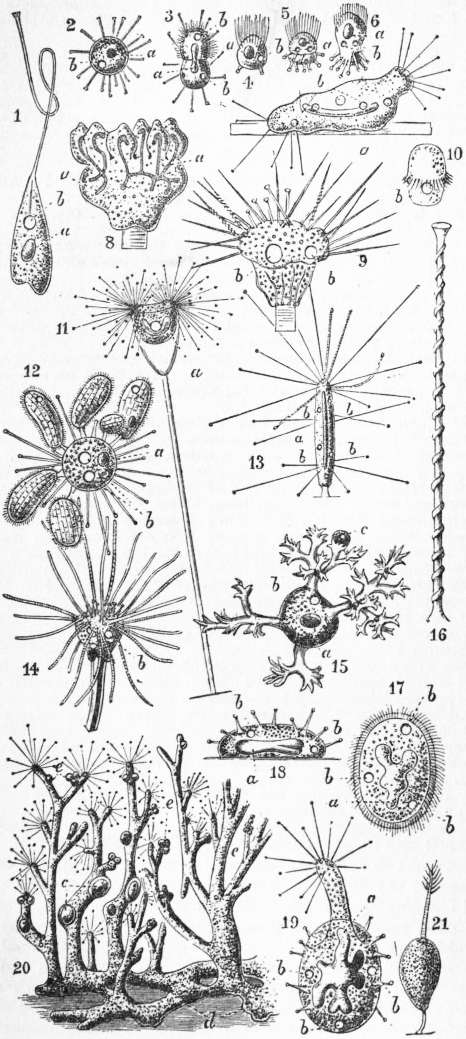
Fig. viii.—Suctoria (in all a, meganucleus; b, contractile vacuole).
1, Rhyncheta cyclopum, Zenker; only a single tentacle and that suctorial; epizoic on Cyclops. 2, Sphaerophrya urostylae, Maupas; normal adult; parasitic in Ciliate Urostyla. 3, The same dividing by transverse fission, the anterior moiety with temporarily developed cilia. 4, 5, 6, Sphaerophrya stentorea, Maupas. Parasitic in Stentor, and at one time mistaken for its young. 7, Trichophrya epistylidis, Cl. and L. 8, Hemiophrya gemmipara, Hertwig. Example with six buds, into each of which a branch of the meganucleus a is extended. 9, The same species, showing the two kinds of tentacles (the suctorial and the pointed), and two contractile vacuoles b. 10, Ciliated embryo of Podophrya steinii, Cl. and L. 11, Acineta grandis, Saville Kent; showing pedunculated cup, and animal with two bunches of entirely suctorial tentacles. 12, Sphaerophrya magna, Maupas. It has seized with its tentacles, and is in the act of sucking out the juices of six examples of the Ciliate Colpoda parvifrons. 13, Podophrya elongata, Cl. and L. 14, Hemiophrya benedenii, Fraip.; the suctorial tentacles retracted. 15, Dendrocometes paradoxus, Stein. Parasitic on Gammarus pulex; captured prey. 16, A single tentacle of Podophrya. R. Hertwig. 17-20, Dendrosoma radians, Ehr.:—17, free-swimming ciliated embryo. 18, Earliest fixed condition of the embryo. 19, Later stage, a single tentaculiferous process now developed. 20, Adult colony; c, enclosed ciliated embryos; d, branching stolon; e, more minute reproductive (?) bodies. 21, Ophryodendron pedicellatum, Hincks.
The meganucleus and the micronucleus are both usually single, but in Dendrosoma (fig. viii. 20), of which the body is branched, and the meganucleus with it, there are numerous micronuclei. In most cases the micronucleus has not been recorded, though from the similarity of conjugation, and its presence in most cases of fission and budding that have been accurately described, we may infer that it is always present. In unequal fission the meganucleus sends a process into the bud, while the micronucleus divides as in Ciliata. The bud may be nearly equal to the remains of the original animal, or much smaller, and in that case a depression surrounds it which may deepen so as to form a brood-cavity, either communicating by a mere “birth-pore” with the outside or entirely closed. In some cases the budding is multiple (fig. viii. 8), and a large number of buds are formed and liberated at the same time. In all cases the bud escapes without tentacles, and possesses a characteristic supply of cilia, whose arrangement is constant for the species.
In some cases an adult may withdraw its tentacles, moult its pellicle and develop an equipment of cilia and swim away: this is the case with Dendrocometes, parasitic on Gammarus, when its host moults.
The numerous species of Suctoria, often so abundant on various species of Cyclops, are not found on the other freshwater Copepoda, Diaptomus and Canthocamptus, belonging indeed to other families. Again, these Suctoria affect different positions, those found on the antennae not being present on the mouth parts; the ventral part of the thorax has another set; and the inside of the pleural fold another. Rhyncheta occupies the front of the “couplers” or median downgrowths uniting the coxopodites of the swimming legs, and Choanophrya settles in the immediate neighbourhood of the mouth, preferably on the epistoma, labrum and metastomatic region, but also on the adoral appendages and in rare cases extends, when the settlement is extensive, to the bases of the two pairs of antennae; while distinct species of Podophrya settle on the antennae, the front of the thorax and the inside of the pleural folds. Dendrocometes is common on the gills of the freshwater shrimp (Amphipod) Gammarus and Stylocometes on the gills and gill-covers of the Isopod Asellus, the water-slater. The independence of the Acinetaria was threatened by the erroneous view of Stein that they were phases in the life-history of Vorticellidae. Small parasitic forms (Sphaerophrya) were also regarded erroneously as the “acinetiform young” of Ciliata. They now must be regarded as an extreme modification of the Protozoon series, in which the differentiation of organs in a unicellular animal reaches its highest point.
Principal Genera.
1. Unstalked simple forms. Urnula Cl. and L., permanently ciliate; Rhyncheta Zenker (fig. viii. 1), on the limb couplers of Cyclops; Sphaerophrya Cl. and L. (fig. viii. 2-6, 12), endoparasitic in Ciliata and formerly taken for embryos thereof, never attached; Trichophrya Cl. and L. (fig. viii. 7), of similar habits, but temporarily attached, sessile.
2. Stalked simple forms; Podophrya Ehr. (fig. viii. 10, 13, 16), tentacles all knobbed or flared; Ephelota Strethill Wright, tentacles all pointed; Hemiophrya S. Kent (fig. viii. 8, 9, 14), tentacles of both kinds; Choanophrya Hartog, tentacles thick, truncate, very retractile, when expanded opening into funnels for aspiration of floating prey, never for attachment—epizoic on antero-ventral parts of Cyclops.
3. Cupped forms; Solenophrya Cl. and L., cup sessile; Acineta Ehr., cup stalked; Acinetopsis Bütschli, like Acineta, but the cup flattened, closed distally with only slit-like apertures (“pylomes”) for the bundles of tentacles; Podocyathus, like Acineta, but with pointed as well as knobbed tentacles.
4. Tentacles in bundles at the tips of one or more processes or branches of the body. Ophryodendron Cl. and L., tentaculiferous process single (fig. viii. 21); Dendrocometes Stein (fig. viii. 15), body rounded, processes repeatedly branched, epizoic on gills of Gammarus pulex; Dendrosoma Ehr. (fig. viii. 17-20), body freely branched from a basal attached stolon, meganucleus branching with the body.
Bibliography.—(a) Infusoria in the widest sense: C. E. Ehrenberg. Die Infusionstierchen als vollkommene Organismen (1838); F. Dujardin, Zoophytes infusoires (1841). (b) Infusoria, including Mastigophora: M. Perty, Zur Kenntniss Kleinster Lebensformen (1852); E. Claparède and J. Lachmann, Études sur les infusoires et les Rhizopodes (1858-1861); F. von Stein, Der Organismus der Infusionstiere (1859-1883); W. Saville Kent, A Manual of the Infusoria, including a description of all known Flagellate, Ciliate and Tentaculiferous Protozoa (1880-1882). (c) Infusoria, as limited by Bütschli. O. Bütschli, Bronn’s Tierreich, vol. i. Protozoa, pt. 3 Infusoria (1887-1889), the most complete work existing, but without specific diagnoses; S. J. Hickson, “The Infusoria” in Lankester’s Treatise on Zoology, vol. i. fasc. 2 (1903), a general account, well illustrated, with a diagnosis of all genera. See also Delage and Hérouard, Traité de Zoologie concrète, vol. i. “La Cellule et les Protozoaires” (1896), with an illustrated conspectus of the genera; E. Maupas, “Recherches expérimentales sur la multiplication des Infusoires ciliés,” Arch. zool. exp. vi. (1888); and “Le Rajeunissement karyogomique chez les Ciliés,” ib. vii. (1889); R. Sand, Étude monographique sur le groupe des Infusoires tentaculifères (Suctoria), (1899), with diagnoses of species; A. Lang, Lehrb. der vergleich, Anatomie der wirbellosen Tiere, vol. i. “Protozoa” (1901) (a view of comparative anatomy, physiology and bionomics); Marcus Hartog, “Protozoa,” in Cambridge Natural History, i. (1906); H. S. Jennings, Contributions to the Study of the Behaviour of Lower Organisms (1904); G. N. Calkins, “Studies on the Life History of Protozoa” (Life cycle of Paramecium), I. Arch. Entw. xv. (1902), II. Arch. Prot. i. (1902), III. Biol. Bull. iii. (1902), IV. J. Exp. Zool. i. (1904). Numerous papers dealing especially with advances in structural knowledge have appeared in the Archiv für Protistenkunde, founded by F. Schaudinn in 1902.
(M. Ha.)
INGEBORG [Ingeburge, Ingelburge, Ingelborg, Isemburge, Dan. Ingibjörg] (c. 1176-1237 or 1238), queen of France, was the daughter of Valdemar I., king of Denmark. She married in 1193 Philip II. Augustus, king of France, but on the day after his marriage the king took a sudden aversion to her, and wished to obtain a separation. During almost twenty years he strained every effort to obtain from the church the declaration of nullity of his marriage. The council of Compiègne acceded to his wish on the 5th of November 1193, but the popes Celestine III. and Innocent III. successively took up the defence of the unfortunate queen. Philip, having married Agnes of Meran in June 1196, was excommunicated, and as he remained obdurate, the kingdom was placed under an interdict. Agnes was finally sent away, but Ingeborg, shut up in the château of Étampes, had to undergo all sorts of privations and vexations. The king attempted to induce her to solicit a divorce herself, or to enter a convent. At last, however (1213), hoping perhaps to justify by his wife’s claims his pretensions to England, Philip was reconciled with Ingeborg, whose life from henceforth was devoted to religion. She survived him more than fourteen years, passing the greater part of the time in the priory of St Jean at Corbeil, which she had founded.
See Robert Davidson, Philip II. August von Frankreich und Ingeborg (Stuttgart, 1888); and E. Michael, “Zur Geschichte der Königin Ingelborg” in the Zeitschrift für Katholische Theologie (1890).
INGELHEIM (Ober-Ingelheim and Nieder-Ingelheim), the name of two contiguous market-towns of Germany, in the grand-duchy of Hesse-Darmstadt, on the Selz, near its confluence with the Rhine, 9 m. W.N.W. of Mainz on the railway to Coblenz. Ober-Ingelheim, formerly an imperial town, is still surrounded by walls. It has an Evangelical church with painted windows representing scenes in the life of Charlemagne, a Roman Catholic church and a synagogue. Its chief industry is the manufacture of red wine. Pop. (1900) 3402. Nieder-Ingelheim has an Evangelical and a Roman Catholic church, and, in addition to wine, manufactories of paper, chemicals, cement and malt. Pop. 3435.
Nieder-Ingelheim is, according to one tradition, the birthplace of Charlemagne, and it possesses the ruins of an old palace built by that emperor between 768 and 774. The building contained one hundred marble pillars, and was also adorned with sculptures and mosaics sent from Ravenna by Pope Adrian I. It was extended by Frederick Barbarossa, and was burned down in 1270, being restored by the emperor Charles IV. in 1354. Having passed into the possession of the elector palatine of the Rhine, the building suffered much damage during a war in 1462, the Thirty Years’ War, and the French invasion in 1689. Only few remains of it are now standing; but of the pillars, several are in Paris, one is in the museum at Wiesbaden and another on the Schillerplatz in Mainz. Inside its boundaries there is the restored Remigius Kirche, apparently dating from the time of Frederick I.
See Hilz, Der Reichspalast zu Ingelheim (Ober-Ingelheim, 1868); and Clemen, “Der Karolingische Kaiserpalast zu Ingelheim,” in Westdeutsche Zeitschrift, Band ix. (Trier, 1890).
INGELOW, JEAN (1820-1897), English poet and novelist, was born at Boston, in Lincolnshire, on the 17th of March 1820. She was the daughter of William Ingelow, a banker of that town. As a girl she contributed verses and tales to the magazines under the pseudonym of “Orris,” but her first (anonymous) volume, A Rhyming Chronicle of Incidents and Feelings, did not appear until her thirtieth year. This Tennyson said had “very charming things” in it, and he declared he should “like to know” the author, who was later admitted to his friendship. Miss Ingelow followed this book of verse in 1851 with a story, Allerton and Dreux, but it was the publication of her Poems in 1863 which suddenly raised her to the rank of a popular writer. They ran rapidly through numerous editions, were set to music, and sung in every drawing-room, and in America obtained an even greater hold upon public estimation. In 1867 she published The Story of Doom and other Poems, and then gave up verse for a while and became industrious as a novelist. Off the Skelligs appeared in 1872, Fated to be Free in 1873, Sarah de Berenger in 1880, and John Jerome in 1886. She also wrote Studies for Stories (1864), Stories told to a Child (1865), Mopsa the Fairy (1869), and other excellent stories for children. Her third series of Poems was published in 1885. She resided for the last years of her life in Kensington, and somewhat outlived her popularity as a poet. She died on the 20th of July 1897. Her poems, which were collected in one volume in 1898, have often the genuine ballad note, and as a writer of songs she was exceedingly successful. “Sailing beyond Seas” and “When Sparrows build” in Supper at the Mill were deservedly among the most popular songs of the day; but they share, with the rest of her work, the faults of affectation and stilted phraseology. Her best-known poem was the “High Tide on the Coast of Lincolnshire,” which reached the highest level of excellence. The blemishes of her style were cleverly indicated in a well-known parody of Calverley’s; a false archaism and a deliberate assumption of unfamiliar and unnecessary synonyms for simple objects were among the most vicious of her mannerisms. She wrote, however, in verse with a sweetness which her sentiment and her heart inspired, and in prose she displayed feeling for character and the gift of narrative; while a delicate underlying tenderness is never wanting in either medium to her sometimes tortured expression. Miss Ingelow was a woman of frank and hospitable manners, with a look of the Lady Bountiful of a country parish. She had nothing of the professional authoress or the “literary lady” about her, and, as with characteristic simplicity she was accustomed to say, was no great reader. Her temperament was rather that of the improvisatore than of the professional author or artist.
INGEMANN, BERNHARD SEVERIN (1789-1862), Danish poet and novelist, was born at Torkildstrup, in the island of Falster, on the 28th of May 1789. He was educated at the grammar school at Slagelse, and entered the university of Copenhagen in 1806. His studies were interrupted by the English invasion, and on the first night of the bombardment of the city Ingemann stood with the young poet Blicher on the walls, while the shells whistled past them, and comrades were killed on either side. All his early and unpublished writings were destroyed when the English burned the town. In 1811 he published his first volume of poems, and in 1812 his second, followed in 1813 by a book of lyrics entitled Procne and in 1814 the verse romance, The Black Knights. In 1815 he published two tragedies, Masaniello and Blanca, followed by The Voice in the Desert, The Shepherd of Tolosa, and other romantic plays. After a variety of publications, all very successful, he travelled in 1818 to Italy. At Rome he wrote The Liberation of Tasso, and returned in 1819 to Copenhagen. In 1820 he began to display his real power in a volume of delightful tales. In 1821 his dramatic career closed with the production of an unsuccessful comedy, Magnetism in a Barber’s Shop. In 1822 the poet was nominated lector in Danish language and literature at Sorö College, and he now married. Valdemar the Great and his Men, an historical epic, appeared in 1824. The next few years were occupied with his best and most durable work, his four great national and historical novels of Valdemar Seier, 1826; Erik Menved’s Childhood, 1828; King Erik, 1833; and Prince Otto of Denmark, 1835. He then returned to epic poetry in Queen Margaret, 1836, and in a cycle of romances, Holger Danske, 1837. His later writings consist of religious and sentimental lyrics, epic poems, novels, short stories in prose, and fairy tales. His last publication was The Apple of Gold, 1856. In 1846 Ingemann was nominated director of Sorö College, a post from which he retired in 1849. He died on the 24th of February 1862. Ingemann enjoyed during his lifetime a popularity unapproached even by that of Öhlenschläger. His boundless facility and fecundity, his sentimentality, his religious melancholy, his direct appeal to the domestic affections, gave him instant access to the ear of the public. His novels are better than his poems; of the former the best are those which are directly modelled on the manner of Sir Walter Scott. As a dramatist he outlived his reputation, and his unwieldy epics are now little read.
Ingemann’s works were collected in 41 vols. at Copenhagen (1843-1865). His autobiography was edited by Galskjöt in 1862; his correspondence by V. Heise (1879-1881); and his letters to Grundtvig by S. Grundtvig (1882). See also H. Schwanenflügel, Ingemanns Liv og Digtning (1886); and Georg Brandes, Essays (1889).
INGERSOLL, ROBERT GREEN (1833-1899), American lawyer and lecturer, was born in Dresden, New York, on the 11th of August 1833. His father was a Congregational minister, who removed to Wisconsin in 1843 and to Illinois in 1845. Robert, who had received a good common-school education, was admitted to the bar in 1854, and practised law with success in Illinois. Late in 1861, during the Civil War, he organized a cavalry regiment, of which he was colonel, until captured at Lexington, Tennessee, on the 18th of December 1862, by the Confederate cavalry under General N. B. Forrest. He was paroled, waited in vain to be exchanged, and in June 1863 resigned from the service. He was attorney-general of Illinois in 1867-1869, and in 1876 his speech in the Republican National Convention, naming James G. Blaine for the Presidential candidate, won him a national reputation as a public speaker. As a lawyer he distinguished himself particularly as counsel for the defendants in the “Star-Route Fraud” trials. He was most widely known, however, for his public lectures attacking the Bible, and his anti-Christian views were an obstacle to his political advancement. Ingersoll was an eloquent rhetorician rather than a logical reasoner. He died at Dobbs Ferry, N.Y., on the 21st of July 1899.
His principal lectures and speeches were published under the titles: The Gods and Other Lectures (1876); Some Mistakes of Moses (1879); Prose Poems (1884); Great Speeches (1887). His lectures, entitled “The Bible,” “Ghosts,” and “Foundations of Faith,” attracted particular attention. His complete works were published in 12 vols. in New York in 1900.
INGERSOLL, a town and port of entry of Oxford county, Ontario, Canada, 19 m. E. of London, on the river Thames and the Grand Trunk and Canadian Pacific railways. Pop. (1901) 4572. The principal manufactures are agricultural implements, furniture, pianos and screws. There is a large export trade in cheese and farm produce.
INGHAM, CHARLES CROMWELL (1796-1863), American artist, was born in Dublin, Ireland. He was a pupil of the Dublin Academy, emigrated to the United States at the age of twenty-one, and immediately became identified with the art life of that country, being one of the founders of the National Academy of New York in 1826 and its vice-president from 1845 to 1850. He painted portraits of the reigning beauties of New York and acquired considerable reputation, continuing to practise his profession until his death, in New York, on the 10th of December 1863.
INGHIRAMI, the name of an Italian noble family of Volterra. The following are its most important members:
Tommaso Inghirami (1470-1516), a humanist, is best known for his Latin orations, seven of which were published in 1777. His success in the part of Phaedra in a presentation of Seneca’s Hippolytus (or Phaedra) led to his being generally known as Fedra. He received high honours from Alexander VI., Leo X. and Maximilian I.
Francesco Inghirami (1772-1846), a distinguished archaeologist, fought in the French wars (1799), and afterwards devoted himself especially to the study of Etruscan antiquities. He founded a college at Fiesole and collected, though without critical insight, a mass of valuable material in his Monumenti etruschi (10 vols., 1820-1827), Galleria omerica (3 vols., 1829-1851), Pitture di vasi fittili (1831-1837), Museo etrusco chiusino (2 vols., 1833), and the incomplete Storia della Toscana (1841-1845): these works were elaborately illustrated.
His brother, Giovanni Inghirami (1779-1851), was an astronomer of repute. He was professor of astronomy at the Institute founded by Ximenes in Florence and published beside a number of text-books Effemeridi dell’ occultazione delle piccole stelle sotto la luna (1809-1830); Effemeridi di Venese e Giove all’ uso de’ naviganti (1821-1824); Tavole astronomichi universali portatili (1811); Base trigonometrica misurata in Toscana (1818); Carta topografica e geometrica della Toscana (1830).
INGLEBY, CLEMENT MANSFIELD (1823-1886), English Shakespearian scholar, was born at Edgbaston, Birmingham, on the 29th of October 1823, the son of a solicitor. After taking his degree at Trinity College, Cambridge, he entered his father’s office, eventually becoming a partner. In 1859 he abandoned the law and left Birmingham to live near London. He contributed articles on literary, scientific and other subjects to various magazines, but from 1874 devoted himself almost entirely to Shakespearian literature. His first work in this field had been an exposure of the manipulations of John Payne Collier, entitled The Shakespeare Fabrications (1859); his work as a commentator began with The Still Lion (1874), enlarged in the following year into Shakespeare Hermeneutics. In this book many of the then existing difficulties of Shakespeare’s text were explained. In the same year (1875) he published the Centurie of Prayse, a collection of references to Shakespeare and his works between 1592 and 1692. His Shakespeare: the Man and the Book was published in 1877-1881; he also wrote Shakespeare’s Bones (1882), in which he suggested the disinterment of Shakespeare’s bones and an examination of his skull. This suggestion, though not due to vulgar curiosity, was regarded, however, by public opinion as sacrilegious. He died on the 26th of September 1886, at Ilford, Essex. Although Ingleby’s reputation now rests solely on his works on Shakespeare, he wrote on many other subjects. He was the author of hand-books on metaphysic and logic, and made some contributions to the study of natural science. He was at one time vice-president of the New Shakspere Society, and one of the original trustees of the “Birthplace.”
INGLEFIELD, SIR EDWARD AUGUSTUS (1820-1894), British admiral and explorer, was born at Cheltenham, on the 27th of March 1820, and educated at the Royal Naval College, Portsmouth. His father was Rear-Admiral Samuel Hood Inglefield (1783-1848), and his grandfather Captain John Nicholson Inglefield (1748-1828), who served with Lord Hood against the French. The boy went to sea when fourteen, took part in the naval operations on the Syrian Coast in 1840, and in 1845 was promoted to the rank of commander for gallant conduct at Obligado. In 1852 he commanded Lady Franklin’s yacht “Isabel” on her cruise to Smith Sound, and his narrative of the expedition was published under the title of A Summer Search for Sir John Franklin (1853). He received the gold medal of the Royal Geographical Society on his return and was given command of the “Phoenix,” in which he made three trips to the Arctic, bringing home part of the Belcher Arctic expedition in 1854. In that year he was again sent out on the last attempt made by the Admirally to find Sir John Franklin.
In the Crimean War Captain Inglefield took part in the siege of Sevastopol. He was knighted in 1877, and nominated a Knight Commander of the Bath ten years later. He was promoted admiral in 1879. Besides being an excellent marine artist, he was the inventor of the hydraulic steering gear and the Inglefield anchor. He died on the 5th of September 1894. His son, Captain Edward Fitzmaurice Inglefield (b. 1861), became secretary of Lloyds in 1906. Sir Edward Inglefield’s brother, Rear-Admiral V. O. Inglefield, was the father of Rear-Admiral Frederick Samuel Inglefield (b. 1854), director of naval intelligence in 1902-1904, and of two other sons distinguished as soldiers.
INGLE-NOOK (from Lat. igniculus, dim. of ignis, fire), a corner or seat by the fireside, within the chimney-breast. The open Tudor or Jacobean fire-place was often wide enough to admit of a wooden settle being placed at each end of the embrasure of which it occupied the centre, and yet far enough away not to be inconveniently hot. This was one of the means by which the builder sought to avoid the draughts which must have been extremely frequent in old houses. English literature is full of references, appreciatory or regretful, to the cosy ingle-nook that was killed by the adoption of small grates. Modern English and American architects are, however, fond of devising them in houses designed on ancient models, and owners of old buildings frequently remove the modern grates and restore the original arrangement.
INGLIS, SIR JOHN EARDLEY WILMOT (1814-1862), British major-general, was born in Nova Scotia on the 15th of November 1814. His father was the third, and his grandfather the first, bishop of that colony. In 1833 he joined the 32nd Foot, in which all his regimental service was passed. In 1837 he saw active service in Canada, and in 1848-1849 in the Punjab, being in command at the storming of Mooltan and at the battle of Gujrat. In 1857, on the outbreak of the Indian Mutiny, he was in command of his regiment at Lucknow. Sir Henry Lawrence being mortally wounded during the siege of the residency, Inglis took command of the garrison, and maintained a successful defence for 87 days against an overwhelming force. He was promoted to major-general and made K.C.B. After further active service in India, he was, in 1860, given command of the British troops in the Ionian Islands. He died at Hamburg on the 27th of September 1862.
INGLIS, SIR WILLIAM (1764-1835), British soldier, was born in 1764, a member of an old Roxburghshire family. He entered the army in 1781. After ten years in America he served in Flanders, and in 1796 took part in the capture of St Lucia. In 1809 he commanded a brigade in the Peninsula, taking part in the battle of Busaco (1810) and the first siege of Badajoz. At Albuera his regiment, the 57th, occupied a most important position, and was exposed to a deadly fire. “Die hard! Fifty-Seventh,” cried Inglis, “Die hard!” The regiment’s answer has gone down to history. Out of a total strength of 579, 23 officers and 415 rank and file were killed and wounded. Inglis himself was wounded. On recovering, he saw further Peninsular service. In two engagements his horse was shot under him. His services were rewarded by the thanks of parliament and in 1825 he became lieutenant-general, and was made a K.C.B. After holding the governorships of Kinsale and Cork, he was, in 1830, appointed colonel of the 57th. He died at Ramsgate on the 29th of November 1835.
INGOLSTADT, a fortified town of Germany, in the kingdom of Bavaria, on the left bank of the Danube at its confluence with the Schutter, 52 m. north of Munich, at the junction of the main lines of railway, Munich, Bamberg and Regensburg-Augsburg. Pop. (1900) 22,207. The principal buildings are the old palace of the dukes of Bavaria-Ingolstadt, now used as an arsenal; the new palace on the Danube; the remains of the earliest Jesuits’ college in Germany, founded in 1555; the former university buildings, now a school; the theatre; the large Gothic Frauenkirche, founded in 1425, with two massive towers, containing several interesting monuments, among them the tomb of Dr Eck, Luther’s opponent; the Franciscan convent and nunnery; and several other churches and hospitals. Ingolstadt possesses several technical and other schools. In 1472 a university was founded in the town by the Bavarian duke, Louis the Rich, which at the end of the 16th century was attended by 4000 students. In 1800 it was removed to Landshut, whence it was transferred to Munich in 1826. Its newer public buildings include an Evangelical church, a civil hospital, an arsenal and an orphanage. The industries are cannon-founding, manufacture of gunpowder and cloth, and brewing.
Ingolstadt, known as Aureatum or Chrysopolis, was a royal villa in the beginning of the 9th century, and received its charter of civic incorporation before 1255. After that date it grew in importance, and became the capital of a dukedom which merged in that of Bavaria-Munich. The fortifications, erected in 1539, were put to the test during the contests of the Reformation period and in the Thirty Years’ War. Gustavus Adolphus vainly besieged Ingolstadt in 1632, when Tilly, to whom there is a monument in the Frauenkirche, lay mortally wounded within the walls. In the War of the Spanish Succession it was besieged by the margrave of Baden in 1704. In 1743 it was surrendered by the French to the Austrians, and in 1800, after three months’ siege, the French, under General Moreau, took the town, and dismantled the fortifications. They were rebuilt on a much larger scale under King Louis I., and since 1870 Ingolstadt has ranked as a fortress of the first class. In 1872 even more important fortifications were constructed, which include têtes-de-pont with round towers of massive masonry, and the redoubt Tilly on the right bank of the river.
See Gerstner, Geschichte der Stadt Ingolstadt (Munich, 1853); and Prantl, Geschichte der Ludwig Maximilians Universität (Munich, 1872).