VOLUME V SLICE VII Cerargyrite to Charing Cross (continued 4)
Reading edition
There are, however, anatomical and histological differences to be seen at any rate at the extremes between the undoubted nephridia of Goodrich, Meyer and Lankester, and the coelomoducts of the same authors.
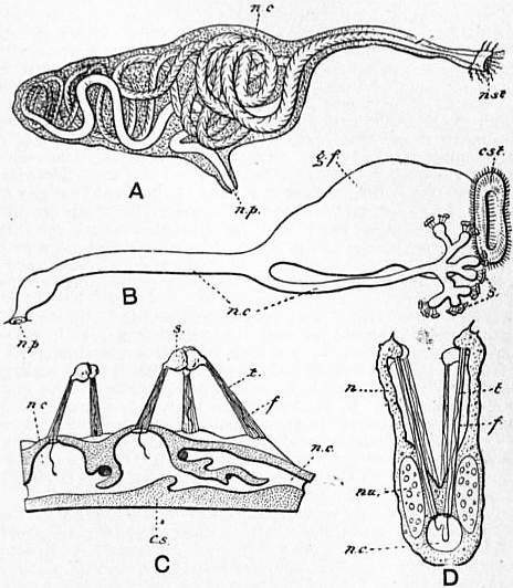
Fig. 2. (from Goodrich).
A, Diagram of the nephridium of Nereis diversicolor. B, Diagram of the nephridium of Alciope, into which opens the large genital funnel (coelomostome). C, Small portion of the nephridium of Glycera siphonostoma, showing the canal cut through, and the solenocytes on the outer surface. D, Optical section of a branch of the nephridium of Nephthys scolopendroides. c.s, Cut surface. cst, Coelomostome. f, Flagellum. g.f, Genital funnel. n, Neck of solenocyte. n.c, Nephridial canal. n.p, Nephridiopore. nst, Nephridiostome. nu, Nucleus of solenocyte. s, Solenocytes. t, Tube.
I. Nephridia.—Excretory organs which are undisputed nephridia are practically universal among the Oligochaeta, Hirudinea and Archiannelida, and occur in many Polychaeta. Their total absence has been asserted definitely only in Paranais littoralis. Usually these organs are present to the number of a single pair per somite, and are commonly present in the majority of the segments of the body, failing often among the Oligochaeta in a varying number of the anterior segments. They are considerably reduced in number in certain Polychaeta. Essentially, a nephridium is a tube, generally very long and much folded upon itself, composed of a string of cells placed end to end in which the continuous lumen is excavated. Such cells are termed “drain pipe” cells. Frequently the lumen is branched and may form a complicated anastomosing network in these cells. Externally, the nephridium opens by a straight part of the tube, which is often very wide, and here the intracellular lumen becomes intercellular. Rarely the nephridium does not communicate with the coelom; in such cases the nephridium ends in a single cell, like the “flame cell” of a Platyhelminth worm, in which there is a lumen blocked at the coelomic end by a tuft of fine cilia projecting into the lumen. This is so with Aeolosoma (Vezhdovský). The condition is interesting as a persistence of the conditions obtaining in the provisional nephridia of e.g. Rhynchelmis, which afterwards become by an enlargement and opening up of the funnel the permanent nephridia of the adult worm. In some Polychaets (e.g. Glycera, see fig. 2) there are many of these flame cells to a single nephridium which are specialized in form, and have been termed “solenocytes” (Goodrich). They are repeated in Polygordius, and are exactly to be compared with similarly-placed cells in the nephridia of Amphioxus.
More usually, and indeed in nearly every other case among the Oligochaeta and Hirudinea, the coelomic aperture of the nephridium consists of several cells, ciliated like the nephridium itself for a greater or less extent, forming a funnel. The funnel varies greatly in size and number of its component cells. There are so many differences of detail that no line can be drawn between the one-celled funnel of Aeolosoma and the extraordinarily large and folded funnel of the posterior nephridia in the Oligochaete Thamnodrilus. In the last-mentioned worm the funnels of the anterior nephridia are small and but few celled; it is only the nephridia in and behind the 17th segment of the body which are particularly large and with a sinuous margin, which recall the funnels of the gonad ducts (i.e. coelomoducts).
Among the Polychaeta the nephridium of Nereis (see fig. 2) is like that of the Oligochaeta and Hirudinea in that the coiled glandular tube has an intracellular duct which is ciliated in the same way in parts. The Polychaeta, however, present us with another form of nephridium seen, for example, in Arenicola, where a large funnel leads into a short and wide excretory tube whose lumen is intercellular. In the young stages of this worm which have been investigated by W.B. Benham, the tube, though smaller, and with a but little pronounced funnel, has still an intercellular duct. That these organs in Polychaeta serve for the removal of the generative products to the exterior is proved not only by the correspondence in number to them of the gonads, but by actual observation of the generative products in transit. This form of nephridia leads to the shorter but essentially similar organs in the Polychaete Sternaspis, and to those of the Echiuroidea (q.v.) and of the Gephyrea (q.v.).
Though the paired arrangement of the nephridia is the prevalent one in the Chaetopoda, there are many examples, among the Oligochaeta, of species and genera in which there are several, even many, nephridia in each segment of the body, which may or may not be connected among themselves, but have in any case separate orifices on to the exterior.
2. Coelomoducis.—In this category are included (by Goodrich and Lankester) the gonad ducts of the Oligochaeta, certain funnels without any aperture to the exterior that have been detected in Nereis, &c., funnels with wide and short ducts attached to nephridia in other Polychaeta, gonad ducts in the Capitellidae, the gonad ducts of the leeches. In all these cases we have a duct which has a usually wide, always intercellular, lumen, generally, if not always, ciliated, which opens directly into the coelom on the one hand and on to the exterior of the body on the other. These characters are plain in all the cases cited, excepting only the leeches which will be considered separately.
There is not a great deal of difference between most of these structures and true nephridia. It is not clear, for example, to which category it is necessary to refer the excretory organs of Arenicola, or Polynoe. Both series of organs consist essentially of a ciliated tube leading from the coelom to the exterior. Both series of organs grow back centrifugally from the funnel. In both the cavity originally or immediately continuous with the coelom appears first in the funnel and grows backwards. In some cases, e.g. oviducts of Oligochaeta, sperm ducts of Phreoryctes, the coelomoducts occupy, like the nephridia, two segments, the funnel opening into that in front of the segment which carries the external pore. It is by no means certain that a hard and fast line can be drawn between intra- and intercellular lumina. Finally, in function there are some points of likeness. The gonad ducts of Lumbricus, &c., must perform one function of nephridia; they must convey to the exterior some of the coelomic fluid with its disintegrated products of waste. There is no possibility that sperm and ova can escape by these tubes not in company with coelomic fluid. In the case of many Oligochaeta where there is no vascular network surrounding the nephridium, this function must be the chief one of those glands, the more elaborate process of excretion taking place in the case of nephridia surrounded by a rich plexus of blood capillaries. A consideration of the mode of development and appearance of the coelomoducts that have thus far been enumerated (with the possible exception of those of the leeches) seems to show that there is a distinct though varying relation between them and the nephridia. It has been shown that in Tubifex, and some other aquatic Oligochaeta, the genital segments are at first provided with nephridia, and that these disappear on the appearance of the generative ducts, which are coelomoducts. In Lumbricus the connexion is a little closer; the funnel of the nephridium, in the segments in which the funnels of the gonad ducts are to be developed, persists and is continuous with the gonad duct funnels on their first appearance. In the development of the Acanthodrilid earthworm Octochaetus (F.E. Beddard) the funnels of the pronephridia disappear except in the genital segments, where they seem to be actually converted into the genital funnels. At the least there is no doubt that the genital funnels are developed precisely where the nephridial funnels formerly existed. If the genital funnels are not wholly or partly formed out of the nephridial funnels they have replaced them. In the genital segments of Eudrilus the nephridia are present, but the funnels have not been found though they are obvious in other segments. Here also the genital funnels have either replaced or been formed out of nephridial funnels. In Haplotaxis heterogyne (W.B. Benham) the sperm ducts are hardly to be distinguished from nephridia; they are sinuous tubes with an intra-cellular duct. But the funnel is large and thus differs from the funnels of the nephridia in adjoining segments. Here again the nephridial funnel seems to have been converted into or certainly replaced by a secondarily developed funnel. This example is similar to cases among the Polychaeta where a true nephridium is provided with a large funnel, coelomostome, according to the nomenclature of Lankester. The whole organ, having, as is thought but not known, this double origin, is termed a nephromixium. The various facts, however, seem to be susceptible of another interpretation. It may be pointed out that the several examples described recall a phenomenon which is not uncommon and is well known to anatomists. That is the replacement of an organ by, sometimes coupled with its partial conversion into, a similar or slightly different organ performing the same or an analogous function. Thus the postcaval vein of the higher vertebrata is partly a new structure altogether, and is partly formed out of the pre-existing posterior cardinals. The more complete replacements, such as the nephridia of the genital segment of Tubifex by a subsequently formed genital duct, may be compared with the succession of the nesonephros to the pronephros in vertebrates, and of the metanephros to the mesonephros in the higher vertebrates. It might be well to term these structures, mostly serving as gonad ducts, which have an undoubted resemblance to nephridia, and for the most part an undoubted connexion with nephridia, “Nephrodinia,” to distinguish them from another category of “ducts” which are communications between the coelom and the exterior, and which have no relation whatever to nephridia or to the organs just discussed. For these latter, the term coelomoducts might well be reserved. To this category belong certain sacs and pouches in many, perhaps most, genera of the Oligochaeta family, Eudrilidae, and possibly the gonad ducts in the Hirudinea. As an example of the former it has been shown (Beddard) that a large median sac in Lybiodrilus is at first freely open to the coelom, that it later becomes shut off from the same, that it then acquires an external orifice, and, finally, that it encloses the ovary or ovaries, between which and the exterior a passage is thus effected. To this category will belong the oviducts in Teleostean fishes and probably the gonad ducts in several groups of invertebrates.
Polychaeta.—This group may be thus defined and the definition contrasted and compared with those of the other divisions of the Chaetopoda. Setae always present and often very large, much varied in form and very numerous, borne by the dorsal and ventral parapodia (when present). The prostomium and the segments generally often bear processes sensory and branchial. Eyes often present and comparatively complicated in structure. Clitellum not present as a definite organ, as in Oligochaeta. The anus is mostly terminal, and there are no anterior and posterior suckers. Nervous system often imbedded in the epidermis. Vascular system generally present forming a closed system of tubes. Alimentary canal rarely coiled, occasionally with glands which are simple caeca and sometimes serve as air reservoirs; jaws often present and an eversible pharynx. Nephridia sometimes of the type of those of the Oligochaeta; in other cases short, wide tubes with a large funnel serving also entirely or in part as gonad ducts. Frequently reduced in number of pairs; rarely (Capitellidae) more than one pair per segment. Gonads not so restricted in position as in Oligochaets, and often more abundant; the individuals usually unisexual. No specialized system of spermathecae, sperm reservoirs, and copulatory apparatus, as in Oligochaeta; development generally through a larval form; reproduction by budding also occurs. Marine (rarely fresh-water) in habit.
The Polychaeta contrast with the Oligochaeta by the great variety of outward form and by the frequency of specialization of different regions of the body. The head is always recognizable and much more conspicuous than in other Chaetopoda. As in the Oligochaeta the peristomial segment is often without setae, but this character is not by any means so constant as in the Oligochaeta. The prostomium bears often processes, both dorsal and ventral, which in the Sabellids are split into the circle of branchial plumes, which surround or nearly surround the mouth in those tube-dwelling Annelids. Tomopteris is remarkable for the fact that the hammer-shaped prostomium has paired ventral processes each with a single seta. It is held, however, that these are a pair of parapodia which have shifted forwards. The presence of parapodia distinguish this from other groups of Chaetopoda. Typically, the parapodium consists of two processes of the body on each side, each of which bears a bundle of setae; these two divisions of the “limb” are termed respectively notopodium and neuropodium. The notopodium may be rudimentary or absent and the entire parapodium reduced to the merest ridge or even completely unrepresented. Naturally, it is among the free living forms that the parapodium is best developed, and least developed among the tubicolous Polychaeta. To each division of the parapodium belongs typically a long tentacle, the cirrus, which may be defective upon one or other of the notopodium or neuropodium, and may be developed into an arborescent gill or into a flat scale-like process, the elytron (in Polynoe, &c.). There are other gills developed in addition to those which represent the cirri.

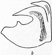
Fig. 3.—a, Bristle of Pionosyllis Malmgreni; b, Hook of Terebella.
Setae.—The setae of the Polychaeta are disposed in two bundles in many genera, but in only one bundle in such forms as have no notopodium (e.g. Syllis). In some genera the setae are in vertical rows, and in certain Capitellidae these rows so nearly meet that an arrangement occurs reminiscent of the continuous circle of setae in the perichaetous Oligochaeta. The setae vary much in form and are often longer and stronger than in the Oligochaetes. Jointed setae and very short hooks or “uncini” (see fig. 3) are among the most remarkable forms. Simple bifid setae, such as those of Oligochaetes, are also present in certain forms.
Among the burrowing and tubicolous forms it is not uncommon for the body to be distinguishable into two or more regions; a “thorax,” for example, is sharply marked off from an “abdomen” in the Sabellids. In these forms the bundles of setae are either capilliform or uncinate, and the dorsal setae of the thorax are like the ventral setae of the abdomen. It is a remarkable and newly-ascertained fact that in regeneration (in Potamilla) the thorax is not replaced by the growth of uninjured thoracic segments; but that the anterior segments of the abdomen take on the same characters, the setae dropping out and being replaced in accordance with the plan of the setae in the thorax of uninjured worms. Among the Oligochaeta the sexually mature worm is distinguished from the immature worm by the clitellum and by the development of genital setae. Among the Polychaeta the sexual worm is often more marked from the asexual form, so much so that these latter have been placed in different species or even genera. The alteration in form does not only affect structures used in generation; but the form of the parapodia, &c., alter. There are even dimorphic forms among the Syllids where the sexes are, as in many Polychaets, separate.
Nephridia.—The nephridia of the Polychaeta have been generally dealt with above in considering the nephridial system of the Chaetopoda as a whole. They contrast with those of the Oligochaeta and Hirudinea by reason of their frequently close association with the gonads, the same organ sometimes serving the two functions of excretion and conveyance of the ova and spermatozoa out of the body. On the hypothesis that such a form as Dinophilus (see Haplodrili) has preserved the characters of the primitive Chaetopod more nearly than any existing Polychaet or Oligochaet, it is clear that the nephridia in the Oligochaeta have preserved the original features of those organs more nearly than most Polychaeta. Thus Nereis among the latter worms, from the resemblance which its excretory system bears to that of the Oligochaeta, may be made the starting-point of a series. In this worm the paired nephridia exist in most of the segments of the body, and their form (see fig. 2) is much like that of the nephridia in the Enchytraeidae. The funnel, which is not large, appears to open, as a rule at least, into the segment in front of that which bears the external orifice. Quite independent of these are certain large dorsally situate funnel-like folds of the coelomic epithelium, ciliated, but of which no duct has been discovered leading to the exterior. It is possible that we have here gonad ducts distinct from nephridia which at the time of sexual maturity do open on to the exterior.
In Polynoe the nephridia are short tubes with a slightly folded funnel whose lumen is intercellular, and this intercellular lumen is characteristic of the Polychaetes as contrasted with leeches and Oligochaetes. Among the Terebelloidea there is a remarkable differentiation of the nephridia into two series. One set lies in front of the diaphragm, which is the most anterior and complete septum, the rest having disappeared or being much less developed. The anterior nephridia, of which there are one to three pairs, contrast with the posterior series by their small funnels and large size, the posterior nephridia having a large funnel followed by a short tube. In Chaetozone setosa the anterior nephridia occupy five segments. There is usually a gap between the two series, several segments being without nephridia. It seems that the posterior nephridia are mainly gonad ducts, and the gonads are developed in close association with the funnels. The same arrangement is found in some other Polychaetes; for instance, in Sabellaria there is a single pair of large anterior nephridia, which open by a common pore, followed after an interval by large-funnelled and short nephridia. This differentiation is not, however, peculiar to the Polychaetes; for in several Oligochaetes the anterior nephridia are of large size, and opening as they do into the buccal cavity clearly play a different function to those which follow. In Thamnodrilus, as has been pointed out, there are two series of nephridia which resemble those of the Terebelloidea in the different sizes of their funnels. In Lanice conchilega the posterior series of nephridia are connected by a thick longitudinal duct, which seems to be seen in its most reduced form in Owenia, where a duct on each side runs in the epidermis, being in parts a groove, and receives one short tubular nephridium only and occupies only one segment. This connexion of successive nephridia (in Lanice) has its counterpart in Allolobophora, Lybiodrilus, and apparently in the Lumbriculids Teleuscolex and Styloscolex, among the Oligochaeta. Among the Capitellidae, which in several respects resemble the Oligochaeta, wide and short gonad ducts coexist in the same segments with nephridia, the latter being narrower and longer. It is noteworthy that in this family only among the Polychaeta, the nephridia are not restricted to a single pair in each segment; so that the older view that the gonad ducts are metamorphosed nephridia is not at variance with the anatomical facts which have been just stated.
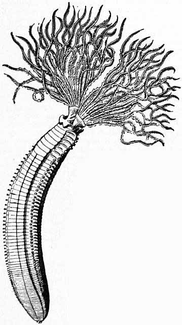
Fig. 4.—Dasychone infracta, Kr. (After Malmgren.)
Alimentary Canal.—The alimentary canal of Polychaetes is usually a straight tube running from the anterior mouth to the posterior anus. But in some forms, e.g. Sternaspis, the gut is coiled. In others, again, e.g. Cobangia, the anus is anterior and ventral. A gizzard is present in a few forms. The buccal cavity is sometimes armed with jaws. The oesophagus is provided often with caeca which in Syllids and Hesionidae have been found to contain air, and possibly therefore perform the function of the fish’s air-bladder. In other Polychaetes one or more pairs of similar outgrowths are glandular. The intestine is provided with numerous branched caeca in Aphrodite.
Reproduction.—As is the case with the Oligochaeta, the Polychaeta furnish examples of species which multiply asexually by budding. There is a further resemblance between the two orders of Chaetopoda in that this budding is not a general phenomenon, but confined to a few forms only. Budding, in fact, among the Polychaetes is limited to the family Syllidae. In the Oligochaetes it is only the families Aeolosomatidae and Naididae that show the same phenomenon. It has been mentioned that in the Nereids a sexual form occurs which differs structurally from the asexual worms, and was originally placed in a separate genus, Heteronereis; hence the name “Heteronereid” for the sexual worm. In Syllis there is also a “Heterosyllid” form in which the gonads are limited to a posterior region of the body which is further marked off from the anterior non-sexual segments by the oak-like setae. In some Syllids this posterior region separates off from the rest, producing a new head; thus a process of fission occurs which has been termed schizogamy. A similar life history distinguishes certain Sabellid worms, e.g. Filigrana. Among the Syllids this simple state of affairs is further complicated. In Autolytus there is, to begin with, a conversion of the posterior half of the body to form a sexual zooid. But before this separates off a number of other zooids are formed from a zone of budding which appears between the two first-formed individuals. Ultimately, a chain of sexual zooids is thus formed. A given stock only produces zooids of one sex. In Myrianida there is a further development of this process. The conversion of the posterior end of the simple individual into a sexual region is dispensed with; but from a preanal budding segment a series of sexual buds are produced. The well-known Syllid, discovered during the voyage of the “Challenger,” shows a modification of this form of budding. Here, however, the buds are lateral, though produced from a budding zone, and they themselves produce other buds, so that a ramifying colony is created.
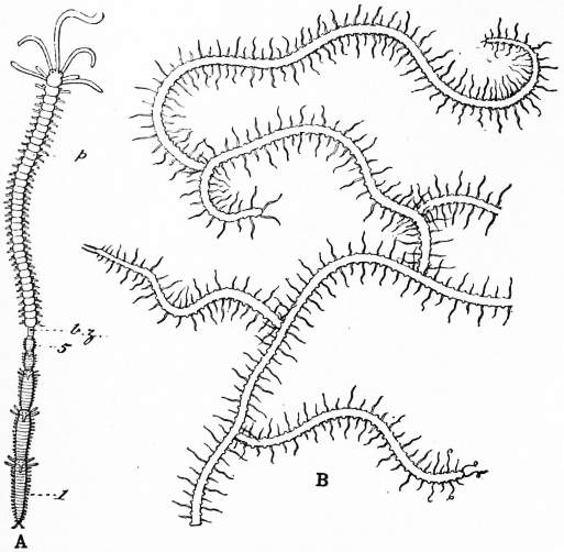
Fig. 5.—A, Autolytus (after Mensch) with numerous buds. B, Portion of a colony of Syllis ramosa (from M‘Intosh). b.z, Budding zone; p, anterior region of the parent worm; 1-5, buds.
Quite recently, another mode of budding has been described in Trypanosyllis gemmipara, where a crowd of some fifty buds arising symmetrically are produced at the tail end of the worm. In some Syllids, such as Pionosyllis gestans, the ova are attached to the body of the parent in a regular line, and develop in situ; this process, which has been attributed to budding, is an “external gestation,” and occurs in a number of species.
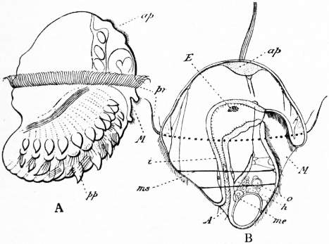
Fig. 6.—A, Side view of the larva of Lopadorhynchus (from Kleinenberg), showing the developing trunk region. B, Side view of the trochophore larva of Eupomatus uncinatus (from Hatschek).
A, Anus. E, Eye. M, Mouth. ap, Apical organ. h, “Head Kidney.” i, Intestine. me, Mesoblast. ms, Larval muscle. o, Otocyst. pp, Parapodium. pr, Praeoral ciliated ring, or prototroch.

Fig. 7.—Nereis pelagica, L. (After Oersted.)
As is very frequently the case with marine forms, as compared with their fresh-water and terrestrial allies, the Polychaeta differ from the Oligochaeta and Hirudinea in possessing a free living larval form which is hatched at an early stage in development. This larva is termed the Trochosphere larva, and typically (as it is held) is an egg-shaped larva with two bands of cilia, one preoral and one postoral, with an apical nervous plate surmounted by a tuft of longer cilia, and with a simple bent alimentary canal, with lateral mouth and posterior anus, between which and the ectoderm is a spacious cavity (blastocoel) traversed by muscular strands and often containing a larval kidney. The segmentation is of the mesoblast to begin with, and appears later behind the mouth, the part anterior to this becoming the prostomium of the adult. The chief modifications of this form are seen in the Mitraria larva of Ammochares with only the preoral band, which is much folded and which has provisional and long setae; the atrochous larva, where the covering of cilia is uniform and not split into bands; and the polytrochous larva where there are several bands surrounding the body. There are also other modifications.
Classification.—The older arrangement of the Polychaeta into Errantia or free living and Tubicola or tube-dwelling forms will hardly fit the much increased knowledge of the group. W.B. Benham’s division into Phanerocephala in which the prostomium is plain, and Crytocephala in which the prostomium is hidden by the peristomium adopted by Sedgwick, can only be justified by the character used; for the Terebellids, though phanerocephalous, have many of the features of the Sabellids. It is perhaps safer to subdivide the Order into 6 Suborders (in the number of these following Benham, except in combining the Sabelliformia and Hermelliformia). Of these 6, the two first to be considered are very plainly separable and represent the extremes of Polychaete organization, (1) Nereidiformia.—“Errant” Polychaetes with well-marked prostomium possessing tentacles and palps with evident and locomotor parapodia, supported (with few exceptions) by strong spines, the aciculi; muscular pharynx usually armed with jaws; septa and nephridia regularly metameric and similar throughout body; free living and predaceous. (2) Cryptocephala.—Tube-dwelling with body divided into thorax and abdomen marked by the setae, which are reversed in position in the neuropodium and notopodium respectively in the two regions. Parapodia hardly projecting; palps of prosomium forming branched gills; no pharynx or eversible buccal region; no septa in thorax, septa in abdomen regularly disposed. Nephridia in two series; large, anterior nephridia followed by small, short tubes in abdomen. The remaining groups are harder to define, with the exception of the (3) Capitelliformia, which are mud-living worms of an “oligochaetous” appearance, and with some affinities to that order. The peristomium has no setae, and the setae generally are hair-like or uncinate, often forming almost complete rings. The genital ducts are limited to one segment (the 8th in Capitella capitata), and there are genital setae on this and the next segment. In other forms genital ducts and nephridia coexist in the same segment. The nephridia are sometimes numerous in each segment. There is no blood system, and the coelomic corpuscles contain haemoglobin. (4) Terebelliformia. These worms are in some respects like the Sabellids (Cryptocephala). The parapodia, as in the Capitellidae, are hardly developed. The buccal region is unarmed and not eversible. The prostomium has many long filaments which recall the gills of the Sabellids, &c. The nephridia are specialized into two series, as in the last-mentioned worms. (5) Spioniformia (including Chaetopterus, Spio, &c.) and (6) Scoleciformia (Arenicola, Chloraema, Sternaspis) are the remaining groups. In both, the nephridia are all alike; there are no jaws; the prostomium rarely has processes. The body is often divisible into regions.
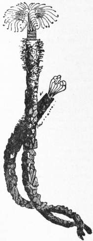
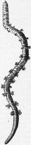
Fig. 8.—Sabella vesiculosa, Mont. (After Montagu.) Fig. 9. Arenicola marina, L.
Literature.—W.B. Benham, “Polychaeta” in Cambridge Natural History; E. Claparède, Annélides chétopodes du golfe de Naples (1868 and 1870); E. Ehlers, Die Börstenwürmer (1868); H. Eisig, Die Capitelliden (Naples Monographs), and development of do. in Mitth. d. zool. Stat. Neapel (1898); W.C. M’Intosh, ”Challenger” Reports (1885); E.R. Lankester, Introductory Chapter in A Treatise on Zoology; E.S. Goodrich, Quart. Journ. Mic. Sci. (1897-1900); E. Meyer, Mitth. d. zool. Stat. Neapel (1887, 1888), as well as numerous other memoirs by the above and by J.T. Cunningham, de St Joseph, A. Malaquin, A. Agassiz, A.T. Watson, Malmgren, Bobretsky and A.F. Marion, E.A. Andrews, L.C. Cosmovici, R. Horst, W. Michaelsen, G. Gilson, F. Buchanan, H. Levinsen, Joyeux-Laffuie, F.W. Gamble, &c.
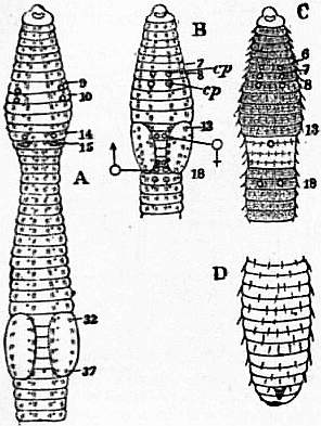
Fig. 10.—Diagrams of various Earthworms, to illustrate external characters. A, B, C, anterior segments from the ventral surface; D, hinder end of body of Urochaeta.
A, Lumbricus: 9, 10, segments containing spermathecae, the orifices of which are indicated; 14, segment bearing oviducal pores; 15, segment bearing male pores; 32, 37, first and last segments of clitellum. B, Acanthodrilus: cp, orifices of spermathecae; ♀, oviducal pores; ♂, male pores; on 17th and 19th segments are the apertures of the atria. C, Perichaeta: the spermathecal pores are between segments 6 and 7, 7 and 8, 8 and 9, the oviducal pores upon the 14th and the male pores upon the 18th segment.
In all the figures the nephridial pores are indicated by dots and the setae by strokes.
Oligochaeta.—As contrasted with the other subdivisions of the Chaetopoda, the Oligochaeta may be thus defined. Setae very rarely absent (genus Achaeta) and as a rule not so large or so numerous in each segment as in the Polychaeta, and different in shape. Eyes rarely present and then rudimentary. Prostomium generally small, sometimes prolonged, but never bearing tentacles or processes. Appendages of body reduced to branchiae, present only in four species, and to the ventral copulatory appendages of Alma and Criodrilus. Clitellum always present, extending over two (many limicolous forms) to forty-five segments (Alma). Segments of body numerous and not distinctive of species, being irregular and not fixed in numbers. In terrestrial forms dorsal pores are usually present; in aquatic forms a head pore only. Anus nearly always terminal, rarely dorsal, at a little distance from end of body. Suckers absent. Nervous system rarely (Aeolosoma) in continuity with epidermis. Vascular system always present, forming a closed system, more complicated in the larger forms than in the aquatic genera. Several specially large contractile trunks in the anterior segments uniting the dorsal and ventral vessels. Nephridia generally paired, often very numerous in each segment, in the form of long, much-coiled tubes with intracellular lumen. Gonads limited in number of pairs, testes and ovaries always present in the same individual. Special sacs developed from the intersegmental septa lodge the developing ova and sperm. Special gonad ducts always present. Male ducts often open on to exterior through a terminal chamber which is variously specialized, and sometimes with a penis.
Generative pores usually paired, sometimes single and median. Spermathecae nearly always present. Alimentary canal straight, often with appended glands of complicated or simpler structure; no jaws. Eggs deposited in a cocoon after copulation. Development direct. Reproduction by budding also occurs. Fresh-water (rarely marine) and terrestrial.
The Oligochaeta show a greater variety of size than any other group of the Chaetopoda. They range from a millimetre or so (smaller species of Aeolosoma) to 6 ft. or even rather more (Microchaeta rappi, &c.) in length.
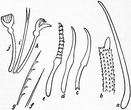
Fig. 11.—Setae of Oligochaeta.
a, Penial seta of Perichaeta ceylonica. b, Extremity of penial seta of Acanthodrilus (after Horst). c, Seta of Urochaeta (Perier). d, Seta of Lumbricus. e, Seta of Criodrilus. f, g, Setae of Bohemilla comata. h, i, j, Setae of Psammoryctes barbatus (f to j after Vezhdovský).
Setae.—The setae, which are always absent from the peristomial segment, are also sometimes absent from a greater number of the anterior segments of the body, and have completely disappeared in Achaeta cameranoi. When present they are either arranged in four bundles of from one to ten or even more setae, or are disposed in continuous lines completely encircling each segment of the body. This latter arrangement characterizes many genera of the family Megascolicidae and one genus (Periscolex) of the Glossoscolicidae. It has been shown (Bourne) that the “perichaetous” condition is probably secondary, inasmuch as in worms which are, when adult, “perichaetous” the setae develop in pairs so that the embryo passes through a stage in which it has four bundles of setae, two to each bundle, the prevalent condition in the group. Rarely there is an irregular disposition of the setae which are not paired, though the total number is eight to a segment (fig. 10), e.g. Pontoscolex. The varying forms of the setae are illustrated in fig. 11.
Structure.—The body wall consists of an epidermis which secretes a delicate cuticle and is only ciliated in Aeolosoma, and in that genus only on the under surface of the prostomium. The epidermis contains numerous groups of sense cells; beneath the epidermis there is rarely (Kynotus) an extensive connective tissue dermis. Usually the epidermis is immediately followed by the circular layer of muscles, and this by the longitudinal coat. Beneath this again is a distinct peritoneum lining the coelom, which appears to be wanting as a special layer in some Polychaetes (Benham, Gilson). The muscular layers are thinner in the aquatic forms, which possess only a single row of longitudinal fibres, or (Enchytracidae) two layers. In the earthworms, on the other hand, this coat is thick and composed of many layers.
The clitellum consists of a thickening of the epidermis, and is of two forms among the Oligochaeta. In the aquatic genera the epidermis comes to consist entirely of glandular cells, which are, however, arranged in a single layer. In the earthworms, on the other hand, the epidermis becomes specialized into several layers of cells, all of which are glandular. It is therefore obviously much thicker than the clitellum in the limicolous forms. The position of the clitellum, which is universal in occurrence, varies much as does the number of component segments. As a rule—to which, however, there are exceptions—the clitellum consists of two or three segments only in the small aquatic Oligochaeta, while in the terrestrial forms it is as a general rule, to which again there are exceptions, a more extensive, sometimes much more extensive, region.
In the Oligochaeta there is a closer correspondence between external metamerism and the divisions of the coelom than is apparent in some Chaetopods. The external segments are usually definable by the setae; and if the setae are absent, as in the anterior segments of several Geoscolicidae, the nephridiopores indicate the segments; to each segment corresponds internally a chamber of the coelom which is separated from adjacent segments by transverse septa, which are only unrecognizable in the genus Aeolosoma and in the head region of other Oligochaeta. In the latter case, the numerous bands of muscle attaching the pharynx to the parietes have obliterated the regular partition by means of septa.
Nephridia.—The nephridia in this group are invariably coiled tubes with an intracellular lumen and nearly invariably open into the coelom by a funnel. There are no renal organs with a wide intercellular lumen, such as occur in the Polychaeta, nor is there ever any permanent association between nephridia and ducts connected with the evacuation of the generative products, such as occur in Alciope, Saccocirrus, &c. In these points the Oligochaeta agree with the Hirudinea. They also agree in the general structure of the nephridia. It has been ascertained that the nephridia of Oligochaeta are preceded in the embryo by a pair of delicate and sinuous tubes, also found in the Hirudinea and Polychaeta, which are larval excretory organs. It is not quite certain whether these are to be regarded as the remnant of an earlier excretory system, replaced among the Oligochaeta by the subsequently developed paired structures, or whether these “head kidneys” are the first pair of nephridia precociously developed. The former view has been extensively held, and it is supported by the fact that in Octochaetus the first segment of the body has a pair of nephridia which is exactly like those which follow, and, like them, persists. On the other hand, in most Oligochaeta the first segment has in the adult no nephridium, and in the case of Octochaetus the existence of a “head kidney” antedating the subsequently developed nephridia of the first and other segments has neither been seen nor proved to be absent. In any case the nephridia which occupy the segments of the body generally are first of all represented by paired structures, the “pronephridia,” in which the funnel is composed of but one cell, which is flagellate. This stage has at any rate been observed in Rhynchelmis and Lumbricus (in its widest sense) by Vezhdovský. It is further noticeable that in Rhynchelmis the covering of vesicular cells which clothes the drain-pipe cells of the adult nephridium is cut off from the nephridial cells themselves and is not a peritoneal layer surrounding the nephridium. Thus the nephridia, in this case at least, are a part of the coelom and are not shut off from it by a layer of peritoneum, as are other organs which lie in it, e.g. the gut. A growth both of the funnel, which becomes multicellular, and of the rest of the nephridium produces the adult nephridia of the genera mentioned. The paired disposition of these organs is the prevalent one among the Oligochaeta, and occurs in all of twelve out of the thirteen families into which the group is divided.
Among the Megascolicidae, however, which in number of genera and species nearly equals the remaining families taken together, another form of the excretory system occurs. In the genera Pheretima, Megascolex, Dichogaster, &c., each segment contains a large number of nephridia, which, on account of the fact that they are necessarily smaller than the paired nephridia of e.g. Lumbricus, have been termed micronephridia, as opposed to meganephridia; there is, however, no essential difference in structure, though micronephridia are not uncommonly (e.g. Megascolides, Octochaetus) unprovided with funnels. It is disputed whether these micronephridia are or are not connected together in each segment and from segment to segment. In any case they have been shown in three genera to develop by the growth and splitting into a series of original paired pronephridia. A complex network, however, does occur in Lybiodrilus and certain other Eudrilidae, where the paired nephridia possess ducts leading to the exterior which ramify and anastomose on the thickness of the body wall. The network is, however, of the duct of the nephridium, possibly ectodermic in origin, and does not affect the glandular tubes which remain undivided and with one coelomic funnel each.
The Oligochaeta are the only Chaetopods in which undoubted nephridia may possess a relationship with the alimentary canal. Thus, in Octochaetus multiporus a large nephridium opens anteriorly into the buccal cavity, and numerous nephridia in the same worm evacuate their contents into the rectum. The anteriorly-opening and usually very large nephridia are not uncommon, and have been termed “peptonephridia.”
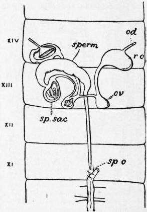
Fig. 12.—Female reproductive system of Heliodrilus.—XI-XIV, eleventh to fourteenth segments, sperm, spermatheca; sp.o, its external orifice; sp.sac, spermathecal sac; ov, sac containing ovary; r.o, egg sac; od, oviduct.
Gonads and Gonad Ducts.—The Oligochaeta agree with the leeches and differ from most Polychaeta in that they are hermaphrodite. There is no exception to this generalization. The gonads are, moreover, limited and fixed in numbers, and are practically invariably attached to the intersegmental septa, usually to the front septum of a segment, more rarely to the posterior septum. The prevalent number of testes is one pair in the aquatic genera and two pairs in earthworms. But there are exceptions; thus a species of Lamprodrilus has four pairs of testes. The ovaries are more usually one pair, but two are sometimes present. The segments occupied by the gonads are fixed, and are for earthworms invariably X, XI, or one of them for the testes, and XIII for the ovaries The position varies in the aquatic Oligochaeta. The Oligochaeta contrast with the Polychaeta in the general presence of outgrowths of the septa in the genital segments, which are either close to, or actually involve, the gonads, and into which may also open the funnels of the gonad ducts. These sacs contain the developing sperm cells or eggs, and are with very few exceptions universal in the group. The testes are more commonly thus involved than are the ovaries. It is indeed only among the Eudrilidae that the enclosure of the ovaries in septal sacs is at all general. Recently the same thing has been recorded in a few species of Pheretima (= Perichaeta), but details are as yet wanting. We can thus speak in these worms of gonocoels, i.e. coelomic cavities connected only with the generative system. These cavities communicate with the exterior through the gonad ducts, which have nothing to do with them, but whose coelomic funnels are taken up by them in the course of their growth. There are, however, in the Eudrilidae, as already mentioned, sacs envolving the ovaries which bore their own way to the exterior, and thus may be termed coelomoducts. These sacs are dealt with later under the description of the spermathecae, which function they appear to perform. The gonad ducts are male and female, and open opposite to or, rarely, alongside of the gonads, whose products they convey to the exterior. The oviducts are always short trumpet-shaped tubes and are sometimes reduced (Enchytraeidae) to merely the external orifices. It is possible, however, that those oviducts belong to a separate morphological category, more comparable to the dorsal pores and to abdominal pores in some fishes. The sperm ducts are usually longer than the oviducts; but in Limicolae both series of tubes opening by the funnel into one segment and on to the exterior in the following segment. While the oviducts always open directly on to the exterior, it is the rule for the sperm ducts to open on to the exterior near to or through certain terminal chambers, which have been variously termed atrium and prostate, or spermiducal gland. The distal extremity of this apparatus is sometimes eversible as a penis. Associated with these glands are frequently to be found bundles or pairs of long and variously modified setae which are termed penial setae, to distinguish them from other setae sometimes but not always associated with rather similar glands which are found anteriorly to these, and often in the immediate neighbourhood of the spermathecae; the latter are spoken of as genital setae.
Spermathecae.—These structures appear to be absolutely distinctive of the Oligochaeta, unless the sacs which contain sperm and open in common with the nephridia of Saccocirrus (see Haplodrili) are similar. Spermathecae are generally present in the Oligochaeta and are absent only in comparatively few genera and species. Their position varies, but is constant for the species, and they are rarely found behind the gonads. They are essentially spherical, pear-shaped or oval sacs opening on to the exterior but closed at the coelomic end. In a few Enchytraeidae and Lumbriculidae the spermathecae open at the distal extremity into the oesophagus, which is a fact difficult of explanation. Among the aquatic Oligochaeta and many earthworms (the families Lunibricidae, Geoscolicidae and a few other genera) the spermathecae are simple structures, as has been described. In the majority of the Megascolicidae each sac is provided with one or more diverticula, tubular or oval in form, of a slightly different histological character in the lining epithelium, and in them is invariably lodged the sperm.
The spermathecae are usually paired structures, one pair to each of the segments where they occur. In many Geoscolicidae, however, and certain Lumbricidae and Perichaetidae, there are several, even a large number, of pairs of very small spermathecae to each of the segments which contain them.
In the Eudrilidae there are spermathecae of different morphological value. In figs. 12 and 13 are shown the spermathecae of the genera Hyperiodrilus and Heliodrilus, which are simple sacs ending blindly as in other earthworms, but of which there is only one median opening in the thirteenth segment or in the eleventh. In Heliodrilus the blind extremity of the spermatheca is enclosed in a coelomic sac which is in connexion with the sacs envolving the ovaries and oviducts. In Hyperiodrilus the whole spermatheca is thus included in a corresponding sac, which is of great extent. In such other genera of the family as have been examined, the true spermatheca has entirely disappeared, and the sac which contains it in Hyperiodrilus alone remains. This sac has been already referred to as a coelomoduct. Its orifice on to the exterior is formed by an involution (as it appears) of the epidermis, and that it performs the function of a spermatheca is shown by its containing spermatozoa, or, in Stuhlmannia, a spermatophore. In Polytoreutus, also, spermatophores have been found in these spermathecal sacs. We have thus the replacement of a spermatheca, corresponding to those of the remaining families of Oligochaeta, and derived, as is believed, from the epidermis, by a structure performing the same function, but derived from the mesoblastic tissues, and with a cavity which is coelom.
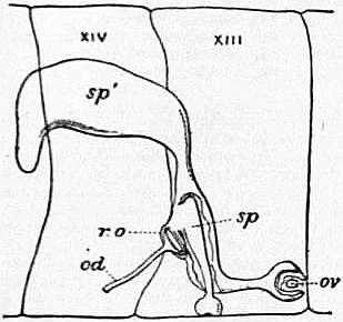
Fig. 13.—Female reproductive system of Hyperiodrilus.—XIII, XIV, thirteenth and fourteenth segments.
sp, Spermatheca. sp’, Spermathecal sac involving the last. ov, Ovary. r.o, Egg sac. od, Oviduct.
Alimentary Canal.—The alimentary canal is always a straight tube, and the anus, save in the genera Criodrilus and Dero, is completely terminal. A buccal cavity, a pharynx, an oesophagus and an intestine are always distinguishable. Commonly among the terrestrial forms there is a gizzard, or two gizzards, or a larger number, in the oesophageal region. There is no armed protrusible pharynx, such as exists in some other Chaetopods. This may be associated with mud-eating habits; but it is not wholly certain that this is the case; for in Chaetogaster and Agriodrilus, which are predaceous worms, there is no protrusible pharynx, though in the latter the oesophagus is thickened through its extent with muscular fibres. The oesophagus is often furnished with glandular diverticula, the “glands of Morren,” which are often of complex structure through the folding of their walls. Among the purely aquatic families such structures are very rare, and are represented by two caeca in the genus Limnodriloides. It is a remarkable fact, not yet understood, that in certain Enchytraeidae and Lumbriculidae the spermathecae open into the oesophagus as well as on to the exterior. The only comparable fact among other worms is the Laurer’s canal or genito-intestinal canal in the Trematoda. The intestine is usually in the higher forms provided with a typhlosole, in which, in Pontoscolex, runs a ciliated canal or canals communicating with the intestine. It is possible that this represents the syphon or supplementary intestine of Capitellidae, which has been shown to develop as a grooving of the intestine ultimately cut off from it. The intestine has a pair of caeca or two or three pairs (but all lie in one segment) in the genus Pheretima and in one species of Rhinodrilus. In Typhoeus and Megascolex there are complex glands appended to the intestine.
In Benhamia caecifera and at least one other earthworm there are numerous caeca, one pair to each segment.
Classification.—The classifications of Adolf Eduard, Grube and Claparède separated into two subdivisions the aquatic and the terrestrial forms. This scheme, opposed by many, has been reinstated by Sedgwick. The chief difficulty in this scheme is offered by the Moniligastridae, which in some degree combine the characters of both the suborders, into neither of which will they fit accurately. The following arrangement is a compromise:—
Group I. Aphaneura.—This group is referred by A. Sedgwick to the Archiannelida. It is, however, though doubtless near to the base of the Oligochaetous series, most nearly allied in the reproductive system to the Oligochaeta. It contains but one family, Aeolosomatidae. There are three pairs of spermathecae situated in segments III-V, a testis in V and an ovary in VI. There are a clitellum and sperm ducts which though like nephridia have a larger funnel and a less complexly wound duct. This family consists of only one well-known genus, Aeolosoma, which contains several species. They are minute worms with coloured oil drops (green, olive green or orange) contained in the epidermis. The nervous system is embedded in the epidermis, and the pairs of ganglia are separated as in Serpula, &c.; each pair has a longish commissure between its two ganglia. The intersegmental septa are absent save for the division of the first segment. The large prostomium is ciliated ventrally. The setae are either entirely capillary or there are in addition some sigmoid setae even with bifid free extremities. This genus also propagates asexually, like Ctenodrilus, which may possibly belong to the same family. Asexual reproduction universal.
Group II. Limicolae.—With a few exceptions the Limicolae are, as the name denotes, aquatic in habit. They are small to moderate-sized Oligochaeta, with a smaller number of segments than in the Terricolae. The alimentary canal is simple and a gizzard or oesophageal diverticula rarely developed. The vascular system is simple with as a rule direct communication between dorsal and ventral vessels in each segment. Nerve cord lies in coelom; brain in first segment or prostomium in many forms. Clitellum generally only two or three segments and more anterior in position than in Terricolae. Nephridia always paired and without plexus of blood capillaries. Spermatheca rarely with diverticula; sperm ducts as a rule occupying two segments only, usually opening by means of an atrium. Sperm sacs generally occupying a good many segments and with simple interior undivided by a network of trabeculae. Ova large and with much yolk. Asexual reproduction only in Naids. Egg sacs as large or nearly so as sperm sacs. Testes and ovaries always free. The following families constitute the group, viz. Naididae, Enchytraeidae, Tubificidae, Lumbriculidae, Phreoryctidae, Phreodrilidae, Alluroididae, the latter possibly not referable to this group.
Group III. Moniligastres.—Moderate-sized to very large Oligochaeta, terrestrial in habit, with the appearance of Terricolae. Generative organs anterior in position as in Limicolae. Sperm ducts and atria as in Limicolae; egg sacs large; body wall thick; vascular system and nephridia as in Terricolae. Only one family, Moniligastridae.
Group IV. Terricolae.—Earthworms, rarely aquatic in habit. Of small to very large size. Clitellum commonly extensive and more posterior in position than in other groups. Vascular system complicated without regular connexion between dorsal and ventral vessels, except in anterior segments. Nephridia as a rule with abundant vascular supply. Testes, and occasionally ovaries, enclosed in sacs. Sperm sacs generally limited to one or two segments with interior subdivided by trabeculae. Sperm ducts traverse several segments on their way to exterior. They open in common with, or near to, or, more rarely, into, glands which are not certainly comparable to the atria of the Limicolae. Egg sacs minute and functionless(?). Eggs minute with little yolk. Nephridia sometimes very numerous in each segment. Spermathecae often with diverticula.
Earthworms are divided into the following families, viz. Megascolicidae, Geoscolicidae, Eudrilidae, Lumbricidae.
As an appendix to the Oligochaeta, and possibly referable to that group, though their systematic position cannot at present be determined with certainty, are to be placed the Bdellodrilidae (Discodrilidae auct.), which are small parasites upon crayfish. These worms lay cocoons like the Oligochaeta and leeches, and where they depart from the structure of the Oligochaeta agree with that of leeches. The body is composed of a small and limited number of segments (not more than fourteen), and there is a sucker at each end of the body. There are no setae and apparently only two pairs of nephridia, of which the anterior pair open commonly by a common pore on the third segment after the head, whose segments have not been accurately enumerated. The intervening segments contain the genitalia, which are on the Oligochaeta plan in that the gonads are independent of their ducts and that there are special spermathecae, one pair. The male ducts are either one pair or two pairs, which open by a common and complicated efferent terminal apparatus furnished with a protrusible penis. The ganglia are crowded at the posterior end of the body as in leeches, and there is much tendency to the obliteration of the coelom as in that group. Pterodrilus and Cirrodrilus bear a few, or circles of, external processes which may be branchiae; Bdellodrilus and Astacobdella have none. The vascular system is as in the lower Oligochaeta. There are two chitinous jaws in the buccal cavity, a dorsal and a ventral, which are of specially complicated structure in Cirrodrilus.
Literature.—F.E. Beddard, A Monograph of the Oligochaeta (Oxford, 1895), also Quart. Journ. Micr. Sci., 1886-1895, and Proc. Zool. Soc., 1885-1906; W.B. Benham, Quart. Journ. Micr. Sci., 1886-1905; W. Michaelsen, “Oligochaeta” in Das Tierreich, 1900, and Mitth. Mus. (Hamburg, 1890-1906); A.G. Bourne, Quart. Journ. Micr. Sci., 1894; H.J. Moore, Journ. Morph., 1895; F. Vezhdovský, System d. Oligochaeten (Prague, 1884), and Entwicklungsgeschichtliche Untersuchungen; and numerous papers by the above and by G. Eisen, E. Perrier, D. Rosa, R. Horst, L. Cognetti, U. Pierantoni, W. Baldwin Spencer, H. Ude, &c., and embryological memoirs by R.S. Bergh, E.B. Wilson, N. Kleinenberg, &c.
Hirudinea.—The leeches are more particularly to be compared with the Oligochaeta, and the following definition embraces the main features in which they agree and disagree with that group. Setae are only present in the genus Acanthobdella. Eyes are present, but hardly so complex as in certain genera of Polychaetes. The appendages of the body are reduced to branchiae, present in certain forms. A clitellum is present. The segments of body are few (not more than thirty-four) and fixed in number. The anus is dorsal. One or two (anterior and posterior) suckers always present. Nervous system always in coelom. Coelom generally reduced to a system of tubes, sometimes communicating with vascular system; in Acanthobdella and Ozobranchus a series of metamerically arranged chambers as in Oligochaeta. Nephridia always paired, rarely (Pontobdella) forming a network communicating from segment to segment; lumen of nephridia always intracellular, funnels pervious or impervious. Alimentary canal sometimes with protrusible proboscis; never with gizzard or oesophageal glands; intestine with caeca as a rule. Jaws often present. Testes several pairs, rarely one pair, continuous with sperm ducts; ovaries, one pair, continuous with oviducts; generative pores single and median. No separate spermathecae or septal chambers for the development of the ova and sperm. Eggs deposited in a cocoon. Development direct. No asexual generation. Fresh-water, marine and terrestrial. Parasitic or carnivorous.
In external characters the Hirudinea are unmistakable and not to be confused with other Annelids, except perhaps with the Bdellodrilidae, which resemble them in certain particulars. The absence of setae—save in Acanthobdella, where five of the anterior segments possess each four pairs of setae with reserve setae placed close behind them (fig. 14), and the presence of an anterior and posterior sucker, produce a looping mode of progression similar to that of a Geometrid larva. The absence of setae and the great secondary annulation render the mapping of the segments a subject of some difficulty. The most reliable test appears to be the nerve ganglia, which are more distinct from the intervening connectives than in other Annelids.
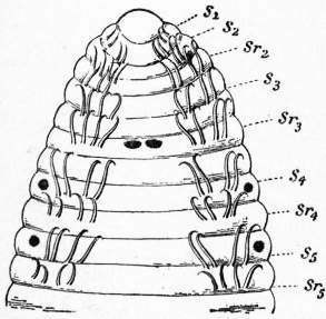
Fig. 14.—Acanthobdella, from the ventral surface, showing the five sets of setae (S1 to S5) and the replacing setae (Sr) behind them. The three pairs of pigmented spots show the position of the eyes on the dorsal surface. (After Kovalevsky.)
In the middle of the body, where the limits of the somites can be checked by a comparison with the arrangement of the nephridia and the gonads, and where the ganglia are quite distinct and separated by long connectives, each ganglion is seen to consist of six masses of cells enclosed by capsules and to give off three nerves on each side. This corresponds to the usual presence (in the Rhynchobdellidae) of three annuli to each segment. Anteriorly and posteriorly separate ganglia have fused. The brain consists not only of a group of six capsules corresponding to the archicerebrum of the Oligochaeta, but of a further mass of cells surrounding and existing below the alimentary canal, which can be analysed into five or six more separate ganglia. The whole mass lies in the seventh or eighth segment. At the posterior end of the body there are likewise seven separate ganglia partially fused to form a single ganglionic mass, which innervates the segments lying behind the anus and corresponding to the posterior sucker. So that a leech in which only twenty-seven segments are apparent by the enumeration of the annuli, separate ganglia, nephridia, lines of sensillae upon the body, really possesses an additional seven lying behind that which is apparently the last of the series and crowded together into a minute space. The annuli into which segments are externally divided are so deeply incised as to render it impossible to distinguish, as can be readily done in the Oligochaeta as a rule, the limits of an annulus from that of a true segment. As remarked, the prevalent number of annuli to a segment is three in the Rhynchobdellidae. But in that group (Cystobranchus) there may be as many as eight annuli. In the Gnathobdellidae the prevailing number of annuli to a segment is five; but here again the number is often increased, and Trocheta has no less than eleven. The reason for this excessive annulation has been seen in the limited number of segments (thirty-four) of which the body is composed, which are laid down early and do not increase. In the Oligochaeta, on the other hand, there is growth of new segments. It is important to notice that the metameric plan of growth of Chaetopods is still preserved.
The nephridia are like those of the Oligochaeta in general structure; that is to say, they consist of drain-pipe cells which are placed end to end and are perforated by their duct. The internal funnel varies in the same way as in the Oligochaeta in the number of cells which form it. In Clepsine (Glossiphonia) there are only three cells, and in Nephelis five to eight cells. In Hirudo the funnel is not pervious and is composed of a large number of cells. Externally, the nephridium opens by a vesicle, as in many Oligochaetes whose lumen is intercellular. In Pontobdella and Branchellion the nephridia form a network extending from segment to segment, but there is only one pair of funnels in each segment. Slight differences in form have been noted between nephridia of different segments; but the Hirudinea do not show the marked differentiation that is to be seen in some other Chaetopods; nor do the nephridia ever acquire any relations to the alimentary canal.
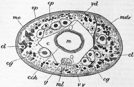
Fig. 15.—Section of Acanthobdella (after Kovalevsky).
c, Coelom. c.ch, Coelomic epithelium (yellow-cells). cg, Glandular cells. cl, Muscle cells of lateral line. cp, Pigment cells. ep, Ectoderm. g, Nerve cord. m, Intestine. mc, Circular muscle. ml, Longitudinal muscle. vd, Dorsal vessel. vv, Ventral vessel.
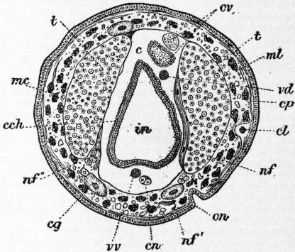
Fig. 16.—Section of Acanthobdella (after Kovalevsky). Identical letters as in fig. 2; in addition, cn, nerve cord; in, intestine; nf, parts of nephridium; on, external opening of nephridium; ov, ova; t, testis.
Coelom.—The coelom of the Hirudinea differs in most genera from that of the Oligochaeta and Polychaeta. The difference is that it is broken up into a complex sinus system. The least modified type is shown by Acanthobdella, a leech, parasitic upon fishes, in which transverse sections (see figs. 15 and 16) show the gut, the nervous system, &c., lying in a spacious chamber which is the coelom. This coelom is lined by peritoneal cells and is divided into a series of metameres by septa which correspond to the segmentation of the body, the arrangement being thus precisely like that of typical Chaetopoda. Moreover, upon the intestine the coelomic cells are modified into chloragogen cells. In Acanthobdella the testes are, however, not contained in the general coelom, and the nephridia lie in the septa. It is remarkable, in view of the spaciousness of the coelom, that the funnels of the latter have not been seen. Ozobranchus possesses a coelom which is less typically chaetopodous than that of Acanthobdella, but more so than in other leeches. There is a spacious cavity surrounding the gut and containing also blood-vessels, and to some extent the generative organs, and the nervous cord. Furthermore, in the mid region of the body this coelom is broken up by metamerically arranged septa, as in Acanthobdella. These septa are, however, rather incomplete and are not fastened to the gut; and, as in Acanthobdella, the nephridia are embedded in them. In addition to the median lacuna there are two lateral lacunae, one upon each side. These regions of the coelom end at the ends of the body and communicate with each other by means of a branched system of coelomic sinuses, which are in places very fine tubes. Neither in this genus nor in the last is there any communication between coelom and vascular system. In Clepsine (Glossiphonia) there is a further breaking up of the coelom. The median lacuna no longer exists, but is represented by a dorsal and ventral sinus. The former lodges the dorsal, the latter the ventral, blood-vessel. The gut has no coelomic space surrounding it. A complex network places these sinuses and the lateral sinuses in communication. Here also the blood system has no communication with the sinus system of the coelom. In Hirudo and the Gnathobdellidae there is only one system of cavities which consist of four principal longitudinal trunks, of which the two lateral are contractile, which communicate with a network ramifying everywhere, even among the cells of the epidermis. The network is partly formed out of pigmented cells which are excavated and join to form tubes, the so-called botryoidal tissue, not found among the Rhynchobdellidae at all. It seems clear from the recent investigations of A.G. Bourne and E.S. Goodrich that the vascular system and the coelom are in communication (as in vertebrates by means of the lymph system). On the other hand, it has been held that in these leeches there is no vascular system at all and that the entire system of spaces is coelom. In favour of regarding the vascular system as totally absent, is the fact that the median coelomic channels contain no dorsal and ventral vessel. In favour of seeing in the lateral trunks and their branches a vascular system, is the contractility of the former, and the fact of the intrusion of the latter into the epidermis, matched among the Oligochaeta, where undoubted blood capillaries perforate the epidermis. A further fact must be considered in deciding this question, which is the discovery of ramifying coelomic tubes, approaching close to, but not entering, the epidermis in the Polychaete Arenicola. These tubes are lined by flattened epithelium and often contain blood capillaries; they communicate with the coelom and are to be regarded as prolongation of it into the thickness of the body wall.
Gonads and Gonad Ducts.—The gonads and their ducts in the Hirudinea invariably form a closed system of cavities entirely shut off from the coelom in which they lie. There is thus a broad resemblance to the Eudrilidae, to which group of Oligochaeta the Hirudinea are further akin by reason of the invariably unpaired condition of the generative apertures, and the existence of a copulatory apparatus (both of which characters, however, are present occasionally in other Oligochaeta).
The testes are more numerous than the ovaries, of which latter there are never more than one pair. The testes vary in numbers of pairs. Four (Ozobranchus) to six (Glossiphonia) or ten (Philaemon) are common numbers. In Acanthobdella, however, the testes of each side of the body have grown together to form a continuous band, which extends in front of external pore. Each testis communicates by means of an efferent duct with a common collecting duct of its side of the body, which opens on to the exterior by means of a protrusible penis, and to which is sometimes appended a seminal vesicle. The efferent ducts are ciliated, and there is a patch of cilia at the point where they communicate with the cavity of each testis. The ovaries are more extensive in some forms (e.g. Ozobranchus) than in others, where they are small rounded bodies. The two ducts continuous with the gonads open by a common vagina on to the exterior behind the male pores. This “vagina” is sometimes of exaggerated size. Thus, in Philaemon pungens (Lambert) it has the form of a large sac, into which open by a single orifice the conjoined oviducts. From this vagina arises a narrow duct leading to the exterior. In Ozobranchus the structures in question are still more complicated. The two long ovarian sacs communicate with each other by a transverse bridge before uniting to form the terminal canal. Into each ovarian sac behind the transverse junction opens a slender tube, which is greatly coiled, and, in its turn, opens into a spherical “spermathecal sac.” From this an equally slender tube proceeds, which joins its fellow of the opposite side, and the two form a thick, walled tube, which opens on to the exterior within the bursa copulatrix through which the penis protrudes. These two last-mentioned types show features which can be, as it seems, matched in the Eudrilidae.
The gonads develop (O. Bürger) in coelomic spaces close to nephridial funnels, which have, however, no relation to the gonad ducts. The ovaries are solid bodies, of which the outer layer becomes separated from the plug of cells lying within; thus a cavity is formed which is clearly coelom. This cavity and its walls becomes prolonged to form the oviducts. A stage exactly comparable to the stage in the leeches, where the ovary is surrounded by a closed sac, has been observed in Eudrilus. In this Annelid later the sac in question joins its fellow, passing beneath the nerve cord exactly as in the leech, and also grows out to reach the exterior. The sole difference is therefore that in Eudrilus the ovarian sac gives rise to a tube which bifurcates, one branch meeting a corresponding branch of the other ovary of the pair, while the second branch reaches the exterior. In the leech the two branches are fused into one. We have here clearly a case of a true coelomoduct performing the function of an oviduct in both leeches and Eudrilidae. The facts just referred to suggest further comparisons between the Hirudinea and Eudrilidae. The large sacs which have been termed vagina are suggestive of the large coelomic spermathecae in Eudrilids, a comparison which needs, however, embryological data, not at present forthcoming, for its justification. It is at least clear that in Ozobranchus this comparison is justifiable; but only probable, or perhaps possible, in the case of Philaemon. In the former, the duct, leading from the ovarian sac, and swelling along its course into the spherical sac, the “spermatheca,” is highly suggestive of the oviduct and receptaculum of the Eudrilidae.
The testes during development become hollowed out and are prolonged into the vasa efferentia. These ducts therefore have not their exact counterparts in the Oligochaeta, unless we are to assume that they collectively are represented by the seminal vesicles of earthworms and the vasa deferentia. It is to be noted that the Hirudinea differ from the Oligochaeta in that the male pore is in advance of the gonads (except in Acanthobdella, which here, as in so many points, approximates to the Oligochaeta), whereas in Oligochaeta that pore is behind the gonads (again with an exception, Allurus).
Classification.—The Hirudinea may be divided into three families:—
(i.) Rhynchobdellidae.—A protrusible proboscis exists, but there are no jaws. The blood is colourless. Pontobdella, Glossiphonia, &c.
(ii.) Gnathobdellidae.—A proboscis absent, but jaws usually present. Blood coloured red with haemoglobin. Hirudo, Nephelis, &c.
(iii.) Acanthobdellidae.—Proboscis present, but short. Paired setae of Oligochaetous pattern present in anterior segments. Blood red. Acanthobdella.
Literature.—A.O. Kovalevsky, Bull. Imp. Sci. (St Petersburg, November 1896) (Acanthobdella); A.G. Bourne, Quart. Journ. Micr. Sci., 1884; A. Oka, Zeitschr. wiss. Zool., 1894; E.S. Goodrich, Quart. Journ. Micr. Sci., 1899; W.E. Castle, Bull. Mus. Comp. Zool., 1900; A.M. Lambert, Proc. Roy. Soc. (Victoria, 1897); C.O. Whitman, Journ. Morph., 1889 and 1891; O. Bürger, Zeitschr. wiss. Zool., 1902, and other memoirs by the above, and by St V. Apáthy, R. Blanchard, H. Bolsius, A. Dendy, R.S. Bergh, &c.
(F. E. B.)
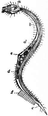
From Cambridge Natural History, vol. ii. “Worms.” by permission of Macmillan & Co., Ltd.
Mature female of Chaetosoma daparedii, (From Mechnikov.) a, Oesophagus; b, intestine; c, anus; d, ovary; e, generative pore; f, ventral bristles.
CHAETOSOMATIDA, a small group of minute, free-living, aquatic organisms which are usually placed as an annex to the Nematoda. Indeed Mechnikov, to whom we owe much of our knowledge of these forms, calls them “creeping Nematoda.” They are usually found amongst seaweed in temperate seas, but they are probably widely distributed; some are fresh-water. The genus Chaetosoma, with the two species Ch. claparedii and Ch. ophicephalum and the genus Tristicochaeta, have swollen heads. The third genus Rhabdogaster has no such distinct head, though the body may be swollen anteriorly. The mouth is terminal and anterior and surrounded by a ring of spicules or a half-ring of hooks. Scattered hairs cover the body. Just in front of the anus there is in Chaetosoma a double, and in Tristicochaeta a triple row of about fifteen stout cylindrical projections upon which the animals creep. The females are a little larger than the males; in Ch. claparedii the former attain a length of 1.5 mm., the latter of 1.12 mm. The mouth opens into an oesophagus which passes into an intestine; this opens by a ventral anus situated a little in front of the posterior end. The testis is single, and its duct opens with the anus, and is provided with a couple of spicules. The ovary is double, and the oviducts open by a median ventral pore about the middle of the body; in this region there is a second swelling both in Chaetosoma and in Rhabdogaster. The last-named form is in the female 0.36 mm. in length. In it the hairs are confined to the dorsal middle line and the creeping setae are hooked, of a finer structure than in Chaetosoma, and situated so far forward that the vagina opens amongst them. Ch. ophicephalum has been taken in the English Channel.
See E. Mechnikov, Zeitschr. wiss. Zool. xvii., 1867, p. 537; Panceri, Atti Acc. Napoli, vii., 1878, p. 7.
(A. E. S.)
CHAFER, a word used in modern speech to distinguish the beetles of the family Scarabaeidae, and more especially those species which feed on leaves in the adult state. The word is derived from the O. Eng. ceafor, and it is interesting to note that the cognate Ger. Käfer is applied to beetles of all kinds. For the characters of the Scarabaeidae see Coleoptera. This family includes a large number of beetles, some of which feed on dung and others on vegetable tissues. The cockchafers and their near allies belong to the subfamily Melolonthinae, and the rose-chafers to the Cetoniinae; in both the beetles eat leaves, and their grubs spend a long life underground devouring roots. In Britain the Melolonthines that are usually noted as injurious are the two species of cockchafer (Melolontha vulgaris and M. hippocastani), large heavy beetles with black pubescent pro-thorax, brown elytra and an elongated pointed tail-process; the summer-chafer (Rhizotrogus solstitialis), a smaller pale brown chafer; and the still smaller garden-chafer or “cocker-bundy” (Phyllopertha horticola), which has a dark green pro-thorax and brown elytra. Of the Cetoniines, the beautiful metallic green rose-chafer, Cetonia aurata, sometimes causes damage, especially in gardens. The larvae of the chafers are heavy, soft-skinned grubs, with hard brown heads provided with powerful mandibles, three pairs of well-developed legs, and a swollen abdomen. As they grow, the larvae become strongly flexed towards the ventral surface, and lie curled up in their earthen cells, feeding on roots. The larval life lasts several years, and in hard frosts the grubs go deep down away from the surface. Pupation takes place in the autumn, and though the perfect insect emerges from the cuticle very soon afterwards, it remains in its underground cell for several months, not making its way to the upper air until the ensuing summer. After pairing, the female crawls down into the soil to lay her eggs. The grubs of chafers, when turned up by the plough, are greedily devoured by poultry, pigs and various wild birds. When the beetles become so numerous as to call for destruction, they are usually shaken off the trees where they rest on to sheets or tarred boards. On the continent of Europe chafers are far more numerous than in the United Kingdom, and the rural governments in France give rewards for their destruction. D. Sharp states that in the department of Seine-inférieure 867,173,000 cockchafers and 647,000,000 larvae were killed in the four years preceding 1870.
The anatomy of Melolontha is very fully described in a classical memoir by H.E. Strauss-Dürckheim (Paris, 1828).
(G. H. C.)
CHAFF (from the A.S. ceaf, allied to the O. High Ger. cheva, a husk or pod), the husks left after threshing grain, and also hay and straw chopped fine as food for cattle; hence, figuratively, the refuse or worthless part of anything. The colloquial use of the word, to chaff, in the sense of to banter or to make fun of a person, may be derived from this figurative sense, or from “to chafe,” meaning to vex or irritate.
CHAFFARINAS, or Zaitarines, a group of islands belonging to Spain off the north coast of Morocco, near the Algerian frontier, 2½ m. to the north of Cape del Agna. The largest of these isles, Del Congreso, is rocky and hilly. It has a watch-house on the coast nearest to Morocco. Isabella II., the central island, contains several batteries, barracks and a penal convict settlement. The Spanish government has undertaken the construction of breakwaters to unite this island with the neighbouring islet of El Rey, with a view to enclose a deep and already sheltered anchorage. This roadstead affords a safe refuge for many large vessels. The Chaffarinas, which are the Tres Insulae of the Romans and the Zafrān of the Arabs, were occupied by Spain in 1848. The Spanish occupation anticipated by a few days a French expedition sent from Oran to annex the islands to Algeria. The population of the islands is under 1000.
CHAFFEE, ADNA ROMANZA (1842- ), American general, was born at Orwell, Ohio, on the 14th of April 1842. At the outbreak of the Civil War he entered the United States cavalry as a private, and he rose to commissioned rank in 1863, becoming brevet captain in 1865. He remained in the army after the war and took part with distinction in many Indian campaigns. His promotion was, however, slow, and he was at the age of fifty-six still a lieutenant-colonel of cavalry. But in 1898, at the outbreak of the Spanish-American War, he was made brigadier-general and soon afterwards major-general of volunteers. In the Cuban campaign he won particular distinction, and the victory of the Americans in the action of El Caney was in large measure due to his careful personal reconnaissances of the ground to be attacked and to the endurance of his own brigade. After reverting for a time to the rank of brigadier-general, he was made a major-general U.S.V. again in 1900 and was appointed to command the United States contingent in China. He took a brilliant and successful part in the advance on Peking and the relief of the Legations. In 1901 he became a major-general in the regular army, and in 1901-1902 commanded the Division of the Philippines. In 1902-1903 he commanded the Department of the East, and from 1904 to 1906 was chief of the general staff of the army. In 1904 he received the rank of lieutenant-general in the United States army, being the first enlisted man of the regular army to attain this, the highest rank in the service. He was retired at his own request on the 1st of February 1906, after more than forty years’ service.
CHAFFINCH (Fringilla coelebs), the common English name of a bird belonging to the family Fringillidae (see Finch), and distinguished, in the male sex, by the deep greyish blue of its crown feathers, the yellowish green of its rump, the white of the wing coverts, so disposed as to form two conspicuous bars, and the reddish brown passing into vinous red of the throat and breast. The female is drab, but shows the same white markings as the male, and the young males resemble the females until after the first autumn moult, when they gradually assume the plumage of their sex. The chaffinch breeds early in the season, and its song may often be heard in February. Its nest, which is a model of neatness and symmetry, it builds on trees and bushes, preferring such as are overgrown with moss and lichens. It is chiefly composed of moss and wool, lined internally with grass, wool, feathers, and whatever soft material the locality affords. The outside consists of moss and lichens, and according to Selby, “is always accordant with the particular colour of its situation.” When built in the neighbourhood of towns the nest is somewhat slovenly and untidy, being often composed of bits of dirty straw, pieces of paper and blackened moss; in one instance, near Glasgow, the author of the Birds of the West of Scotland found several postage-stamps thus employed. It lays four or five eggs of a pale purplish buff, streaked and spotted with purplish red. In spring the chaffinch is destructive to early flowers, and to young radishes and turnips just as they appear above the surface; in summer, however, it feeds principally on insects and their larvae, while in autumn and winter its food consists of grain and other seeds. On the continent of Europe the chaffinch is a favourite song-bird, especially in Germany, where great attention is paid to its training.
CHAFING-DISH (from the O. Fr. chaufer, to make warm), a kind of portable grate heated with charcoal, and used for cooking or keeping food warm. In a light form, and heated over a spirit lamp, it is also used for cooking various dainty dishes at table. The employment of the chafing-dish for the latter purpose has been largely restored in modern cookery.
CHAGOS, a group of atolls in the Indian Ocean, belonging to Britain, disposed in circular form round the Chagos bank, in 4° 44′ to 7° 39′ S., and 70° 55′ to 72° 52′ E. The atolls on the south and east side of the bank, which has a circumference of about 270 m., have disappeared through subsidence; a few—Egmont, Danger, Eagle, and Three Brothers—still remain on the east side, but most of the population (about 700) is centred on Diego Garcia, which lies on the south-east side, and is nearly 13 m. long by 6 m. wide. The lagoon, which is enclosed by two coral barriers and accessible to the largest vessels on the north side, forms one of the finest natural harbours in the world. The group, which has a total land area of 76 sq. m., is dependent for administrative purposes on Mauritius, and is regularly visited by vessels from that colony. The only product is cocoa-nut oil, of which about 106,000 gallons are annually exported. The French occupied the islands in 1791 from Mauritius, and the oil industry (from which the group is sometimes called the Oil Islands) came into the hands of French Creoles.
CHAGRES, a village of the Republic of Panama, on the Atlantic coast of the Isthmus, at the mouth of the Chagres river, and about 8 m. W. of Colon. It has a harbour from 10 to 12 ft. deep, which is difficult to enter, however, on account of bars at its mouth. The port was discovered by Columbus in 1502, and was opened for traffic with Panama, on the Pacific coast, by way of the Chagres river, in the 16th century. With the decline of Porto Bello in the 18th century Chagres became the chief Atlantic port of the Isthmus, and was at the height of its importance during the great rush of gold-hunters across the Isthmus to California in 1849 and the years immediately following. With the completion of the Panama railway in 1855, however, travel was diverted to Colon, and Chagres soon became a village of miserable huts, with no evidence of its former importance. On a high rock at the mouth of the river stands the castle of Lorenzo, which was destroyed by Sir Henry Morgan when he captured the town in 1671, but was rebuilt soon afterwards by the Spaniards. Chagres was again captured in 1740 by British forces under Admiral Edward Vernon.
CHAIN (through the O. Fr. choeine, choene, &c., from Lat. catena), a series of links of metal or other material so connected together that the whole forms a flexible band or cord. Chains are used for a variety of purposes, such as fastening, securing, or connecting together two or more objects, supporting or lifting weights, transmitting mechanical power, &c.; or as an ornament to serve as a collar, as a symbol of office or state, or as part of the insignia of an order of knighthood; or as a device from which to hang a jewelled or other pendant, a watch, &c. (see Collar). Ornamental chains are made with a great variety of links, but those intended for utilitarian purposes are mostly of two types. In stud chains a stud or brace is inserted across each link to prevent its sides from collapsing inwards under strain, whereas in open link chains the links have no studs. The addition of studs is reckoned to increase the load which the chain can safely bear by 50%. Small chains of the open-link type are to a great extent made by machinery. For larger sizes the smith cuts off a length of iron rod of suitable diameter, forms it while hot to the shape of the link by repeated blows of his hammer, and welds together the two ends of the link, previously slipped inside its fellow, by the aid of the same tool; in some cases the bending is done in a mechanical press and the welding under a power hammer (see also Cable). Weldless chains are also made; in A.G. Strathern’s process, for instance, cruciform steel bars are pressed, while hot, into links, each without join and engaging with its neighbours. Chains used for transmitting power are known as pitch-chains; the chain of a bicycle (q.v.) is an example.
From the use of the chain as employed to bind or fetter a prisoner or slave, comes the figurative application to anything which serves as a constraining or restraining force; and from its series of connected links, to any series of objects, events, arguments, &c., connected by succession, logical sequence or reasoning. Specific uses are for a measuring line in land-surveying, consisting of 100 links, i.e. iron rods, 7.92 in. in length, making 22 yds. in all, hence a lineal measure of that length; and, as a nautical term, for the contrivance by which the lower shrouds of a mast are extended and secured to the ship’s sides, consisting of dead-eyes, chain-plates, and chain-wale or “channel.”
CHAIR (in. Mid. Eng. choere, through O. Fr. chaëre or chaiere, from Lat. cathedra, later caledra, Gr. καθέδρα, seat, cf. “cathedral”; the modern Fr. form chaise, a chair, has been adopted in English with a particular meaning as a form of carriage; chaire in French is still used of a professorial or ecclesiastical “chair,” or cathedra), a movable seat, usually with four legs, for a single person, the most varied and familiar article of domestic furniture. The chair is of extreme antiquity, although for many centuries and indeed for thousands of years it was an appanage of state and dignity rather than an article of ordinary use. “The chair” is still extensively used as the emblem of authority in the House of Commons and in public meetings. It was not, in fact, until the 16th century that it became common anywhere. The chest, the bench and the stool were until then the ordinary seats of everyday life, and the number of chairs which have survived from an earlier date is exceedingly limited; most of such examples are of ecclesiastical or seigneurial origin. Our knowledge of the chairs of remote antiquity is derived almost entirely from monuments, sculpture and paintings. A few actual examples exist in the British Museum, in the Egyptian museum at Cairo, and elsewhere. In ancient Egypt they appear to have been of great richness and splendour. Fashioned of ebony and ivory, or of carved and gilded wood, they were covered with costly stuffs and supported upon representations of the legs of beasts of the chase or the figures of captives. An arm-chair in fine preservation found in a tomb in the Valley of the Kings is astonishingly similar, even in small details, to that “Empire” style which followed Napoleon’s campaign in Egypt. The earliest monuments of Nineveh represent a chair without a back but with tastefully carved legs ending in lions’ claws or bulls’ hoofs; others are supported by figures in the nature of caryatides or by animals. The earliest known form of Greek chair, going back to five or six centuries before Christ, had a back but stood straight up, front and back. On the frieze of the Parthenon Zeus occupies a square seat with a bar-back and thick turned legs; it is ornamented with winged sphinxes and the feet of beasts. The characteristic Roman chairs were of marble, also adorned with sphinxes; the curule chair was originally very similar in form to the modern folding chair, but eventually received a good deal of ornament.
The most famous of the very few chairs which have come down from a remote antiquity is the reputed chair of St Peter in St Peter’s at Rome. The wooden portions are much decayed, but it would appear to be Byzantine work of the 6th century, and to be really an ancient sedia gestatoria. It has ivory carvings representing the labours of Hercules. A few pieces of an earlier oaken chair have been let in; the existing one, Gregorovius says, is of acacia wood. The legend that this was the curule chair of the senator Pudens is necessarily apocryphal. It is not, as is popularly supposed, enclosed in Bernini’s bronze chair, but is kept under triple lock and exhibited only once in a century. Byzantium, like Greece and Rome, affected the curule form of chair, and in addition to lions’ heads and winged figures of Victory and dolphin-shaped arms used also the lyre-back which has been made familiar by the pseudo-classical revival of the end of the 18th century. The chair of Maximian in the cathedral of Ravenna is believed to date from the middle of the 6th century. It is of marble, round, with a high back, and is carved in high relief with figures of saints and scenes from the Gospels—the Annunciation, the Adoration of the Magi, the flight into Egypt and the baptism of Christ. The smaller spaces are filled with carvings of animals, birds, flowers and foliated ornament. Another very ancient seat is the so-called “Chair of Dagobert” in the Louvre. It is of cast bronze, sharpened with the chisel and partially gilt; it is of the curule or faldstool type and supported upon legs terminating in the heads and feet of animals. The seat, which was probably of leather, has disappeared. Its attribution depends entirely upon the statement of Suger, abbot of St Denis in the 12th century, who added a back and arms. Its age has been much discussed, but Viollet-le-Duc dated it to early Merovingian times, and it may in any case be taken as the oldest faldstool in existence. To the same generic type belongs the famous abbots’ chair of Glastonbury; such chairs might readily be taken to pieces when their owners travelled. The faldisterium in time acquired arms and a back, while retaining its folding shape. The most famous, as well as the most ancient, English chair is that made at the end of the 13th century for Edward I., in which most subsequent monarchs have been crowned. It is of an architectural type and of oak, and was covered with gilded gesso which long since disappeared.
Passing from these historic examples we find the chair monopolized by the ruler, lay or ecclesiastical, to a comparatively late date. As the seat of authority it stood at the head of the lord’s table, on his dais, by the side of his bed. The seigneurial chair, commoner in France and the Netherlands than in England, is a very interesting type, approximating in many respects to the episcopal or abbatial throne or stall. It early acquired a very high back and sometimes had a canopy. Arms were invariable, and the lower part was closed in with panelled or carved front and sides—the seat, indeed, was often hinged and sometimes closed with a key. That we are still said to sit “in” an arm-chair and “on” other kinds of chairs is a reminiscence of the time when the lord or seigneur sat “in his chair.” These throne-like seats were always architectural in character, and as Gothic feeling waned took the distinctive characteristics of Renaissance work. It was owing in great measure to the Renaissance that the chair ceased to be an appanage of state, and became the customary companion of whomsoever could afford to buy it. Once the idea of privilege faded the chair speedily came into general use, and almost at once began to reflect the fashions of the hour. No piece of furniture has ever been so close an index to sumptuary changes. It has varied in size, shape and sturdiness with the fashion not only of women’s dress but of men’s also. Thus the chair which was not, even with its arms purposely suppressed, too ample during the several reigns of some form or other of hoops and farthingale, became monstrous when these protuberances disappeared. Again, the costly laced coats of the dandy of the 18th and early 19th centuries were so threatened by the ordinary form of seat that a “conversation chair” was devised, which enabled the buck and the ruffler to sit with his face to the back, his valuable tails hanging unimpeded over the front. The early chair almost invariably had arms, and it was not until towards the close of the 16th century that the smaller form grew common.
The majority of the chairs of all countries until the middle of the 17th century were of oak without upholstery, and when it became customary to cushion them, leather was sometimes employed; subsequently velvet and silk were extensively used, and at a later period cheaper and often more durable materials. Leather was not infrequently used even for the costly and elaborate chairs of the faldstool form—occasionally sheathed in thin plates of silver—which Venice sent all over Europe. To this day, indeed, leather is one of the most frequently employed materials for chair covering. The outstanding characteristic of most chairs until the middle of the 17th century was massiveness and solidity. Being usually made of oak, they were of considerable weight, and it was not until the introduction of the handsome Louis XIII. chairs with cane backs and seats that either weight or solidity was reduced. Although English furniture derives so extensively from foreign and especially French and Italian models, the earlier forms of English chairs owed but little to exotic influences. This was especially the case down to the end of the Tudor period, after which France began to set her mark upon the British chair. The squat variety, with heavy and sombre back, carved like a piece of panelling, gave place to a taller, more slender, and more elegant form, in which the framework only was carved, and attempts were made at ornament in new directions. The stretcher especially offered opportunities which were not lost upon the cabinet-makers of the Restoration. From a mere uncompromising cross-bar intended to strengthen the construction it blossomed, almost suddenly, into an elaborate scroll-work or an exceedingly graceful semicircular ornament connecting all four legs, with a vase-shaped knob in the centre. The arms and legs of chairs of this period were scrolled, the splats of the back often showing a rich arrangement of spirals and scrolls. This most decorative of all types appears to have been popularized in England by the cavaliers who had been in exile with Charles II. and had become familiar with it in the north-western parts of the European continent. During he reign of William and Mary these charming forms degenerated into something much stiffer and more rectangular, with a solid, more or less fiddle-shaped splat and a cabriole leg with pad feet. The more ornamental examples had cane seats and ill-proportioned cane backs. From these forms was gradually developed the Chippendale chair, with its elaborately interlaced back, its graceful arms and square or cabriole legs, the latter terminating in the claw and ball or the pad foot. Hepplewhite, Sheraton and Adam all aimed at lightening the chair, which, even in the master hands of Chippendale, remained comparatively heavy. The endeavour succeeded, and the modern chair is everywhere comparatively slight. Chippendale and Hepplewhite between them determined what appears to be the final form of the chair, for since their time practically no new type has lasted, and in its main characteristics the chair of the 20th century is the direct derivative of that of the later 18th.
The 18th century was, indeed, the golden age of the chair, especially in France and England, between which there was considerable give and take of ideas. Even Diderot could not refrain from writing of them in his Encyclopédie. The typical Louis Seize chair, oval-backed and ample of seat, with descending arms and round-reeded legs, covered in Beauvais or some such gay tapestry woven with Boucher or Watteau-like scenes, is a very gracious object, in which the period reached its high-water mark. The Empire brought in squat and squabby shapes, comfortable enough no doubt, but entirely destitute of inspiration. English Empire chairs were often heavier and more sombre than those of French design. Thenceforward the chair in all countries ceased to attract the artist. The art nouveau school has occasionally produced something of not unpleasing simplicity; but more often its efforts have been frankly ugly or even grotesque. There have been practically no novelties, with the exception perhaps of the basket-chair and such like, which have been made possible by modern command over material. So much, indeed, is the present indebted to the past in this matter that even the revolving chair, now so familiar in offices, has a pedigree of something like four centuries (see also Sedan-chair).
(J. P.-B.)
CHAISE (the French for “chair,” through a transference from a “sedan-chair” to a wheeled vehicle), a light two- or four-wheeled carriage with a movable hood or “calash”; the “post-chaise” was the fast-travelling carriage of the 18th and early 19th centuries. It was closed and four-wheeled for two or four horses and with the driver riding postillion.
CHAKRATA, a mountain cantonment in the Dehra Dun district of the United Provinces of India, on the range of hills overlooking the valleys of the Jumna and the Tons, at an elevation of 7000 ft. It was founded in 1866 and first occupied in April 1869.
CHALCEDON, more correctly Calchedon (mod. Kadikeui), an ancient maritime town of Bithynia, in Asia Minor, almost directly opposite Byzantium, south of Scutari. It was a Megarian colony founded on a site so obviously inferior to that which was within view on the opposite shore, that it received from the oracle the name of “the City of the Blind.” In its early history it shared the fortunes of Byzantium, was taken by the satrap Otanes, vacillated long between the Lacedaemonian and the Athenian interests, and was at last bequeathed to the Romans by Attalus III. of Pergamum (133 B.C.). It was partly destroyed by Mithradates, but recovered during the Empire, and in A.D. 451 was the seat of the Fourth General Council. It fell under the repeated attacks of the barbarian hordes who crossed over after having ravaged Byzantium, and furnished an encampment to the Persians under Chosroes, c. 616-626. The Turks used it as a quarry for building materials for Constantinople. The site is now occupied by the village of Kadikeui (“Village of the Judge”), which forms the tenth “cercle” of the municipality of Constantinople. Pop. about 33,000, of whom 8000 are Moslems. There is a large British colony with a church, and also Greek and Armenian churches and schools, and a training college for Roman Catholic Armenians. To the S. are the ruins of Panteichion (mod. Pendik), where Belisarius is said to have lived in retirement.