VOLUME IV SLICE IV Bradford, William to Brequigny, Louis (continued 4)
Reading edition
There seems a connexion between consciousness and the power to modify reflex action to meet the exigencies of the occasion. Pure reflexes are admirably adapted to certain ends. They are reactions which have long proved advantageous to the phylum of which the existent animal is the representative embodiment. But the reflexes have a machine-like fatality, and conscious aim does not forerun their execution. The subject as active agent does not direct them. Yet they lie under the control of higher centres. The cough, the eye-closure, the impulse to smile, all these can be suppressed. The innate respiratory rhythm can be modified to meet the requirements of vocal utterance. In other words, the reaction of reflex arcs is controllable by the mechanism to whose activity consciousness is adjunct. The reflexes controlled are often reactions but slightly affecting consciousness, but consciousness is very distinctly operative with the centres which exert the control. It may be that the primary aim, object and purpose of consciousness is control. “Consciousness in a mere automaton,” writes Professor Lloyd Morgan, “is a useless and unnecessary epiphenomenon.” As to how this conscious control is operative on reflexes, how it intrudes its influence on the running of the reflex machinery, little is known.
The Cerebrum an Organ giving Adaptation and Readjustment of Motor Acts.—The exercise of this control and the acquirement of skilled actions have obviously elements in common. By skilled actions, we understand actions not innately given, actions acquired by training in individual experience. The controlling centres pick out from an ancestral motor action some part, and isolate and enhance that until it becomes a skilled act. The motor co-ordination ancestrally provided for the ring finger gives an extending of it only in company with extension of the fingers on either side of it. The isolated lifting of the ring finger can, however, soon be acquired by training. In such cases the higher centre with conscious effort is able to dissociate a part from an ancestral co-ordination, and in that way to add a skilled adapted act to the powers of the individual.
The nervous organs of control form, therefore, a special instrument of adaptation and of readjustment of reaction, for better accommodation to requirements which may be new. The attainment of more precision and speed in the use of a tool, or the handling of a weapon, means a process in which nervous organs of control modify activities of reflex centres themselves already perfected ancestrally for other though kindred actions. This process of learning is accompanied by conscious effort. The effort consists not so much in any course of reasoning but rather in the acquiring of new sensorimotor experience. To learn swimming or skating by simple cogitation or mere visual observation is of course impossible. The new ideas requisite cannot be constructed without motor experience, and the training must include that motor experience. Hence the training for a new skilled motor manoeuvre must be simply ad hoc, and is of itself no training for another motor co-ordination.
The more complex an organism the more points of contact does it have with its environment, and the more does it need readjustment amid an environment of shifting relationships. Hence the organs of consciousness and control, being organs of adaptation and readjustment of reaction, will be more pronounced the farther the animal scale is followed upward to its crowning species, man. The cerebrum and especially the cerebral cortex may be regarded as the highest expression of the nervous organ of individual adaptation of reactions. Its high development in man makes him the most successful animal on earth’s surface at the present epoch. The most important part of all this adjustment in his case, as he stands now, consists doubtless in that nervous activity which is intellectual. The mentality attached to his cerebrum includes reason in higher measure than is possessed by the mentality of other animals. He, therefore, more than they, can profitably forecast the future and act suitably to meet it from memory of the past. The cerebrum has proved itself by his case the most potent weapon existent for extending animal dominance over the environment.
Means and Present Aims of Physiological Study of the Brain.—The aspects of cerebral activity are therefore twofold. There is the contribution which it makes to the behaviour of the animal as seen in the creature’s doings. On the other hand there is its product in the psychical life of the animal. The former of these is subject matter for physiology; the latter is especially the province of psychology. Physiology does, however, concern itself with the psychical aspect of cerebral functions. Its scope, embracing the study of the bodily organs in regard to function, includes the psychic as well as the material, because as just shown the former inextricably interlace with the latter. But the relation between the psychic phenomena and the working of the brain in regard to any data of fundamental or intimate character connecting the two remains practically as unknown to us as to the Greek philosophers. What physiology has at present to be content with in this respect is the mere assigning of certain kinds of psychic events to certain local regions of the cerebrum. This primitive quest constitutes the greater part of the “neurology” of our day, and some advance has been made along its lines. Yet how meagre are really significant facts will be clear from the brief survey that follows. Before passing finally from these general considerations, we may note that it becomes more and more clear that the brain, although an organ than can be treated as a whole, is complex in the sense that separable functions belong in some measure to its several parts.
The means principally adopted in studying the functions of the brain—and it must be remembered that this study in its present phase is almost exclusively a mere search for localization—are four. These are the physiological, the clinico-pathological, the histological and the zoological. The first named proceeds by observing the effects of artificial excitation, chiefly electric, of various parts of the brain, and the defects produced by destruction or removal of circumscribed portions. The clinico-pathological proceeds by observing the disturbances of body and mind occurring in disease or injury, and ascertaining the extent of the disease or injury, for the most part post mortem. The histological method examines the microscopic structure of the various regions of the brain and the characters and arrangement of the nerve-cells composing it. The zoological follows and compares the general features of the brain, as represented in the various types of animal creation.
It is on the functions of the fore-brain that interest now mainly focuses, for the reasons mentioned above. And the interest in the fore-brain itself chiefly attaches to the functions of its cortex. This is due to several causes. In man and the animals nearest him the cortex forms by far the larger part of the whole cerebral hemisphere. More than any other part it constitutes the distinctively human feature. It lies accessible to various experimental observations, as also to traumatic lesions and to the surgeon’s art. It is composed of a great unbroken sheet of grey matter; for that reason it is a structure wherein processes of peculiar interest for the investigation in view are likely to occur. To make this last inference more clear a reference to the histology of nervous tissue must be made. The whole physiological function of the nervous system may be summed up in the one word “conduction.” This “conduction” may be defined as the transmission of states of excitement (nerve-impulses) along the neural arcs composing the system. The whole nervous system is built up of chains of nerve-cells (neurones) which are nervous conductors, the chains often being termed arcs. Each neurone is an elongated cell which transmits nerve-impulses from its one end to its other, without so far as is known modifying the impulses in transit, unless in that part of the nerve-cell where the nucleus lies. That part of the neurone or nerve-cell is called the perikaryon or cell-body, and from that part usually many branches of the cell (each branch being a nerve-fibre) ramify. There is no evidence that impulses are modified in transit along a branch of a nerve-cell, but there is clear evidence of manifold modification of nerve-impulses in transit along the nerve-arcs of the nervous system. These nerve-arcs are neurone-chains. In them one neurone continues the line of conduction where the immediately foregoing neurone left it. That is, the neurones are laid in conductive series, the far end of one apposed to the near end of its precursor. The place of juxtaposition of the end of one neurone against the beginning of another is called the synapse. At it the conduction which has so far been wholly intra-neuronic is replaced by an inter-neuronic process, in which the nerve impulse passes from one neurone to the next. The process there, it is natural to think, must be physiologically different from that conductive process that serves for transmission merely within the neurone itself. It may be that to this inter-neuronic conduction are due the differences between conduction in nerve-arcs and nerve-trunks (nerve-fibres) respectively. Significant of the former are changes in rhythm, intensity, excitability and modifications by summation and inhibition; in fact a number of the main features of nervous reaction. These characters impressed upon conduction in nerve arcs (neurone-chains) would therefore be traceable to the intercalation of perikarya and synapses, for both these structures are absent from nerve-trunks. It is therefore probably to perikarya and synapses that the greater part of the co-ordination, elaboration and differentiation of nervous reactions is due. Now, perikarya and synapses are not present in the white matter of the central nervous organ, any more than they are in nerve-trunks. They are confined exclusively to those portions of the central organ which consist of grey matter (so called from its naked-eye appearance). Hence it is to the great sheet of grey matter which enfolds the cerebrum that the physiologist turns, as to a field where he would expect to find evidences of the processes of cerebral co-ordination at work. It is therefore to items regarding the functions of the great sheet of cerebral cortex that we may now pass.
The Cerebral Cortex and its Functions.—The main question which vexed the study of the physiology of the cerebral hemispheres in the 19th century was whether differences of function are detectible in the different regions of the hemisphere and especially in those of its cortex. One camp of experimenters and observers held that the cortex was identical in function throughout its extent. These authorities taught that the various faculties and senses suffer damage in proportion to the amount of cortex removed or injured, and that it is a matter of indifference what may be the particular region wherein the destruction takes place. Against this an opposed set of observers held that different regions perform different functions, and this latter “differential” view was raised in two wholly dissimilar forms in the first and last quarters of the 19th century respectively. In the first quarter of the century, a school, with which the name of Gall is prominently associated, held that each faculty of a set of particular so-called “faculties,” which it assumed constituted intelligence, has in the brain a spatially separate organ proper to itself. Gall’s doctrine had two fundamental propositions. The first was that intelligence resides exclusively in the brain: the second, that intelligence consists of twenty-seven “faculties,” each with a separate local seat in the brain. The first proposition was not new. It is met with in Hippocrates, and it had been elaborated by Descartes and others. But Bichat in his Anatomie generale had partly wandered from the gradually established truth and referred the emotions to the visceral organs, returning to a naive view popularly prevalent. Gall’s first proposition was probably raised especially in reaction against Bichat. But Gall’s proposition was retrograde from the true position of the science of his time. Flourens and others of his contemporaries had already shown not only that intelligence was resident exclusively in the brain, but that it was resident exclusively in that part of the brain which is the fore-brain. Now Gall placed certain of his twenty-seven intellectual faculties in the cerebellum, which is part of the hind-brain.
Phrenology.—As to Gall’s second proposition, the set of faculties into which he analysed intelligence shows his power of psychological analysis to have been so weak that it is matter of surprise his doctrine could obtain even the ephemeral vogue it actually did. Among his twenty-seven faculties are, for instance, “l’amour de la progéniture, l’instinct carnassier, l’amitié, la ruse, la sagacité comparative, l’esprit métaphysique, le talent poétique, la mimique,” &c. Such crudity of speculation is remarkable in one who had undoubtedly considerable insight into human character. Each of the twenty-seven faculties had its seat in a part of the brain, and that part of the brain was called its “organ.” The mere spatial juxtaposition or remoteness of these organs one from another in the brain had, according to Gall, an influence on the constitution of the mind. “Comme l’organe des arts est placé loin de l’organe du sens des couleurs, cette circonstance explique pourquoi les peintres d’histoire ont été rarement coloristes.” All these “faculty-organs” were placed by Gall at the surface of the brain. “This explains the correspondence which exists between craniology and the doctrine of the functions of the brain (cerebral physiology), the single aim of my researches.” Gall wrote that he found the bump of pride (la bosse de l’orgueil) as far down in the animal series as the goat. Broussais traced the “organ” of veneration as far down as the sheep. Gall found the bump of murder (bosse du meurtre) in the carnivora. Later it was traced also in herbivora. Broussais added apologetically that “the herbivora cause a real destruction of plants.”
Gall’s doctrine enjoyed enormous vogue. He himself had the gifts and the demerits of quackery. His doctrine possessed, apart from its falsity, certain other mischievous qualities. “Que ces hommes si glorieux, qui font égorger les nations par millions, sachent qu’ils n’agissent point de leur propre chef, que c’est la nature qui a placé dans leur coeur la rage de la destruction.” One of his scientific opponents rejoined, “Nay, it is not that which they should know. What they should know is that if providence has allowed to man the possibility of doing evil, it has also endowed him with the power to do good.” The main cause of the success of phrenology (q.v.) has been no doubt the common desire of men to read the characters and hidden thoughts of others by external signs. Each bump or “bosse” on the cranium was supposed to indicate the existence and degree of development of one or other of the twenty-seven “faculties.” One such “bosse” showed the development of the organ of “goodness,” and another the development of the organ of “murder.” Such an easy means to arrive at information so curious delighted many persons, and they were not willingly undeceived.
Modern Localization Doctrines.—The crude localization of the phrenologists is therefore too clumsy to possess an interest it might otherwise have had as an early expression of belief in cerebral localization, a belief which other labours have subsequently justified, although on facts and lines quite different from these imagined by Gall and his followers. Patient scientific toil by the hands of E. Hitzig and D. Ferrier and their followers has slowly succeeded in obtaining certain facts about the cortex cerebri which not only show that different regions of it are concerned with different functions, but, for some regions at least, outline to some extent the kind of function exercised. It is true that the greater part of the cortex remains still terra incognita unless we are content with mere descriptive features concerning its coarse anatomy. For several scattered regions some knowledge of their function has been gained by physiological investigation. These scattered regions are the visual, the auditory, the olfactory and the precentral.
The grey matter of the cerebral cortex is broadly characterized histologically by the perikarya (nerve-cells bodies) which lie in it possessing a special shape; they are pyramidal. The dendrite fibres of these cells—that is, their fibres which conduct towards the perikarya—are branches from the apex and corners of the pyramid. From the base often near its middle arises one large fibre—the axone fibre, which conducts impulses away from the perikaryon. The general appearance and arrangement of the neurones in a particle of cortical grey matter are shown in fig. 15, above. The apices of the pyramidal perikarya are turned towards the free surface of the cortex. The figure as interpreted in terms of functional conduction means that the cortex is beset with conductors, each of which collects nerve-impulses, from a minute but relatively wide field by its branched dendrites, and that these nerve-impulses converge through its perikaryon, issue by its axone, and are carried whithersoever the axone runs. In some few cells the axone breaks up into branches in the immediate neighbourhood of its own perikaryon in the cortex. In most cases, however, the axone runs off into the subjacent white matter, leaving the cortex altogether. On reaching the subjacent white matter it mingles with other fibres and takes one of the following courses:—(1) to the grey matter of the cortex of the same hemisphere, (2) to the grey matter of the cortex of the opposite hemisphere, (3) to the grey matter of the pons, (4) to the grey matter of the bulb or spinal cord. It is noteworthy that the dendrite fibres of these cortical neurones do not transgress the limits of the grey cortex and the immediate neighbourhood of the perikaryon to which they belong; whereas the discharging or axone fibre does in the vast majority of cases transgress the limits of the grey matter wherein its perikaryon lies. The cortical neurone therefore collects impulses in the region of cortex just about its perikaryon and discharges them to other regions, some not cortical or even cerebral, but spinal, &c. One question which naturally arises is, do these cells spontaneously generate their impulses or are they stirred to activity by impulses which reach them from without? The tendency of physiology is to regard the actions of the cortex as reactions to impulses communicated to the cortical cells by nerve-channels reaching them from the sense organs. The neurone conductors in the cortex are in so far considered to resemble those of reflex centres, though their reactions are more variable and complex than in the use of the spinal. The chains of neurones passing through the cortex are more complex and connected with greater numbers of associate complex chains than are those of the spinal centres. But just as the reflex centres of the cord are each attached to afferent channels arriving from this or that receptive-organ, for instance, tactile-organs of the skin, or spindles of muscle-sense, &c., so the regions of cortex whose function is to-day with some certainty localized seem to be severally related each to some particular sense-organ. The localization, so far as ascertained, is a localization which attaches separate areas of cortex to the several species of sense, namely the visual, the auditory, the olfactory, and so on. This being so, we should expect to find the sensual representation in the cortex especially marked for the organs of the great distance-receptors, the organs which—considered as sense organs—initiate sensations having the quality of projicience into the sensible environment. The organs of distance-receptors are the olfactory, the visual and the auditory. The environmental agent which acts as stimulus in the case of the first named is chemical, in the second is radiant, and in the last is mechanical.
Olfactory Region of Cortex.—There is phylogenetic evidence that the development of the cortex cerebri first occurred in connexion with the distance-receptors for chemical stimuli—that is, expressed with reference to psychosis, in connexion with olfaction. The olfactory apparatus even in mammals still exhibits a neural architecture of primitive pattern. The cell which conducts impulses to the brain from the olfactory membrane in the nose resembles cells in the skin of the earthworm, in that its cell-body lies actually amid the epithelium of the skin-surface and is not deeply buried near or in the central nervous organ. Further, it has at its external end tiny hairlets such as occur in specially receptive-cells but not usually in purely nervous cells. Hence we must think that one and the same cell by its external end receives the environmental stimulus and by its deep end excites the central nervous organ. The cell under the stimulation of the environmental agent will therefore generate in itself a nervous impulse. This is the clearest instance we have of a neurone being actually excited under natural circumstances by an agent of the environment directly, not indirectly. The deep ends of these olfactory neurones having entered the central nervous organ come into contact with the dendrites of large neurones, called, from their shape, mitral. In the dog, an animal with high olfactory sense, the axone of each olfactory neurone is connected with five or six mitral cells. In man each olfactory neurone is connected with a single mitral cell only. We may suppose that the former arrangement conduces to intensification of the central reaction by summation. At the same time it is an arrangement which could tend to smother sharp differentiation of the central reaction in respect to locality of stimulus at the receptive surface. Considering the diffuse way in which olfactory stimuli are applied in comparison, for instance, with visual, the exact localization of the former can obviously yield little information of use for locating the exact position of their source. On the other hand, in the case of visual stimuli the locus of incidence, owing to the rectilinear propagation of light, can serve with extraordinary exactitude for inferences as to the position of their source. The adaptation of the neural connexions of the two organs in this respect is therefore in accord with expectation.
The earliest cerebral cortex is formed in connexion with the neurone-chains coming into the central nervous organ from the patch of olfactory cells on the surface of the head. The region of cerebrum thus developed is the so-called olfactory lobe and hippocampal formation. The greater part of the cerebral hemisphere is often termed the pallium, because as its development extends it folds cloak-wise over the older structures at the base of the brain. The olfactory lobe, from its position, is sometimes called the pallium basale, and the hippocampal formation the pallium marginale; and these two parts of the pallium form what, on account of their phylogenetic history, Elliott Smith well terms the archipallium. A fissure, the limbic fissure, marks off more or less distinctly this archipallium from the rest of the pallium, a remainder which is of later development and therefore designated by Elliott Smith the neopallium. Of the archipallium, the portion which constitutes the olfactory lobe is well formed in the selachian fish. In the reptilian cerebrum the hippocampal region, the pallium marginale, coexists in addition. These are both of them olfactory in function. Even so high up in the animal scale as the lowest mammals they still form one half of the entire pallium. But in the higher apes and in man the olfactory portion of the pallium is but a small fraction of the pallium as a whole. It is indeed so relatively dwarfed and obscured as to be invisible when the brain is regarded from the side or above. The olfactory part of the pallium exhibits little variation in form as traced up through the higher animals. It is of course small in such animals as Cetaceans, which are anosmatic. In highly osmatic such as the dog it is large. The uncus, and subiculum cornu ammonis of the human brain, belong to it. Disease of these parts has been accompanied by disturbance of the sense of smell. When stimulated electrically (in the rabbit) the olfactory pallium occasions peculiar torsion of the nose and lips (Ferrier), and change, often slowing or arrested, of the respiratory rhythm. P.E. Flechsig has shown that the nerve-fibres of this part of the pallium attain the final stage of their growth, that is to say, acquire their sheaths of myelin, early in the ontogenetic development of the brain. In the human brain they are myelinate before birth. This is significant from the point of view of function, for reasons which have been made clear especially by the researches of Flechsig himself.
The completion of the growth of the nerve-fibres entering and leaving the cortex occurs at very various periods in the growth of the brain. Study of the development of the fibres entering and leaving the various regions of the pallium in the human brain, discovers that the regions may be conveniently grouped into those whose fibres are perfected before birth and those whose fibres are perfected during the first post-natal month, and those whose fibres are perfected after the first but before the end of the fourth post-natal month. The regions thus marked out by completion before birth are five in number, and are each connected, as also shown by collateral evidence, with one or other particular species of sense-organ. And these regions have another character in common recognizable in the nerve-fibres entering and leaving them, namely, they possess fibres projected to or from parts of the nervous system altogether outside the cortex itself. These fibres are termed “projection” fibres. Other regions of the cortex possess fibres coming from or going to various regions of the cortex itself, but do not possess in addition, as do the five primitive cortical fields, the fibres of projection. So that the facts established by Flechsig for the regions of pallium, which other evidence already indicated as connected with the sense-organ of smell, support that evidence and bring the olfactory region of cortex into line with certain other regions of cortex similarly primarily connected with organs of sense.
It will be noted that what has been achieved by these various means of study in regard to the region of the cortex to which olfactory functions are attributed amounts at present to little more than the bare ascertainment of the existence there of nervous mechanisms connected with olfaction, and to the delimiting roughly of their extent and of their ability to influence certain movements, and in man sensations, habitually associated with exercise of the olfactory organ. As to what part the cortical mechanism has in the elaboration or association of mental processes to which olfaction contributes, no evidence worth the name seems as yet forthcoming. In this respect our knowledge, or rather our want of knowledge, of the functions of the olfactory region of the cortex, is fairly typical of that to which we have to confess in regard to the other regions of the cortex, even the best known.
Visual Region of the Cortex.—There is a region of the cortex especially connected with vision. The optic nerve and tract constitute the second link in the chain of neurones joining the retina to the brain. They may therefore be regarded as the equivalent of an intraspinal tract connecting the deep ends of the afferent neurones from the skin with higher nervous centres. In the bony fishes the optic tract reaches the grey matter of the optic lobe, a part of the mid-brain, to which the so-called anterior colliculus is equivalent in the mammalian brain. In the optic lobe the axones of the neurones of the optic tract meet neurones whose axones pass in turn to the motor neurones of the muscles moving the eyeballs, and also to other motor neurones. But in these fish the optic tract has no obvious connexion with the fore-brain or with any cerebral pallium. Ascending, however, to the reptilian brain is found an additional arrangement: a small portion of the optic tract passes to grey matter in front of the optic lobe. This grey matter is the lateral geniculate body. From this geniculate body a number of neurones extend to the pallial portion of the cerebrum, for in the reptilian brain the pallium is present. The portion of pallium connected with the lateral geniculate body lies above and behind the olfactory or archipallium. It is a part of what was mentioned above as neopallium.
In the mammalian brain the portion of the optic tract which goes to the optic lobe (ant. colliculus of the mammal) is dwarfed by great development of the part which goes to the geniculate body and an adjoining grey mass, the pulvinar (part of the optic thalamus). From these latter pass large bands of fibres to the occipital region of the neopallium. In mammals this visual region of the cortex is distinguished in its microscopic features from the cortex elsewhere by a layer of myelinate nerve-fibres, many of which are the axones of neurones of the geniculate body and pulvinar. Thus, whereas in the bony fishes all the third links of the conductive chain from the retina lead exclusively to the final neurones of motor centres for muscles, in the mammal the majority of the third links conduct to grey matter of the cortex cerebri.
The application of electric stimuli to the surface of the cortex does not for the greater part of the extent of the cortex evoke in higher mammalian brains any obvious effect; no muscular act is provoked. But from certain limited regions of the cortex such stimulation does evoke muscular acts, and one of these regions is that to which the neurones forming the third link of the conductive chain from the retina pass. The muscular acts thus provoked from that region are movements of the eyeballs and of the neck turning the head. In the monkey the movement is the turning of both eyeballs and the head away from the side stimulated. In short, the gaze is directed as to an object on the opposite side. The newer conductive chain traceable through the cortex does therefore, after all, like the older one through the optic lobe, lead ultimately to the motor neurones of the eye muscles and the neck, only it takes a longer course thither and is undoubtedly much more complex. What gain is effected by this new and as it were alternative and longer route, which takes a path up to the cerebral cortex and down again, we can only conjecture, but of one point we may rest well assured, namely, that a much richer inter-connexion with other arcs of the nervous system is obtained by the path that passes via the cortex. The functional difference between the old conductive circuit and the new can at present hardly indeed be stated even in outline. A natural inference might be that the phylogenetically older and less complex path is concerned with functions purely reflex-motor, not possessing sensation as an attribute. But fish, which possess only the older path, can be trained to seize bait of one colour and not of another colour, even against what appeared to be an original colour-preference in them. Such discrimination individually acquired seems to involve memory, though it may be rudimentary in kind. Where motor reaction to visual stimuli appears to involve memory—and without memory the training could hardly be effective—some germ of consciousness can hardly be denied to the visual reactions, although the reactions occurred in complete absence of a cortical path and indeed of a visual cortex altogether.
Removal of the visual pallium in the tortoise produces little or no obvious defect in vision; but in the bird such a lesion greatly impairs the vision of the eye of the side opposite to the lesion. The impairment does not, however, amount to absolute blindness. Schrader’s hawk, after removal of the pallium, reacted to movements of the mice with which it was caged. But the reactions were impaired: they lacked the sustained purpose of the normal reactions. The bird saw the mice; that was certain, for their movements across its field of vision made it turn its gaze towards them. But on their ceasing to move, the reaction on the part of the bird lapsed. Neither did their continuing to move excite the attack upon them which would have been the natural reaction on the part of the bird of prey towards its food. The bird apparently did not recognize them as prey, but saw them merely as moving objects. It saw them perhaps as things to which mental association gave no significance. Similarly, a dog after ablation of the occipital lobes of the cortex is able to see, for it avoids obstacles in its path; but if food is offered to it or the whip held up to it, it does not turn towards the food or away from the whip. It sees these things as if it saw them for the first time, but without curiosity, and as if it had no experience of their meaning. It gives no hint that it any longer understands the meaning of even familiar objects so long as these are presented to it through the sense of vision. Destruction of the visual cortex of one hemisphere alone produces in the dog impairment of vision, not as in the bird practically exclusively in the opposite eye, but in one lateral half of each eye, and that half the half opposite the hemisphere injured. Thus when the cortex destroyed is of the right cerebral hemisphere, the resultant visual defect is in the left half of the field of vision of both eyes. And this is so in man also.
In man disturbances of sensation can be better studied because it is possible to obtain from him his description of his condition. The relation of the cortex cerebri to human vision can be summarized briefly as follows. The visual cortex is distinguishable in higher mammals by a thin white stripe, the stripe of Gennari, seen in its grey matter when that is sectioned. This stripe results from a layer of nerve-fibres, many of which are axones from the neurones of the lateral geniculate body and the pulvinar, the grey masses directly connected with the optic nerve-fibres. In the dog, and in such monkeys as the Macaque, the region of cortex containing this stripe traceable to optic fibres forms practically the whole occipital lobe. But in the man-like apes and in man this kind of cortex is confined to one region of the occipital lobe, namely, that of the calcarine fissure and the cuneus behind that. This region of cortex thus delimited in man is one of Flechsig’s areas of earlier myelinization. It is also one of his areas possessing projection fibres; and this last fact agrees with the yielding by this area, when under electrical stimulation, of movements indicating that impulses have been discharged from it into the motor neurones of the muscles of the eyes and neck. Evidence from cases of disease show that destruction of the cortex of the upper lip of the calcarine fissure, say in the right half of the brain, causes in man impairment in the upper right-hand quadrant of both retinae: destruction of the lower lip of the fissure causes impairment in the lower right-hand quadrants. Destruction of the calcarine region of one hemisphere produces therefore “crossed hemianopia,” that is, loss of the opposite half of the field of vision. But in this hemianopia the region of central vision is always spared. That is, the piece of visual field which corresponds with the yellow spot of the retina is not affected in either eye, unless the calcarine regions of both hemispheres are destroyed. This central point of vision is connected therefore not with one side of the brain only but with both.
The impairment of sight is more severe in men than in lower animals. Where the destruction of the visuo-sensory cortex in one calcarine region is complete, a candle-flame offered in the hemianopic field cannot even be perceived. It may hardly excite a reflex contraction of the pupil. In such cases the visual defect amounts to blindness. But this is a greater defect than is found in the dog even after entire removal of both occipital lobes. The dog still avoids obstacles as it walks. Its defect is rather, as said above, a complete loss of interest in the visual images of things. But a dog or monkey after loss of the visual cortex hesitates more and avoids obstacles less well in a familiar place than it does when entirely blind from loss of the peripheral organ of vision. In man extensive destruction of the visual cortex has as one of its symptoms loss of memory of localities, thus, of the paths of a garden, of the position of furniture, and of accustomed objects in the patient’s own room. This loss of memory of position does not extend to spatial relations ordinarily appreciated by touch, such as parts of the patient’s own person or clothing. There is nothing like this in the symptoms following blindness by loss of the eye itself. Those who lose their sight by disease of the retina retain good memorial pictures of positions and directions appreciated primarily by vision.
Cases of disease are on record in which loss of visual memory has occurred without hemianopia. Visual hallucinations referred to the hemianopic side have been observed. This suggests that the function of visual memory in regard to certain kinds of percepts must belong to localities of cortex different from those pertaining to other visual percepts. The area of cortex characterized by the stripe of Gennari occupies in man, as mentioned, the calcarine and cuneate region. It is surrounded by a cortical field which, though intimately connected with it by manifold conducting fibres, &c., is yet on various grounds distinct from it. This field of cortex surrounding the visuo-sensory of the calcarine-cuneate region is a far newer part of the neopallium than the region it surrounds. Both in the individual (Flechsig) and in the phylum (Bolton, Campbell, Mott) its development occurs far later than that of the visuo-sensory which it surrounds. Flechsig finds that it has no “projection” fibres, that is, that it receives none of the optic radiations from the lower visual centres and gives no centrifugal fibres in the reverse direction. This field encompassing the visuo-sensory region differs from the latter in its microscopic structure by absence of the lower layer of stellate cells and by the presence in it of a third or deep layer of pyramidal cells (Mott). Its fibres are on the average smaller than are those of the visuo-sensory (W.A. Campbell). This zonal field is small in the lower apes, and hardly discoverable in the dog. In the anthropoid apes it is much larger. In man it is relatively much larger still. The impairment of visual memory and visual understanding in regard to direction and locality is said to be observed in man only when the injury of the cortex includes not only the calcarine-cuneate region but a wide area of the occipital lobe. From this it is argued that the zonal field is concerned with memories and recognitions of a kind based on visual perceptions. It has therefore been termed the visuo-psychic area. It is one of Flechsig’s “association-areas” of the cortex.
Adjoining the antero-lateral border of the just-described visuo-psychic area lies another region separate from it and yet related to it. This area is even later in its course of development than is the visuo-psychic. It is one of Flechsig’s “terminal fields,” and its fibres are among the last to ripen in the whole cortex. This terminal field is large in man. It runs forward in the parietal lobe above and in the temporal lobe below. Its wide extent explains, in the opinion of Mott, the displacement of the visuo-sensory field from the outer aspect of the hemisphere in the lower monkeys to the median aspect in man. To this terminal field all the more interest attaches because it includes the angular gyrus, which authorities hold to be concerned with the visual memory of words. Study of diseased conditions of speech has shown that the power to understand written words may be lost or severely impaired although the words may be perfectly distinct to the sight and although the power to understand heard words remains good. This condition is asserted by many physicians to be referable to destruction of part of the angular gyrus. Close beneath the cortex of the angular gyrus runs a large tract of long fibres which pass from the visual cortex (see above) to the auditory cortex (see below) in the superior temporal gyrus and to the lower part of the frontal lobe. This lower part of the frontal lobe is believed—and has long been believed—to be concerned intimately with the production of the movements of speech. A difficulty besetting the investigation of the function of the angular gyrus is the fact that lesion of the cortex there is likely to implicate the underlying tract of fibres in its damage. It cannot be considered to have been as yet clearly ascertained whether the condition of want of recognition of seen words—”word-blindness”—is due to cortical injury apart from subcortical, to the angular gyrus itself apart from the underlying tract. Word-blindness seems, in the right-handed, to resemble the aphasia believed to be connected with the lower part of the frontal lobe, in that it ensues upon lesions of the left hemisphere, not of the right. In left-handed persons, on the contrary, it seems to attach to the right hemisphere.
Auditory Region of the Cortex.—Besides the two great organs of distance-receptors, namely, the nose and eye, whose cerebral apparatus for sensation has just been mentioned, those of a third great distance-receptor have to be considered. The agents of stimulation of the two former are respectively chemical (olfactory) and radiant (visual); the mode of stimulation of the third is mechanical, and the sensations obtained by it are termed auditory. Their cerebral localization is very imperfectly ascertained. Electric stimuli applied to a part of the uppermost temporal gyrus excites movements of the ears and eyes in the dog. Destruction of the same region when executed on both hemispheres is argued by several observers to impair the sense of hearing. To this region of cortex fibres have been traced from the lower centres connected with the nerve-fibres coming from the cochlea of the ear. From each cochlear nerve a path has been traced which passes to the insulae and the above-mentioned temporal region of cortex of both the cerebral hemispheres. The insula is a deeper-seated area of cortex adjoining the uppermost temporal convolution. To it Flechsig’s chronological studies also impute a connexion with the nerves of the ear. Early myelinization of fibres, presence of ascending and descending “projection” tracts to and from lower centres outside the cortex, calibre of fibres, microscopic characters of its cortical cells, all those kinds of indirect items of evidence that obtain for the visual cortex likewise mark out this insular-temporal area as connected fairly directly with a special sense-organ, as in fact a sensory field of the cortex; and the suspicion is that it is auditory. Clinical observation supports the view in a striking way, but one requiring, in the opinion of some, further confirmation. It is widely believed that destruction of the upper and middle part of the uppermost temporal convolution produces “word-deafness,” that is, an inability to recognize familiar words when heard, although the words are recognized when seen.
More precise information regarding this auditory region of the cortex has recently been obtained by the experiments of Kalischer. These show that after removal of this region from both sides of the brain in the dog the animal shows great defect in answering to the call of its master. Whereas prior to the operation the animal will prick its ears and attend at once to the lightest call, it requires after the removal of the auditory regions great loudness and insistence of calling to make it attend and react as it did. This is the more striking in view of other experimental results obtained. Kalischer trained a number of his dogs not to take meat offered them except at the sound of a particular note given by an organ pipe or a harmonium. The dogs rapidly learned not to take the food on the sounding of notes of other pitch than the one taught them as the permissive signal. This reaction on the part of the animal was not impaired by the removal of the so-called auditory regions of the cortex. Kalischer suggests that this reaction taught by training is not destroyed by the operation which so greatly impairs the common reaction to the master’s call, because the former is a simpler process more allied to reflex action. In it the attention of the dog is already fastened upon the object, namely the food, and the stimulus given by the note excites a reaction which simply allows the act of seizing the food to take place, or on the other hand stops it. In the case of answering the call of the master the stimulus has to excite attention, to produce perception of the locality whence it comes, and to invoke a complicated series of movements of response. He finds that destruction of the posterior colliculi of the mid-brain, which have long been known to be in some way connected with hearing, likewise destroys the response to the call of the master, but did not destroy the trick taught to his dogs of taking meat offered at the sound of a note of one particular pitch but not at notes of other pitch given by the same instrument.
Other Senses and Localization in the Cortex Cerebri.—Turning now to the connexion between the function of the cortex and the senses other than those of the great distance-receptors just dealt with, even less is known. Disturbance and impairment of skin sensations are observable both in experiments on the cerebrum of animals and in cases of cerebral disease in man. But the localization in the cortex of regions specially or mainly concerned with cutaneous sensation has not been made sufficiently clear to warrant statement here. Still less is there satisfactory knowledge regarding the existence of cortical areas concerned with sensations originated in the alimentary canal. The least equivocal of such evidence regards the sense of taste. There is some slight evidence of a connexion between this sense and a region of the hippocampal gyrus near to but behind that related to smell.
As to the sensations excited by the numerous receptors which lie not in any of the surface membranes of the body but embedded in the masses of the organs and between them, the proprioceptors, buried in muscles, tendons and joints, there is little doubt that these sensations may be disturbed or impaired by injury of the cortex cerebri. They may probably also be excited by cortical stimulation. But evidence of localization of their seat in, and their details of connexion with, the cortex, is at present uncertain. Many authorities consider it probable that sensations of touch and the sensations initiated by the proprioceptors of muscles and joints (the organs of the so-called muscular sense) are specially related to the post-central gyrus and perhaps to the pre-central gyrus also. The clearest items on this point are perhaps the following.
Besides the regions instanced above, in the limbic (olfactory), occipital (visual), and temporal (auditory) lobes, as exhibiting precocity of development, there is a region showing similar precocity in the fronto-parietal portion of the hemisphere. This is the region which in the Primates includes the large central fissure (sometimes called the fissure of Rolando). To it fibres are traced which seem to continue a path of conduction that began with afferent tracts belonging to the spinal cord, and tracts which there is reason to think conduct impulses from the receptor-organs of skin and muscles. The part of the cortex immediately behind the central fissure seems to be the main cortical goal for these upward-conducting paths. That post-central strip of cortex would in this view bear to these paths a relation similar to that which the occipital and temporal regions bear to afferent tracts from the retina and the cochlea. There are observations which associate impaired tactual sense and impaired perception of posture and movement of a limb with injury of the central region of the cortex. But there are a number also which show that the motor defect which is a well-ascertained result of injury of the pre-central gyrus is sometimes unaccompanied by any obvious defect either of touch or of muscular sense. It seems then that the motor centres of this region are closely connected with the centres for cutaneous and muscular sense, yet are not so closely interwoven with them that mechanical damage inflicted on the one of necessity heavily damages the other as well. There is evidence that the sensory cortex in this region lies posterior to that which has been conveniently termed the “motor.” These latter in the monkey and the man-like apes and man lie in front of the central fissure: the sensory lie probably behind it. A.W. Campbell has found changes in the cortex of the post-central convolution ensuing in the essentially sensory disease, tabes dorsalis, a disease in which degeneration of sensory nerve-fibres of the muscular sense and of the skin senses is prominent. He considers that in man and the man-like apes the part of the post-central gyrus which lies next to and enters into the central fissure is concerned with simpler sensual recognitions, while the adjoining part of that convolution farther back is a “psychic region” concerned with more complex psychosis connected with the senses of skin and muscle. His subdivision of the post-central gyrus is based on histological differences which he discovers between its anterior and its posterior parts and on the above-described analogous differentiation of a “sensory” from a “psychic” part in the visual region of cortex.
It will be noted that although certain regions of the cortex are found connected closely with certain of the main sense organs, there are important receptive organs which do not appear to have any special region of cortex assigned to their sensual products. Thus, there is the “vestibular labyrinth” of the ear. This great receptive organ, so closely connected in function with the movements and adjustment of the postures of the head and eyes, and indeed of the whole body, is prominent in the co-ordination necessary for the equilibrium of the body, an essential part of the fundamental acts of progression, standing, &c. Yet neither structural nor functional connexion with any special region of the cortex has been traced as yet for the labyrinthine receptors. Perceptions of the position of the head and of the body are of course part of our habitual and everyday experience. It may perhaps be that these perceptions are almost entirely obtained through sense organs which are not labyrinthine, but visual, muscular, tactual, and so on. The labyrinth may, though it controls and adjusts the muscular activities which maintain the balance of the body, operate reflexly without in its operation exciting of itself sensations. The results of the unconscious reflexes it initiated and guided would be perceptible through other organs of sense. But against this purely unconscious functioning of the labyrinth and its nervous apparatus stands the fact that galvanic stimulation of the labyrinth is accompanied by well-known distinctive sensations—including giddiness, &c. Moreover, the prominent factor in sea-sickness, a disorder richly suffused with sensations, is probably the labyrinth. Yet there is marked absence of evidence of any special and direct connexion between the cortex cerebri and the labyrinth organs.
Also there is curiously little evidence of connexion of the cortex with the nervous paths of conduction concerned with pain. As far as the present writer can find from reference to books and from the clinical experience of others, “pain” is unknown as an aura in cortical epilepsy, or at most is of equivocal occurrence.
The preceding brief exposition of some of the main features of the localization of function in the cortex cerebri, gradually deciphered by patient inquiry, shows that the scheme of partition of function so far perceptible does not follow the quaint lines of analysis of the phrenologists with their supposed mental entities, so-called “faculties.” On the contrary it is based, as some of those who early favoured a differential arrangement of function in the cerebrum had surmised, on the separateness of the incoming channels from peripheral organs of sense. These organs fall into groups separate one from another not only by reason of their spatial differentiation at the surface and in the thickness of the body, but also because each group generates sensations which introspection tells us are of a species unbridgeably separate from those generated by the other groups. Between sensations of hearing and sensations of sight there is a dissimilarity across which no intermediate series of sensual phenomena extend. The two species of sensations are wholly disparate. Similarly there is a total and impassable gap between sensations of touch and sensations of sight and sound. In other words the sensations fall into groups which are wholly disparate and are hence termed species. But within each species there exist multifold varieties of the specific sensation, e.g. sensations of red, of yellow, &c. We should expect, therefore, that the conducting paths from the receptive organs which in their function as sense-organs yield wholly disparate sensations would in so far as subserving sensation diverge and pass to separate neural mechanisms. That these sense-organs should in fact be found to possess in the cortex of the cerebrum separate fields for their sensual nervous apparatus is, therefore, in harmony with what would be the a priori supposition.
But, as emphasized at the beginning of this article, the receptive organs belonging to the surfaces and the depths of the body and forming the starting-points for the whole system of the afferent nerves, have two functions more or less separate. One of these functions is to excite sensations and the other is to excite movements, by reflex action, especially in glands and muscles. In this latter function, namely the reflexifacient, all that the receptive organs effect is effected by means of the efferent nerves. They all have to use the efferent, especially the motor, nerves of the body. So rich is the connexion of the receptive organs with the efferent nerves that it is not improbable that, through the central nervous organ, each receptive organ is connected with every motor nerve of the whole nervous system,—the facts of strychnine poisoning show that if this is not literally true it is at least approximately so. Hence one of the goals to which each afferent fibre from a receptive organ leads is a number of motor nerves. Their conducting paths must, therefore, converge in passing to the starting-points of the motor nerves; because these latter are instruments common to the use of a number of different receptive organs in so far as they excite reflex actions. On the other hand those of their conducting paths which are concerned in the genesis of sensation, instead of converging, diverge, at least as far as the cortex cerebri, or if not divergent, remain separate. These considerations would make it appear likely that the conducting path from each receptive organ divides in the central nervous system into two main lines, one of which goes off to its own particular region of the cortex cerebri whither run conductors only of similar sensual species to itself, while the other main line passes with many others to a great motor station where, as at a telephone exchange, coordinate use of the outgoing lines is assured to them all. Now there is in fact a portion of the cortex in mammals the functions of which are so pre-eminently motor, as judged by our present methods, that it is commonly designated the motor cortex (see fig. 24). This region of the cortex occupies in the Primates, including Man, the pre-central gyrus. Among the items of evidence which reveal its motor capabilities are the following.
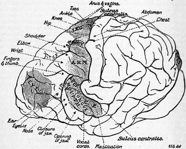
Fig. 24.—Diagram of the Topography of the Main Groups of Foci in the Motor Field of Chimpanzee.
The Precentral or Motor Region of the Cortex.—The application to it of electric currents excites movements in the skeletal muscles. The movements occur in the half of the body of the side crossed from that of the hemisphere excited. The “motor representation,” as it is termed, is in the cortex better described as a representation of definite actions than of particular muscles. The actions “represented” in the top part of the gyrus, namely next the great longitudinal fissure, move the leg; those in the lowest part of the gyrus belong to the tongue and mouth. The topical distribution along the length of the gyrus may be described in a general way as following a sequence resembling that of the motor representation in the spinal cord, the top of the gyrus being taken as corresponding with the caudal end of the spinal cord. The sequence as the gyrus is followed downwards runs: perineum, foot, knee, hip, abdomen, chest, shoulder, elbow, wrist, hand, eyelids and ear, nose, mouth and tongue. The nature of the movement is very fairly constant for separate points of this motor cortex as observed both in the same and in similar experiments. Thus flexion of the arm will be excitable from one set of points, and extension of the arm from another set of points; opening of the jaw from one set and closure from another, and so on. These various movements if excited strongly tend to have characters like those of the movements seen in an epileptic convulsion. Strong stimulation excites in fact a convulsion like that of epilepsy, beginning with the movement usual for the point stimulated and spreading so as to assume the proportions of a convulsion affecting the entire skeletal musculature of one half or even of the whole body. The resemblance to an epileptic seizure is the closer because the movement before it subsides becomes clonic (rhythmic) as in epilepsy. The determination of the exact spots of cortex in which are represented the various movements of the body has served a useful practical purpose in indicating the particular places in the cortex which are the seat of disease. These the physician can localize more exactly by reason of this knowledge. Hence the surgeon, if the nature of the disease is such as can be dealt with by surgical means, can without unnecessarily damaging the skull and brain, proceed directly to the point which is the seat of the mischief.
The motor representation of certain parts of the body is much more liberal than is that of others. There is little correspondence between the mere mass of musculature involved and the area of the cortex devoted to its representation. Variety of movement rather than force or energy of movement seems to demand extent of cortex. The cortical area for the thumb is larger than those for the whole abdomen and chest combined. The cortical area for the tongue is larger than that for the neck. Different movements of one and the same part are very unequally represented in the cortex. Thus, flexion of the leg is more extensively represented than is extension, opening of the jaw has a much larger cortical area than has closure of the jaws. It is interesting that certain agents, for instance strychnine, and the poison of the bacilli which cause the disease known as tetanus or lock-jaw, upset this normal topography, and replace in the cortex flexion of the limb by extension of the limb, and opening of the jaw by closure of the jaw. There is, however, no evidence that they do this by changing in any way the cortical mechanisms themselves. It is more likely that their action is confined to the lower centres, bulbar and spinal, upon which the discharge excited from the cortex plays. The change thus induced in the movement excited by the cortex does, however, show that the point of cortex which causes for instance opening of the mouth is connected with the motor nerves to the closing muscles as well as with those of the opening muscles. This is an item of evidence that the “centres” of the cortex are connected with the motor nerves of antagonistic muscles in such a way that when the “centre” excites one set of the muscles to contract, it simultaneously under normal circumstances causes inhibition of the motor neurones of the opposed set of muscles (reciprocal innervation). In the great majority of movements excited from the motor cortex of a single hemisphere of the cerebrum, the movement evoked is confined to one side of the body, namely to that opposite to the hemisphere stimulated. There are, however, important exceptions to this. Thus, adduction of both vocal cords is excited from the cortex of either hemisphere. The movement of closure of the eyelids is usually bilateral, unless the stimulation be very weak; then the movement is of the eyelids of the opposite side only. The same holds true for the movements of the jaw. It, therefore, seems clear that with many movements which are usually bilaterally performed in ordinary life, such as opening of the jaw, blinking, &c., the symmetrical areas of the motor regions of both hemispheres are simultaneously in action.
In regard to all these movements elicitable by artificial stimuli from the motor cortex it is obvious that were there clearer evidence that the pallial region from which they are elicitable is fairly directly connected with corticopetal paths subserving cutaneous sensation or “muscular sense,” the movements might be regarded as falling into the category of higher reflexes connected with the organs of touch, muscular sense, &c., just as the movements of the eyeball excitable from the visual cortex may be regarded as higher reflexes connected with vision. The evidence of the connexion of the reactions of the motor cortex with cutaneous and muscular senses appears, however, scarcely sufficient to countenance at present this otherwise plausible view, which has on general grounds much to commend it.
It is remarkable that movements of the eyeball itself, i.e. apart from movement of the lids, are not in the category of movements elicitable from the precentral gyrus, the “motor” cortex. They are found represented in a region farther forward, namely in front of the precentral gyrus altogether, and occupying a scattered set of points in the direction frontal from the areas for movements of arm and face. This frontal area yields on excitation conjugate movements of both eyeballs extremely like if not exactly similar to those yielded by excitation of the occipital (visual) region of the cortex. It is supposed by some that this frontal area yielding eye-movements has its function in this respect based upon afferent conductors from other parts of the eyeball than the retina, for instance upon kinaesthetic (Bastian) impressions or upon sensual impressions derived from the cornea and the coats of the eyeball including the ciliary and iris muscles. The ocular muscles are certainly a source of centripetal impulses, but their connexion with the cortex is not clear as to either their nature or their seat. The question seems for the present to allow no clearer answer. It is certain, however, that the frontal area of eye movements has corticofugal paths descending from it to the lower motor centres of the eyeballs quite independent of those descending from the occipital (visual) area of eye-movements. Further, it seems clear that in many animals there is another cortical region, a third region, the region which we saw above might be considered auditory, where movements of the eyeball similar to those elicitable in the occipital and frontal cortex can be provoked. A. Tschermak is inclined to give the eyeball movements of the frontal region the significance of reflex movements which carry the visual field in various directions in answer to demands made by sensory data derived from touch, &c., as for instance from the hand. The movements of the eyeballs elicitable from the occipital region of the cortex he regards as probably concerned with directing the gaze toward something seen, for instance, in the peripheral field of vision. The occipital movement would, therefore, be excited through the retina, and would result in bringing the yellow spot region of the retinae of both eyes to bear upon the object. This view has much to justify it. The movements of the eyeballs excited from the cortex of the auditory region would in a similar way be explicable as bringing the gaze to bear upon a direction in which a sound had been located, auditory initiation replacing the visual and tactual of the occipital and the frontal regions respectively.
Turning from these still speculative matters to others less suggestive but of actual ascertainment, we find that the motor nature of the precentral cortex as ascertained by electric stimuli is further certified by the occurrence of disturbance and impairment of motor power and adjustment following destruction of that region of the cortex. The movements which such a part as a limb executes are of course manifold in purpose. The hind limb of a dog is used for standing, for stepping, for scratching, for squatting, and, where a dog, for instance, has been trained to stand or walk on its hind legs alone, for skilled acts requiring a special training for their acquisition. It is found that when the motor area of the brain has been destroyed, the limb is at first paralysed for all these movements, but after a time the limb recovers the ability to execute some of them, though not all. The scratching movement suffers little, and rapid improvement after cerebral injury soon effaces the impairment, at first somewhat pronounced, in the use of the limb for walking, running, &c., and ordinary movements of progression. Even when both hemispheres have been destroyed the dog can still stand and walk and run. Destruction of the motor region of the cortex renders the fore limbs of the dog unable to execute such skilled movements as the steadying of a bone for gnawing or the trained act of offering the paw in answer to the command of the master. Skilled acts of the limb, apart from conjoined movements in which it, together with all the other limbs, takes part, assume of course a larger share of the office of the limb in the Primates than in the dog; and this is especially true for the fore limb. It is when the fore-foot becomes a hand that opportunity is given for its more skilled individual use and for its training in movements as a tool, or for the handling of tools and weapons. It is these movements which suffer most heavily and for the longest period after injury of the motor region of the cortex. Hence the disablement ensuing upon injury to the cortex would be expected to be most apparent in the Primates; and it is so, and most of all in Man. Further, in Man there ensues a condition called “contracture,” which is not so apparent or frequent a result in other animals,—indeed, does not occur at all in other animals except the monkey. In contracture the muscles of the paretic limb are not flaccid, as they are usually in paralysis, but they are tense and the limb is more or less rigidly fixed by them in a certain position, usually one of flexion at elbow and wrist. This condition does not occur at first, but gradually supervenes in the course of a number of weeks. In Man the destruction of the motor area of the cortex cripples the limb even for the part it should play in the combined limb movements of walking, &c., and cripples it to an extent markedly contrasting with the slight disturbances seen in the lower mammals, e.g. the dog.
As regards the recovery of motor power after lesions of the motor cortex, two processes seem at work which are termed respectively restitution and compensation. By the former is understood the recovery obtained when a part of a “centre” is destroyed, and the rest of the centre, although thrown out of function at first, recovers and supplements the deficiency later. An example of restitution would be the recovery from temporary hemianopia caused by a small injury in one occipital lobe. By compensation is understood the improvement of an impaired nervous function, traceable to other centres different from those destroyed supplying means to compass the reaction originally dependent on the centres subsequently destroyed. Instances of such compensation are the recovery of taxis for equilibrium subsequent to destruction of the labyrinth of the ear, where the recovery is traceable to assistance obtained through the eye. It will be noted that these instances of recovery by restitution and by compensation respectively are taken, from cases of injury inflicted on receptive rather than on motor centres. It is doubtful how far they really apply to the undoubted improvement that does within certain limits progress and succeed in partially effacing the paresis immediately consequent on lesions of the motor area. It has to be remembered that in all cases of traumatic injury to the nervous system, especially where the trauma implicates the central nervous organ, the first effects and impairment of function resulting are due to a mixed cause, namely on the one hand the mechanical rupture of conducting paths actually broken by solution of their continuity, and on the other hand the temporary interruption of conducting paths by “shock.” Shock effects are not permanent: they pass off. They are supposed to be due to a change at the synapses connecting neurone with neurone in the grey matter. They amount in effect to a long-lasting and gradually subsiding inhibition.
For diseases of the brain see Neuropathology, Insanity, Skull (Surgery), &c.
(C. S. S.)
1 The literature of the pineal region is enormous. Studnicka (in Oppels Vergleichende mikrosk. Anat. Teile 4-5, 1904, 1905) gives 285 references. The present conception of the generalized arrangement is: (α) A single glandular median organ from the fore-brain called the paraphysis. (β) A pouch of the ependymal roof of the ventricle called the dorsal sac. (γ) A right and left epiphysis, one of which may be wholly or partially suppressed. These may change their position to anterior and posterior in some animals.
BRAINERD, DAVID (1718-1747), American missionary among the Indians, was born at Haddam, Connecticut, on the 20th of April 1718. He was orphaned at fourteen, and studied for nearly three years (1739-1742) at Yale. He then prepared for the ministry, being licensed to preach in 1742, and early in 1743 decided to devote himself to missionary work among the Indians. Supported by the Scottish “Society for Promoting Christian Knowledge,” he worked first at Kaunaumeek, an Indian settlement about 20 m. from Stockbridge, Massachusetts, and subsequently, until his death, among the Delaware Indians in Pennsylvania (near Easton) and New Jersey (near Cranbury). His heroic and self-denying labours, both for the spiritual and for the temporal welfare of the Indians, wore out a naturally feeble constitution, and on the 19th of October 1747 he died at the house of his friend, Jonathan Edwards, in Northampton, Massachusetts.
His Journal was published in two parts in 1746 by the Scottish Society for Promoting Christian Knowledge; and in 1749, at Boston, Jonathan Edwards published An Account of the Life of the Late Rev. David Brainerd, chiefly taken from his own Diary and other Private Writings, which has become a missionary classic. A new edition, with the Journal and Brainerd’s letters embodied, was published by Sereno E. Dwight at New Haven in 1822; and in 1884 was published what is substantially another edition, The Memoirs of David Brainerd, edited by James M. Sherwood.
BRAINERD, a city and the county-seat of Crow Wing county, Minnesota, U.S.A., on the E. bank of the Mississippi river, about 127 m. N.W. of Minneapolis. Pop. (1890) 5703; (1900) 7524, of whom 2193 were foreign-born; (1905) 8133; (1910) 8526. It is served by the Minnesota & International and the Northern Pacific railways. The latter maintains here large car and repair shops, and a sanatorium for its employees. There are also the Sisters of St Joseph hospital, a county court house, a public library and a Y.M.C.A. building. A dam across the Mississippi provides water power (about 60,000 H.P.) which is utilized extensively for manufacturing purposes. Lumbering is an important industry, and there are saw mills and planing mills, and an extensive creosote plant for treating railway ties and timber. There are also flour mills, paper and pulp mills, cigar factories, a brewery, a large foundry and a grain elevator. In 1906 large quantities of iron ore were discovered in the vicinity, the new range, the Cuyuna, running through the city from north-east to south-west. Brainerd, named in honour of David Brainerd, was settled in 1870, and chartered as a city in 1883.
BRAINTREE, a market town in the Maldon parliamentary division of Essex, England; 45 m. N.E. of London by a branch line from Witham of the Great Eastern railway. Pop. of urban district, 5330. The parish church of St Michael is a fine edifice of Early English work with later additions. A corn exchange, mechanics’ institute and public hall may also be mentioned. The bishops of London had formerly a palace in the town, but there are no remains of the building. The manufactures of silk and crape have superseded that of woollen cloth, which was introduced by the Flemings who fled to England to escape the persecution of the duke of Alva. Matting and brushes are also made. On the north lies the large village of BOCKING, with the Perpendicular parish church of St Mary, similar industries, and a population of 3347.
BRAINTREE, a township of Norfolk county, Massachusetts, U.S.A., on the Monatiquot river about 10 m. S. of Boston. Pop. (1890) 4848; (1900) 598l, including 1250 foreign-born; (1905, state census) 6879; (1910) 8066. The New York, New Haven & Hartford railway crosses the town and has stations at its villages of Braintree, South Braintree and East Braintree, which are also served by suburban electric railways. In South Braintree are the Thayer Academy (co-educational; opened 1877) and the Thayer public library, both founded by and named in honour of General Sylvanus Thayer (1785-1872), a well-known military engineer born in Braintree, who was superintendent of the United States Military Academy in 1817-1833 and has been called the “father of West Point.” There are large shoe factories and other manufactories. Bog iron was early found in Braintree, and iron-works, among the first in America, were established here in 1644. Braintree was first incorporated in 1640 from land belonging to Boston and called Mount Wollaston, and was named from the town in England. At Merry Mount, in that part of Braintree which is now Quincy, a settlement was established by Thomas Morton in 1625, but the gay life of the settlers and their selling rum and firearms to the Indians greatly offended the Pilgrims of Plymouth, who in 1627 arrested Morton; soon afterward Governor John Endecott of Massachusetts Bay visited Merry Mount, rebuked the inhabitants and cut down their Maypole. Later the place was abandoned, and in 1634 a Puritan settlement was made here. In 1708 the town was divided into the North Precinct and the South Precinct, and it was in the former, now Quincy, that John Adams, John Hancock and John Quincy Adams were born. Quincy was separated from Braintree in 1792 (there were further additions to Quincy from Braintree in 1856), and Randolph in 1793.
See D.M. Wilson, Quincy, Old Braintree and Merry Mount (Boston, 1906); C.F. Adams, Jr., Three Episodes of Massachusetts History (Boston, 1892 and 1896); W.S. Pattee, History of Old Braintree and Quincy (Quincy, 1878).
BRAKE, a town of Germany, in the grand duchy of Oldenburg, on the left bank of the Weser, about halfway between Bremen and the mouth of the river. Pop. 5000. It was for centuries the port of Bremen; and though, since the founding of Bremerhaven, it no longer possesses a monopoly of the river traffic as before, it still continues to flourish. Large docks have been constructed, and the place has a considerable import trade in English coal. Shipbuilding and weaving are carried on to some extent.
Brake in Oldenburg must be distinguished from the village of the same name in the principality of Lippe, known as Brake bei Limgo, which gave its name to the cadet line of the counts of Lippe-Brake (1621-1709).
BRAKE. (1) A term for rough-tangled undergrowth, connected, according to the New English Dictionary, with “break,” to separate. The “brake-fern” (Pteris aquilina) is the common “bracken,” and is a shortened form of that northern Eng. word, derived from a Scand. word for “fern” (cf. Swed. bräken), though often confused with “brake,” undergrowth. (2) A term applied to many implements and mechanical and other appliances, often spelled “break.” Here there are probably several words, difficult to separate in origin, connected either with “break,” to separate, and its derived meanings, or with the Fr. braquer (appearing in such expressions as braquer un canon, to turn or point a gun), from O. Fr. brac, modern bras, an arm, Lat. bracchium. The word is thus used of a toothed instrument for separating the fibre of flax and hemp; of the “break-rolls” employed in flour manufacture; of a heavy wheeled vehicle used for “breaking in” horses, and hence of a large carriage of the wagonette type; of an arm or lever, and so of the winch of a crossbow and of a pump handle, cf. “brake-pump”; of a curb or bridle for a horse; and of a mechanical appliance for checking the speed of moving vehicles, &c. It is noteworthy that the two last meanings are also possessed by the Fr. frein and the Ger. Bremse.
Brakes, in engineering, are instruments by means of which mechanical energy may be expended in overcoming friction. They are used for two main classes of purpose: (1) to limit or decrease the velocity of a moving body, or to bring it completely to rest; and (2) to measure directly the amount of frictional resistance between two bodies, or indirectly the amount of energy given out by a body or bodies in motion. Machines in which brakes are employed for purposes of the second class are commonly known as dynamometers (q.v.). The other class is exemplified in the brakes used on wheeled vehicles and on cranes, lifts, &c. Here a body, or system of bodies, originally at rest, has been set in motion and has received acceleration up to a certain velocity, the work which has been done in that acceleration being stored up as “actual energy” in the body itself. Before the body can be brought to rest it must part with this energy, expending it in overcoming some external resistance. If the energy be great in proportion to the usual resistance tending to stop the body, the motion will continue for a long time, or through a long distance, before the energy has been completely expended and the body brought to rest. But in certain cases considerations of safety or convenience require that this time or distance be greatly shortened, and this is done by artificially increasing the external resistance for the time being, by means of a brake.
A simple method of obtaining this increased resistance is by pressing a block or shoe of metal or wood against the rim of a moving wheel, or by tightening a flexible strap or band on a rotating pulley or drum. In wheeled road vehicles, a wheel may be prevented from rotating by a chain passed through its spokes and attached to the body of the vehicle, when the resistance is increased by the substitution of a rubbing for a rolling action; or the same effect may be produced by fixing a slipper or skid under the wheel. Other forms of brake depend, not on the friction between two solid bodies, but on the frictional resistance of a fluid, as in “fan” and “pump” brakes. Thus the motion of revolving blades may be opposed by the resistance of the air or of a liquid in which they are made to work, or the motion of a plunger fitting tightly in a cylinder filled with a fluid may be checked by the fluid being prevented from escape except through a narrow orifice. The fly used to regulate the speed of the striking train in a clock is an example of a fan brake, while a pump brake is utilized for controlling the recoil of guns and in the hydraulic buffers sometimes fitted at terminal railway stations to stop trains that enter at excessive speed. On electric tramcars a braking effect is sometimes obtained by arranging the connexions of the motors so that they act as generators driven by the moving car. In this way a counter-torque is exerted on the axles. The current produced is expended by some means, as by being made to operate some frictional braking device, or to magnetize iron shoes carried on the car just over, but clear of, the running rails, to which they are then magnetically attracted (see Traction).
The simplest way of applying a brake is by muscular force, exerted through a hand or foot lever or through a screw, by which the brake block is pressed against the rim of the wheel or the band brake tightened on its drum. This method is sufficient in the case of most road vehicles, and is largely used on railway vehicles. But the power thus available is limited, and becomes inadequate for heavy vehicles moving at high speeds. Moreover, on a train consisting of a number of vehicles, the hand brakes on each of which are independent of all others, either a brakesman must be carried on each, or a number of the brakes must be left unused, with consequent loss of stopping power; while even if there is a brakesman on every vehicle it is impossible to secure that all the brakes throughout the train are applied with the promptness that is necessary in case of emergency.
Considerations of this sort led to the development of power brakes for railway trains. Of these there are five main classes:—
(1) Mechanical brakes, worked by springs, friction wheels on the axle, chains wound on drums, or other mechanical devices, or by the force produced when, by reason of a sudden checking of the speed of the locomotive, the momentum Railway power brakes. of the cars causes pressure on the draw-bars or buffing devices. (2) Hydraulic brakes, worked by means of water forced through pipes into proper mechanism for transmitting its force to the brake-shoes. (3) Electric brakes. (4) Air and vacuum brakes, worked by compressed air or by air at atmospheric pressure operating on a vacuum. (5) Brakes worked by steam or water from the boiler of the engine, operating by means of a cylinder; the use of these is generally limited to the locomotive. Of this kind is the counter-pressure or water brake of L. le Chatelier. If the valve gear of a locomotive in motion be reversed and the steam regulator be left open, the cylinders act as compressors, pumping air from the exhaust pipe into the boiler against the steam pressure. A retarding effect is thus exercised, but at the cost of certain inconveniences due to the passage of hot air and cinders from the smoke box through the cylinders. To remedy these, le Chatelier arranged that a jet of hot water from the boiler should be delivered into the exhaust pipe, so that steam and not the hot flue gases should be pumped back.
Power brakes may be either continuous or independent—continuous if connected throughout the train and with the locomotive by pipes, wires, &c., as the compressed air, vacuum and electric brakes; independent if not so connected, as the buffer-brakes and hand-brakes. Continuous brakes may be divided into two other great classes—automatic and non-automatic. The former are so arranged that they are applied automatically on all the coaches of the train if any important part of the apparatus is broken, or the couplings between cars are ruptured; in an emergency they can be put on by the guard, or (in some cases) by a passenger. Non-automatic brakes can be applied only by the person (usually the engine-driver) to whom the management of them is given; they may become inoperative on all the coaches, and always on those which have become detached, if a coupling or other important and generally essential part is broken. Many mechanical and several hydraulic and electrical continuous brakes have been invented and tried; but experience has shown them so inadequate in practice that they have all practically disappeared, leaving the field to the air and the vacuum brakes. At first these were non-automatic, but in 1872 the automatic air-brake was invented by George Westinghouse, and the automatic vacuum-brake was developed a few years later.
Those respects in which non-automatic brakes are inadequate will be understood from the following summary of the requirements most important in a train-braking apparatus: (1) It must be capable of application to every wheel throughout the train. (2) It must be so prompt in action that the shortest possible time shall elapse between its first application and the moment when the full power can be exerted throughout the train. (3) It must be capable of being applied by the engine-driver or by any of the officials in charge of the train, either in concert or independently. (4) The motion of the train must be arrested in the shortest possible distance. (5) The failure of a vital part must declare itself by causing the brake to be applied and to remain applied until the cause of failure is removed. (6) The breaking of the train in two or more parts must cause immediate automatic application of the brakes on all the coaches. (7) When used in ordinary service stops it must be capable of gradual and uniform application (followed, if necessary, by a full emergency application at any part of the service application) and of prompt release under all conditions of application. (8) It must be simple in operation and construction, not liable to derangement, and inexpensive in maintenance.
The Westinghouse non-automatic or “straight” air-brake, patented in 1869, consists in its simplest form of a direct-acting, steam-driven air-pump, carried on the locomotive, which forces compressed air into a reservoir, usually placed Simple air-brake. under the foot-plate of the locomotive. From this reservoir a pipe is led through the engine cab, where it is fitted with a three-way cock, to the rear of the locomotive tender, where it terminates in a flexible hose, on the end of which is a coupling. The coaches are furnished with a similar pipe, having hose and coupling at each end, which communicates with one end of a cylinder containing a piston, to the rod of which the brake-rods and levers are connected. The application of the brakes is effected by the engine-driver turning the three-way cock, so that compressed air flows through the pipe and, acting against one side of the brake-cylinder piston, applies the brake-shoes to the wheels by the movement of this piston and the rods and levers connected to it. To release the brakes the three-way cock is turned to cut off communication between the main reservoir and the train-pipe, and to open a port permitting the escape of the compressed air in the train-pipe and brake-cylinders. This brake was soon found defective and inadequate in many ways. An appreciable time was required for the air to flow through the pipes from the locomotive to the car-cylinders, and this time increased quickly with the length of the trains. Still more objectionable, however, was the fact that on detached coaches the air-brakes could not be applied, the result being sometimes serious collisions between the front and rear portions of the train.
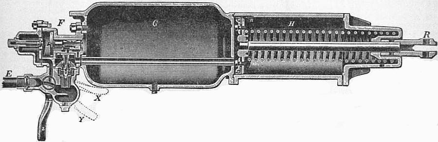
Fig. 1.—Westinghouse Air-Brake. Section through Triple-Valve and Brake-Cylinder.
In the Westinghouse “ordinary” automatic air-brake a main air reservoir on the engine is kept charged with compressed air at 80 ℔ per sq. in. by means of the steam-pump, which may be controlled by an automatic governor. On electric Automatic air-brake. railways a pump, driven by an electric motor, is generally employed; but occasionally, on trains which run short distances, no pump is carried, the main reservoir being charged at the terminal points with sufficient compressed air for the journey. Conveniently placed to the driver’s hand is the driver’s valve, by means of which he controls the flow of air from the main reservoir to the train-pipe, or from the train-pipe to the atmosphere. A reducing-valve is attached to the driver’s valve, and in the normal or running position of the latter reduces the pressure of the air flowing from the main reservoir to the train-pipe by 10 or 15 ℔ per sq. in. From the engine a train-pipe runs the whole length of the train, being rendered continuous between each vehicle and between the engine and the rest of the train by flexible hose couplings. Each vehicle is provided with a brake-cylinder H (fig. 1), containing a piston, the movement of which applies the brake blocks to the wheels, an “auxiliary air-reservoir” G, and an automatic “triple-valve” F. The auxiliary reservoir receives compressed air from the train-pipe and stores it for use in the brake-cylinder of its own vehicle, and both the auxiliary reservoir and the triple-valve are connected directly or indirectly with the train-pipe through the pipe E. The automatic action of the brake is due to the construction of the triple-valve, the principal parts of which are a piston and slide-valve, so arranged that the air in the auxiliary reservoir acts at all times on the side of the piston to which the slide-valve is attached, while the air in the train-pipe exerts its pressure on the opposite side. So long as the brakes are not in operation, the pressures in the train-pipe, triple-valve and auxiliary reservoir are all equal, and there is no compressed air in the brake-cylinder. But when, in order to apply the brake, the driver discharges air from the train-pipe, this equilibrium is destroyed, and the greater pressure in the auxiliary reservoir forces the triple-valve to a position which allows air from the auxiliary reservoir to pass directly into the brake-cylinder. This air forces out the piston of the brake-cylinder and applies the brakes, connexion being made with the brake-rigging at R. The purpose of the small groove n which establishes communication between the two sides of the piston when the brakes are off, is to prevent their unintended application through slight leakage from the train-pipe. To release the brakes, the driver, by moving the handle of his valve to the release position, admits air from the main reservoir to the train-pipe, the pressure in which thus becomes greater than that in the auxiliary reservoir; the piston and slide-valve of the triple-valve are thereby forced back to their normal position, the compressed air in the brake-cylinder is discharged, and the piston is brought back by the coiled spring, thus releasing the brakes. At the same time the auxiliary reservoir is recharged.
With this “ordinary” brake, since an appreciable time is required for the reduction of pressure to travel along the train-pipe from the engine, the brakes are applied sensibly sooner at the front than at the end of the train, and with long trains this Quick-acting air-brake. difference in the time of application becomes a matter of importance. The “quick-acting” brake was introduced to remedy this defect. For it the triple valve is provided with a supplementary mechanism, which, when the air pressure in the train-pipe is suddenly or violently reduced, opens a passage whereby air from the train-pipe is permitted to enter the brake-cylinder directly. The result is twofold: not only is the pressure from the auxiliary reservoir acting in the brake-cylinder reinforced by the pressure in the train-pipe, but the pressure in the train-pipe is reduced locally in every vehicle in extremely rapid succession instead of at the engine only, and in consequence all the brakes are applied almost simultaneously throughout the train. The same effect is produced should the train break in two, or a hose or any part of the train-pipe burst; but during ordinary or “service” stops the triple-valve acts exactly as in the ordinary brake, the quick-acting portion, that is, the vertical piston and valve seen in fig. 1, not coming into operation. When the handle Z is turned to the position X the quick-acting mechanism is rendered inoperative, and when it is at Y the brake on the vehicle concerned is wholly cut out of action.
A further improvement introduced in the Westinghouse brake in 1906 was designed to give quick action for service as well as emergency stops. In this the triple-valve is substantially the same as in the ordinary brake. The additional mechanism of the quick-acting portion is dispensed with, but instead, a small chamber, normally containing air at atmospheric pressure, is provided on each vehicle, and is so arranged that it is put into communication with the train-pipe by the first movement of the triple-valve. As soon, therefore, as the driver, by lowering the pressure in the train-pipe, causes the triple-valve in the foremost vehicle of the train to operate, a certain quantity of air rushes out of the train-pipe into the small chamber; a further local reduction in the pressure of the train-pipe in that vehicle is thereby effected, and this almost instantaneously actuates the triple-valve of the succeeding vehicle, and so on throughout the train. In this way, on a train 1800 ft. long, consisting of sixty 30-ft. vehicles, the brake-blocks may be applied, with equal force, on the last vehicle about 2½ seconds later than on the first.
Brake-blocks can be applied, without skidding the wheels, with greater pressure at high speeds than at low. Advantage is taken of this fact in the design of the Westinghouse “high-speed” brake, invented in 1894, which consists of High-speed air-brake. attachments enabling the pressure in the train-pipe and reservoirs to be increased at the will of the driver. The increased pressure acting in the brake-cylinder increases in the same proportion the pressure of the brake-shoes against the wheels. Attached to the brake cylinder is a valve for automatically reducing the pressure therein proportionately to the reduction in speed, until the maximum pressure under which the brakes are operated in making ordinary stops is reached, when this valve closes and the maximum safe pressure for operating the brakes at ordinary speeds is retained until a stop is made.
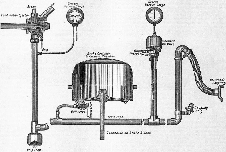
Fig. 2—Automatic Vacuum-Brake, showing its general arrangement.
In the automatic vacuum-brake, the exhausting apparatus generally consists of a combined large and small ejector (a form of jet-pump) worked by steam and under the control of the driver, though sometimes a mechanical air-pump, driven Automatic Vacuum-Brake. from the crosshead of the locomotive, is substituted for the small ejector. These ejectors, of which the small one is at work continuously while the large one is only employed when it is necessary to create vacuum quickly, e.g. to take off the brakes after a short stop, produce in the train-pipe a vacuum equal to about 20 in. of mercury, or in other words reduce the pressure within it to about one-third of an atmosphere. The train-pipe extends the whole length of the train and communicates under each vehicle with a cylinder, to the piston of which, by suitable rods and levers, the brake-shoes are connected. The communication between the train-pipe and the cylinder is controlled by a ball-valve, one form of which is shown in fig. 2. The release-valve is for the purpose of withdrawing the ball from its seat when it is necessary to take off the brakes by hand; it is made air-tight by a small diaphragm, the pressure of which, when there is vacuum in the pipe, pulls in the spindle and allows the ball to fall freely into its seat. When air is exhausted through the train-pipe it travels out from below the piston direct, and from above it past the ball, which is thus forced off its seat, to roll back again when the exhaustion is complete. In this state of affairs the piston is held in equilibrium and the brake-blocks are free of the wheels. To apply them, air is admitted to the train-pipe, either purposely by the guard or driver, or accidentally by the rupture of the train-pipe or coupling-hose between the vehicles. The air passes to the lower side of the piston, but is prevented from gaining access to the upper side by the ball-valve which blocks the passage; hence the pressure becomes different on the two sides of the piston, which in consequence is forced upwards and thus applies the brakes. They are released by the re-establishment of equilibrium (by the use of the large ejector if necessary); when this is done the piston falls and the brakes drop off. The general arrangement of the apparatus is shown in fig. 2. To render the application of the brakes nearly simultaneous throughout a long train, the valve in the guard’s van is arranged to open automatically when the driver suddenly lets in air to the train-pipe. This valve has a small hole through its stem, and is secured at the top by a diaphragm to a small dome-like chamber, which is exhausted when a vacuum is created in the train-pipe. A gradual application destroys the vacuum in the chamber as quickly as in the pipe and the diaphragm remains unmoved; but with a sudden one the vacuum below the valve is destroyed more quickly, and with the difference of pressure the diaphragm lifts the valve and admits air. A rapid-acting valve (fig. 3) is sometimes interposed between the train-pipe and the cylinder on each vehicle. In the normal or running position, a vacuum is maintained below the valve A and above the diaphragm B, while the chamber below B and above A is at atmospheric pressure. For an emergency application of the brake, air is suddenly admitted to the train-pipe and thus to the lower side of A, and the pressure acting on the under side of B is sufficient to cause it to lift the valve A, and to admit air from the atmosphere, both to the brake-cylinder and the train-pipe, through the clappet-valve D, which also rises because of the difference of pressure on its two sides. In a graduated application, neither D nor A rises from its seat, but air from the train-pipe finds access to the brake-cylinder by passing around the peg C, which is so proportioned as to allow the necessary amount of air to enter the brake-cylinder, and so obtain simultaneous action of the brake throughout the train. When the handle E is turned so as to prevent the clappet D from rising, the rapid action is cut out and the brake acts as an ordinary vacuum automatic brake. A modification of the device for obtaining accelerated action, described above in connexion with the Westinghouse brake, is also applicable. Accelerating chambers, again containing air at atmospheric pressure, are provided on each vehicle and are connected with the train-pipe by valves which open as the vacuum in the latter begins to decrease with the operation of the driver’s valve. The air thus admitted into the train-pipe effects a still further local reduction of the vacuum, which is sufficient to actuate the accelerating valve of each next succeeding vehicle and is thus rapidly propagated throughout the train.