VOLUME III SLICE VII Bible to Bisectrix (continued 11)
Reading edition
3. Its tendency to undergo cyclical changes.
In the ordinary course of nature, all living matter proceeds from pre-existing living matter, a portion of the latter being detached and acquiring an independent existence. The new form takes on the characters of that from which it arose; exhibits the same power of propagating itself by means of an offshoot; and, sooner or later, like its predecessor, ceases to live, and is resolved into more highly oxidated compounds of its elements.
Thus an individual living body is not only constantly changing its substance, but its size and form are undergoing continual modifications, the end of which is the death and decay of that individual; the continuation of the kind being secured by the detachment of portions which tend to run through the same cycle of forms as the parent. No forms of matter which are either not living, or have not been derived from living matter, exhibit these three properties, nor any approach to the remarkable phenomena defined under the second and third heads. But in addition to these distinctive characters, living matter has some other peculiarities, the chief of which are the dependence of all its activities upon moisture and upon heat, within a limited range of temperature, and the fact that it usually possesses a certain structure or organization.
As has been said, a large proportion of water enters into the composition of all living matter; a certain amount of drying arrests vital activity, and the complete abstraction of this water is absolutely incompatible with either Life conditioned by moisture. actual or potential life. But many of the simpler forms of life may undergo desiccation to such an extent as to arrest their vital manifestations and convert them into the semblance of not-living matter, and yet remain potentially alive. That is to say, on being duly moistened they return to life again. And this revivification may take place after months, or even years, of arrested life.
The properties of living matter are intimately related to temperature. Not only does exposure to heat sufficient to coagulate protein matter destroy life, by demolishing the molecular structure upon which life depends; but Life conditioned by temperature. all vital activity, all phenomena of nutritive growth, movement and reproduction are possible only between certain limits of temperature. These limits may be set down as from a little above the freezing point of water to a little below the boiling point It is to be noted, however, that these limits apply to the living matter itself, and many of the apparent exceptions are due to cases in which the living matter is enclosed in protective wrappings capable of resisting heat and cold. In many low organisms, such as the spores of bacteria, the thick, non-conducting wall may preserve the living protoplasm from subjection to external temperatures below freezing point, or above boiling point, but all the evidence goes to show that applications of such cold or heat, if prolonged or arranged so as to penetrate to the living matter, destroy life. In warm-blooded animals, such as birds and mammals, protective mechanisms for the regulation of temperature enable them to endure exposure to extreme heat or cold, but in such cases the actually living cells do not appreciably rise or fall in temperature. A variation of a very few degrees in the blood itself produces death.
Recent investigations point to the conclusion that the immediate cause of the arrest of vitality, in the first place, and of its destruction, in the second, is the coagulation of certain substances in the protoplasm, and that the latter contains various coagulable matters, which solidify at different temperatures. And it remains to be seen, how far the death of any form of living matter, at a given temperature, depends on the destruction of its fundamental substance at that heat, and how far death is brought about by the coagulation of merely accessory compounds.
It may be safely said of all those living things which are large enough to enable us to trust the evidence of microscopes, that they are heterogeneous optically, and that their different parts, and especially the surface layer, as Life and organization. contrasted with the interior, differ physically and chemically; while, in most living things, mere heterogeneity is exchanged for a definite structure, whereby the body is distinguished into visibly different parts, which possess different powers or functions. Living things which present this visible structure are said to be organized; and so widely does organization obtain among living beings, that organized and living are not unfrequently used as if they were terms of co-extensive applicability. This, however, is not exactly accurate, if it be thereby implied that all living things have a visible organization, as there are numerous forms of living matter of which it cannot properly be said that they possess either a definite structure or permanently specialized organs: though, doubtless, the simplest particle of living matter must possess a highly complex molecular structure, which is far beyond the reach of vision.
The broad distinctions which, as a matter of fact, exist between every known form of living substance and every other component of the material world, justify the separation of the biological sciences from all others. But it must not be supposed that the differences between living and not-living matter are such as to justify the assumption that the forces at work in the one are different from those which are to be met with in the other. Considered apart from the phenomena of consciousness, the phenomena of life are all dependent upon the working of the same physical and chemical forces as those which are active in the rest of the world. It may be convenient to use the terms “vitality” and “vital force” to denote the causes of certain great groups of natural operations, as we employ the names of “electricity” and “electrical force” to denote others; but it ceases to be proper to do so, if such a name implies the absurd assumption that “electricity” and “vitality” are entities playing the part of efficient causes of electrical or vital phenomena. A mass of living protoplasm is simply a molecular machine of great complexity, the total results of the working of which, or its vital phenomena, depend—on the one hand, upon its construction, and, on the other, upon the energy supplied to it; and to speak of “vitality” as anything but the name of a series of operations is as if one should talk of the “horologity” of a clock.
Living matter, or protoplasm and the products of its metamorphosis, Classification of the phenomena of life. may be regarded under four aspects:—
1. It has a certain external and internal form, the latter being more usually called structure;
2. It occupies a certain position in space and in time;
3. It is the subject of the operation of certain forces in virtue of which it undergoes internal changes, modifies external objects, and is modified by them; and
4. Its form, place and powers are the effects of certain causes.
In correspondence with these four aspects of its subject, biology is logically divisible into four chief subdivisions—I. Morphology; II. Distribution; III. Physiology; IV. Aetiology.
Various accidental circumstances, however, have brought it about that the actual distribution of scientific work does not correspond with the logical subdivisions of biology. The difference in technical methods and the historical evolution of teaching posts (for in all civilized countries the progress of biological knowledge has been very closely associated with the existence of institutions for the diffusion of knowledge and for professional education) have been the chief contributory causes to this practical confusion. Details of the morphology of plants will be found in the articles relating to the chief groups of plants, those of animals in the corresponding articles on groups of animals, while the classification of animals adopted in this work will be found in the article Zoology. Distribution is treated of under Zoological Distribution, Plankton, Palaeontology and Plants: Distribution. Physiology and its allied articles deal with the subject generally and in relation to man, while the special physiology of plants is dealt with in a section of the article Plants. Aetiology is treated of under the heading Evolution. But practical necessity has given rise to the existence of many other divisions; see Cytology, for the structure of cells; Embryology, for the development of individual organisms; Heredity and Reproduction, for the relations between parents and offspring.
(T. H. H.; P. C. M.)
BION, Greek bucolic poet, was born at Phlossa near Smyrna, and flourished about 100 B.C. The account formerly given of him, that he was the contemporary and imitator of Theocritus, the friend and tutor of Moschus, and lived about 280 B.C., is now generally regarded as incorrect. W. Stein (De Moschi et Bionis aetate, Tübingen, 1893) puts Bion, chiefly on metrical grounds, in the first half of the 1st century B.C. Nothing is known of him except that he lived in Sicily. The story that he died of poison, administered to him by some jealous rivals, who afterwards suffered the penalty of their crime, is probably only an invention of the author of the Ἐπιτάφιος Βίωνος (see Moschus). Although his poems are included in the general class of bucolic poetry, the remains show little of the vigour and truthfulness to nature characteristic of Theocritus. They breathe an exaggerated sentimentality, and show traces of the overstrained reflection frequently observable in later developments of pastoral poetry. The longest and best of them is the Lament for Adonis (Ἐπιτάφιος Ἀδώνιδος). It refers to the first day of the festival of Adonis (q.v.), on which the death of the favourite of Aphrodite was lamented, thus forming an introduction to the Adoniazusae of Theocritus, the subject of which is the second day, when the reunion of Adonis and Aphrodite was celebrated. Fragments of his other pieces are preserved in Stobaeus; the epithalamium of Achilles and Deidameia is not his.
Bion and Moschus have been edited separately by G. Hermann (1849) and C. Ziegler (Tübingen, 1869), the Epitaphios Adonidos by H.L. Ahrens (1854) and E. Hiller in Beiträge zur Textegeschichte der griechischen Bukoliker (1888). Bion’s poems are generally included in the editions of Theocritus. There are English translations by J. Banks (1853) in Bohn’s Classical Library, and by Andrew Long (1889), with Theocritus and Moschus; there is an edition of the text by U. Wilamowitz-Möllendorff in the Oxford Scriptorum Classicorum Bibliotheca (1905). On the date of Bion see F. Bücheler in Rheinisches Museum, xxx. (1875), pp. 33-41; also G. Knaack in Pauly-Wissowa’s Realencyclopädie, s.v.; and F. Susemihl, Geschichte der griechischen Litteratur in der Alexandrinerzeit, i. (1891), p. 233.
BION, of Borysthenes (Olbia), in Sarmatia, Greek moralist and philosopher, flourished in the first half of the 3rd century B.C. He was of low origin, his mother being a courtesan and his father a dealer in salt fish, with which he combined the occupation of smuggling. Bion, when a young man, was sold as a slave to a rhetorician, who gave him his freedom and made him his heir. After the death of his patron, Bion went to Athens to study philosophy. Here he attached himself in succession to the Academy, the Cynics, the Cyrenaics and the Peripatetics. One of his teachers was the Cyrenaic Theodorus, called “the atheist,” whose influence is clearly shown in Bion’s attitude towards the gods. After the manner of the sophists of the period, Bion travelled through Greece and Macedonia, and was admitted to the literary circle at the court of Antigonus Gonatas. He subsequently taught philosophy at Rhodes and died at Chalcis in Euboea. His life was written by Diogenes Laertius. Bion was essentially a popular writer, and in his Diatribae he satirized the follies of mankind in a manner calculated to appeal to the sympathies of a low-class audience. While eulogizing poverty and philosophy, he attacked the gods, musicians, geometricians, astrologers, and the wealthy, and denied the efficacy of prayer. His influence is distinctly traceable in succeeding writers, e.g. in the satires of Menippus. Horace (Epistles, ii. 2. 60) alludes to his satires and caustic wit (sal nigrum). An idea of his writings can be gathered from the fragments of Teles, a cynic philosopher who lived towards the end of the 3rd century, and who made great use of them. Specimens of his apophthegms may be found in Diogenes Laertius and the florilegium of Stobaeus, while there are traces of his influence in Seneca.
See Hoogvliet, De Vita, Doctrina, et Scriptis Bionis (1821); Rossignol, Fragmenta Bionis Borysthenitae (1830); Heinze, De Horatio Bionis Imitatore (1889).
BIOT, JEAN BAPTISTE (1774-1862), French physicist, was born at Paris on the 21st of April 1774. After serving for a short time in the artillery, he was appointed in 1797 professor of mathematics at Beauvais, and in 1800 he became professor of physics at the Collège de France, through the influence of Laplace, from whom he had sought and obtained the favour of reading the proof sheets of the Mécanique céleste. Three years later, at an unusually early age, he was elected a member of the Academy of Sciences, and in 1804 he accompanied Gay Lussac on the first balloon ascent undertaken for scientific purposes. In 1806 he was associated with F.J.D. Arago, with whom he had already carried out investigations on the refractive properties of different gases, in the measurement of an arc of the meridian in Spain, and in subsequent years he was engaged in various other geodetic determinations. In 1814 he was made chevalier and in 1849 commander, of the Legion of Honour. He failed in his ambition of becoming perpetual secretary of the Academy of Sciences, but was somewhat consoled by his election as a member of the French Academy in 1856. He died in Paris on the 3rd of February 1862. His researches extended to almost every branch of physical science, but his most important work was of an optical character. He was especially interested in questions relating to the polarization of light, and his observations in this field, which gained him the Rumford medal of the Royal Society in 1840, laid the foundations of the polarimetric analysis of sugar.
Biot was an extremely prolific writer, and besides a great number of scientific memoirs, biographies, &c., his published works include: Analyse de la mécanique céleste de M. Laplace (1801); Traité analytique des courbes et des surfaces du second degré (1802); Recherches sur l’intégration des équations différentielles partielles et sur les vibrations des surfaces (1803); Traité de physique (1816); Recueil d’observations géodésiques, astronomiques et physiques exécutées en Espagne et Écosse, with Arago (1821); Mémoire sur la vraie constitution de l’atmosphère terrestre (1841); Traité élementaire d’astronomie physique (1805); Recherches sur plusieurs points de l’astronomie égyptienne (1823); Recherches sur l’ancienne astronomie chinoise (1840); Études sur l’astronomie indienne et sur l’astronomie chinoise (1862); Essai sur l’histoire générale des sciences pendant la Révolution (1803); Discours sur Montaigne (1812); Lettres sur l’approvisionnement de Paris et sur le commerce des grains (1835); Mélanges scientifiques et littéraires (1858).
His son, Edouard Constant Biot (1803-1850), after amassing a competence from railway engineering, turned to the study of Chinese subjects, and published Causes de l’abolition de l’esclavage ancien en occident (1840); Dictionnaire des noms anciens et modernes des villes et des arrondissements compris dans l’empire chinois (1842); Essai sur l’histoire de l’instruction publique en Chine et de la corporation des lettres (1847); Mémoire sur les colonies militaires et agricoles des chinois (1850).
BIOTITE, an important rock-forming mineral belonging to the group of micas (q.v.). The name was given by J.F.L. Hausmann in 1847 in honour of the French physicist, J.B. Biot, who in 1816 found the magnesia-micas to be optically uniaxial or nearly so. The magnesia-micas are now referred to the species biotite and phlogopite, which differ in that the former contains a considerable but widely varying amount of iron. Biotite is an orthosilicate of aluminium, magnesium, ferrous and ferric iron, potassium and basic hydrogen, with small amounts of calcium, sodium, lithium, fluorine, titanium, &c., and ranges in composition between (H, K)2(Mg, Fe)4(Al, Fe)2(SiO4)4 and (H, K)2(Mg, Fe)2Al2(SiO4)3.
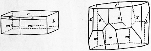
Fig. 1. Fig. 2.
Like the other micas, it is monoclinic with pseudo-hexagonal symmetry (figs. 1, 2) and possesses a perfect cleavage in one direction (c). Biotite is, however, readily distinguished by its darker colour, strong pleochroism, and small optic axial angle. The colour is usually dark-green or brown; thick crystals are often deep-black and opaque. The absorption of light-rays vibrating parallel to the cleavage is much greater than of rays vibrating in a direction perpendicular thereto, and in dark-coloured crystals the former are almost completely absorbed. The angle between the optic axes is usually very small, the crystals being often practically uniaxial; an axial angle of 50° has, however, been recorded in a dark-coloured biotite. The specific gravity of biotite is, as a rule, higher than that of other micas, varying from 2.7 to 3.1 according to the amount of iron present. The hardness is 2½ to 3.
Several varieties of biotite are distinguished. By G. Tschermak it is divided into two classes, meroxene and anomite; in the former the plane of the optic axis coincides with the plane of symmetry, whilst in the latter it is perpendicular thereto. Meroxene includes nearly all ordinary biotite, and is the name given by A. Breithaupt in 1841 to the Vesuvian crystals; on the other hand, anomite (named from ἄνομος, “contrary to law”) is of rare occurrence. Haughtonite and siderophyllite are black varieties rich in ferrous iron, and lepidomelane (from λεπίς, a scale, and μέλας, black) is a variety rich in ferric iron. In barytobiotite and manganophyllite the magnesia is partly replaced by baryta and manganous oxide respectively. Rubellane, hydrobiotite, pseudobiotite, and others are altered forms of biotite, which is a mineral particularly liable to decomposition with the production of chlorites and vermiculites.
Biotite is a common constituent of igneous and crystalline rocks; in granite, gneiss and mica-schist it is often associated with muscovite (white mica), the two kinds having sometimes grown in parallel position. In volcanic rocks, and in nearly all other kinds of igneous rocks with the exception of granite, biotite occurs to the exclusion of the muscovite. In the dyke-rocks known as mica-traps or mica-lamprophyres biotite is especially abundant. It is also one of the most characteristic products of contact-metamorphism, being developed in sedimentary and other rocks at their contact with granite masses. In the ejected blocks of crystalline limestone of Monte Somma, Vesuvius, the most perfectly developed crystals of biotite (figs. 1, 2), or indeed of any of the micas, are found in abundance, associated with brilliant crystals of augite, olivine, humite, &c.
Although biotite (black mica) is much more common and widely distributed than white mica, yet it is of far less economic importance. The small size of the sheets, their dark colour and want of transparency render the material of little value. Large, cleavable masses yielding fine smoky-black and green sheets, sufficiently elastic for industrial purposes, are, however, found in Renfrew county, Ontario.
(L. J. S.)
BIPARTITE (from the Lat. bi-, two, and partire, to divide). In a general sense, the word means having two corresponding parts or in duplicate. In geometry, a bipartite curve consists of two distinct branches (see Parabola, figs. 3, 5). In botany, the word is applied to leaves divided into two parts near the base. A bipartient factor is a number whose square exactly divides another number. In zoology, the Bipartiti was a name given by P.A. Latreille to a group of carnivorous Coleoptera.
BIPONT EDITIONS, the name of a famous series of editions, in 50 volumes, of Greek and Latin classical authors, so called from Bipontium, the modern Latin name of Zweibrücken or Deux-Ponts in Bavaria, where they were first issued in 1779. Their place of publication was afterwards transferred to Strassburg.
See Butters, Ûber die Editiones Bipontinae (1877).
BIQUADRATIC (from the Lat. bi-, bis, twice, and quadratus, squared). In mathematics, the biquadratic power or root of a quantity is its fourth power or root (see Algebra); a biquadratic equation is an equation in which the highest power of the unknown is the fourth (see Equation: Biquadratic).
BIQUINTILE (from Lat. prefix bi-, twice, quintilis, fifth), the aspect of two planets which are distant from each other twice the fifth part of a great circle, i.e. 144°. It was one of the new aspects introduced by Kepler.
BIRBHUM, a district of British India in the Burdwan division of Bengal, situated in the Gangetic plain and partly on the hills, being bounded on the south by the river Ajai. The administrative headquarters are at Suri, which is the only town in the district. The area comprises 1752 sq. m. The eastern portion of the district is the ordinary alluvial plain of the Gangetic delta; the western part consists of undulating beds of laterite resting on a rock basis, and covered with small scrub jungle. The Ajai, Bakheswar and Mor or Maurakshi, are the principal rivers of the district, but they are merely hill streams and only navigable in the rains. In 1901 the population was 902,280, showing an increase of 13% in the decade. The principal industry is the spinning and weaving of silk, chiefly from tussur or jungle silkworms. There are also several lac factories. The loop-line of the East Indian railway runs through the district, with a junction at Nalhati for Murshidabad.
History.—Birbhum in the early part of the 13th century was a Hindu state, with its capital at Rajnagar or Nagar. In the course of the century it was conquered by the Pathans and formed part of the Pathan kingdom of Bengal. At the beginning of the 18th century it appears as a kind of military fief held under the nawab of Murshidabad by one Asadullah Pathan, whose family had probably been its chieftains since the fall of the Pathan dynasty of Bengal in 1600. It passed into British possession in 1765, but the East India Company did not assume its direct government until 1787, when that course became necessary. In the interval it had been a prey to armed bands from the highlands of Chota Nagpur, with whom the raja was unable to cope, and who practically brought the trade of the Company in the district to a standstill. The two border principalities of Birbhum and Bankura were accordingly united into a district under a British collector, being, however, separated again in 1793. By 1789, after considerable trouble, the marauders were driven back into their mountains, and since that time (except during the Santal rising of 1855) the district has been one of the most peaceful and prosperous in India.
See Imperial Gazetteer of India (Oxford, 1908), vol. viii. s.v.
BIRCH, SAMUEL (1813-1885), English Egyptologist and antiquary, was born on the 3rd of November 1813, being the son of the rector of St Mary Woolnoth, London. From an early age he manifested a tendency to the study of out-of-the-way subjects, and after a brief employment in the Record Office obtained in 1836 an appointment in the antiquities department of the British Museum on account of his knowledge of Chinese. He soon extended his researches to Egyptian, and when the cumbrous department came to be divided he was appointed to the charge of the Egyptian and Assyrian branch. In the latter language he had assistance, but for many years there was only one other person in the institution—in a different department—who knew anything of ancient Egyptian, and the entire arrangement of the department devolved upon Birch. He found time nevertheless for Egyptological work of the highest value, including a hieroglyphical grammar and dictionary, translations of The Book of the Dead and the Harris papyrus, and numerous catalogues and guides. He further wrote what was long a standard history of pottery, investigated the Cypriote syllabary, and proved by various publications that he had not lost his old interest in Chinese. Paradoxical in many of his views on things in general, he was sound and cautious as a philologist; while learned and laborious, he possessed much of the instinctive divination of genius. He died on the 27th of December 1885.
BIRCH, THOMAS (1705-1766), English historian, son of Joseph Birch, a coffee-mill maker, was born at Clerkenwell on the 23rd of November 1705. He preferred study to business, but as his parents were Quakers he did not go to the university. Notwithstanding this circumstance, he was ordained deacon in the Church of England in 1730 and priest in 1731. As a strong supporter of the Whigs, he gained the favour of Philip Yorke, afterwards lord chancellor and first earl of Hardwicke, and his subsequent preferments were largely due to this friendship. He held successively a number of benefices in different counties, and finally in London. In 1735 he became a member of the Society of Antiquaries, and was elected a fellow of the Royal Society, of which he was secretary from 1752 to 1765. In 1728 he had married Hannah Cox, who died in the following year. Birch was killed on the 9th of January 1766 by a fall from his horse, and was buried in the church of St Margaret Pattens, London, of which he was then rector. He left his books and manuscripts to the British Museum, and a sum of about £500 to increase the salaries of the three assistant librarians.
Birch had an enormous capacity for work and was engaged in a large number of literary undertakings. In spite of their dulness many of his works are of considerable value, although Horace Walpole questioned his “parts, taste and judgment.” He carried on an extensive correspondence with some of the leading men of his time, and many of his letters appear in Literary Anecdotes of the 18th Century (London, 1812-1815) and Illustrations of the Literary History of the 18th Century (London, 1817-1858) by J. Nichols, in the Bibliotheca Topographica Britannica, vol. iii. (London, 1780-1790), and in Boswell’s Life of Johnson. Birch wrote most of the English lives in the General Dictionary, Historical and Critical, 10 vols. (London, 1734-1741), assisted in the composition of the Athenian Letters (London, 1810), edited the State Papers of John Thurloe (London, 1742) and the State Papers of W. Murdin (London, 1759). He also wrote a Life of the Right Honourable Robert Boyle (London, 1744); Inquiry into the share which King Charles I. had in the transactions of the Earl of Glamorgan for bringing over a body of Irish rebels (London, 1756); Historical view of Negotiations between the Courts of England, France and Brussels 1592-1617 (London, 1749); Life of Archbishop Tillotson (London, 1753); Memoirs of the Reign of Queen Elizabeth from 1581 (London, 1754); History of the Royal Society of London (London, 1756-1757); Life of Henry, Prince of Wales (London, 1760), and many other works. Among the papers left at his death were some which were published in 1848 as the Court and Times of James I. and the Court and Times of Charles I.
See W.P. Courtney in the Dictionary of National Biography, vol. v. (1886); A. Kippis, Biographia Britannica (London, 1778-1793); Horace Walpole, Letters (London, 1891).
BIRCH (Betula), a genus of plants allied to the alder (Alnus), and like it a member of the natural order Betulaceae. The various species of birch are mostly trees of medium size, but several of them are merely shrubs. They are as a rule of a very hardy character, thriving best in northern latitudes—the trees having round, slender branches, and serrate, deciduous leaves, with barren and fertile catkins on the same tree, and winged fruits, the so-called seeds. The bark in most of the trees occurs in fine soft membranous layers, the outer cuticle of which peels off in thin, white, papery sheets.
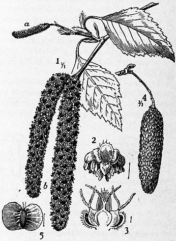
From Strasburger, Lerbuch der Botanik.
Betula alba. 1, Branch with male (a) and female (b) inflorescences; 2, bract with three male flowers; 3, bract with three female flowers; 4, infrutescence; 5, fruit. (After Wossidlo.)
The common white or silver birch (B. alba) (see fig.) grows throughout the greater part of Europe, and also in Asia Minor, Siberia and North America, reaching in the north to the extreme limits of forest vegetation, and stretching southward on the European continent as a forest tree to 45° N. lat., beyond which birches occur only in special situations or as isolated trees. It is well known in England for its graceful habit, the slender, grey—or white—barked stem, the delicate, drooping branches and the quivering leaves, a bright, clear green in spring, becoming duller in the summer, but often keeping their greenness rather late into the autumn. The male and female flowers are borne on separate catkins in April and May. It is a shortlived tree, generally from 40 to 50 ft. high with a trunk seldom more than 1 ft. in diameter. It flourishes in light soils and is one of the few trees that will grow amongst heather; owing to the large number of “winged seeds” which are readily scattered by the wind, it spreads rapidly, springing up where the soil is dry and covering clearings or waste places.
The birch is one of the most wide-spread and generally useful of forest trees of Russia, occurring in that empire in vast forests, in many instances alone, and in other cases mingled with pines, poplars and other forest trees. The wood is highly valued by carriage-builders, upholsterers and turners, on account of its toughness and tenacity, and in Russia it is prized as firewood and a source of charcoal. A very extensive domestic industry in Russia consists in the manufacture of wooden spoons, which are made to the extent of 30,000,000 annually, mostly of birch. Its pliant and flexible branches are made into brooms; and in ancient Rome the fasces of the lictors, with which they cleared the way for the magistrates, were made up of birch rods. A similar use of birch rods has continued among pedagogues to times so recent that the birch is yet, literally or metaphorically, the instrument of school-room discipline. The bark of the common birch is much more durable, and industrially of greater value, than the wood. It is impermeable to water, and is therefore used in northern countries for roofing, for domestic utensils, for boxes and jars to contain both solid and liquid substances, and for a kind of bark shoes, of which it is estimated 25 millions of pairs are annually worn by the Russian peasantry. The jars and boxes of birch bark made by Russian peasants are often stamped with very effective patterns. By dry distillation the bark yields an empyreumatic oil, called diogott in Russia, used in the preparation of Russia leather; to this oil the peculiar pleasant odour of the leather is due. The bark itself is used in tanning; and by the Samoiedes and Kamchatkans it is ground up and eaten on account of the starchy matter it contains. A sugary sap is drawn from the trunk in the spring before the opening of the leaf-buds, and is fermented into a kind of beer and vinegar. The whole tree, but especially the bark and leaves, has a very pleasant resinous odour, and from the young leaves and buds an essential oil is distilled with water. The leaves are used as fodder in northern latitudes.
The species which belong peculiarly to America (B. lenta, excelsa, nigra, papyracea, &c.) are generally similar in appearance and properties to B. alba, and have the same range of applications. The largest and most valuable is the black birch (B. lenta) found abundantly over an extensive area in British North America, growing 60 to 70 ft. high and 2 to 3 ft. in diameter. It is a wood most extensively used for furniture and for carriage-building, being tough in texture and bearing shocks well, while much of it has a handsome grain and it is susceptible of a fine polish. The bark, which is dark brown or reddish, and very durable, is used by Indians and backwoodsmen in the same way as the bark of B. alba is used in northern Europe.
The canoe or paper birch (B. papyracea) is found as far north as 70° N. on the American continent, but it becomes rare and stunted in the Arctic circle. Professor Charles Sprague Sargent says: “It is one of the most widely distributed trees of North America. From Labrador it ranges to the southern shores of Hudson’s Bay and to those of the Great Bear Lake, and to the valley of the Yukon and the coast of Alaska, forming with the aspen, the larch, the balsam poplar, the banksian pine, the black and white spruces and the balsam fir, the great subarctic transcontinental forest; and southward it ranges through all the forest region of the Dominion of Canada and the northern states.” It is a tree of the greatest value to the inhabitants of the Mackenzie river district in British North America. Its bark is used for the construction of canoes, and for drinking-cups, dishes and baskets. From the wood, platters, axe-handles, snow-shoe frames, and dog sledges are made, and it is worked into articles of furniture which are susceptible of a good polish. The sap which flows in the spring is drawn off and boiled down to an agreeable spirit, or fermented with a birch-wine of considerable alcoholic strength. The bark is also used as a substitute for paper. A species (B. Bhojputtra) growing on the Himalayan Mountains, as high up as 9000 ft., yields large quantities of fine thin papery bark, extensively sent down to the plains as a substitute for wrapping paper, for covering the “snakes” of hookahs and for umbrellas. It is also said to be used as writing paper by the mountaineers; and in Kashmir it is in general use for roofing houses.
BIRCH-PFEIFFER, CHARLOTTE (1800-1868), German actress and dramatic writer, was born at Stuttgart on the 23rd of June 1800, the daughter of an estate agent named Pfeiffer. She received her early training at the Munich court theatre, and in 1818 began to play leading tragic rôles at various theatres. In 1825 she married the historian Christian Birch of Copenhagen, but continued to act. From 1837 to 1843 she managed the theatre at Zürich. In 1844 she accepted an engagement at the royal theatre in Berlin, to which she remained attached until her death on the 24th of August 1868. Her intimate knowledge of the technical necessities of the stage fitted her for the successful dramatization of many popular novels, and her plays, adapted and original, make twenty-three volumes, Gesammelte dramatische Werke (Leip. 1863-1880). Many still retain the public favour. Her novels and tales, Gesammelte Novellen und Erzählungen, were collected in three volumes (Leip. 1863-1865).
Her daughter, Wilhelmine von Hillern (b. 1836), born at Munich, went on the stage, but retired upon her marriage in 1857. After 1889 she lived in Oberammergau and won a reputation as a novelist. Her most popular works are Ein Arzt der Seele (1869, 4th ed. 1886); and Die Geier-Wally (1883), which was dramatized and translated into English as The Vulture Maiden (Leip. 1876).
BIRD, the common English name for feathered vertebrates, members of the class Aves. The word in Old Eng. is brid and in Mid. Eng. byrd or bryd, and in early uses meant the young or nestlings only. It is partly due to this early meaning that the derivation from the root of “brood” has been usually accepted; this the New English Dictionary regards as “inadmissible.” The word does not occur in any other Teutonic language. As a generic name for the feathered vertebrates “bird” has replaced the older “fowl,” a common Teutonic word, appearing in German as Vogel. “Bird,” when it passed from its earliest meaning of “nestlings,” seems to have been applied to the smaller, and “fowl” to the larger species, a distinction which was retained by Johnson. In modern usage “fowl,” except in “wild-fowl” or “water-fowl,” is confined to domestic poultry.
The scope of the anatomical part of the following article is a general account of the structure of birds (Aves) in so far as they, as a class, differ from other vertebrates, notably reptiles and mammals, whilst features especially characteristic, peculiar or unique, have been dwelt upon at greater length so far as space permitted. References to original papers indicate further sources of information. For a comprehensive account the reader may be referred to Prof. M. Fürbringer’s enormous work Untersuchungen zur Morphologie und Systematik der Vögel, 4to., 2 vols. (1888); H.G. Bronn’s Klassen und Ordnungen des Thierreichs, vol. vi., “Aves,” Leipzig, completed 1893 by Gadow; and A. Newton’s Dictionary of Birds, London, 1896. For the history of the classification of birds see the article Ornithology, where also the more important ornithological works are mentioned. Egg, Feather (including Moult), Migration, &c., also form separate articles to which reference should be made. In this article (A) the general anatomy of birds is discussed, (B) fossil birds, (C) the geographical distribution of birds, (D) the latest classification of birds.
A. Anatomy of Birds
1. Skeleton.
Skull.—When W.K. Parker wrote the account of the skull in the article Birds for the 9th edition of the Encyclopaedia Britannica, he had still to wrestle with the general problem of the composition and evolution of the skull. That chapter of comparative anatomy (together with other anatomical details, for which see the separate articles) is now dealt with in the article Skull; here only the most avine features are alluded to, and since some of Parker’s original illustrations have been retained, the description has been shortened considerably.
One general feature of the adult bird’s skull is the almost complete disappearance of the sutures between the bones of the cranium proper, whilst another is the great movability of the whole palatal and other suspensorial apparatus. The occipital condyle (fig. 1) is a single knob, being formed almost wholly by the basioccipital, while the lateral occipitals (often perversely called exoccipitals) take but little share in it. Part of the membranous roof between the supra-occipital and parietal bones frequently remains unossified and presents in the macerated skull a pair of fontanelles. The squamosals form the posterior outer margin of the orbits and are frequently continued into two lateral downward processes across the temporal fossa. One of these, the processus orbitatis posterior, often combines with an outgrowth of the alisphenoid, and may be, e.g. in cockatoos, continued forwards to the lacrymal bone, so as to form a complete infraorbital bridge. The posterior, so-called processus Zygomaticus is very variable; in many Galli it encloses a foramen by distally joining the orbital process. The ethmoid frequently appears on the dorsal surface between the frontals. There are three periotic bones (pro-, epi-, opisth-otic). The proötic encloses between it and the lateral occipital the fenestra ovalis, into which fits the columella of the ear. The epiotic is often small, ossifies irregularly, and fuses with the supra-occipital. The opisthotic lies between the epiotic and the lateral occipital with which it ultimately fuses; in some birds, e.g. in Larus, it extends far enough to help to bound the foramen magnum. The basisphenoids are ventrally overlaid, and later on fused with, a pair of membrane bones, the basi-temporals, homologous in part with the parasphenoid of lower vertebrates. They contribute to the formation of the auditory meatus, and of the right and left carotid canals which accompany the eustachian tubes. In many birds the basisphenoids send out a pair of basipterygoid processes by which they articulate with the pterygoids. Dorso-laterally the basisphenoid is joined by the alisphenoid, which forms most of the posterior wall of the orbit. The orbito-sphenoids diverge only posteriorly, otherwise they are practically unpaired and form the median interorbital septum, which is very large in correlation with the extraordinary size of the eyeballs.,
Fig. 1.—End view of skull of a Chicken three weeks old. Here the opisthotic bone appears in the occipital region, as in the adult Chelonian. (After W.K. Parker.)
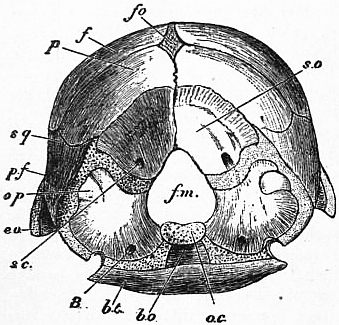
bo, Basi-occipital. bt, Basi-temporal. eo, Opisthotic. f, Frontal. fm, Foramen magnum. fo, Fontanella. oc, Occipital condyle. op, Opisthotic. p, Parietal. pf, Post-frontal. sc, Sinus canal in supra-occipital. so, Supra-occipital. sq, Squamosal. 8, Exit of vagus nerve.
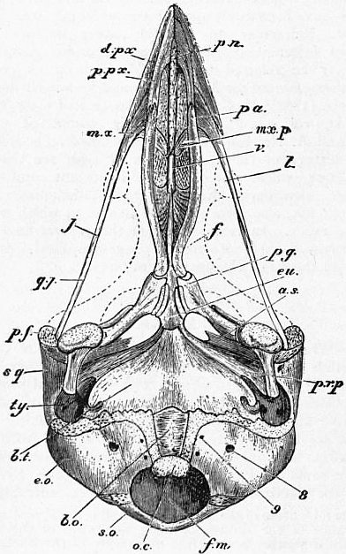
Fig. 2.—Ripe Chick’s head, 1¼ in. long. (After W.K. Parker.)
as, Alisphenoid. bo, Basi-occipital. bt, Basi-temporal. dpx, Dentary process of premaxilla. eo, Opisthotic. eu, Eustachian tube. f, Frontal. fm, Foramen magnum. j, Jugal. l, Lacrymal. mx, Maxilla. mxp, Maxillo-palatine process. oc, Occipital condyle. pa, Palatine. pf, Post-frontal. pg, Pterygoid. pn, Prenasal cartilage. ppx, Palatine process of pre-maxillary. prp, Pterygoid process of sphenoid. qj, Quadratojugal. so, Supra-occipital. sq, Squamosal. ty, Tympanic cavity. v, Vomer. 8, Exit of vagus nerve. 9, Exit of hypoglossal nerve.
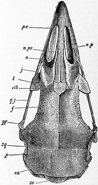
Fig. 3.—Skull of an old Fowl, upper view. (After W.K. Parker.)
eo, Lateral occipital. eth, Ethmoid. f, Frontal. j, Jugal. l, Lacrymal. n, Nostril. np, Upper process of nasal. npx, Nasal process of premaxillary. p, Parietal. pf, Post-frontal. px, Premaxilla. qj, Quadratojugal. so, Supra-occipital. sq, Squamosal.
Prefrontal bones are absent; post-frontals are possibly indicated by a frequently occurring separate centre of ossification in the post-orbital process, to which the frontals always contribute. The lacrymal is always present, and perforated by a glandular duct. Attached to it or the neighbouring frontal is often a supraorbital; infraorbitals occur also, attached to the jugal or downward process of the lacrymal. The nasals were used by A.H. Garrod to distinguish the birds as holorhinal (fig. 2) where the anterior margin of the nasal is concave, and schizorhinal where this posterior border of the outer nares is continued backwards into a slit which extends beyond the frontal processes of the premaxilla. Many birds possess a more or less well developed cross-joint in front of the frontals and lacrymals, perhaps best developed in Anseres and Psittaci. Owing to this joint the whole upper beak can be moved up and down with extra facility, according to the shoving forwards or backwards of the palato-pterygo-quadrate apparatus which moves sledge-like upon the cranial basis. The premaxilla is always unpaired, but each half has three long processes directed backwards; one fuses with the maxillary bone, another helps to form the anterior part of the palate, while the third, together with its fellow, forms the “culmen” and extends backwards to the frontals, or rather to the ethmoid which there crops up on the surface. The maxillaries (fig. 3) have besides others, a maxillo-palatine process directed inwards in a transverse horizontal direction. The palatines are long, always fused anteriorly with the premaxilla, and frequently with the maxillo-palatine processes; posteriorly they slide upon the presphenoidal rostrum, and articulate in most birds with the pterygoids; they form the greater part of the palatal roof and border the choanae or inner nares. Between these, resting vertically upon the rostrum, appears the vomer; very variable in shape and size, often reduced to a mere trace, as in the Galli, or even absent, broken up into a pair of tiny splints in Pici.
The taxonomic importance of the configurations of the palate was first pointed out by J. de Cornay. T.H. Huxley, in 1868, divided the carinate birds into Dromaeo-, Schizo-, Desmo-, and Aegithognathae, an arrangement which for many years had a considerable influence upon classification. However, subsequent additions and corrections have detracted much from its value, especially when it became understood that the above sub-orders are by no means natural groups. Dromaeognathae have a struthious palate, with a broad vomer meeting in front the broad maxillo-palatal plates, while behind it reaches the pterygoids. The only representatives are the Tinamous. Schizognathae, e.g. fowls (fig. 4), pigeons, gulls, plovers, rails and penguins, have the vomer pointed in front while the maxillo-palatines are free, leaving a fissure between the vomer and themselves. The schizognathous formation is doubtless the most primitive, and its representatives form a tolerably natural assembly. Desmognathae (fig. 5) were supposed to have the maxillo-palatines united across the middle line, either directly or by the intermediation of ossifications in the nasal septum. This is a hopeless assembly. Parker and Fürbringer have demonstrated that desmognathism has been produced in half a dozen ways, implying numerous cases of convergence without any nearer relationship than that they are all derived from some schizognathous group or other. The Aegithognathae, meant to comprise the passeres, woodpeckers and swifts, &c., are really schizognathous but with a vomer which is broadly truncated in front.
Fig. 4.—Skull of adult Fowl. This skull is unusually schizognathous, the vomer (v.) being very small, and the maxillo-palatine process (mxp) much aborted.
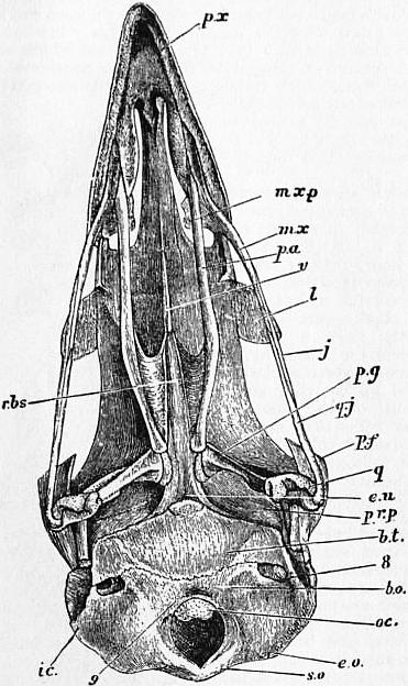
bo, Basi-occipital. bt, Basi-temporal. eo, Lateral occipital. eu, Eustachian tube. ic, Internal carotid. j, Jugal. l, Lacrymal. mx, Maxilla. mxp, Maxillo-palatine process. oc, Occipital condyle. pa, Palatine. pf, Post-frontal. pg, Pterygoid. prp, Pterygoid process of sphenoid. px, Premaxilla. q, Quadrate. qj, Quadratojugal. rbs, Rostrum of basisphenoid. so, Supra-occipital. v, Vomer. 8, Exit of vagus nerve. 9, Exit of hypoglossal nerve. (After W.K. Parker.)
Fig. 5.—Skull of a nestling Sparrow-hawk (Accipiter nisus), palatal view. The circular space on each side of the basi-temporal (bt.) is the opening of the anterior tympanic recess. The basi-pterygoids (bpg) are mere knobs, and the common eustachian opening is seen between them. The maxillo-palatine plates (mxp) are dotted to show their spongy character.
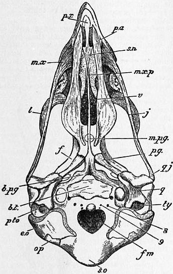
bt, Basi-temporal. bpg, Basi-pterygoid. eo, Lateral occipital. f, Frontal. fm, Foramen magnum. j, Jugal. l, Lacrymal. mpg, Mesopterygoid process of W.K. Parker. mx, Maxillary. mxp, Maxillo-palatine process. op, Opisthotic. pa, Palatine. pg, Pterygoid. px, Premaxilla. pto, Prootic. q, Quadrate. qj, Quadratojugal. sn, Nasal septum. so, Supra-occipital. ty, Tymapanic cavity. v, Vomer. 8, Exit of vagus nerve. 9, Exit of hypoglossal nerve. (After W.K. Parker.)
The remainder of the appendicular skeleton (fig. 6) of the head requires little description. The maxillaries are connected with the distal anterior corner of the quadrate by the thin, splint-like jugal and quadratojugal. The quadrate is invariably a conspicuous bone and movably articulating with the cranium and by a special process with the pterygoid. The mandible is composed of several bones as in reptiles. The os articulare bears on its inner side the inner mandibular process which serves for the insertion of part of the digastric muscle or opener of the mouth; another portion of this muscle is attached to the os angulare, which frequently forms a posterior mandibular process. The greater part of the under-jaw is formed by the right and left dentaries, which in all recent birds are fused together in front. Supra-angular and coronoid splint-bones serve for the insertion of part of the temporal or masseter muscle. Additional splints rest on the inner side of the jaw. Like the crocodiles, birds possess a siphonium, i.e. a membranous, or ossified, tube which rises from a pneumatic foramen in the os articulare, on the median side of the articulation, and passes upwards between the quadrate and lateral occipital bone, opening into the cavity of the middle ear.
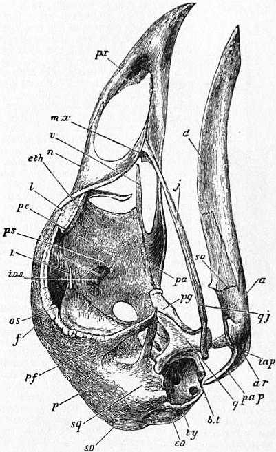
Fig. 6.—Skull of adult Fowl. Here the temporal fossa is bridged over by the junction of the post-frontal and squamosal processes (pf., sq.). The processes of the mandible (iap, pap) are characteristic of this type, and of the anseres.
a, Angular of mandible. ar, Articular. bt, Basi-temporal. d, Dentary. eo, Lateral occipital. eth, Ethmoid. f, Frontal. iap, Interangular process of mandible. ios, Interorbital septum. j, Jugal. l, Lacrymal. mx, Maxillar. n, Nasal. os, Orbito-sphenoid. p, Parietal. pa, Palatine. pap, Posterior angular process of mandible. pe, Ethmoid. pf, Post-frontal. pg, Pterygoid. ps, Pre-sphenoid. px, Premaxilla. q, Quadrate. qj, Quadratojugal. sa, Supra-angular or coronoid. so, Supra-occipital. sq, Squamosal. ty, Tympanic cavity. v, Vomer. 1, Exit of olfactory nerve.
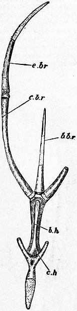
Fig. 7.—Oshyoides of adult Fowl.
c.h, Ceratohyals (confluent). b.h, The so-called basihyal, answering to the first basibranchial of a fish. b.br, Basibranchial, or urohyal, answering to the rest of the basibranchial series. c.br, e.br, together form the thyrohyal, answering to the first cerato- and epi-branchials.
The Hyoid apparatus is, in its detail, subject to many variations in accord with the very diverse uses to which the tongue of birds is put. It consists of (1) the basihyal variously called copula, or corpus linguae, or unpaired middle portion. (2) The urohyal likewise unpaired, rested ventrally on the larynx. (3) The os entoglossum originally paired, but coalescing into an arrow-headed piece, attached to the anterior end of the basihyal and lodged in the tongue proper. It is homologous with the distal ends of the ceratohyals or ventral elements of the hyoidean or second visceral arch. The dorsal or hyomandibular portion of this same arch is transformed into the auditory chain, ending in the fenestra ovalis. (4) A pair of thyrohyals, homologous with the posterior hyoid horns of mammals, i.e. third visceral or first branchial arch. As the most developed pair in birds they are commonly, although wrongly, called the hyoid horns. They articulate upon facets of the hinder outer corners of the basihyal.
The vertebrae are stereospondylous, the centrum or body and the arch being completely fused into one mass, leaving not even a neuro-central suture. The arch alone sends out processes, viz. the spinous process, the anterior and posterior oblique (commonly called pre- and post-zygapophyses), and the transverse processes. The latter articulate with the tuberculum of the corresponding rib, while the capitulum articulates by a knob on the side of the anterior end of the centrum. In the cervical region the ribs are much reduced, fused with their vertebrae and enclosing the transverse canal or foramen. When the vertebrae are free their centra articulate with each other by complicated joints, exhibiting four types. (1) Amphicoelous; each end of the centrum is concave; this, the lowest condition, is embryonic, but was retained in Archaeopteryx and in the thoracic vertebrae of Ichthyornis. (2) Procoelous, concave in front; only in the atlas, for the reception of the occipital condyle. (3) Opisthocoelous, or concave behind, only occasionally found in the thoracic region, e.g. Sphenisci. (4) Heterocoelous (fig. 8) or saddle-shaped; the anterior surface is concave in a transverse, but convex in a vertical direction, which on posterior surface shows the conditions reversed. This is the most perfect arrangement attained by the vertebral column, and is typical of, and restricted to, birds. The intervertebral joints are further complicated by the interposition of a cartilaginous or fibrous pad or ring. This pad varies much; it is morphologically the homologue of the pair of basiventral elements which by their lateral extension give origin to the corresponding ribs. Later those pads fuse with the anterior end of the centrum of the vertebra to which they belong; where the vertebral column is rendered inflexible, the disks are ossified with the centra and all trace of them is lost. Sometimes the pad is reduced to a ventral semi-ring or meniscus; it retains its largest almost original shape and size in the second vertebra, the axis or epistropheus, where it forms a separately ossifying piece which connects, and coössifies with, the odontoid process (the centrum of the atlas) and the centrum of the second vertebra. Sometimes the ventral portions of these pads form paired or unpaired little ossifications, then generally described as intercentra; such are not uncommon on the tail. The atlas is composed of three pieces; a pair of lateral elements (the right and left dorsal arch pieces) joining above the spinal cord, and a ventral piece equivalent to the first basiventral elements, i.e. serially homologous with the intervertebral pads. In the adults the atlas forms a more or less solid ring. A remnant of the chorda dorsalis and its sheath persists as the ligamentum suspensorium between the central portions of the successive vertebrae.
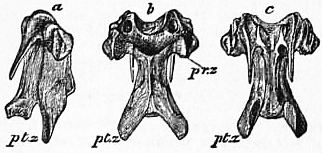
Fig. 8.—A cervical vertebra from the middle of the neck of a Fowl; natural size. a, Side view; b, upper view; c, lower view; pr.z, pre-zygapophyses; pt.z, post-zygapophyses.
In birds we distinguish between the following regions of the axial skeleton. (1) Cervical vertebrae, or those between the skull and the first vertebra which is connected with the sternum by a pair of complete ribs. The last 1 to 5 of these vertebrae have movable ribs which do not reach the sternum, and are called cervico-dorsals. (2) Dorsals, those which begin with the first thoracic rib, and end at the last that is not fused with the ilium. The term “lumbar” vertebrae is inapplicable to birds. (3) Pelvic, all those which are fused with the iliac portion of the pelvis, generally a considerable number. (4) Caudal, those which are not connected with the pelvis. It is to be noted that often no absolute line of demarcation can be drawn in regard to these regions, their definitions being rather convenient than morphological.
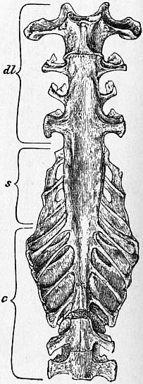
Fig. 9.—The “sacrum” of a young Fowl; natural size, seen from below. d.l, Dorso-lumbar, s, sacral, c, caudal vertebrae.
In comparison with all other vertebrates the number of neck-vertebrae of the birds is considerably increased; the lowest number, 14 to 15, is that of most Passeres and many other Coraciomorphae; the largest numbers, 20 or 21, are found in the ostrich, 23 in Cygnus olor and 25 in the black swan. Dorsal vertebrae frequently have a ventral outgrowth of the centrum; these hypapophyses may be simple vertical blades, ⊥-shaped, or paired knobs; they serve for the attachment of the thoracic origin of the longus collianticus muscle, reaching their greatest development in Sphenisci and Colymbidae. In many birds some of the thoracic vertebrae are more or less coössified, in most pigeons for instance the 15th to 17th; in most Galli the last cervical and the next three or four thoracics are coalesced, &c. The pelvic vertebrae include of course the sacrum. There are only two or three vertebrae which are equivalent to those of the reptiles; these true sacrals are situated in a level just behind the acetabulum; as a rule between these two primary sacral vertebrae issues the last of the spinal nerves which contributes to the composition of the sciadic plexus. These true sacrals alone are connected with the ilium by processes which are really equivalent to modified ribs; but the pelvis of birds extends considerably farther forwards and backwards, gradually coming into contact with other vertebrae, which in various ways send out connecting transverse processes or buttresses, and thus become pre- and post-sacral vertebrae (fig. 9). The most anterior part of the ilium often overlaps one or more short lumbar ribs and fuses with them, or even a long, complete thoracic rib. Similarly during the growth of the bird the posterior end of the ilium connects itself with the transverse processes of vertebrae which were originally free, thus transforming them from caudals into secondary post-sacrals. Individual, specific and generic variations are frequent.
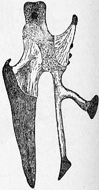
Fig. 10.—A side view of the Chick’s sternum.
The last six or seven caudal vertebrae coalesce into the pygostyle, an upright blade which carries the rectrices. Such a pygostyle is absent in Archaeopteryx, Hesperornis, Tinami and Ratitae, but it occurs individually in old specimens of the ostrich and the kiwi. In Ichthyornis it is very small. In all the Neornithes the total number of caudal vertebrae, inclusive of those which coalesce, is reduced to at least 13.
Sternum (figs. 10 and 11).—Characteristic features of the sternum are the following. There is a well-marked processus lateralis anterior (the right and left together equivalent to the mammalian manubrium), which is the product of two or three ribs, the dorsal parts of which reduced ribs remain as cervico-dorsal ribs. Then follows the rib-bearing portion and then the processus lateralis posterior; this also is the product of ribs, consequently the right and left processes together are equivalent to the xiphoid process or xiphisternum of the mammals. The lateral process in most birds sends out an outgrowth, directed out and upwards, overlapping some of the ribs, the processus obliquus. The median and posterior extension of the body of the sternum is a direct outgrowth of the latter, therefore called meta-sternum. The anterior margin of the sternum, between the right and left anterior lateral processes receives in sockets the feet of the coracoids. Between them arises a median crest, which varies much in extent and composition, and is of considerable taxonomic value. It is represented either by a spina interna or by a spina externa, or by both, or they join to form a spina communis which is often very large and sometimes ends in a bifurcation. Eventually, when the right and left feet of the coracoids overlap each other, the anterior sternal spine contains a foramen. The keel, or carina sterni, is formed as a direct cartilaginous outgrowth of the body of the sternum, ossifying from a special centre. This keel is much reduced in the New Zealand parrot, Stringops, less in various flightless rails, in the dodo and solitaire. It is absent in the Ratitae, which from this feature have received their name, but considerable traces of a cartilaginous keel occur in the embryo of the ostrich, showing undeniably that the absence of a keel in the recent bird is not a primitive, fundamental feature. The keel has been lost, and is being lost, at various epochs and by various groups of birds. The swimming Hesperornis (see Odontornithes) was also devoid of such a structure. In many birds the spaces between the meta-sternum and the posterior processes and again the spaces between this and the oblique process are filled up by proceeding ossification and either remain as notches, or as fenestrae, or they are completely abolished so that the breastbone is turned into one solid more or less oblong plate.
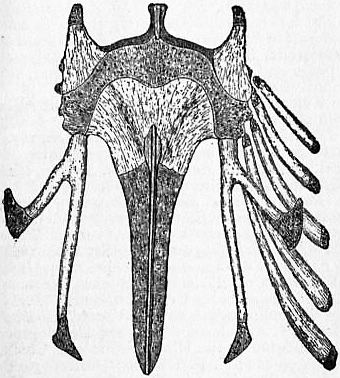
Fig. 11.—Sternum of a Chick (Gallus domesticus) three days old, lower view. The cartilage is shaded and dotted, and the bony centers are light and striated.
Shoulder Girdle.—Scapula, coracoid and clavicle, meet to form the foramen triosseum, through which passes the tendon of the supracoracoideus, or subclavius muscle to the tuberculum superius of the humerus. The coracoid is one of the most characteristic bones of the bird’s skeleton. Its upper end forms the acrocoracoid process, against the inner surface of which leans the proximal portion of the clavicle. From the inner side of the neck of the coracoid arises the precoracoidal process, the remnant of the precoracoid. Only in the ostrich this element is almost typically complete, although soon fused at either end with the coracoid. Near the base of the precoracoidal process is a small foramen for the passage of the nervus supracoracoideus. In most birds the feet of the coracoids do not touch each other; in some groups they meet, in others one overlaps the other, the right lying ventrally upon the left. The scapula is sabre-shaped, and extends backwards over the ribs, lying almost parallel to the vertebral column. This is a peculiar character of all birds. The clavicles, when united, as usual, form the furcula; mostly the distal median portion is drawn out into a hypocleidium of various shape. Often it reaches the keel of the sternum, with subsequent syndosmosis or even synostosis, e.g. in the gannet. In birds of various groups the clavicles are more or less degenerated, the reduction beginning at the distal end. This condition occurs in the Ratitae as well as in the well-flying Platyrcecinae amongst parrots.
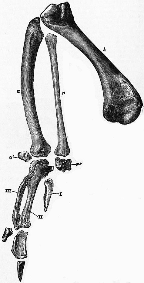
Fig. 12.—Bones of Fowl’s right wing, adult, nat. size.
h, Humerus. r, Radius. u, Ulnar. r′, u′, Radial and ulnar carpal bones; with the three digits I., II., III.
The fore-limb or wing (fig. 12); highly specialized for flight, which, initiated and made possible mainly by the strong development of quill-feathers, has turned the wing into a unique organ. The humerus with its crests, ridges and processes, presents so many modifications characteristic of the various groups of birds, that its configuration alone is not only of considerable taxonomic value but that almost any genus, excepting, of course, those of Passeres, can be “spotted” by a close examination and comparison of this bone. When the wing is folded the long glenoid surface of the head of the humerus is bordered above by the tuberculum externum or superius, in the middle and below by the tuberculum medium or inferius for the insertion of the coraco-brachialis posterior muscle. From the outer tuberculum extends the large crista superior (insertion of pectoralis major and of deltoideus major muscles). The ventral portion of the neck is formed by the strong crista inferior, on the median side of which is the deep fossa subtrochanterica by which air sacs enter the humerus. On the outer side of the humerus between the head and the crista inferior is a groove lodging one of the coraco-humeral ligaments. The distal end of the humerus ends in a trochlea, with a larger knob for the ulna and a smaller oval knob for the radius. Above this knob is often present an ectepicondylar process whence arise the tendons of the ulnar and radial flexors. The radius is the straighter and more slender of the two forearm bones. Its proximal end forms a shallow cup for articulation with the outer condyle of the humerus; the distal end bears a knob which fits into the radial carpal. The ulna is curved and rather stout; it articulates with both carpal bones; the cubital quills often cause rugosities on its dorsal surface. Of wrist-bones only two remain in the adult bird; the original distal carpals coalesce with the proximal end of the metacarpals. These are reduced, in all birds, to three, but traces of the fourth have been observed in embryos. The first metacarpal is short and fuses throughout its length with the second. This and the third are much longer and fuse together at their upper and distal ends, leaving as a rule a space between the shafts. The pollex and the third finger are as a rule reduced to one phalanx each, while the index still has two. The first and second fingers frequently carry a little claw. The greatest reduction of the hand-skeleton is met with in Dromaeus and in Apteryx, which retain only the index finger. It is of importance for our understanding of the position of the Ratitae in the system, that the wing-skeleton of the ostrich and rhea is an exact repetition of that of typical flying birds; the bones are much more slender, and the muscles are considerably reduced in strength also to a lesser extent in numbers, but the total length of the wing of an ostrich or a rhea is actually and comparatively enormous. Starting with the kiwi and cassowary, people have got into the habit of confounding flightless with wingless conditions. It is absolutely certain that the wings of the Ratitae bear the strongest testimony that they are the descendants of typical flying birds.
The pelvis (fig. 13), consisting of the sacrum (already described) and the pelvic arch, namely ilium, ischium and pubis, it follows that only birds and mammals possess a pelvis proper, whilst such is entirely absent in the Amphibia and in reptiles with the exception of some of the Dinosaurs. The ventral inner margin of the preacetabular portion of the ilium is attached to the pre-sacral vertebrae, whilst the inner and dorsal margin of the postacetabular portion is attached to the primary sacral and the postsacral vertebrae. In rare cases the right and left preacetabular blades fuse with each other above the spinous processes. In front of the acetabulum a thick process of the ilium descends to meet the pubis, and a similar process behind meets the ischium. The acetabulum is completely surrounded by these three bones, but its cup always retains an open foramen; from its posterior rim arises the strong antitrochanter. The ischium and postacetabular ilium originally enclose the ischiadic notch or incisura ischiadica. This primitive condition occurs only in the Odontornithes (q.v.), Ratitae and Tinami; in all others this notch becomes converted into a foramen ischiadicum, through which pass the big stems of the ischiadic nerves and most of the blood-vessels of the hind-limb. The pubis consists of a short anterior portion (spina pubica or pectineal process, homologous with the prepubic process of Dinosaurs) and the long and slender pubis proper (equivalent to the processus lateralis pubis of most reptiles). The shaft of the pubis runs parallel with that of the ischium, with which it is connected by a short ligamentous or bony bridge; this cuts off from the long incisura pubo-ischiadica a proximal portion, the foramen obturatum, for the passage of the obturator nerve. Only in the ostrich the distal ends of the pubes meet, forming a dagger-shaped symphysis, which is curved forwards. The pectineal process is variable; it may grow entirely from the pubis, or both pubis and ilium partake of its formation, or lastly its pubic portion may be lost and the process is entirely formed by the ilium. It is largest in the Galli and some of the Cuculi, in others it is hardly indicated. It served originally for the origin of the ambiens muscle (see Muscular System below); shifting or disappearance of this muscle, of course, influences the process.
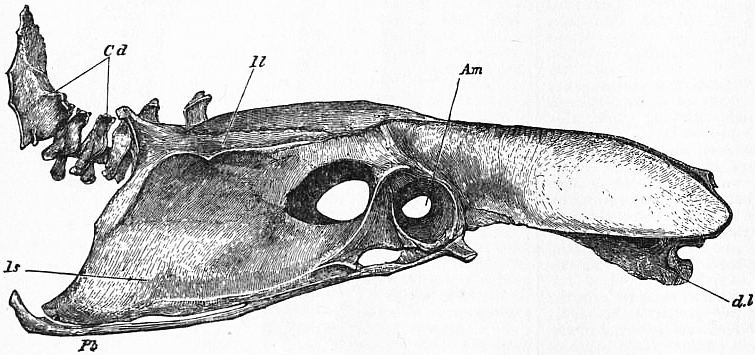
Fig. 13.—Pelvis and caudal vertebrae of adult Fowl, side view, natural size. Il. Ilium; Is, ischium; Pb, pubis; d.l, dorso-lumbar vertebrae; Cd, caudal vertebrae; Am, acetabulum.
The Hind Limb.—The femur often possesses a well visible pneumatic foramen on the median side of the proximal end of its shaft. The inner condyle, the intercondylar sulcus, and a portion only of its outer condyle, articulate with corresponding facets of the tibia. The outer condyle articulates mainly with the fibula. There is a patella, intercalated in the tendon of the femori-tibialis or extensor cruris muscle. In Colymbus the patella is reduced to a small ossicle, its function being taken by the greatly developed pyramidal processus tibialis anterior; in Podiceps and Hesperornis the patella itself is large and pyramidal. The distal half of the fibula is very slender and normally does not reach the ankle-joint; it is attached to the peroneal ridge of the tibia. On the anterior side of the tibia, is the intercondylar sulcus, which is crossed by an oblique bridge of tendon or bone, acting as a pulley for the tendon of the extensor digitorum communis muscle. The condyles of the tibia are in reality not parts of this bone, but are the three proximal tarsalia which fuse together and with the distal end of the tibia. The distal tarsalia likewise fuse together, and then on to the upper ends of the metatarsals; the tarsale centrale remains sometimes as a separate osseous nodule, buried in the inter-articular pad. Consequently the ankle-joint of birds is absolutely cruro-tarsal and tarso-metatarsal, i.e. intertarsal, an arrangement absolutely diagnostic of birds if it did not also occur in some of the Dinosaurs. Of the metatarsals the fifth occurs as an embryonic vestige near the joint; the first is reduced to its distal portion, and is, with the hallux, shoved on to the inner and posterior side of the foot, at least in the majority of birds. The three middle metatarsals become fused together into a cannon bone; the upper part of the third middle metatarsal projects behind and forms the so-called hypotarsus, which in various ways, characteristic of the different groups of birds (with one or more sulci, grooved or perforated), acts as guiding pulley to the tendons of the flexor muscles of the toes. Normally the four toes have two, three, four and five phalanges respectively, but in Cypselus the number is reduced to three in the front toes. Reduction of the number of toes (the fifth shows no traces whatever, not even in Archaeopteryx) begins with the hallux, which is completely or partly absent in many birds; the second toe is absent in Struthio only. The short feet of the penguins are quite plantigrade, in adaptation to which habit the metatarsals lie in one plane and are incompletely co-ossified, thus presenting a pseudo-primitive condition.
Literature.—Only a mere fraction of the enormous literature dealing with the skeleton of birds can here be mentioned.
M.E. Alix Essai sur l’appareil locomoteur des oiseaux (Paris, 1874); E. Blanchard, “Recherches sur les caractères ostéologiques des oiseaux appliquées à la classification,” Ann. Sci. Nat. Ser. iv., t. xi.; W. Dames, “Über Brustbein Schulter- und Beckengürtel der Archaeopteryx,” Math. Naturw. Mitsh., Berlin, vii., 1897, pp. 476-492; T.C. Eyton, Osteologia avium (London, 1858-1881), with many plates; C. Gegenbaur, Untersuch. z. vergl. Anat. d. Wirbelthiere, I. Carpus und Tarsus, II. Schultergurtel (Leipzig, 1864-1865); P. Harting, L’Appareil épisternal des oiseaux (Utrecht, 1864); T.H. Huxley, “On the Classification of Birds and on the Taxonomic Value of the Modifications of certain of the Cranial Bones...” P.Z.S., 1867; G. Jaeger, “Das Wirbelkorpergelenk der Vögel,” Sitzb. K. Ak. Wiss., Wien, xxxiii., 1858; A. Johnson, “On the Development of the Pelvic Girdle and Skeleton of the Hind-limb in the Chick,” Q.J.M.S., xxiii., 1883, pp. 399-411; K.F. Kessler, “Osteologie der Vogelfüsse,” Bull. Soc. Imp. Nat., Moscow, xiv., 1841; B. Lindsay, “On the Avian Sternum,” P.Z.S., 1885; E. Mehnert, “Entwickelung des Ospelvis der Vögel,” Morph. Jahrb., xiii., 1877; A.B. Meyer, Abbildungen van Vögel-Skeletten (Dresden, 1879); St G. Mivart, “On the Axial Skeleton of the Ostrich, Struthionidae, Pelecanidae,” Trans. Zool. Soc. viii., 1874; x., 1877; E.S. Morse, “On the Carpus and Tarsus of Birds,” Ann. Lyc. N.H., New York, x., 1874; J.S. Parker, “Observations on the Anatomy and Development of Apteryx,” Phil. Trans., 1890, pp. 1-110, 17 pls.; W. K. Parker, numerous papers in Trans. L.S., R.S. and Z.S., e.g. “Osteology of Gallinaceous Birds,” T.Z.S., v., 1863; “Rhinochetus,” ibid. vi.; “Skull of Aegithognathous Birds,” ibid., x., 1878; “Skull in the Ostrich Tribe,” Phil. Trans. vol. 156, 1866; “Skull of Common Fowl,” ibid. vol. 159, 1870; “Skull of Picidae,” T. Linn. Soc., 1875; “Monograph on the Structure and Development of the Shoulder-girdle and Sternum,” Ray Soc. London, 1868; W.P. Pycraft, “On the Morphology and Phylogeny of the Palaeognathae (Ratitae and Crypturi) and Neognathae,” Trans. Zool. Soc. xv., 1900, pp. 149-290, pis. 42-45; id. “Some points in the morphology of the Palate of the Neognathae,” T. Linn. Soc. 28, pp. 343-357, pls. 31-32; P. Suschkin, “Zur Morphologie des Vogelskelets. I. Schädel von Tinnunculus,” Mem. Soc., Moscow, xvi., 1900, pp. 1-63, pls.
2. Muscular System.
Of the muscles of the stem or axis, those of the neck and tail are well-developed and specialized, while those of the lower back are more or less reduced, or even completely degenerated owing to the rigidity of this region, brought about by the great antero-posterior extent of the pelvis.
The muscles of the limbs show a great amount of specialization, away from the fundamental reptilian and mammalian conditions. The muscles of the fore limbs are most aberrant, but at the same time more uniformly developed than those of the hinder extremities. The reasons are obvious. The whole wing is a unique modification, deeply affecting the skeletal, muscular and tegumentary structures, but fluttering, skimming, sailing, soaring are motions much more akin to one another than climbing and grasping, running, scratching, paddling and wading. The modifications of the hind-limbs are in fact many times greater (such as extremely long legs, with four, three or only two toes; very short legs, almost incapable of walking, with all four toes directed forwards, or two or one backwards, and two or more connected and therefore bound to act together, in various ways). Thus it has come to pass that the muscles of the hind limbs are, like their framework, more easily compared with those of reptiles and mammals than are the wings, whilst within the class of birds they show an enormous amount of variation in direct correlation with their manifold requirements. The only really aberrant modifications of the wing-muscles are found in the Ratitae, where they are, however, all easily explained by reduction, and in the penguins, where the wings are greatly specialized into blades for rowing with screw-like motions.
The wing of the bird is folded in a unique way, namely, the radius parallel with the humerus, and the whole wrist and hand with their ulnar side against the ulna; upper and forearm in a state of supination, the hand in that of strong abduction. Dorsal and ventral bending, even in the extended wing, is almost impossible. Consequently only a few of the original extensor muscles have been preserved, but these are much modified into very independent organs, notably the extensor metacarpi radialis longus, the ext. metac. ulnaris and the two radio- and ulnari-metacarpi muscles, all of which are inserted upon the metacarpus by means of long tendons. The chief muscular mass, arising from the sternum in the shape of a U, is the pectoralis muscle; its fibres converge into a strong tendon, which is inserted upon the greater tubercle and upper crest of the humerus, which it depresses and slightly rotates forwards during the downstroke. This great muscle covers completely the supracoracoideus, generally described as the second pectoral, or subclavius muscle, in reality homologous with the mammalian supraspinatus muscle. This arises mostly from the angle formed by the keel with the body of the sternum, passes by a strong tendon through the foramen triosseum, and is inserted upon the upper tubercle of the humeral crest, which it rotates and abducts. The extent of the origin of this muscle from the sternum, on which it leaves converging, parallel or diverging impressions, is of some taxonomic value.
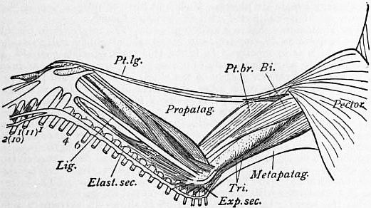
From Newton’s Dictionary of Birds, by permission of A. & C. Black.
Fig. 14.—Wing muscles of a Goose. Bi, Biceps; Elast. sec., elastic vinculum and Exp.sec., expansor secundariorum; Pt.br and Pt.lg, short and long propatagial muscles; Tri, triceps.
Much labour has been bestowed by A.H. Garrod and Max Fürbringer upon the investigation of the variations of the inserting tendons of the patagial muscles (fig. 14), mainly from a taxonomic point of view. The propatagialis longus muscle is composed of slips from the deltoid, pectoral, biceps and cucullaris muscles. Its strong belly originates near the shoulder joint from clavicle, coracoid and scapula. Its elastic tendon runs directly to the carpus, forming thereby the outer margin of the anterior patagium, or fold of skin between the upper and forearm, which it serves to extend, together with the propatagialis brevis muscle. This runs down the anterior and outer side of the upper arm, and is attached to the proximal tendon of the extensor metacarpi radialis longus, a little below the outer condyle of the humerus. In most birds the tendon is split into several portions, one of which is often attached to the outer side of the ulna, below the elbow joint, while others are in variable but characteristic ways connected with similar slips of the propatagialis longus. The posterior patagium, the fold between trunk and inner surface of the upper arm, is stretched by the metapatagialis muscle, which is composed of slips from the serratus, superficialis, latissimus dorsi and the expansor secundariorum muscles. This, the stretcher of the cubital quills, is a very interesting muscle. Arising as a long tendon from the sterno-scapular ligament, it passes the axilla by means of a fibrous pulley, accompanies the axillary vessels and nerves along the humerus, and is inserted by a few fleshy fibres on the base of the last two or three cubital quills. Here, alone, at the distal portion of the tendon, occur muscular fibres, but these are unstriped, belonging to the category of cutaneous muscles. We have here the interesting fact that a muscle (portion of the triceps humeri of the reptiles) has been reduced to a tendon, which in a secondary way has become connected with cutaneous muscles, which, when strongly developed, represent its belly.
The flexor digitorum sublimis muscle arises fleshy from the long elastic band which extends from the inner humeral condyle along the ventral surface of the ulna to the ulnar carpal bone, over which the tendon runs to insert itself on the radial anterior side of the first phalanx of the second digit. Owing to the elasticity of the humerocarpal band the wing remains closed without any special muscular exertion, while, when the wing is extended, this band assists in keeping it taut. The arm-muscles have been studied in an absolutely exhaustive manner by Fürbringer, who in his monumental work has tabulated and then scrutinized the chief characters of fourteen selected muscles. The results are as interesting from a morphological point of view (showing the subtle and gradual modifications of these organs in their various adaptations), as they are sparse in taxonomic value, far less satisfactory than are those of the hind-limb. He was, however, the first to show clearly that the Ratitae are the retrograde descendants of flying ancestors, that the various groups of surviving Ratitae are, as such, a polyphyletic group, and he has gone fully into the interesting question of the development and subsequent loss of the power of flight, a loss which has taken place not only in different orders of birds but also at various geological periods, and is still taking place. Very important are also the investigations which show how, for instance in such fundamentally different groups as petrels and gulls, similar bionomic conditions have produced step by step a marvellously close convergence, not only in general appearance, but even in many details of structure.
Of the muscles of the hind-limbs likewise only a few can be mentioned. The ambiens muscle, long and spindle-shaped, lying immediately beneath the skin, extending from the pectineal process or ilio-pubic spine to the knee, is the most median of the muscles of the thigh. When typically developed its long tendon passes the knee-joint, turning towards its outer side, and lastly, without being anywhere attached to the knee, it forms one of the heads of the flexor perforatus digit, ii. or iii. One of the functions of this peculiar muscle (which is similarly developed in crocodiles, but absent, or not differentiated from the ilio-tibial and ilio-femoral mass, in other vertebrates) is that its contraction helps to close the second and third toes. Too much has been made of this feature since Sir R. Owen (Cyclop. Anat. Phys. i. p. 296, 1835), following G.A. Borelli (De motu animalium, Rome, 1680), explained that birds are enabled to grasp the twig on which they rest whilst sleeping, without having to make any muscular exertion, because the weight of the body bends the knee and ankle-joints, over both of which pass the tendons of this compound muscle. There are many perching birds, e.g. all the Passeres, which do not possess this muscle at all, whilst many of those which have it fully developed, e.g. Anseres, can hardly be said to “perch.”
Garrod went so far as to divide all the birds into Homalogonatae and Anomalogonatae, according to the presence or absence of the ambiens muscle. This resulted in a failure. To appreciate this, it is sufficient to enumerate the birds without the critical muscle: Passeriformes and Coraciiformes, without exception; Ardeae and Podiceps; lastly various genera of storks, pigeons, parrots, petrels and auks. The loss has taken place, and still takes place, independently in widely different groups. It follows, first, that the absence of this muscle does not always indicate relationship; secondly that we can derive birds that are without it from a group which still possess it, but not vice versa. The absence of the ambiens muscle in all owls, which apparently use their feet in the same way as the Accipitres (all of which possess it), indicates that owls are not developed from the latter, but from a group which, like the other Coraciiformes, had already lost their muscle.
Garrod further attributed much taxonomic value to the caudilio-femoralis muscle (fig. 15). This, when fully developed, consists of two parts, but inserted by a single ribbon-like tendon upon the hinder surface of the femur, near the end of its first third; the caudal part, femoro-caudalis, expressed by Garrod by the symbol A, arises from transverse processes of the tail; the iliac part (accessoro-femoro-caudal of Garrod, with the symbol B), arises mostly from the outer surface of the postacetabular ilium. Of course this double-headed condition is the more primitive, and as such exists in most nidifugous birds, but in many of these, as well as in many nidicolous birds, either the caudal or the iliac head is absent, and in a very few (Cancroma, Dicholophus, Steatornis and some Cathartes) the whole muscle is absent. The caud-ilio flexorius (semitendinosus of most authors) arises from the transverse processes of the tail, and from the distal half of the postacetabular ilium, thence passing as a broad ribbon to the popliteal region, where it splits into two portions. One of these, broad and fleshy, is inserted upon the posterior surface of the distal third of the femur. This portion, morphologically the original, was named the “accessory semitendinosus” with the symbol Y; the other portion descends on the hinder aspect of the leg and joins the fascia of the inner femoral head of the gastrocnemius muscle. In many birds the insertion is shifted from the femur to the neck of the tibia, in which case the “accessory head” is said to be absent, a condition expressed by Garrod by the symbol X. By combining the four symbols A, B, X, Y, according to their presence or absence, Garrod got a considerable number of formulae, each of which was overruled, so to speak, by the two categories of the presence or absence of the ambiens muscle. It needs hardly to be pointed out why such a purely mechanical scheme was doomed to failure. Its author, with a considerable mathematical and mechanical bias, reckoned entirely with the quantity, not with the quality of his units, and relied almost implicitly upon his formulae. It is, however, fair to state that his system was not built entirely upon these muscular variations, but rather upon a more laborious combination of anatomical characters, which were so selected that they presumably could not stand in direct correlation with each other, notably the oil-gland, caeca, carotids, nasal bones and above all, the muscles of the thigh. He was, indeed, the first to show clearly the relationship of the heron-like birds with the Steganopodes; of stork-like birds with the American vultures; the great difference between the latter and the other birds of prey; the connexion of the gulls and auks with the plovers, and that of the sand-grouse with the pigeons—discoveries expressed in the new terms of the orders Ciconiiformes and Charadriiformes. These are instances, now well understood, that almost every organic system, even when studied by itself, may yield valuable indications as to the natural affinities of the various groups of birds. That Garrod has so very much advanced the classification of birds is ultimately due to his comprehensive anatomical knowledge and general insight.
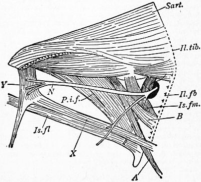
From Newton’s Dictionary of Birds.
Fig. 15.—Left thigh-muscles of a Rail. Outer view after removal of the Il.fb, ilio-fibularis and Il.tib, ilio-tibialis.
A, Caudal. B, Iliac portion of caud-ilio-femoralis. X, Caud-ilio-flexorius. Y, “Accessory” portion of the same. Pif, Pubischio-femoralis. N, Sciatic nerve. Is.fm, Ischio-femoralis. Is.fl, Ischio-fibularis. Sart. Sartorius.
To return to these thigh muscles. The most primitive combination, ambiens and A B X Y, is the most common; next follows that of A X Y, meaning the reduction of B, i.e. the iliac portion of the caud-ilio-femoralis; A B X and B X Y are less common; A X and X Y are rare and occur only in smaller groups, as in subfamilies or genera; B X occurs only in Podiceps. But the greatest reduction, with only A remaining, is characteristic of such a heterogeneous assembly as Accipitres, Cypselidae. Trochilidae, Striges and Fregata. This fact alone is sufficient proof that these conditions, or rather reductions, have been acquired independently of the various groups. A B Y, A Y, A B, X Y and B do not occur at all, some of them for obvious reasons. Occasionally there is an instructive progressive evolution expressed in these formula; for instance Phaethon, in various other respects the lowest of the Steganopodes, has A X Y, Sula and Phalacrocorax have A X, Fregata, the most specialized of these birds, has arrived at the reduced formula A. Further, the combinations B X Y and A X Y cannot be derived from each other, but both directly from A B X Y in two different directions. Keeping this in mind, we may fairly conclude that the flamingo with B X Y points to an ancestral condition A B X Y, which is still represented by Platalea and Ibis, whilst the other storks proper have taken a different line, leading to A X Y.
Literature.—Well nigh complete lists of the enormous myological literature are contained in Fürbringer’s Untersuchungen zur Morphologie und Systematik der Vögel, and in Gadow’s vol. Vögel of Bronn’s Klassen und Ordnungen des Tierreichs. Only a few papers and works can be mentioned here, with the remark that few authors have paid attention to the all-important innervation of the muscles. A. Carlsson, Beiträge zur Kenntniss der Anatomieder Schiwmmvögel; K. Svensk, Vet. Ak. Handlinger. J.G. No. 3 (1884); A. Alix, Essai sur l’appareil locomoteur des oiseaux (Paris, 1874); H. Gadow, Zur vergl. Anat. der Muskulatur des Beckens und der hinteren Gliedmasse der Ratiten,4° (Jena, 1880); A.H. Garrod, “On Certain Muscles of the Thigh of Birds and on their value in Classification,” P.Z.S., 1873, pp. 624-644; 1874, pp. 111-123. Other papers by Garrod, 1875, pp. 339-348 (deep planter tendons); 1876, pp. 506-519 (wing-muscles of Passeres), &c.; J.G. de Man, Vergelijkende myologische en neurologische Studien over Amphibien en Vögels (Leiden, 1873), (Corvidae); A. Milne-Edwards, Recherches anatomiques et paléontologiques pour servir à l’histoire des oiseaux fossiles de la France (Paris, 1867-1868), tom. i. pls. ix.-x. (Aquila and Gallus); R. Owen, article “Aves,” Todds’ Cydopaed. of Anat. and Phys. i. (London, 1835); “On the Anatomy of the Southern Apteryx,” Trans. Zool. Soc., iii., 1849; A. Quennerstedt, “Studier i foglarnas anatomi,” Lunds Univers. Aarsk., ix., 1872 (hind-limb of swimming birds); G. Rolleston, “On the Homologies of Certain Muscles connected with the Shoulder-joint,” Trans. Linn. Soc., xxvi., 1868; R.W. Shufeldt, The Myology of the Raven (London, 1891); M. Watson, “Report on the Anatomy of the Spheniscidae,” Challenger Reports, 1883.
3. Nervous System.
Brain.—The more characteristic features of the bird’s brain show clearly a further development of the reptilian type, not always terminal features in a direct line, but rather side-departures, sometimes even a secondary sinking to a lower level, and in almost every case in a direction away from those fundamentally reptilian lines which have led to the characters typical of, and peculiar to, the mammals.
The forebrain forms the bulk of the whole brain, but the large size of the hemispheres is due to the greater development of the basal and lateral portions (pedunculi cerebri and corpora striata), while the pallium (the portion external to the lateral ventricles) is thin, and restricted to the median side of each hemisphere. As a direct result of this undoubtedly secondary reduction of the pallium—due to the excessive preponderance of the basal and lateral parts—the corpus callosum (i.e. the transverse commissure of the right and left pallium) is in birds reduced to a narrow flat bundle of a few white fibres; it is situated immediately above and behind the much stronger anterior commissure, i.e. the connexion between the corpora striata, or chief remaining part of the hemispheres. Owing to the small size of the olfactory lobes the anterior arms of the latter commissure are wanting. There is very little grey matter in the cortex of the hemispheres, the surface of which is devoid of convolutions, mostly quite smooth; in others, for instance pigeons, fowls and birds of prey, a very slight furrow might be compared with the Sylvian fissure.
The Thalamencephalon is much reduced. The epiphysis, or pineal body, is quite as degenerate as in mammals, although still forming a long stalk as in reptiles. In birds, this stalk consists entirely of blood-vessels, which in the adult enclose no terminal vesicle, and fuse with the membranous linings of the skull. The midbrain is represented chiefly by the optic lobes, the cortex of which alone is homologous with the corpora quadragemina of the mammals. Their transverse dorsal connexion is the posterior commissure; otherwise the whole roof portion of the midbrain is reduced to a thin membrane, continuous with that which covers the Sylvian aqueduct, and this ventricle sends a lateral cavity into each optic lobe, as is the case in reptiles. The right and left lobes themselves are rent asunder (so to speak), so that they are freely visible from above, filling the corners formed by the hemispheres and the cerebellum. The latter is, in comparison with mammals; represented by its middle portion only, the vermis; in a sagittal section it shows an extremely well developed arbor vitae, produced by the transverse, repeated folding of the whole organ. In comparison with reptiles the cerebellum of birds shows high development. Forwards it covers, and has driven asunder, the optic lobes; backwards it hides the much shortened medulla oblongata.
Several futile attempts have been made to draw conclusions as to the intelligence of various birds, from comparison of the weight of the whole brain with that of the body, or the weight of the hemispheres with that of other parts of the central nervous system.
The brachial plexus is formed by four or five of the lowest cervical nerves; the last nerve of this plexus often marks the boundary of the cervical and thoracic vertebrae. The composition of the plexus varies much, not only in different species, but even individually. The most careful observations are those by Fürbringer. The serial number of these nerves depends chiefly upon the length of the neck, the extremes being represented by Cypselus (10th-14th cervical) and Cygnus (22nd-24th), the usual numbers of the common fowl being the 13th-17th nerves.
The Crural Plexus is divided into a crural, ischiadic and pubic portion. The first is generally composed of three nerves, the hindmost of which, the furcalis, issues in most birds between the last two lumbo-sacral vertebrae, and then divides, one half going to the crural, the other to the sciatic portions. The obturatorius nerve invariably comes from the two main stems of the crural. The ischiadic portion consists generally of five or six nerves, which leave the pelvis as one thick system through the ilio-ischiadic foramen. The last nerve which contributes to the ischiadic plexus leaves the spinal column in most birds either between the two primary sacral vertebrae, or just below the hindmost of them, and sends a branch to the pubic portion which is composed of post-ischiadic nerves, partly imbedded in the kidneys, and innervates the ventral muscles between the tail and pubis, together with those of the cloaca and copulatory organs.
The Sympathetic System forms a chain on either side of the vertebral column. In the region of the neck lateral strands pass through the transverse canal of the cervical vertebrae; but from the thoracic region onwards, where the cardiac branch to the heart is given off, each strand is double and the basal ganglia are successively connected with the next by a branch which runs ventrally over the capitulum of the rib, and by another which passes directly through the foramen or space formed between capitulum and tuberculum. In the pelvic region, from about the level of the posterior end of the ischiadic plexus, the strand of each side becomes single again, passing ventrally over the transverse processes. Lastly, towards the caudal region the right and left strands approach and anastomose, eventually coalescing in the mid line.
Literature.—A. Bumm, “Das Grosshirn der Vögel,” Zeitschr. wiss. Zool., 38, 1883, pp. 430-466, pls. 24-25; F. Leuret and P. Gratiolet, Anatomie comparée du système nerveux (Paris, 1839-1857), with atlas; A. Meckel, “Anatomie des Gehirns der Vögel,” in Meckel’s Archiv f. Physiol. vol. ii.; H.F. Osborn, “The Origin of the Corpus Callosum, a contribution upon the Cerebral Commissures of the Vertebrata,” Morphol. Jahrbuch, 1886, xii. pp. 223-251, pls. 13-14; M.A. Schulgin, “Lobi optici der Vögel,” Zool. Anzeig. iv. pp. 277 and 303; E.R.A. Serres, Anatomie comparée du cerveau (Paris, 1824, 4 pls.); L. Stieda, “Studien uber das centrale Nervensystem der Vögel und Säugethiere,” Zeitschr. wiss. Zool. xix., 1869, pp. 1-92, pls.; J. Swan, Illustrations of the Comparative Anatomy of the Nervous System (London, 1835, 4to, with plates).
Concerning the spinal nerves and their plexus: H. v. Jhering, Das peripherische Nervensystem der Wirbeltiere (Leipzig, 1871); W.A. Haswell, “Notes on the Anatomy of Birds,” Proc. Linn. Soc. N.S.W. iii., 1879; M. Fürbringer, “Zur Lehre von den Umbildungen der Nervenplexus,” Morph. Jahrb. v., 1879, p. 358.
4. Organs of Sense.
The Eye is essentially reptilian, but in sharpness of vision, power and quickness of accommodation it surpasses that of the mammals. The eyeball, instead of being globular, resembles rather the tube of a short and thick opera-glass.
The anterior half of the sclerotic is composed of a ring of some ten to seventeen cartilaginous or bony scales which partly overlap each other. Another cartilage or ossification, the posterior sclerotic ring, occurs within the walls of the posterior portion of the cup, and surrounds, especially in the Pici and in the Passeres, the entrance of the optic nerve. The iris is in most young birds at first brown or dull-coloured, but with maturity attains often very bright tints which add considerably to the charm of the bird; sexual dimorphism is in this respect of common occurrence. The iris contains a sphincter and a dilator muscle; the former, supplied by branches from the oculomotorius nerve, is under control of the will, whilst the dilator fibres belong to the sympathetic system. When fully dilated, the pupil is round in all birds; when contracted it is usually round, rarely oval as in the fowl. From near the entrance of the optic nerve, through the original choroidal fissure, arises the much-folded pecten, deeply pigmented and very vascular, far into the vitreous humour. The number of its folds varies considerably, from three in Caprimulgus to nearly thirty in crow (Corvus). Apteryx, which since Owen has generally been stated to be devoid of such an organ, likewise possesses a pecten; its base is, however, trumpet-shaped, covers almost the whole of the optic disk, and extends nearly to the lens in the shape of a thick, densely pigmented cone, without any plications, resembling in these respects the pecten of many Lacertilia (see G.L. Johnson, Phil. Trans., 1901, p. 54). In the retina the cones prevail in numbers over the rods as in the mammals, and their tips contain, as in other Sauropsida, coloured drops of oil, mostly red or yellow. Near the posterior pole of the fundus, but somewhat excentrically placed towards the temporal or outer side, is the fovea centralis, a slight depression in the retina, composed almost entirely of cones, the spot of most acute vision. Many birds possess besides this temporal fovea a second fovea nearer the nasal side. It is supposed that the latter serves monocular, the other the binocular vision, most birds being able to converge their eyes upon one spot. Consequently the whole field of vision of these birds possesses three points where vision is most acute. It may here be remembered that of the mammalia man and monkeys alone are capable of convergence, and have a circumscribed macular area.
Of the outer eyelids, the lower alone is movable in most birds, as in reptiles, and it frequently contains a rather large saucer-shaped cartilage, the tarsus palpebralis. The margins of the lids are sometimes furnished with eyelashes, e.g. in the ostrich and in the Amazon parrots, which are vestigial feathers without barbs. During the embryonic stage the lids are fused together, and either become separated shortly before the bird is hatched, as is the case with most Nidifugae, or else the blind condition prevails for some time, in the young Nidicolae. All birds have, like most reptiles, a well-developed third lid or “nictitating membrane,” which moves from the inner canthus obliquely upwards and backwards over the cornea. The moving mechanism is a further and much higher development of that which prevails in reptiles, there being two muscles completely separate from each other. Both are supplied by the abducens nerve, together with the rectus externus muscle. One, the quadratus or bursalis muscle, arises from the hinder surface of the eyeball, and forms with its narrow margin, which is directed towards the optic nerve, a pulley for the long tendon of the pyramidalis muscle. This arises from the nasal surface of the ball, and its tendon passes into the somewhat imperfectly transparent nictitating membrane. The quadrate muscle adjusts the motion, and prevents pressure upon the optic nerve; during the state of relaxation of both muscles the nictitans withdraws through its own elasticity.