VOLUME II SLICE VI Armour Plates to Arundel, Earls of (continued 9)
Reading edition
The axillary artery supplies thoracic branches to the wall of the chest, the pectoral muscles, and the fat and glands of the axilla; an acromio-thoracic to the parts about the acromion; anterior and posterior circumflex branches to the shoulder joint and deltoid muscle; a subscapular branch to the muscles of the posterior fold of the axilla.
The brachial artery supplies muscular branches to the muscles of the upper arm; a nutrient branch to the humerus; superior and inferior profunda branches and an anastomotic to the muscles of the upper arm and the region of the elbow joint.
The ulnar artery extends down the ulnar side of the front of the fore-arm to the palm of the hand, where it curves outward toward the thumb, and anastomoses with the superficial volar or other branch of the radial artery to form the superficial palmar arch. In the fore-arm the ulnar gives off the interosseous arteries, which supply the muscles of the fore-arm and give nutrient branches to the bones; two recurrent branches to the region of the elbow; carpal branches to the wrist joint: in the hand it gives a deep branch to the deep muscles of the hand, and from the superficial arch arise digital branches to the sides of the little, ring, and middle fingers, and the ulnar border of the index finger.
The radial artery extends down the radial side of the front of the fore-arm, turns round the outer side of the wrist to the back of the hand, passes between the 1st and 2nd metacarpal bones to the palm, where it joins the deep branch of the ulnar, and forms the deep palmar arch. In the fore-arm it gives off a recurrent branch to the elbow joint; carpal branches to the wrist joint; and muscular branches, one of which, named superficial volar, supplies the muscle of the thumb and joins the ulnar artery: in the hand it gives off a branch to the thumb, and one to the radial side of the index, interosseous branches to the interosseous muscles, perforating branches to the back of the hand, and recurrent branches to the wrist.
The branches of the aorta which supply the viscera of the thorax are the coronary, the oesophageal, the bronchial and the pericardiac. The coronary arteries, two in number, are the first branches of the aorta, and arise opposite the anterior and left Visceral branches. posterior segments of the semilunar valve, from the wall of the aorta, where it dilates into the sinuses of Valsalva. They supply the tissue of the heart.
The oesophageal, bronchial and pericardiac branches are sufficiently described by their names.
The branches of the aorta which supply the viscera of the abdomen arise either singly or in pairs. The single arteries are the coeliac axis, the superior mesenteric, and the inferior mesenteric, which arise from the front of the aorta; the pairs are the capsular, the two renal, and the two spermatic or ovarian, which arise from its sides. The single arteries supply viscera which are either completely or almost completely invested by the peritoneum, and the veins corresponding to them are the roots of the vena portae. The pairs of arteries supply viscera developed behind the peritoneum, and the veins corresponding to them are rootlets of the inferior vena cava.
The coeliac axis is a thick, short artery, which almost immediately divides into the gastric, hepatic and splenic branches. The gastric gives off oesophageal branches and then runs along the lesser curvature of the stomach. The hepatic artery ends in the substance of the liver; but gives off a cystic branch to the gall bladder, a pyloric branch to the stomach, a gastro-duodenal branch, which divides into a superior pancreatico-duodenal for the pancreas and duodenum, and a right gastro-epiploic for the stomach and omentum. The splenic artery ends in the substance of the spleen; but gives off pancreatic branches to the pancreas, vasa brevia to the left end of the stomach, and a left gastro-epiploic to the stomach and omentum.
The superior mesenteric artery gives off an inferior pancreatico-duodenal branch to the pancreas and duodenum; about twelve intestinal branches to the small intestines, which form in the substance of the mesentery a series of arches before they end in the wall of the intestines; an ileocolic branch to the end of the ileum, the caecum, and beginning of the colon; a right colic branch to the ascending colon; and a middle colic branch to the transverse colon.
The inferior mesenteric artery gives off a left colic branch to the descending colon, a sigmoid branch to the iliac and pelvic colon, and ends in the superior haemorrhoidal artery, which supplies the rectum. The arteries which supply the coats of the alimentary tube from the oesophagus to the rectum anastomose freely with each other in the wall of the tube, or in its mesenteric attachment, and the anastomoses are usually by the formation of arches or loops between adjacent branches.
The capsular arteries, small in size, run outward from the aorta to end in the supra-renal capsules.
The renal arteries pass one to each kidney, in which they for the most part end, but in the substance of the organ they give off small perforating branches, which pierce the capsule of the kidney, and are distributed in the surrounding fat. Additional renal arteries are fairly common.
The spermatic arteries are two long slender arteries, which descend, one in each spermatic cord, into the scrotum to supply the testicle. The corresponding ovarian arteries in the female do not leave the abdomen.
The branches of the aorta which supply the walls of the thorax, abdomen and pelvis, are the intercostal, the Parietal branches. lumbar, the phrenic, and the middle sacral.
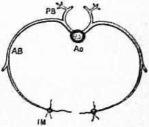
Fig. 1.—Diagram of a pair of intercostal arteries.
Ao, The aorta transversely divided, giving off at each side an intercostal artery. PB, The posterior or dorsal branch. AB, The anterior or proper intercostal branch. IM, A transverse section through the internal mammary artery.
The intercostal arteries arise from the back of the thoracic aorta, and are usually nine pairs. They run round the sides of the vertebral bodies as far as the commencement of the intercostal spaces, where each divides into a dorsal and a proper intercostal branch; the dorsal branch passes to the back of the thorax to supply the deep muscles of the spine; the proper intercostal branch (AB.) runs outward in the intercostal space to supply its muscles, and the lower pairs of intercostals also give branches to the diaphragm and wall of the abdomen. Below the last rib a subcostal artery runs.
The lumbar arteries arise from the back of the abdominal aorta, and are usually four pairs. They run round the sides of the lumbar vertebrae, and divide into a dorsal branch which supplies the deep muscles of the back of the loins, and an abdominal branch which runs outward to supply the wall of the abdomen. The distribution of the lumbar and intercostal arteries exhibits a transversely segmented arrangement of the vascular system, like the transversely segmented arrangement of the bones, muscles and nerves met with in these localities, but more especially in the thoracic region.
The phrenic arteries, two in number, pass to supply the under surface of the diaphragm.
The middle sacral artery, as it runs down the front of the sacrum, gives branches to the back of the pelvic wall.
Injections made by Sir W. Turner have shown that, both in the thoracic and abdominal cavities, slender anastomosing communications exist between the visceral and parietal branches.
The arteries to the pelvis and hind limbs begin at the bifurcation of the aorta into the two common iliacs.
The common iliac artery, after a short course, divides into the internal and external iliac arteries. The internal iliac enters the pelvis and divides into branches for the supply of the pelvic walls and viscera, including the organs of generation, and for the Iliac system. great muscles of the buttock. The external iliac descends behind Poupart’s ligament into the thigh, where it takes the name of femoral artery. The femoral descends along the front and inner surface of the thigh, gives off a profunda or deep branch, which, by its circumflex and perforating branches, supplies the numerous muscles of the thigh; most of these extend to the back of the limb to carry blood to the muscles situated there. The femoral artery then runs to the back of the limb in the ham, where it is called popliteal artery. The popliteal divides into two branches, of which one, called anterior tibial, passes between the bones to the front of the leg, and then downward to the upper surface of the foot; the other, posterior tibial, continues down the back of the leg to the sole of the foot, and divides into the internal and external plantar arteries; branches proceed from the external plantar artery to the sides of the toes, and constitute the digital arteries. From the large arterial trunks in the leg many branches proceed, to carry blood to the different structures in the limb.
The wall of an artery consists of several coats (see fig. 2). The outermost is the tunica adventitia, composed of connective tissue; immediately internal to this is the yellow elastic coat; within this again the muscular coat, formed of involuntary. Structure of arteries. muscular tissue, the contractile fibre-cells of which are for the most part arranged transversely to the long axis of the artery; in the larger arteries the elastic coat is much thicker than the muscular, but in the smaller the muscular coat is relatively strong; the vaso-motor nerves terminate in the muscular coat. In the first part of the aorta, pulmonary artery and arteries of the retina there is no muscular coat. Internal to the muscular coat is the elastic fenestrated coat, formed of a smooth elastic membrane perforated by small apertures. Most internal of all is a layer of endothelial cells, which form the free surface over which the blood flows. The arteries are not nourished by the blood which flows through them, but by minute vessels, vasa vasorum, distributed in their external, elastic and muscular coats.
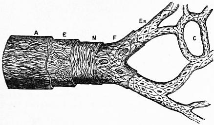
Fig. 2.—Diagram of the structure of an artery. A, tunica adventitia; E, elastic coat; M, muscular coat; F, fenestrated coat; En, endothelium continuous with the endothelial wall of C, the capillaries.
Embryology
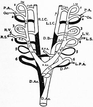
Fig. 3.—Diagram of the Embryonic Arterial Arches. 1, 2, 3, 4, 5, 6, point to the six arches. (The black parts are obliterated in the adult human subject.)
V.Ao. Ventral Aorta. A.Ao. Arch of Aorta. D.Ar. Ductus Arteriosus. In. Innominate Artery. R.I.C.-L.I.C. Right and Left Internal Carotid Arteries. D.B. Duct of Botalli. R.S.-L.S. Right and Left Subclavian Arteries. R.V.-L.V. Right and Left Vertebral Arteries. P.A. Posterior Auricular Artery. Oph. Ophthalmic Artery. D.Ao. Dorsal Aorta. P.T. Pulmonary trunk. R.P.A.-L.P.A. Right and Left Pulmonary Arteries. R.C.C.-L.C.C. Right and Left Common Carotid Arteries. E.C. External Carotid Artery. Oc. Occipital Artery. I.M. Internal Maxillary Artery.
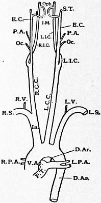
Fig. 4.—Diagram of the Human Aorta and its branches. S.T., Superficial Temporal Artery.
The earliest appearance of the blood vessels is dealt with under Vascular System. Here will be briefly described the fate of the main vessel which carries the blood away from the truncus arteriosus of the developing heart (q.v.). This ventral aorta, if traced forward, soon divides into two lateral parts, the explanation being that there were originally two vessels, side by side, which fused to form the heart, but continued separate anteriorly. The two parts run for a little distance toward the head of the embryo, ventral to the alimentary canal, and then turn toward the dorsum, passing one on either side of that tube to form the first aortic arch. Having reached the dorsum they turn backward toward the tail end and form the dorsal aortae; here, according to A.H. Young (Studies in Anatomy, Owens College, 1891 and 1900) they again turn toward the ventral side and become, after a transitional stage, the hypogastric, placental, allantoic or umbilical arteries. This authority does not believe that the middle sacral artery of the adult is the real continuation of the single median dorsal aorta into which the two parallel dorsal vessels just mentioned soon coalesce, though until recently it has always been so regarded. The anterior loop between the ventral and dorsal aortae already described as the first aortic arch is included in the maxillary or first visceral arch of the soft parts (see fig. 3, 1). Later, four other well-marked aortic arches grow behind this in the more caudal visceral arches, so that there are altogether five arterial arches on each side of the pharynx, through which the blood can pass from the ventral to the dorsal aorta. Of these arches the first soon disappears, but is probably partly represented in the adult by the internal maxillary artery, one branch of which, the infraorbital, is enclosed in the upper jaw, while another, the inferior dental, is surrounded by the lower jaw. Possibly the ophthalmic artery also belongs to this arch. The second arch also disappears, but the posterior auricular and occipital arteries probably spring from it, and at an early period it passed through the stapes as the transitory stapedial artery. The third arch forms the beginning of the internal carotid. The fourth arch becomes the arch of the adult aorta, between the origins of the left carotid and left subclavian, on the left side, and the first part of the right subclavian artery on the right. The apparent fifth arch on the left side (fig. 3, 6) remains all through foetal life as the ductus arteriosus, and, as the lungs develop, the pulmonary arteries are derived from it. J.E.V. Boas and W. Zimmermann have shown that this arch is in reality the sixth, and that there is a very transitory true fifth arch in front of it (fig. 3, 5). The part of the ventral aorta from which this last arch rises is a single median vessel due to the same fusion of the two primitive ventral aortae which precedes the formation of the heart, but a spiral septum has appeared in it which divides it in such a way that while the anterior or cephalic arches communicate with the left ventricle of the heart, the last one communicates with the right (see Heart). The fate of the ventral and dorsal longitudinal vessels must now be followed. The fused part of the two ventral aortae, just in front of the heart, forms the ascending part of the adult aortic arch, and where this trunk divides between the fifth and fourth arches (strictly speaking, the sixth and fifth), the right one forms the innominate (fig. 3, In.) and the left one a very short part of the transverse arch of the aorta until the fourth arch comes off (see fig. 4). From this point to the origin of the third arch is common carotid, and after that, to the head, external carotid on each side. The dorsal longitudinal arteries on the head side of the junction with the third arch form the internal carotids. Between the third and fourth arches they are obliterated, while on the caudal side of this, until the point of fusion is reached on the dorsal side of the heart, the left artery forms the upper part of the dorsal aorta while the right entirely disappears. Below this point the thoracic and abdominal aortae are formed by the two primitive dorsal aortae which have fused to form a single median vessel. As the limbs are developed, vessels bud out in them. The subclavian for the arm comes from the fourth aortic arch on each side, while in the leg the main artery is a branch of the caudal arch which is curving ventralward to form the umbilical artery. From the convexity of this arch the internal iliac and sciatic at first carry the blood to the limb, as they do permanently in reptiles, but later the external iliac and femoral become developed, and, as they are on the concave side of the bend of the hip, while the sciatic is on the convex, they have a mechanical advantage and become the permanent main channel.
For further details see O. Hertwig, Handbuch der vergleichenden und experimentellen Entwickelungslehre der Wirbeltiere (Jena, 1905).
Comparative Anatomy
In the Acrania the lancelet (Amphioxus) shows certain arrangements of its arteries which are suggestive of the embryonic stages of the higher vertebrates and Man. There is a median ventral aorta below the pharynx, from which branchial arteries run up on each side between the branchial clefts, where the blood is aerated, to join two dorsal aortae which run back side by side until the hind end of the pharynx is reached; here they fuse to form a median vessel from which branches are distributed to the straight intestine. There is no heart, but the ventral aorta is contractile, and the blood is driven forward in it and backward in the dorsal aortae. The branchial arteries are very numerous, and cannot be homologized closely with the five (originally six) pairs of aortic arches in Man.
In the fish the ventral aorta gives rise to five afferent branchial arteries carrying the blood to the gills, though these may not all come off as independent trunks from the aorta. From the gills the afferent branchials carry the blood to the median dorsal aorta. As pectoral and pelvic fins are now developed, subclavian and iliac arteries are found rising from the dorsal aorta, though the aorta itself is continued directly backward as the caudal artery into the tail. In the Dipnoi or mud fish, in which the swim bladder is converted into a functional lung, the hindmost afferent branchial artery, corresponding to the fifth (strictly speaking the sixth) aortic arch of the human embryo, gives off on each side a pulmonary artery to that structure.
The arrangement of the branchial aortic arches in the tailed Amphibia (Urodela), and in the tadpole stage of the tailless forms (Anura), makes it probable that the generalized vertebrate has six (if not more) pairs of these instead of the five which are evident in the human embryo. Four pairs of arches are present, the first of which is the carotid and corresponds to the third of Man; the second is the true aortic arch on each side; the third undergoes great reduction or disappears when the gills atrophy, and is very transitory in the Mammalia (fig. 3, 5), while the fourth is the one from which the pulmonary artery is developed when the lungs appear, and corresponds to the nominal fifth, though really the sixth arch, of the higher forms (fig. 3, 6). The dorsal part of this sixth arch remains as a pervious vessel in the Urodela, joining the pulmonary arch to the dorsal aorta. In the central part of the carotid arch the vessel breaks up into a plexus, for a short distance forming the so-called carotid gland, which has an important effect upon the adult circulation of the Amphibia. In the Reptilia the great arteries are arranged on the same plan as in the adult Amphibia, but the carotid arch retains its dorsal communication with the systematic aortic arch on each side, and this communication is known as the duct of Botalli (fig. 3, D.B.). In this class, as in the Amphibia, one great artery, the coeliaco-mesenteric, usually supplies the liver, spleen, stomach and anterior part of the intestines; this is a point of some interest when it is noticed how very close together the coeliac axis and superior mesenteric arteries rise from the abdominal aorta in Man.
In the Birds the right fourth arch alone remains as the aorta, the dorsal part of the left corresponding arch being obliterated. From the arch of the aorta rise two symmetrical innominates, each of which divides later into a carotid and subclavian. The blood path from the aorta to the hind limb in the Amphibia, Reptilia and Aves, is a dorsal one, and passes through the internal iliac and sciatic to the back of the thigh, and so to the popliteal space; the external iliac is, if it is developed at all, only a small branch to the pelvis.
In the Mammalia the fourth left arch becomes the aorta, the corresponding right one being obliterated, but several cases have been recorded in Man in which both arches have persisted, as they do in the reptiles (H. Leboucq, Ann. Sci. Med. Gand, 1894, p. 7). Examples have also been found of a right aortic arch, as in birds, while a very common human abnormality is that in which the dorsal part of the fourth right arch persists, and from it the right subclavian artery arises (see fig. 3).
The commonest arrangement of the great branches of the aortic arch in Mammals is that in which the innominate and left carotid arise by a single short trunk, while the left subclavian comes off later; this is also Man’s commonest abnormality. Sometimes, especially among the Ungulata, all the branches may rise from one common trunk; at other times two innominate arteries may be present; this is commonest in the Cheiroptera, Insectivora and Cetacea. It is extremely rare to find all four large arteries rising independently from the aorta, though it has been seen in the Koala (F.G. Parsons, “Mammalian Aortic Arch,” Journ. of Anat. vol. xxxvi. p. 389). The human arrangement of the common iliacs is not constant among mammals, for in some the external and internal iliacs rise independently from the aorta, and this is probably the more primitive arrangement. The middle sacral artery has already been referred to. A.H. Young and A. Robinson believe, on embryological grounds, that this artery in mammals is not homologous with the caudal artery of the fish, and is not the direct continuation of the aorta; it is an artery which usually gives off two or more collateral branches, and sometimes, as in the Ornithorynchus and some edentates, breaks up into a network of branches which reunite and so form what is known as a rete mirabile. These retia mirabilia are often found in other parts of the mammalian body, though their function is still not satisfactorily explained. The way in which the blood is carried to the foot in the pronograde mammals differs from that of Man; a large branch called the internal saphenous comes off the common femoral in the lower third of the thigh, and this runs down the inner side of the leg to the foot. This arrangement is quite convenient as long as the knee is flexed, but when it comes to be extended, as in the erect posture, the artery is greatly stretched, and it is much easier for the blood to pass to the foot through the anterior and posterior tibials. A vestige of this saphenous artery, however, remains in Man as the anastomotica magna.
The literature of the Comparative Anatomy of the Arteries up to 1902 will be found in R. Wiedersheim’s Vergleichende Anatomie der Wirbeltiere (Jena, 1902). The morphology of the Iliac Arteries is described by G. Levi, Archivio Italiano di Anat. ed Embriol., vol. i. (1902).
(F. G. P.)
ARTERN, a town of Germany, in Prussian Saxony, on the Unstrut, at the influx of the Helme, at the junction of railways to Erfurt, Naumburg and Sangerhausen, 8 m. S. of the last named. Pop. 5000. It has an Evangelical church, an agricultural college and some manufactures of machinery, sugar and boots. Its brine springs, known as early as the 15th century, are still frequented.
ARTESIAN WELLS, the name properly applied to water-springs rising above the surface of the ground by natural hydrostatic pressure, on boring a small hole down through a series of strata to a water-carrying bed enclosed between two impervious layers; the name is, however, sometimes loosely applied to any deep well, even when the water is obtained by pumping. In Europe this mode of well-boring was first practised in the French province of Artois, whence the name of Artesian is derived. At Aire, in that province, there is a well from which the water has continued steadily to flow to a height of 11 feet above the ground for more than a century; and there is, within the old Carthusian convent at Lillers, another which dates from the 12th century, and which still flows. But unmistakable traces of much more ancient bored springs appear in Lombardy, in Asia Minor, in Persia, in China, in Egypt, in Algeria, and even in the great desert of Sahara. (See Well.)
ARTEVELDE, JACOB VAN (c. 1290-1345), Flemish statesman, was born at Ghent about 1290. He sprang from one of the wealthy commercial families of this great industrial city, his father’s name being probably William van Artevelde. His brother John, a rich cloth merchant, took a leading part in public affairs during the first decades of the 14th century. Jacob, who according to tradition was a brewer by trade, spent three years in amassing quietly a large fortune. He was twice married, the second time to Catherine de Coster, whose family was of considerable influence in Ghent. Not till 1337, when the outbreak of hostilities between France and England threatened to injure seriously the industrial welfare of his native town, did Jacob van Artevelde make his first appearance as a political leader. As the Flemish cities depended upon England for the supply of the wool for their staple industry of weaving, he boldly came forward, as a tribune of the people, and at a great meeting at the monastery of Biloke unfolded his scheme of an alliance of the Flemish towns, with those of Brabant, Holland and Hainaut, to maintain an armed neutrality in the dynastic struggle between Edward III. and Philip VI. of France. His efforts were successful. Bruges, Ypres and other towns formed a league with Ghent, in which town Artevelde, with the title of captain-general, henceforth until his death exercised almost dictatorial authority. His first step was to conclude a commercial treaty with England. The efforts of the count of Flanders to overthrow the power of Artevelde by force of arms completely failed, and he was compelled at Bruges to sign a treaty (June 21, 1338) sanctioning the federation of the three towns, Ghent, Bruges and Ypres, henceforth known as the “Three members of Flanders.” This was the first of a series of treaties, made during the year 1339-1340, which gradually brought into the federation all the towns and provinces of the Netherlands. The policy of neutrality, however, proved impracticable, and the Flemish towns, under the leadership of Artevelde, openly took the side of the English king, with whom a close alliance was concluded. Artevelde now reached the height of his power, concluding alliances with kings, and publicly associating with them on equal terms. Under his able administration trade flourished, and Ghent rose rapidly in wealth and importance. His well-nigh despotic rule awoke at last among his compatriots jealousy and resentment. The proposal of Artevelde to disown the sovereignty of Louis, count of Flanders, and to recognize in its place that of Edward, prince of Wales (the Black Prince), gave rise to violent dissatisfaction. A popular insurrection broke out in Ghent, and Artevelde fell into the hands of the crowd and was murdered on the 24th of July 1345.
The great services that he rendered to Ghent and to his country have in later times been recognized. A statue was erected in his native town on the Marché du Vendredi, and was unveiled by Leopold I., king of the Belgians, on the 13th of September 1863.
See J. Hutten, James and Philip van Artevelde (London, 1882); W.J. Ashley, James and Philip van Artevelde (London, 1883); P. Namèche, Les van Artevelde et leur époque (Louvain, 1887); L. Vanderkindere, Le Siècle des Arteveldes (Brussels, 1879).
ARTEVELDE, PHILIP VAN (c. 1340-1382), youngest son of the above, and godson of Queen Philippa of England, who held him in her arms at his baptism, lived in retirement until 1381. The Ghenters had in that year risen in revolt against the oppression of the count of Flanders, and Philip, now forty years of age, and without any military or political experience, was offered the supreme command. His name awakened general enthusiasm. At first his efforts were attended by considerable success. He defeated Louis de Mâle, count of Flanders, before Bruges, entered that city in triumph, and was soon master of all Flanders. But France took up the cause of the Flemish count, and a splendid French army was led across the frontier by the young king Charles VI. in person. Artevelde advanced to meet the enemy at the head of a burgher army of some 50,000 Flemings. The armies met at Roosebeke near Courtrai, with the result that the Flemings were routed with terrible loss, Philip himself being among the slain. This happened on the 27th of November 1382.
The brief but stirring career of this popular leader is admirably treated in Sir Henry Taylor’s drama, Philip van Artevelde.
ART GALLERIES. An art gallery (by which, as distinguished from more general Museums of Art, q.v., is here meant one specially for pictures) epitomizes so many phases of human thought and imagination that it connotes much more than a mere collection of paintings. In its technical and aesthetic aspect the gallery shows the treatment of colour, form and composition. In its historical aspect we find the true portraits of great men of the past; we can observe their habits of life, their manners, their dress, the architecture of their times, and the religious worship of the period in which they lived. Regarded collectively, the art of a country epitomizes the whole development of the people that produced it. Most important of all is the emotional aspect of painting, which must enter less or more into every picture worthy of notice. To take examples from the British National Gallery: pathos in its most intense degree will be found in Francia’s “Pietà”; dignity in Velasquez’ portrait of Admiral Pareja; homeliness in Van Eyck’s portrait of Jan Arnolfini and his wife; the interpretation of the varying moods of nature in the work of Turner or Hobbema; nothing can be more devotional than the canvases of Bellini or his Umbrian contemporaries. So also the ruling sentiments of mankind—mysticism, drama and imagination—are the keynotes of other great conceptions of the artist. All this may be at the command of those who visit the art gallery; but without patience, care and study the higher meaning will be lost to the spectator. The picture which “tells its own story” is often the least didactic, for it has no inner or deeper lesson to reveal; it gives no stimulus or training to the eye, quick as that organ may be—segnius irritant animos—to translate sight into thought. In brief, the painter asks that his ἦθος may be shared as much as possible by the man who looks at the painting—the art above all others in which it is most needful to share the master’s spirit if his work is to be fully appreciated. So, too, the art gallery, recalling the gentler associations of the past amidst surroundings of harmonious beauty and its attendant sense of comfort, is essentially a place of rest for the mind and eye. In the more famous galleries where the wealth of paintings allows a grouping of pictures according to their respective schools, one may choose the country, the epoch, the style or even the emotion best suited to one’s taste. According to this theory, though imperfectly realized owing to the paucity of examples, the philosophic influence of art galleries is becoming more widely extended; and in its further development will be found an ever-growing source of interest, instruction and scholarship to the community. The most suitable method of describing art galleries is to classify them by their types and contents rather than by the various countries to which they belong. Thus the great representative galleries of the world which possess works of every school are grouped together, followed by state galleries which are not remarkable for more than one school of national art. Municipal galleries are divided into those which have general collections, and those which are notable for special collections. Churches which have good paintings, together with those which are now secularized, are treated separately; while the collections in the Vatican and private houses are described together. The remaining galleries, such as the Salon or the Royal Academy, are periodical or commercial in character, and are important in the development of modern art.
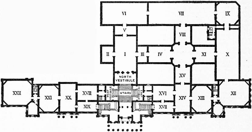
Fig. 1.—Plan of the National Gallery, London.
North Vestibule, Early Italian Schools: I. Tuscan School (15th and 16th centuries). II. Sienese School, &c. III. Tuscan School. IV. Lombard School. V. Ferrarese and Bolognese Schools. VI. Umbrian School, &c. VII. Venetian and Brescian Schools. VIII. Paduan and Early Venetian Schools. IX. Later Venetian School. X. Flemish School. XI. Early Dutch and Flemish Schools. XII. Dutch and Flemish Schools. XIII. Flemish School. XIV. Spanish School. XV. German Schools. XVI. French School. XVII. French School. XVIII. British School. XIX. Old British School. XX. British School. XXI. British School. XXII. Turner Collection. Octagonal Hall: Miscellaneous. East Vestibule: British School. West Vestibule: Italian School.
The collections most worthy of attention are the state galleries representative of international schools. Among these the British National Gallery holds a high place. The collection was founded in 1824 by the acquisition of the Angerstein State galleries of international schools. pictures. Its accessions are mainly governed by the parliamentary grant of £5000 to £10,000 a year, a sum which has occasionally been enlarged to permit special purchases. Thus, in 1871, the Peel collection of seventy-seven pictures was bought for £75,000, and in 1885 the Ansidei Madonna (Raphael) and Van Dyck’s portrait of Charles I. were bought, the one for £70,000 and the other for £17,500. In 1890 the government gave £25,000 to meet a gift of £30,000 made by three gentlemen to acquire three portraits by Moroni, Velazquez and Holbein. The most important private gifts were the Vernon gift in 1847, the Turner bequest in 1856 and the Wynne-Ellis legacy in 1876. Since 1905 the Art Collections Fund, a society of private subscribers, has also been responsible for important additions to the gallery, notably the Venus of Velazquez (1907). The gallery contains very few poor works and all schools are well represented, with the sole exception of the French school. This, however, can be amply studied at Hertford House (Wallace Collection), which, besides Dutch, Spanish and British pictures of the highest value, contains twenty examples of Greuze, fifteen by Pater, nineteen by Boucher, eleven by Watteau and fifteen by Meissonier. The national gallery of pictures at Berlin (Kaiser Friedrich Museum), like the British National Gallery, is remarkable for its variety of schools and painters, and for the select type of pictures shown. During the last twenty-five years of the 19th century, the development of this collection was even more striking than that of the English gallery. Italian and Dutch examples are specially numerous, though every school but the British (here as elsewhere) is really well seen. The purchase grant is considerable, and is well applied. Two other German capitals have collections of international importance—Dresden and Münich. The former is famous for the Sistine Madonna by Raphael, a work of such supreme excellence that there is a tendency to overlook other Italian pictures of celebrity by Titian, Giorgione and Correggio. Münich (Old Pinakothek) has examples of all the best masters, the South German school being particularly noticeable. The arrangement is good, and the methods of exhibition make this one of the most pleasant galleries on the continent. Vienna has the Imperial Gallery, a collection which in point of number cannot be considered large, as there are not more than 1700 pictures. This, however, is in itself a safeguard, like the wise provision in a statute of 1856 for enabling the English authorities to dispose of pictures “unfit for the collection, or not required.” It avoids the undue multiplication of canvases, and the overcrowding so noticeable in many Italian galleries where first-rate pictures hang too high to be examined. Thus the Viennese gallery, besides the intrinsic value of its pictures (Albert Durer’s chief work is there), is admirably adapted for study. The best gallery in Russia (St Petersburg, Hermitage) was made entirely by royal efforts, having been founded by Peter the Great, and much enlarged by the empress Catherine. It contains the collections of Crozat, Brühl and Walpole. There are about 1800 works, the schools of Flanders and Italy being of signal merit; and there are at least thirty-five genuine examples by Rembrandt. The French collection (Louvre Palace, Paris) is one of the most important of all. In 1880 it was undoubtedly the first gallery in Europe, but its supremacy has since been menaced by other establishments where acquisitions are made more frequently and with greater care, and where the system of classification is such that the value of the pictures is enhanced rather than diminished by their display. In 1900 it was partly rearranged with great effect. The feature of the Louvre is the Salon Carré, a room in which the supposed finest canvases in the collection are kept together, pictures of world-wide fame, representing all schools. It is now generally accepted that this system of selection not only lowers the standard of individual schools elsewhere by withdrawing their best pictures, but does not add to the aesthetic or educational value of the masterpieces themselves. In Florence the Tribuna room of the Uffizi gallery is a similar case in point. Probably the two most widely known pictures in the Louvre are Watteau’s second “Embarquement pour Cythère,” and the “Monna Lisa,” a portrait by Leonardo da Vinci, but each school has many unique examples. The original drawings should be noted, being of equal importance to the collection preserved at the British Museum. The last collection to be mentioned under this heading is that known as the Royal Galleries in Florence, housed in the Pitti and Uffizi palaces. In some ways this collection does not represent general painting sufficiently to justify its inclusion with the galleries of Berlin, Paris and London. On the other hand, the great number of Italian pictures of vital importance to the history of international art makes this one of the finest existing collections. The two great palaces, dating from the 15th and 16th centuries, are joined together and contain the Medici pictures. They form the largest gallery in the world, and though many of the rooms are small and badly lighted, and although many paintings have suffered from thoughtless restoration, they have a charm and attraction which certainly make them the most popular galleries in Europe. The Pitti has ten Raphaels and excellent examples of Andrea del Sarto, Giorgione and Perugino. The Uffizi is more representative of non-Italian schools, but is best known for its works by Botticelli, Leonardo da Vinci, Michelangelo and Sodoma, the schools of Tuscany and Umbria forming the bulk of both collections. Admission to the galleries is by payment, and the small income derived from this source is devoted to maintaining and enlarging the collections.
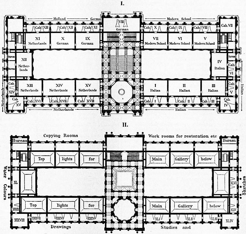
Fig. 2.—Plan of the first and second floors of the Imperial Gallery, Vienna.
As to the ground plans of the National Gallery, London (fig. 1), and of the Imperial Gallery at Vienna (fig. 2), it will be observed that while the former has the advantage of uniform top-light, the galleries at Vienna possess the most ample facilities for minute classification, small rooms or “cabinets” opening from each large room. Special rooms are also provided for drawings and water-colours, while special ranges of rooms are used by copyists and those responsible for the repair and preservation of the pictures.
Though not so comprehensive as the great collections just described, the state galleries showing national schools of painting and little else are of striking interest. In England the National Gallery of British Art (known as the State galleries of national schools. Tate Gallery) contains British pictures. The corresponding collection of modern French art is at Paris (Luxembourg Palace), Berlin, Rome, Dresden, Vienna and Madrid having analogous galleries. The Victoria and Albert Museum has also numerous British pictures, especially in water-colour, and the National Portrait Gallery, founded in 1856, and since 1896 housed in its permanent home, is instructive in this connexion, though many of its pictures are the work of foreign artists. The national collections at Dublin and Edinburgh may be mentioned here, though most schools are represented. Brussels and Antwerp are remarkable for fine examples of Flemish art—Matsys, Memlinc and Van Eyck of the primitive schools, Rubens and Van Dyck of the later period. The collections at Amsterdam (Ryks Museum) and the Hague (Mauritshuis) are a revelation to those who have only studied Rembrandt, Franz Hals, Van der Helst, and other Dutch portrait painters outside Holland; and in the former gallery especially, the pictures are arranged in a manner showing them to the best advantage. The Museo del Prado is even more noteworthy, for the fifty examples of Velasquez (outrivalling the Italian pictures, important as they are) make a visit to Madrid imperative to those who wish to realize the achievements of Spanish art. Christiania, Stockholm and Copenhagen have large collections of Scandinavian art, and the cities of Budapest and Basel have galleries of some importance. In Italy the state maintains twelve collections, mainly devoted to pictorial art. Of these the best are situated at Bologna, Lucca, Parma, Venice, Modena, Turin and Milan. In each case the local school of painting is fully represented. In Rome the Corsini and Borghese Galleries, the latter being the most catholic in the city, contain superb examples, some of them accepted masterpieces of Italian art; there are also good foreign pictures, but their number is limited. The Accademia at Florence should also be noted as the most important state gallery of early Italian art. The central Italian Renaissance can be more adequately studied here than in the Pitti. The “Primavera” of Botticelli, and the “Last Judgment” by Fra Angelico are perhaps the best-known works. The large statue of David by Michelangelo is also in this gallery, which, on the whole, is one of the most remarkable in Italy. Speaking broadly, these national galleries scattered throughout the country are not well arranged or classified; and though some are kept in fine old buildings, beautiful in themselves, the lighting is often indifferent, and it is with difficulty that the pictures can be seen. In nearly every case admission fees are charged every day, festivals and Sundays excepted; few pictures are bought, acquisitions being chiefly made by removing pictures from churches.
Many towns own collections of well-merited repute. In Italy such galleries are common, and among them may be noted Siena, with Sodoma and his school; Venice with Tintoretto (Doge’s Palace); Genoa, with the great Municipal galleries of special schools. palaces Balbi and Rosso; Vicenza (Montagna and school), Ferrara (Dosso and school), Bergamo and Milan (north Italian schools). Other civic collections of Italian art are maintained at Verona, Pisa, Rome, Perugia and Padua. In Holland, Haarlem, Leiden, Rotterdam and the Hague have galleries supplemental to those of the state, and are remarkable in showing the brilliance of artists like Grebber, de Bray and Ravesteyn, who are usually ignored. Birmingham and Manchester have good examples of modern British art. Moscow (Tretiakoff collection) has modern Russian pictures, and contemporary German and French work will be found in all the galleries of these two countries included in the municipal group. Collections of French work are found at Amiens, Rouen, Nancy, Tours, Le Mans and Angers, but large as these civic collections are, sometimes containing six and eight hundred canvases, few of their pictures are really good, many being the enormous patriotic canvases marked “Don de l’État,” which do not confer distinction on the galleries. Cologne has the central collection of the early Rhenish school; Nuremberg is remarkable for early German work (Wohlgemut, &c.). Stuttgart, Cassel (Dutch) and Hamburg (with a considerable number of British pictures) are also noteworthy, together with Brunswick, Hanover, Augsburg, Darmstadt and Düsseldorf, where German and Dutch art preponderate. Seville is famous for twenty-five examples of Murillo, and there are old Spanish paintings at Valencia, Cordova and Cadiz.
In Great Britain the best of the municipal galleries of general schools are at Liverpool (early Flemish and British), and at Glasgow (Scottish painters, Rembrandt, Van der Goes and Venetian schools). In France there are Municipal galleries of general schools. very large galleries at Tours, Montpellier, Lyons (Perugino, Rubens), Dijon and Grenoble (Italian), Valenciennes (Watteau and school), while Rennes, Lille and Marseilles have first-rate collections. Nantes, Orleans, Besançon, Cherbourg and Caen have also many paintings, French for the most part, but with occasional foreign pictures of real importance, presented by the state during the Napoleonic conquests, and not returned on the declaration of peace as were the works of art amassed in Paris. Some of the American collections have in recent years made a great advance in their acquisition of good pictures. At Boston (Museum of Fine Arts) all schools are represented, so too at the Metropolitan Museum of Art in New York, which is strong in Italian and Dutch works. Modern French and Flemish art is a feature of the Academy at Philadelphia, at the Lenox Library (New York), and at Chicago, where there are good examples of Millet, Constable and Rembrandt. The Corcoran bequest at Washington is of minor importance. The best civic collection in Germany of this class is the Städel Institute at Frankfort (Van Eyck, Christus, early Flemish and Italian).
As the great bulk of religious painting was executed for church decoration, there are still numberless churches which may be considered picture galleries. Thus at Antwerp cathedral the Rubens paintings are remarkable; at Churches. Ghent, Van Eyck; at Bruges (hospital of St John), Memlinc; at Pisa, the Campo Santo (early Tuscan schools); at Sant’ Apollinare, Ravenna, primitive Italo-Byzantine mosaics; at Siena, Pinturichio. Examples could be multiplied indefinitely—in Italy alone there are 80,000 churches and chapels, in all of which pictorial art has been employed. In Italy, besides the church “galleries” still used for religious services, there are some which have been secularized and are now used as museums, e.g. Certosa at Pavia, and San Vitale at Ravenna (mosaics); at Florence, the Scalzo (Andrea del Sarto); San Marco (Fra Angelico); the Riccardi and Pazzi chapels (Gozzoli and Perugino); at Milan, in the Santa Maria delle Grazie, the “Last Supper,” by Leonardo, and at Padua, the famous Arena chapel (Giotto).
The Vatican galleries, though best known for their statuary, have fine examples of painting, chiefly of the Italian school; the most famous easel picture is Raphael’s “Transfiguration,” but the Stanze, apartments entirely Private and semi-private galleries. decorated by painting, are even more famous. In England three royal palaces are open to the public—Hampton Court (Mantegna), Windsor (Van Dyck, Zuccarelli), and Kensington (portraits). At Buckingham Palace the Dutch pictures are admirable, and Queen Victoria lent the celebrated Raphael cartoons to the Victoria and Albert Museum. Semi-private collections belong to Dulwich College (Velasquez and Watteau), Oxford University (Italian drawings), the Soane Museum (Hogarth and English school), and the Royal Academy (Leonardo). Among private collections the most important are the Harrach, and Prince Liechtenstein (Vienna), J. Pierpont Morgan (including miniatures), Mrs J. Gardner of Boston (Italian), Prince Corsini (Florence). In Great Britain there are immense riches in private houses, though many collections have been dispersed. The most noteworthy (1909) belong to the dukes of Devonshire and Westminster, Lord Ellesmere, Captain Holford (including the masterpiece of Cuyp), Ludwig Mond, Lord Lansdowne, Miss Rothschild. The finest private collection is at Panshanger, formerly the seat of Lord Cowper, the gallery of Van Dyck’s work being quite the best in the world.
Many galleries are devoted to periodical exhibitions in London; the Royal Academy is the leading agency of this character, having held exhibitions since 1769. Its loan exhibitions of Old Masters are most important. Similar Periodical and commercial. enterprises are the New Gallery, opened in 1888, the Grafton Gallery, and others. There are also old-established societies of etchers, water-colourists, &c. A feature common to these exhibitions is that the public always pays for admission, though they differ from the commercial exhibitions, becoming more common every year, in which the work of a single school or painter is shown for profit. But the annual exhibitions at the Guildhall, under the auspices of the corporation, are free. The great periodical exhibition of French art is known as the Salon, and for some years it has had a rival in the Champ de Mars exhibition. These two societies are now respectively housed in the Grand Palais and Petit Palais, in the Champs Elysees, which were erected in connexion with the Paris Exhibition of 1900, but with the ultimate object of being devoted to the service of the two Salons. Berlin, Rome, Vienna and other Continental towns have regular exhibitions of original work.
The best history of art galleries is found in their official and other catalogues, see article Museums. See also L. Viardot, Les Musees d’Italie, &c. (3 vols., Paris, 1842, 1843, 1844); Annual Reports, official, of National Portrait Gallery, National Galleries of England, Ireland and Scotland; Civil Service Estimates, class iv. official. See also the series edited by Lafenestre and E. Richtenberger: Le Louvre, La Belgique, Le Hollande, Florence, Belgique; A. Lavice, Revue des musees de France,... d’Allemagne,... d’Angleterre,... d’Espagnc,... d’Italie,... de Belgique, de Hollande et de Russie (Paris, 1862-1872); E. Michel, Les Musées d’Allemagne (Paris, 1886); Kate Thompson, Public Picture Galleries of Europe (1880); C.L. Eastlake, Notes on Foreign Picture Galleries; Lord Ronald Gower, Pocket Guide to Art Galleries (public and private) of Belgium and Holland (1875); and many works, albums, and so forth, issued mainly for the sake of the illustrations.
(B.)
ARTHRITIS (from Gr. ἄρθρον, a joint), inflammation of the joints, in various forms of what are generally called gout and rheumatism (qq.v.).
ARTHROPODA, a name, denoting the possession by certain animals of jointed limbs, now applied to one of the three sub-phyla into which one of the great phyla (or primary branches) of coelomocoelous animals—the Appendiculata—is divided; the other two being respectively the Chaetopoda and the Rotifera. The word “Arthropoda” was first used in classification by Siebold and Stannius (Lehrbuch der vergleich. Anatomie, Berlin, 1845) as that of a primary division of animals, the others recognized in that treatise being Protozoa, Zoophyta, Vermes, Mollusca and Vertebrata. The names Condylopoda and Gnathopoda have been subsequently proposed for the same group. The word refers to the jointing of the chitinized exo-skeleton of the limbs or lateral appendages of the animals included, which are, roughly speaking, the Crustacea, Arachnida, Hexapoda (so-called “true insects”), Centipedes and Millipedes. This primary group was set up to indicate the residuum of Cuvier’s Articulata when his class Annelides (the modern Chaetopoda) was removed from that embranchement. At the same time C.T.E. von Siebold and H. Stannius renovated the group Vermes of Linnaeus, and placed in it the Chaetopods and the parasitic worms of Cuvier, besides the Rotifers and Turbellarian worms.1
The result of the knowledge gained in the last quarter of the 19th century has been to discredit altogether the group Vermes (see Worm), thus set up and so largely accepted by German writers even at the present day. We have, in fact, returned very nearly to Cuvier’s conception of a great division or branch, which he called Articulata, including the Arthropoda and the Chaetopoda (Annelides of Lamarck, a name adopted by Cuvier), and differing from it only by the inclusion of the Rotifera. The name Articulata, introduced by Cuvier, has not been retained by subsequent writers. The same, or nearly the same, assemblage of animals has been called Entomozoaria by de Blainville (1822), Arthrozoa by Burmeister (1843), Entomozoa or Annellata by H. Milne-Edwards (1855), and Annulosa by Alexander M‘Leay (1819), who was followed by Huxley (1856). The character pointed to by all these terms is that of a ring-like segmentation of the body. This, however, is not the character to which we now ascribe the chief weight as evidence of the genetic affinity and monophyletic (uni-ancestral) origin of the Chaetopods, Rotifers and Arthropods. It is the existence in each ring of the body of a pair of hollow lateral appendages or parapodia, moved by intrinsic muscles and penetrated by blood-spaces, which is the leading fact indicating the affinities of these great sub-phyla, and uniting them as blood-relations. The parapodia (fig. 8) of the marine branchiate worms are the same things genetically as the “legs” of Crustacea and Insects (figs. 10 and 11). Hence the term Appendiculata was introduced by Lankester (preface to the English edition of Gegenbaur’s Comparative Anatomy, 1878) to indicate the group. The relationships of the Arthropoda thus stated are shown in the subjoined table:—
Phylum—Appendiculata. { Sub-phylum 1. Rotifera.
” 2. Chaetopoda.
” 3. Arthropoda.
The Rotifera are characterized by the retention of what appears in Molluscs and Chaetopods as an embryonic organ, the velum or ciliated prae-oral girdle, as a locomotor and food-seizing apparatus, and by the reduction of the muscular parapodia to a rudimentary or non-existent condition in all present surviving forms except Pedalion. In many important respects they are degenerate—reduced both in size and elaboration of structure.
The Chaetopoda are characterized by the possession of horny epidermic chaetae embedded in the integument and moved by muscles. Probably the chaetae preceded the development of parapodia, and by their concentration and that of the muscular bundles connected with them at the sides of each segment, led directly to the evolution of the parapodia. The parapodia of Chaetopoda are never coated with dense chitin, and are, therefore, never converted into jaws; the primitive “head-lobe” or prostomium persists, and frequently carries eyes and sensory tentacles. Further, in all members of the sub-phylum Chaetopoda the relative position of the prostomium, mouth and peristomium or first ring of the body, retains its primitive character. We do not find in Chaetopoda that parapodia, belonging to primitively post-oral rings or body-segments (called “somites,” as proposed by H. Milne-Edwards), pass in front of the mouth by adaptational shifting of the oral aperture. (See, however, 8.)
The Arthropoda might be better called the “Gnathopoda,” since their distinctive character is, that one or more pairs of appendages behind the mouth are densely chitinized and turned (fellow to fellow on opposite sides) towards one another so as to act as jaws. This is facilitated by an important general change in the position of the parapodia; their basal attachments are all more ventral in position than in the Chaetopoda, and tend to approach from the two sides towards the mid-ventral line. Very usually (but not in the Onychophora = Peripatus) all the parapodia are plated with chitin secreted by the epidermis, and divided into a series of joints—giving the “arthropodous” or hinged character.
There are other remarkable and distinctive features of structure which hold the Arthropoda together, and render it impossible to conceive of them as having a polyphyletic origin, that is to say, as having originated separately by two or three distinct lines of descent from lower animals; and, on the contrary, establish the view that they have been developed from a single line of primitive Gnathopods which arose by modification of parapodiate annulate worms not very unlike some of the existing Chaetopods. These additional features are the following—(1) All existing Arthropoda have an ostiate heart and have undergone “phleboedesis,” that is to say, the peripheral portions of the blood-vascular system are not fine tubes as they are in the Chaetopoda and as they were in the hypothetical ancestors of Arthropoda, but are swollen so as to obliterate to a large extent the coelom, whilst the separate veins entering the dorsal vessel or heart have coalesced, leaving valvate ostia (see fig. 1) by which the blood passes from a pericardial blood-sinus formed by the fused veins into the dorsal vessel or heart (see Lankester’s Zoology, part ii., introductory chapter, 1900). The only exception to this is in the case of minute degenerate forms where the heart has disappeared altogether. The rigidity of the integument caused by the deposition of dense chitin upon it is intimately connected with the physiological activity and form of all the internal organs, and is undoubtedly correlated with the total disappearance of the circular muscular layer of the body-wall present in Chaetopods. (2) In all existing Arthropoda the region in front of the mouth is no longer formed by the primitive prostomium or head-lobe, but one or more segments, originally post-oral, with their appendages have passed in front of the mouth (prosthomeres). At the same time the prostomium and its appendages cease to be recognizable as distinct elements of the head. The brain no longer consists solely of the nerve-ganglion-mass proper to the prostomial lobe, as in Chaetopoda, but is a composite (syncerebrum) produced by the fusion of this and the nerve-ganglion-masses proper to the prosthomeres or segments which pass forwards, whilst their parapodia (= appendages) become converted into eye-stalks, and antennae, or more rarely grasping organs. (3) As in Chaetopoda, coelomic funnels (coelomoducts) may occur right and left as pairs in each ring-like segment or somite of the body, and some of these are in all cases retained as gonoducts and often as renal excretory organs (green glands, coxal glands of Arachnida, not crural glands, which are epidermal in origin); but true nephridia, genetically identical with the nephridia of earthworms, do not occur (on the subject of coelom, coelomoducts and nephridia, see the introductory chapter of part ii. of Lankester’s Treatise on Zoology).
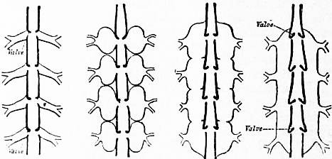
After Lankester, Q. J. Mic. Sci. vol. xxxiv., 1893.
Fig. 1.—Diagram to show the gradual formation of the Arthropod pericardial blood-sinus and “ostiate” heart by the swelling up (phleboedesis) of the veins entering the dorsal vessel or heart of a Chaetopod-like ancestor. The figure on the left represents the condition in a Chaetopod, that on the right the condition in an Arthropod, the other two are hypothetical intermediate forms.
Tabular Statement of the Grades, Classes and Sub-classes of the Arthropoda.—It will be convenient now to give in the clearest form a statement of the larger subdivisions of the Arthropoda which it seems necessary to recognize at the present day. The justification of the arrangement adopted will form the substance of the rest of the present article. The orders included in the various classes are not discussed here, but are treated of under the following titles:—Peripatus (Onychophora), Centipede and Millipede (Myriapoda), Hexapoda (Insecta), Arachnida and Crustacea.
Sub-Phylum ARTHROPODA (of the Phylum Appendiculata). Grade A. Hyparthropoda (hypothetical forms connecting ancestors of Chaetopoda with those of Arthropoda). Grade B. Protarthropoda. Class Onychophora. Ex.—Peripatus. Grade C. Euarthropoda. Class 1. Diplopoda. Ex.—Julus. Class 2. Arachnida. Grade a. Anomomeristica. Ex.—Phacops. Grade b. Nomomeristica. (a) Pantopoda. Ex.—Pycnogonum. (b) Euarachnida. Ex.—Limulus, Scorpio, Mygale, Acarus. Class 3. Crustacea. Grade a. Entomostraca. Ex.—Apus, Branchipus, Cyclops, Balanus. Grade b. Malacostraca. Ex.—Nebalia, Astacus, Oniscus, Gammarus. Class 4. Chilopoda. Ex.—Scolopendra. Class 5. Hexapoda (syn. Insecta Pterygota). Ex.—Locusta, Phryganea, Papilio, Apis, Mnsca, Cimex, Lucanus, Machilis. Incertae sedis—Tardigrada, Pentastomidae (degenerate forms).
The Segmentation of the Body of Arthropoda.—The body of the Arthropoda is more or less clearly divided into a series of rings, segments, or somites which can be shown to be repetitions one of another, possessing identical parts and organs which may be larger or smaller, modified in shape or altogether suppressed in one somite as compared with another. A similar constitution of the body is more clearly seen in the Chaetopod worms. In the Vertebrata also a repetition of units of structure (myotomes, vertebrae, &c.)—which is essentially of the same nature as the repetition in Arthropods and Chaetopods, but in many respects subject to peculiar developments—is observed. The name “metamerism” has been given to this structural phenomenon because the “meres,” or repeated units, follow one another in line. Each such “mere” is often called a “metamere.” A satisfactory consideration of the structure of the Arthropods demands a knowledge of what may be called the laws of metamerism, and reference should be made to the article under that head.
From Goodrich, Q. J. Micr. Sci. vol. xi p. 247.
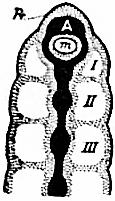
Fig. 2.—Diagram of the head and adjacent region of an Ohgochaet Chaetopod.
Pr, The prostomium. m, The mouth. A, The prostomial ganglion-mass or archi-cerebrum. I, II, III, coelom of the first, second and third somites.
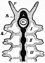
Fig. 3—Diagram of the head and adjacent region of a Polychaet Chaetopod Letters as in fig. 1, with the addition of T, prostomial tentacle; Pa, parapodium. (From Goodrich.)
The Theory of the Arthropod Head.—The Arthropod head is a tagma or group of somites which differ in number and in their relative position in regard to the mouth, in different classes. In a simple Chaetopod (fig. 2) the head consists of the first somite only; that somite is perforated by the mouth, and is provided with a prostomium or prae-oral lobe. The prostomium is essentially a part or outgrowth of the first somite, and cannot be regarded as itself a somite. It gives rise to a nerve-ganglion mass, the prostomial ganglion. In the marine Chaetopods (the Polychaeta) (fig. 3), we find the same essential structure, but the prostomium may give rise to two or more tactile tentacles, and to the vesicular eyes. The somites have well-marked parapodia, and the second and third, as well as the first, may give rise to tentacles which are directed forward, and thus contribute to form “the head.” But the mouth remains as an inpushing of the wall of the first somite.
The Arthropoda are all distinguished from the Chaetopoda by the fact that the head consists of one or more somites which lie in front of the mouth (now called prosthomeres), as well as of one or more somites behind it (opisthomeres). The first of the post-oral somites invariably has its parapodia modified so as to form a pair of hemignaths (mandibles). About 1870 the question arose for discussion whether the somites in front of the mouth are to be considered as derived from the prostomium of a Chaetopod-like ancestor. Milne-Edwards and Huxley had satisfied themselves with discussing and establishing, according to the data at their command, the number of somites in the Arthropod head, but had not considered the question of the nature of the prae-oral somites. Lankester (2) was the first to suggest that (as is actually the fact in the Nauplius larva of the Crustacea) the prae-oral somites or prosthomeres and their appendages were ancestrally post-oral, but have become prae-oral “by adaptational shifting of the oral aperture.” This has proved to be a sound hypothesis and is now accepted as the basis upon which the Arthropod head must be interpreted (see Korschelt and Heider (3)). Further, the morphologists of the ’fifties appear, with few exceptions, to have accepted a preliminary scheme with regard to the Arthropod head and Arthropod segmentation generally, which was misleading and caused them to adopt forced conclusions and interpretations. It was conceived by Huxley, among others, that the same number of cephalic somites would be found to be characteristic of all the diverse classes of Arthropoda, and that the somites, not only of the head but of the various regions of the body, could be closely compared in their numerical sequence in classes so distinct as the Hexapods, Crustaceans and Arachnids.
The view which it now appears necessary to take is, on the contrary, this—viz that all the Arthropoda are to be traced to a common ancestor resembling a Chaetopod worm, but differing from it in having lost its chaetae and in having a prosthomere in front of the mouth (instead of prostomium only) and a pair of hemignaths (mandibles) on the parapodia of the buccal somite. From this ancestor Arthropods with heads of varying degrees of complexity have been developed characteristic of the different classes, whilst the parapodia and somites of the body have become variously modified and grouped in these different classes. The resemblances which the members of one class often present to the members of another class in regard to the form of the limb-branches (rami) of the parapodia and the formation of tagmata (regions) are not hastily to be ascribed to common inheritance, but we must consider whether they are not due to homoplasy—that is, to the moulding of natural selection acting in the different classes upon fairly similar elements under like exigencies.
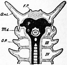
Fig. 4.—Diagram of the head and adjacent region of Peripatus. Monoprosthomerous.
m, Mouth. I, Coelom of the first somite which carries the antennae and is in front of the mouth. II, Coelom of the second somite which carries the mandibles (hence deuterognathous). III and IV, Coelom of the third and fourth somites. FP, Rudimentary frontal processes perhaps representing the prostomial tentacles of Polychaeta. Ant, Antenna or tactile tentacle. Md, Mandible. Op, Oral-papilla. P, Protocerebrum or foremost cerebral mass belonging to the first somite. D, Deuterocerebrum, consisting of ganglion cells belonging to the second or mandibular somite. (After Goodrich.)
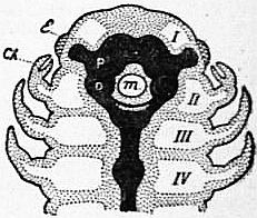
Fig. 5.—Diagram of the head and adjacent region of an Arachnid. Diprosthomerous in the adult condition, though embryologically the appendages of somite II and the somite itself are, as here drawn, not actually in front of the mouth.
E, Lateral eye. Ch Chelicera. m, Mouth. P, Protocerebrum, D, Deuterocerebrum. I, II III, IV. Coelom of the first, second, third and fourth somites. (After Goodrich.)
The structure of the head in Arthropods presents three profoundly separated grades of structure dependent upon the number of prosthomeres which have been assimilated by the prae-oral region. The classes presenting these distinct plans of head-structure cannot be closely associated in any scheme of classification professing to be natural. Penpatus, the type-genus of the class Onychophora, stands at the base of the series with only a single prosthomere (fig. 4). In Peripatus the prostomium of the Chaetopod-like ancestor is atrophied, but it is possible that two processes on the front of the head (FP) represent in the embryo the dwindled prostomial tentacles. The single prosthomere carries the retractile tentacles as its “parapodia.” The second somite is the buccal somite (II, fig. 4); its parapodia have horny jaws on their ends, like the claws on the following legs (fig. 9), and act as hemignaths (mandibles). The study of sections of the embryo establishes these facts beyond doubt. It also shows us that the neuromeres, no less than the embryonic coelomic cavities, point to the existence of one, and only one, prosthomerp in Peripatus, of which the “protocerebrum,” P, is the neuromere, whilst the deuterocerebrum, D, is the neuromere of the second or buccal somite. A brief indication of these facts is given by saying that the Onychophora are “deuterognathous”—that is to say, that the buccal somite carrying the mandibular hemignaths is the second of the whole series.
What has become of the nerve-ganglion of the prostomial lobe of the Chaetopod in Peripatus is not clearly ascertained, nor is its fate indicated by the study of the embryonic head of other Arthropods so far. Probably it is fused with the protocerebrum, and may also be concerned in the history of the very peculiar paired eyes of Peripatus, which are like those of Chaetopods in structure—viz vesicles with an intravesicular lens, whereas the eyes of all other Arthropods have essentially another structure, being “cups” of the epidermis, in which a knob-like or rod-like thickening of the cuticle is fitted as refractive medium.
In Diplopoda (Julus, &c.) the results of embryological study point to a composition of the front part of the head exactly similar to that which we find in Onychophora. They are deuterognathous.
The Arachnida present the first stage of progress. Here embryology shows that there are two prosthomeres (fig. 5), and that the gnathobases of the chelae which act as the first pair of hemignaths are carried by the third somite. The Arachnida are therefore tritognathous. The two prosthomeres are indicated by their coelomic cavities in the embryo (I and II, fig. 5), and by two neuromeres, the protocerebrum and the deuterocerebrum. The appendages of the first prosthomere are not present as tentacles, as in Peripatus and Diplopods, but are possibly represented by the eyes or possibly altogether aborted. The appendages of the second prosthomere are the well-known chelicerae of the Arachnids, rarely, if ever, antenniform, but modified as “retroverts” or clasp-knife tangs in spiders.
The Crustacea (fig. 6) and the Hexapoda (fig. 7) agree in having three somites in front of the mouth, and it is probable, though not ascertained, that the Chilopoda (Scolopendra, &c.) are in the same case. The three prosthomeres or prae-oral somites of Crustacea due to the sinking back of the mouth one somite farther than in Arachnida are not clearly indicated by coelomic cavities in the embryo, but their existence is clearly established by the development and position of the appendages and by the neuromeres.
The eyes in some Crustacea are mounted on articulated stalks, and from the fact that they can after injury be replaced by antenna-like appendages it is inferred that they represent the parapodia of the most anterior prosthomere. The second prosthomere carries the first pair of antennae and the third the second pair of antennae. Sometimes the pair of appendages has not a merely tactile jointed ramus, but is converted into a claw or clasper. Three neuromeres—a proto-, deutero-, and trito-cerebrum—corresponding to those three prosthomeres are sharply marked in the embryo. The fourth somite is that in which the mouth now opens, and which accordingly has its appendages converted into hemignathous mandibles. The Crustacea are tetartognathous.
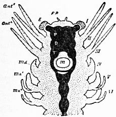
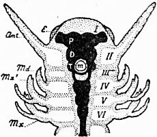
Fig. 6.—Diagram of the head of a Crustacean. Tri-prosthomerous. Fig. 7.—Diagram of the head of a Hexapod insect.
FP, Frontal processes (observed in Cirrhiped nauplius-larvae) probably representing the prostomial tentacles of Chaetopods. e, Eye. Ant1, First pair of antennae. Ant2, Second pair of antennae. md, Mandible. mx1, mx2, First and second pairs of maxillae. m, Mouth. I, II, and III, The three prosthomeres. IV, V, VI, The three somites following the mouth. P, Protocerebrum. D, Deuterocerebrum. T, Tritocerebrum. (After Goodrich.) e, Eye. ant, Antenna. md, Mandible. mx1, First maxilla. mx2, Second maxilla. m, Mouth. I, Region of the first or eye-bearing prosthomere. II, Coelom of the second antenna-bearing prosthomere. III, Coelom of the third prosthomere devoid of appendages. IV, V, and VI, Coelom of the fourth, fifth and sixth somites. P, Protocerebrum belonging to the first prosthomere. D, Deuterocerebrum belonging to the second prosthomere. T, Tritocerebrum belonging to the third prosthomere. (After Goodrich.)
The history of the development of the head has been carefully worked out in the Hexapod insects. As in Crustacea and Arachnida, a first prosthomere is indicated by the paired eyes and the protocerebrum; the second prosthomere has a well-marked coelomic cavity, carries the antennae, and has the deuterocerebrum for its neuromere. The third prosthomere is represented by a well-marked pair of coelomic cavities and the tritocerebrum (III, fig. 7), but has no appendages. They appear to have aborted. The existence of this third prosthomere corresponding to the third prosthomere of the Crustacea is a strong argument for the derivation of the Hexapoda, and with them the Chilopoda, from some offshoot of the Crustacean stem or class. The buccal somite, with its mandibles, is in Hexapoda, as in Crustacea, the fourth: they are tetartognathous.
The adhesion of a greater or less number of somites to the buccal somite posteriorly (opisthomeres) is a matter of importance, but of minor importance, in the theory and history of the Arthropod head. In Peripatus no such adhesion or fusion occurs. In Diplopoda two opisthomeres—that is to say, one in addition to the buccal somite—are united by a fusion of their terga with the terga of the prosthomeres. Their appendages are respectively the mandibles and the gnathochilarium.
In Arachnida the highest forms exhibit a fusion of the tergites of five post-oral somites to form one continuous carapace united with the terga of the two prosthomeres. The five pairs of appendages of the post-oral somites of the head or prosoma thus constituted all primitively carry gnathobasic projections on their coxal joints, which act as hemignaths: in the more specialized forms the mandibular gnathobases cease to develop.
In Crustacea the fourth or mandibular somite never has less than the two following somites associated with it by the adaptation of their appendages as jaws, and the ankylosis of their terga with that of the prosthomeres. But in higher Crustacea the cephalic “tagma” is extended, and more somites are added to the fusion, and their appendages adapted as jaws of a kind.
The Hexapoda are not known to us in their earlier or more primitive manifestations; we only know them as possessed of a definite number of somites arranged in definite numbers in three great tagmata. The head shows two jaw-bearing somites besides the mandibular somite (V, VI, in fig. 7)—thus six in all (as in some Crustacea), including prosthomeres, all ankylosed by their terga to form a cephalic shield. There is, however, good embryological evidence in some Hexapods of the existence of a seventh somite, the supra-lingual, occurring between the somite of the mandibles and the somite of the first maxillae (4). This segment is indicated embryologically by its paired coelomic cavities. It is practically an excalated somite, having no existence in the adult. It is probably not a mere coincidence that the Hexapod, with its two rudimentary somites devoid of appendages, is thus found to possess twenty-one somites, including that which carries the anus, and that this is also the number present in the Malacostracous Crustacea.
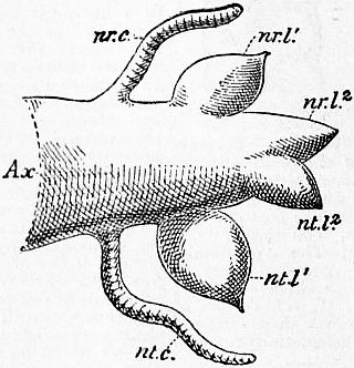
Fig. 8.—Diagram of the somite-appendage or parapodium of a Polychaet Chaetopod. The chaetae are omitted.
Ax, The axis. nr.c, Neuropodial cirrhus. nr.l1, nr.l2, Neuropodial lobes or endites. nt.c. Notopodial cirrhus. nt.l1, nt.l2, Notopodial lobes or exites. The parapodium is represented with its neural or ventral surface uppermost. (Original).
The Segmented Lateral Appendages or Limbs of Arthropoda.—It has taken some time to obtain any general acceptance of the view that the parapodia of the Chaetopoda and the limbs of Arthropoda are genetically identical structures; yet if we compare the parapodium of Tomopteris or of Phyllodoce with one of the foliaceous limbs of Branchipus or Apus, the correspondences of the two are striking. An erroneous view of the fundamental morphology of the Crustacean limb, and consequently of that of other Arthropoda, came into favour owing to the acceptance of the highly modified limbs of Astacus as typical. Protopodite, endopodite, exopodite, and epipodite were considered to be the morphological units of the crustacean limb. Lankester (5) has shown (and his views have been accepted by Professors Korschelt and Heider in their treatise on Embryology) that the limb of the lowest Crustacea, such as Apus, consists of a corm or axis which may be jointed, and gives rise to outgrowths, either leaf-like or filiform, on its inner and outer margins (endites and exites). Such a corm (see figs. 10 and 11), with its outgrowths, may be compared to the simple parapodia of Chaetopoda with cirrhi and branchial lobe (fig. 8). It is by the specialization of two “endites” that the endopodite and exopodite of higher Crustacea are formed, whilst a flabelliform exite is the homogen or genetic equivalent of the epipodite (see Lankester, “Observations and Reflections on Apus Cancriformis,” Q. J. Micr. Sci.). The reduction of the outgrowth-bearing “corm” of the parapodium of either a Chaetopod or an Arthropod to a simple cylindrical stump, devoid of outgrowths, is brought about when mechanical conditions favour such a shape. We see it in certain Chaetopods (e.g. Hesione) and in the Arthropod Peripatus (fig. 9). The conversion of the Arthropod’s limb into a jaw, as a rule, is effected by the development of an endite near its base into a hard, chitinized, and often toothed gnathobase (see figs. 10 and 11, en1). It is not true that all the biting processes of the Arthropod limb are thus produced—for instance, the jaws of Peripatus are formed by the axis or corm itself, whilst the poison-jaws of Chilopods, as also their maxillae, appear to be formed rather by the apex or terminal region of the ramus of the limb; but the opposing jaws (= hemignaths) of Crustacea, Arachnida and Hexapoda are gnathobases, and not the axis or corm. The endopodite (corresponding to the fifth endite of the limb of Apus, see fig. 10) becomes in Crustacea the “walking leg” of the mid-region of the body; it becomes the palp or jointed process of anterior segments. A second ramus, the “exopodite,” often is also retained in the form of a palp or feeler. In Apus, as the figure shows, there are four of these “antenna-like” palps or filaments on the first thoracic limb. A common modification of the chief ramus of the Arthropod parapodium is the chela or nipper formed by the elongation of the penultimate joint of the ramus, so that the last joint works on it—as, for instance, in the lobster’s claw. Such chelate rami or limb-branches are independently developed in Crustacea and in Arachnida, and are carried by somites of the body which do not correspond in position in the two groups. The range of modification of which the rami or limb-branches of the limbs of Arthropoda are capable is very large, and in allied orders or even families or genera we often find what is certainly the palp of the same appendage (as determined by numerical position of the segments)—in one case antenniform, in another chelate, in another pediform, and in another reduced to a mere stump or absent altogether. Very probably the power which the appendage of a given segment has of assuming the perfected form and proportions previously attained by the appendage of another segment must be classed as an instance of “homoeosis,” not only where such a change is obviously due to abnormal development or injury, but also where it constitutes a difference permanently established between allied orders or smaller groups, or between the two sexes.
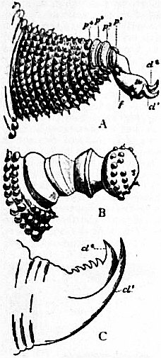
Fig. 9.—Three somite-appendages or parapodia of Peripatus.
A, A walking leg; p1 to p4, the characteristic “pads”; f, the foot; cl1, cl2, the two claws. B, An oral papilla, one of the second pair of post-oral appendages. C, One of the first post-oral pair of appendages or mandibles; cl1, cl2, the greatly enlarged claws. (Compare A.) The appendages are represented with the neural or ventral surface uppermost.
The most extreme disguise assumed by the Arthropod parapodium or appendage is that of becoming a mere stalk supporting an eye—a fact which did not obtain general credence until the experiments of Herbst in 1895, who found, on cutting off the eye-stalk of Palaemon, that a jointed antenna-like appendage was regenerated in its place. Since the eye-stalks of Podophthalmate Crustacea represent appendages, we are forced to the conclusion that the sessile eyes of other Crustacea, and of other Arthropoda generally, indicate the position of appendages which have atrophied.2
From what has been said, it is apparent that we cannot, in attempting to discover the affinities and divergences of the various forms of Arthropoda, attach a very high phylogenetic value to the coincidence or divergence in form of the appendages belonging to the somites compared with one another.
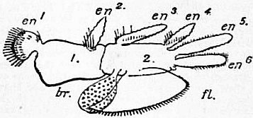
After Lankester, Q. J. Mic. Sci. vol. xxi., 1881.
Fig. 10.—The second thoracic (fifth post-oral) appendage of the left side of Apus cancriformis, placed with its ventral or neural surface uppermost to compare with figs. 8 and 9.
1, 2, The two segments of the axis. en1, The gnathobase. en2 to en6, The five following “endites.” fl, The flabellum or anterior exite. br, The bract or posterior exite.
The principal forms assumed by the Arthropod parapodium and its rami may be thus enumerated:—
(1) Axial corm well developed, unsegmented or with two to four segments; lateral endites and exites (rami) numerous and of various lengths (certain limbs of lower Crustacea).
(2) Corm, with short unsegmented rami, forming a flattened foliaceous appendage, adapted to swimming and respiration (trunk-limbs of Phyllopods).
(3) Corm alone developed; with no endites or exites, but provided with terminal chitinous claws (ordinary leg of Peripatus), with terminal jaw teeth (jaw of Peripatus), or with blunt extremity (oral papilla of same) (see fig. 9).
(4) Three of the rami of the primitive limb (endites 5 and 6, and exite 1) specially developed as endopodite, exopodite, and epipodite—the first two often as firm and strongly chitinized, segmented, leg-like structures; the original axis or corm reduced to a basal piece, with or without a distinct gnathobase (endite 1)—typical tri-ramose limb of higher Crustacea.
(5) One ramus (the endopodite) alone developed—the original axis or corm serving as its basal joint with or without gnathobase. This is the usual uni-ramose limb found in the various classes of Arthropoda. It varies as to the presence or absence of the jaw-process and as to the stoutness of the segments of the ramus, their number (frequently six, plus the basal corm), and the modification of the free end. This may be filiform or brush-like or lamellate when it is an antenna or palp; a simple spike (walking leg of Crustacea, of other aquatic forms, and of Chilopods and Diplopods); the terminal joint flattened (swimming leg of Crustacea and Gigantostraca); the terminal joint provided with two or with three recurved claws (walking leg of many terrestrial forms—e.g. Hexapoda and Arachnida); the penultimate joint with a process equal in length to the last joint, so as to form a nipping organ (chelae of Crustaceans and Arachnids); the last joint reflected and movable on the penultimate, as the blade of a clasp-knife on its handle (the retrovert, toothed so as to act as a biting jaw in the Hexapod Mantis, the Crustacean Squilla and others); with the last joint produced into a needle-like stabbing process in spiders.
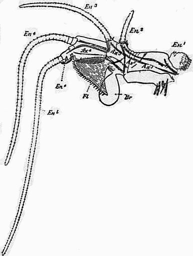
After Lankester, Q. J. Mic. Sci. vol. xxi., 1881.
Fig. 11.—The first thoracic (fourth post-oral) appendage of Apus cancriformis (right side).
Ax1 to Ax4, the four segments of the axis with muscular bands. En1, Gnathobase. En2 to En5, The elongated jointed endites (rami). En6, The rudimentary sixth endite (exopodite of higher Crustacea). Fl, The flabellum which becomes the epipodite of higher forms. Br, The bract devoid of muscles and respiratory in function.
(6) Two rami developed (usually, but perhaps not always, the equivalents of the endopodite and exopodite) supported on the somewhat elongated corm (basal segment). This is the typical “bi-ramose limb” often found in Crustacea. The rami may be flattened for swimming, when it is “a bi-ramose swimmeret,” or both or only one may be filiform and finely annulate; this is the form often presented by the antennae of Crustacea, and rarely by prae-oral appendages in other Arthropods.
(7) The endopoditic ramus is greatly enlarged and flattened, without or with only one jointing, the corm (basal segment) is evanescent; often the plate-like endopodites of a pair of such appendages unite in the middle line with one another or by the intermediary of a sternal up-growth and form a single broad plate. These are the plate-like swimmerets and opercula of Gigantostraca and Limulus among Arachnids and of Isopod Crustaceans. They may have rudimentary exopodites, and may or may not have branchial filaments or lamellae developed on their posterior faces. The simplest form to which they may be reduced is seen in the genital operculum of the scorpion.
(8) The gnathobase becomes greatly enlarged and not separated by a joint from the corm; it acts as a hemignath or half jaw working against its fellow of the opposite side. The endopodite may be retained as a small segmented palp at the side of the gnathobase or disappear (mandible of Crustacea, Chilopoda and Hexapoda).
(9) The corm becomes the seat of a development of a special visual organ, the Arthropod eye (as opposed to the Chaetopod eye). Its jointing (segmentation) may be retained, but its rami disappear (Podophthalmous Crustacea). Usually it becomes atrophied, leaving the eye as a sessile organ upon the prae-oral region of the body (the eye-stalk and sessile lateral eyes of Arthropoda generally, exclusive of Peripatus).
(10) The forms assumed by special modification of the elements of the parapodium in the maxillae, labium, &c., of Hexapods, Chilopods, Diplopods, and of various Crustacea, deserve special enumeration, but cannot be dealt with without ample space and illustration.
It may be pointed out that the most radical difference presented in this list is that between appendages consisting of the corm alone without rami (Onychophora) and those with more or less developed rami (the rest of the Arthropoda). In the latter class we should distinguish three phases: (a) those with numerous and comparatively undeveloped rami; (b) those with three, or two highly developed rami, or with only one—the corm being reduced to the dimensions of a mere basal segment; (c) those reduced to a secondary simplicity (degeneration) by overwhelming development of one segment (e.g. the isolated gnathobase often seen as “mandible” and the genital operculum).
There is no reason to suppose that any of the forms of limb observed in Arthropoda may not have been independently developed in two or more separate diverging lines of descent.
Branchiae.—In connexion with the discussion of the limbs of Arthropods, a few words should be devoted to the gill-processes. It seems probable that there are branchial plumes or filaments in some Arthropoda (some Crustacea) which can be identified with the distinct branchial organs of Chaetopoda, which lie dorsal of the parapodia and are not part of the parapodium. On the other hand, we cannot refuse to admit that any of the processes of an Arthropod parapodium may become modified as branchial organs, and that, as a rule, branchial out-growths are easily developed, de novo, in all the higher groups of animals. Therefore, it seems to be, with our present knowledge, a hopeless task to analyse the branchial organs of Arthropoda and to identify them genetically in groups.
A brief notice must suffice of the structure and history of the Eyes, the Tracheae and the so-called Malpighian tubes of Arthropoda, though special importance attaches to each in regard to the determination of the affinities of the various animals included in this great sub-phylum.
The Eyes.—The Arthropod eye appears to be an organ of special character developed in the common ancestor of the Euarthropoda, and distinct from the Chaetopod eye, which is found only in the Onychophora where the true Arthropod eye is absent. The essential difference between these two kinds of eye appears to be that the Chaetopod eye (in its higher developments) is a vesicle enclosing the lens, whereas the Arthropod eye is a pit or series of pits into which the heavy chitinous cuticle dips and enlarges knobwise as a lens. Two distinct forms of the Arthropod eye are observed—the monomeniscous (simple) and the polymeniscous (compound). The nerve-end-cells, which lie below the lens, are part of the general epidermis. They show in the monomeniscous eye (see article Arachnida, fig. 26) a tendency to group themselves into “retinulae,” consisting of five to twelve cells united by vertical deposits of chitin (rhabdoms). In the case of the polymeniscous eye (fig. 23, article Arachnida) a single retinula or group of nerve-end-cells is grouped beneath each associated lens. A further complication occurs in each of these two classes of eye. The monomeniscous eye is rarely provided with a single layer of cells beneath its lens; when it is so, it is called monostichous (simple lateral eye of Scorpion, fig. 22, article Arachnida). More usually, by an infolding of the layer of cells in development, we get three layers under the lens; the front layer is the corneagen layer, and is separated by a membrane from the other two which, more or less, fuse and contain the nerve-end-cells (retinal layer). These eyes are called diplostichous, and occur in Arachnida and Hexapoda (fig. 24, article Arachnida).
On the other hand, the polymeniscous eye undergoes special elaboration on its lines. The retinulae become elongated as deep and very narrow pits (fig. 12 and explanation), and develop additional cells near the mouth of the narrow pit. Those nearest to the lens are the corneagen cells of this more elaborated eye, and those between the original retinula cells and the corneagen cells become firm and transparent. They are the crystalline cells or vitrella (see Watase, 7). Each such complex of cells underlying the lenticle of a compound eye is called an “ommatidium”; the entire mass of cells underlying a monomeniscous eye is an “ommataeum.” The ommataeum, as already stated, tends to segregate into retinulae which correspond potentially each to an ommatidium of the compound eye. The ommatidium is from the first segregate and consists of few cells. The compound eye of the king-crab (Limulus) is the only recognized instance of ommatidia in their simplest state. Each can be readily compared with the single-layered lateral eye of the scorpion. In Crustacea and Hexapoda of all grades we find compound eyes with the more complicated ommatidia described above. We do not find them in any Arachnida.
It is difficult in the absence of more detailed knowledge as to the eyes of Chilopoda and Diplopoda to give full value to these facts in tracing the affinities of the various classes of Arthropods. But they seem to point to a community of origin of Hexapods and Crustacea in regard to the complicated ommatidia of the compound eye, and to a certain isolation of the Arachnida, which are, however, traceable, so far as the eyes are concerned, to a distant common origin with Crustacea and Hexapoda through the very simple compound eyes (monostichous, polymeniscous) of Limulus.
Fig. 12.—Diagram to show the derivation of the unit or “ommatidium” of the compound eye of Crustacea and Hexapoda, C, from a simple monomeniscous monostichous eye resembling the lateral eye of a scorpion, A, or the unit of the compound lateral eye of Limulus (see article Arachnida, figs. 22 and 23). B represents an intermediate hypothetical form in which the cells beneath the lens are beginning to be superimposed as corneagen, vitrella and retinula, instead of standing side by side in horizontal series. The black represents the cuticular product of the epidermal cells of the ocular area, taking the form either of lens, cl, of crystalline body, cry, or of rhabdom, rhab; hy, hypodermis or epidermal cells; corn1, laterally-placed cells in the simpler stage, A, which like the nerve-end cells, vit1 and ret1, are corneagens or lens-producing; corn, specialized corneagen or lens-producing cells; vit1, potential vitrella cells with cry1, potential crystalline body now indistinguishable from retinula cells and rhabdomeres; vit, vitrella cell with cry, its contained cuticular product, the crystalline cone or body; ret1, rhab1, retinula cells and rhabdom of scorpion undifferentiated from adjacent cells, vit1; ret, retinula cell; rhab, rhabdom; nf, optic nerve-fibres. (Modified from Watase.)
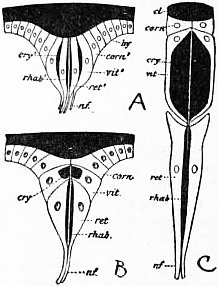
The Tracheae.—In regard to tracheae the very natural tendency of zoologists has been until lately to consider them as having once developed and once only, and therefore to hold that a group “Tracheata” should be recognized, including all tracheate Arthropods. We are driven by the conclusions arrived at as to the derivation of the Arachnida from branchiate ancestors, independently of the other tracheate Arthropods, to formulate the conclusion that tracheae have been independently developed in the Arachnidan class. We are also, by the isolation of Peripatus and the impossibility of tracing to it all other tracheate Arthropoda, or of regarding it as a degenerate offset from some one of the tracheate classes, forced to the conclusion that the tracheae of the Onychophora have been independently acquired. Having accepted these two conclusions, we formulate the generalization that tracheae can be independently acquired by various branches of Arthropod descent in adaptation to a terrestrial as opposed to an aquatic mode of life. A great point of interest therefore exists in the knowledge of the structure and embryology of tracheae in the different groups. It must be confessed that we have not such full knowledge on this head as could be wished for. Tracheae are essentially tubes like blood-vessels—apparently formed from the same tissue elements as blood-vessels—which contain air in place of blood, and usually communicate by definite orifices, the tracheal stigmata, with the atmosphere. They are lined internally by a cuticular deposit of chitin. In Peripatus and the Diplopods they consist of bunches of fine tubes which do not branch but diverge from one another; the chitinous lining is smooth. In the Hexapods and Chilopods, and the Arachnids (usually), they form tree-like branching structures, and their finest branches are finer than any blood-capillary, actually in some cases penetrating a single cell and supplying it with gaseous oxygen. In these forms the chitinous lining of the tubes is thickened by a close-set spiral ridge similar to the spiral thickening of the cellulose wall of the spiral vessels of plants. It is a noteworthy fact that other tubes in these same terrestrial Arthropoda—namely, the ducts of glands—are similarly strengthened by a chitinous cuticle, and that a spiral or annular thickening of the cuticle is developed in them also. Chitin is not exclusively an ectodermal product, but occurs also in cartilaginous skeletal plates of mesoblastic origin (connective tissue). The immediate cavities or pits into which the tracheal stigmata open appear to be in many cases ectodermic in sinkings, but there seems to be no reason (based on embryological observation) for regarding the tracheae as an ingrowth of the ectoderm. They appear, in fact, to be an air-holding modification of the vasifactive connective tissue. Tracheae are abundant just in proportion as blood-vessels become suppressed. They are reciprocally exclusive. It seems not improbable that they are two modifications of the same tissue-elements. In Peripatus the stigmatic pits at which the tracheae communicate with the atmosphere are scattered and not definite in their position. In other cases the stigmata are definitely paired and placed in a few segments or in several. It seems that we have to suppose that the vasifactive tissue of Arthropoda can readily take the form of air-holding instead of blood-holding tubes, and that this somewhat startling change in its character has taken place independently in several instances—viz. in the Onychophora, in more than one group of Arachnida, in Diplopoda, and again in the Hexapoda and Chilopoda.
The Malpighian Tubes.—This name is applied to the numerous fine caecal tubes of noticeable length developed from the proctodaeal invert of ectodermal origin in Hexapods. These tubes are shown to excrete nitrogenous waste products similar to uric acid. Tubes of renal excretory function in a like position occur in most terrestrial Arthropoda—viz. in Chilopoda, Diplopoda and Arachnida. They are also found in some of the semi-terrestrial and purely aquatic Amphipod Crustaceans. But the conclusion that all such tubes are identical in essential character seems to be without foundation. The Malpighian tubes of Hexapods are outgrowths of the proctodaeum, but those of Scorpion and the Amphipod Crustacea are part of the metenteron or endodermal gut, though originating near its junction with the proctodaeum. Hence the presence or absence of such tubes cannot be used as an argument as to affinity without some discrimination. The Scorpion’s so-called Malpighian tubes are not the same organs as those so named in the other Tracheata. Such renal caecal tubes seem to be readily evolved from either metenteron or proctodaeum when the conditions of the out-wash of nitrogenous waste-products are changed by the transference from aquatic to terrestrial life. The absence of such renal caeca in Limulus and their presence in the terrestrial Arachnida is precisely on a parallel with their absence in aquatic Crustacea and their presence in the feebly branchiate Amphipoda.
Group Characters.—We shall now pass the groups of the Arthropoda in review, attempting to characterize them in such a way as will indicate their probable affinities and genetic history.
Sub-Phylum ARTHROPODA.—The characters of the sub-phylum and those of the associated sub-phyla Chaetopoda and Rotifera have been given above, as well as the general characters of the phylum Appendiculata which comprises these great sub-phyla.
Grade A.—Hyparthropoda. Hypothetical forms.
Grade B.—Protarthropoda.
(a) The integument is covered by a delicate soft cuticle (not firm or plated) which allows the body and its appendages great range of extension and contraction.