Volume 1 / Part 1 — CHAPTER IX.
Reading edition
MOLLUSCA[99].
Although the majority of important developmental features are common to the whole of the Mollusca, yet at the same time many of the subdivisions have well-marked larval types of their own. It will for this reason be convenient in considering the larval characters to deal successively with the different subdivisions, but to take the whole group at once in considering the development of the organs.
Formation of the layers and larval characters.
Odontophora.
Gasteropoda and Pteropoda. There is a very close agreement amongst the Gasteropoda and Pteropoda in the general characters of the larva; but owing to the fact that the eggs of the various species differ immensely as to the amount of food-yolk, considerable differences obtain in the mode of formation of the layers and of the alimentary tract.
The spheres at a very early stage of segmentation[100] become divided into two categories, one of them destined to give rise mainly to the hypoblast, the other mainly to the epiblast. According as there is much or little food-yolk the hypoblast spheres are either very bulky or the reverse. In all cases the epiblast cells lie at one pole, which may be called the formative pole, and the hypoblast cells at the opposite pole. When the bulk of the food-yolk is very great, the number of hypoblast spheres is small. Thus in Aplysia there are only two such spheres. In other cases, where there is but little food-yolk, they may be nearly as numerous as the epiblast cells. In all these cases, however, as was first shewn by Lankester and Selenka, a gastrula becomes formed either by normal invagination as in the case of Paludina (fig. 107), or by epibole as in Nassa mutabilis (fig. 105). In both cases the hypoblast becomes completely enclosed by the epiblast. The blastopore is always situated opposite the original formative pole. In the large majority of cases (i.e. Marine Gasteropoda, Heteropoda, and Pteropoda) the blastopore becomes gradually narrowed to a circular opening which eventually occupies the position of the mouth. It either closes or remains permanently open at this point. In some cases the blastopore remains permanently open and becomes the anus. The best authenticated instance of this is Paludina vivipara, as was first shewn by Lankester (No. 263).
In some instances the blastopore assumes before closing a very narrow slit-like form, and would seem to extend along the future ventral region of the body from the mouth to the anus. This appears, according to Lankester (No. 262), to be the condition in Lymnæus, but while Lankester believes that the closure proceeds from the oral towards the anal extremity, other investigators hold that it does so in the reverse direction. Fol (No. 249) has also described a similar type of blastopore. In an undetermined marine Gasteropod, with an embolic gastrula, observed by myself at Valparaiso, the blastopore had the same elongated form as in Lymnæus, but the whole of it soon became closed except the oral extremity; but whether this finally closed could not be determined. It is probable that the typical form of the blastopore is the elongated form observed by Lankester and myself, in which an unclosed portion can indifferently remain at either extremity; and that from this primitive condition the various modifications above described have been derived[101].
Before the blastopore closes or becomes converted into the oral or anal aperture, a number of very important embryonic organs make their appearance; but before describing these it will be convenient to state what is known with reference to the third embryonic layer or mesoblast.
This layer generally originates in a number of cells at the lips of the blastopore, which then gradually make their way dorsalwards and forwards, and form a complete layer between the epiblast and hypoblast. The above general mode of formation of the mesoblast may be seen in fig. 107, representing three stages in the development of Paludina.
In some cases the mesoblast arises from certain of the segmentation spheres intermediate in size between the epiblast and hypoblast spheres. This is the case in Nassa mutabilis, where the mesoblast appears when the epiblast only forms a very small cap at the formative pole of the ovum; and in this case the mesoblast cells accompany the epiblast cells in their growth over the hypoblast (fig. 105).
In other cases the exact derivation of the mesoblast cells is quite uncertain. The evidence is perhaps in favour of their originating from the hypoblast. It is also uncertain whether the mesoblast is bilaterally symmetrical at the time of its origin. It is stated by Rabl to be so in Lymnæus[102].
In the case of Paludina the mesoblast becomes two layers thick, and then splits into a splanchnic and somatic layer, of which the former attaches itself to the hypoblast, and gives rise to the muscular and connective-tissue wall of the alimentary tract, and the latter attaches itself to the epiblast, and forms the muscular and connective-tissue wall of the body and other structures. The two layers remain connected by protoplasmic strands, and the space between them forms the body cavity (fig. 107). In most instances there would appear to be at first no such definite splitting of the mesoblast, but the layer has the form of a scattered network of cells between the epiblast and the hypoblast. Finally certain of the cells form a definite layer over the walls of the alimentary canal, and constitute the splanchnic mesoblast, and the remaining cells constitute the somatic mesoblast.
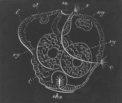
Fig. 101. Diagram of an embryo of Pleurobranchidium. (From Lankester.)
f. foot; ot. otocyst; m. mouth; v. velum; ng. nerve ganglion; ry. residual yolk spheres; shs. shell-gland; i. intestine.
We must now return to the embryo at the time when the blastopore is becoming narrowed. First of all it will be necessary to define the terms to be applied to the various regions of the body—and these will best be understood by taking a fully formed larva such as that represented in fig. 101. The ventral surface I consider to be that comprised between the mouth (m) and the anus, which is very nearly in the position (i) in the figure. As a great protuberance on the ventral surface is placed the foot f. The long axis of the body, at this period though not necessarily in the adult, is that passing through the mouth and the shell-gland (shs.): while the dorsal surface is that opposite the ventral as already defined.
Before the blastopore has attained its final condition three organs make their appearance, which are eminently characteristic of the typical molluscan larva. These organs are (1) the velum, (2) the shell-gland, (3) the foot.
The velum is a provisional larval organ, which has the form of a præoral ring of cilia, supported by a ridge of cells, often in the form of a double row, the ventral end of which lies immediately dorsal to the mouth. Its typical position is shewn in fig. 101, v. There are considerable variations in its mode and extent of development etc., but there is no reason to think that it is entirely absent in any group of Gasteropoda or Pteropoda. In a few individual instances, especially amongst viviparous forms and land Pulmonata, it has been stated to be absent. Semper (No. 274) failed to find it in Vitrina, Bulimus citrinus, Vaginulus luzonicus, and Paludina costata. It is very probably absent in Helix, etc.
In some cases, e.g. Limax (Gegenbaur), Neritina (Claparède), Pterotrachæa (Gegenbaur), the larva is stated to be coated by an uniform covering of cilia before the formation of the velum, but the researches of Fol have thrown very considerable doubt on these statements. In some cases amongst the Nudibranchiata (Haddon) and Pteropoda there are one or two long cilia in the middle of the velar area. In many Nudibranchiata (Haddon) there is present a more or less complete post-oral ring of small cilia, which belongs to the velum.
The cilia on the velum cause a rotation of the larva within the egg-capsule. Cilia are in most cases (Paludina, etc.) developed on the foot and on a small anal area.
The shell-gland arises as an epiblastic thickening on the posterior and dorsal side. In this thickening a deep invagination (fig. 101, shs.) is soon formed, in which a chitinous plug may become developed (Paludina, Cymbulia? etc.), and in abnormal larvæ such a chitinous plug is generally formed.
The foot is a simple prominence of epiblast on the ventral surface, in the cavity of which there are usually a number of mesoblast cells (fig. 101, f). The larval form just described has been named by Lankester the trochosphere larva.
Before considering the further external changes which the larva undergoes, it will be well to complete the history of the invaginated hypoblast.
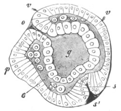
Fig. 102. Embryo of A Heteropod. (From Gegenbaur; after Fol.)
o. mouth; v. velum; g. archenteron; p. foot; c. body cavity; s. shell-gland.
The hypoblast has after its invagination either the form of a sack (fig. 102) or of a solid mass (fig. 101). Whether the mouth be the blastopore or no, the permanent œsophagus is formed of epiblast cells, so that the œsophagus and buccal cavity are always lined by epiblast. When the blastopore remains permanently open the outer part of the œsophagus grows as a prominent ridge round the opening.
The mesenteric sack itself becomes differentiated into a stomach adjoining the œsophagus, a liver opening immediately behind this, and an intestine. The cells forming the hepatic diverticula and sometimes also those of the stomach may during larval life secrete in their interior peculiar albuminous products, similar to ordinary food-yolk.
The proctodæum, except when it is the blastopore, arises later than the mouth. It is frequently developed from a pair of projecting epiblast cells symmetrically placed in the median ventral line behind the foot. It eventually forms a very shallow invagination meeting the intestine. Its opening is the anus. The anus, though at first always symmetrical and ventral, subsequently, on the formation of the pallial cavity, opens into this usually on the right and dorsal side.
In the cases where the hypoblast is not invaginated in the form of a sack the formation of the mesenteron is somewhat complicated, and is described in the sequel.
From the trochosphere stage the larva passes into what has been called by Lankester the veliger stage (fig. 103), which is especially characteristic of Gasteropod and Pteropod Mollusca.
The shell-gland (with a few exceptions to be spoken of subsequently) of the previous stage flattens out, forming a disc-like area, on the surface of which a delicate shell becomes developed, while the epiblast of the edges of the disc becomes thickened. The disc-like area is the mantle. The edge of the area and with it the shell now rapidly extend, especially in a dorsal direction. Up to this time the embryo has been symmetrical, but in most Gasteropods the shell and mantle extend very much more towards the left than towards the right side, and a commencement of the permanent spiral shell is thus produced.
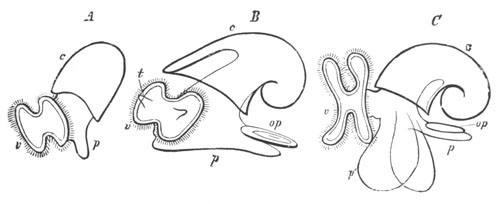
Fig. 103. Larvæ of Cephalophorous Mollusca in the veliger stage. (From Gegenbaur.)
A. and B. Earlier and later stage of Gasteropod. C. Pteropod (Cymbulia). v. velum; c. shell; p. foot; op. operculum; t. tentacle.
The edge of the mantle forms a projecting lip separating the dorsal visceral sack from the head and foot. An invagination appears, usually on the right in Gasteropods, and eventually extends to the dorsal side (fig. 103 B). It gives rise to the pallial or branchial cavity, and receives also the openings of the digestive, generative and urinary organs. In most Pteropods it is also formed to the right, and usually eventually extends afterwards towards the ventral surface (fig. 103 C). In the pallial cavity the gills are formed, in those groups in which they are present, as solid processes frequently ciliated. They are coated by epiblast and contain a core of mesoblast. They soon become hollow and contractile.
The velum in the more typical forms loses its simple circular form, and becomes a projecting bilobed organ, which serves the larva after it is hatched as the organ of locomotion (fig. 103 B and C). The extent of the development of the velum varies greatly. In the Heteropods especially it becomes very large, and in Atlanta it becomes six-lobed, each lateral half presenting three subdivisions. It is usually armed on its projecting edge with several rows of long cilia, and below this with short cilia which bring food to the mouth. It persists in many forms for a very long period. Within the area of the velum there appear the tentacles and eyes (fig. 103 B). The latter are usually formed at the base of the tentacles.
The foot grows in most forms to a very considerable size. On its hinder and dorsal surface is formed the operculum as a chitinos plate which originates in a depression lined by thickened epiblast, much in the same way as the shell (fig. 103 B and C, op). In the typical larval forms it is only possible to distinguish the anterior flattened surface of the foot for locomotion and the posterior opercular region, but special modifications of the foot are found in the Pteropods and Heteropods, which are described with those groups. The foot very often becomes richly ciliated, and otic vesicles are early developed in it (fig. 101, ot).
All the Gasteropods and Pteropods have a shell-bearing larval form like that first described, with the exception of a few forms, such as Limax and perhaps some other Pulmonata, in which the shell-gland closes up and gives rise to an internal shell.
The subsequent metamorphosis in the different groups is very various, but in all cases it is accompanied by the disappearance of the velum, though in some cases remnants of the velum may persist as the subtentacular lobes (Lymnæus, Lankester) or the lip tentacles (Tergipes, Nordmann). In prosobranchiate Gasteropods the larval shell is gradually added to, and frequently replaced by, a permanent shell, though the free-swimming veligerous larva may have a long existence. In many of the Opisthobranchiata the larval shell is lost in the adult and in others reduced. Lankester, who has especially worked at the early stages of this group, has shewn that the larvæ are in almost every respect identical with those of prosobranchiate Gasteropods. They are all provided with a subnautiloid shell, an operculated foot, etc. The metamorphosis has unfortunately been satisfactorily observed in but few instances. In Heteropods and Pteropods the embryonic shell is in many cases lost in the adult.
The following sections contain a special account of the development in the various groups of Gasteropoda and Pteropoda which will complete the necessarily sketchy account of the preceding pages.
Gasteropoda. To illustrate the development of the Gasteropoda I have given a detailed description of two types, viz. Nassa mutabilis and Paludina vivipara.
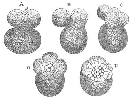
Fig. 104. Segmentation of Nassa mutabilis. (From Bobretzky.)
A. Upper half divided into two segments. B. One of these has fused with the large lower segment. C. Four small and one large segment, one of the former fusing with the large segment. D. Each of the four segments has given rise to a fresh small segment. E. Small segments have increased to thirty-six.
Nassa mutabilis. This form, the development of which has been very thoroughly worked out by Bobretzky (No. 242), will serve as an example of a marine Gasteropod with a large food-yolk. The segmentation has already been described, p. 102. It will be convenient to take up the development at a late stage of the segmentation. The embryo is then formed of a cap of small cells which may be spoken of as the blastoderm resting upon four large yolk cells of which one is considerably larger than the others (fig. 104 A). The small and the large cells are separated by a segmentation cavity. The general features at this stage are shewn in fig. 105 A, representing a longitudinal section through the largest yolk cell and a smaller yolk cell opposite to it. The blastoderm is for the most part one cell thick, but it will be noticed that, at the edge of the blastoderm adjoining the largest yolk cell, there are placed two cells underneath the edge of the blastoderm (me). These cells are the commencement of the mesoblast. In the later stages of development the blastoderm continues to grow over the yolk cells, and as it grows the three smaller yolk cells travel round the side of the largest yolk cell with it. As they do so they give rise to a layer of protoplasmic cells (fig. 105, hy) which form a thickened layer at the edge of the blastoderm and therefore round the lips of the blastopore. These cells form the hypoblast. The whole of the protoplasmic matter of the yolk cells is employed in the formation of the hypoblast. The rest of them remains as a mass of yolk. A longitudinal section of the embryo at a slightly later stage, when the blastopore has become quite narrowed, is represented in fig. 105 C. The greater part of the dorsal surface is not represented.
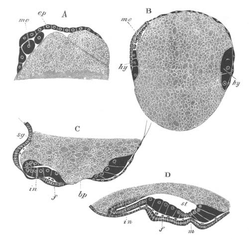
Fig. 105. Longitudinal section through the embryo of Nassa mutabilis. (After Bobretzky.)
A. Stage when the mesoblast is commencing to be formed. B. Stage when the yolk is half enclosed. The hypoblast is seen at the lips of the blastopore. C. Stage when the blastopore (bp) is nearly obliterated. D. The blastopore is closed.
ep. epiblast; me. mesoblast; hy. hypoblast; bp. blastopore; in. intestine; st. stomach; f. foot; sg. shell-gland; m. mouth.
Two definite organs have already become established. One of these is a pit lined by thickened epiblast on the posterior and dorsal side (sg). This is the shell-gland. The other is the foot (f) which arises as a ventral prominence of thickened epiblast immediately behind the blastopore. The hypoblast forms a ring of columnar cells round the blastopore. On the posterior side its cells have bent over so as to form a narrow tube (in), the rudiment of the intestine.
In the next stage (fig. 105 D) the blastopore completely closes, but its position is marked by a shallow pit (m) where the stomodæum is eventually formed. The foot (f) is more prominent, and on its hinder border is formed the operculum. The shell-gland (not shewn in the figure) has flattened out, and its thickened borders commence to extend especially over the dorsal side of the embryo. A delicate shell has become formed. In front of and dorsal to the mouth, a ciliated ring-shaped ridge of cells, which is however incomplete dorsally, gives rise to the velum. On each side of the foot there appears a protuberance of epiblast cells, which forms a provisional renal organ. The hypoblast now forms a complete layer ventrally, bounding a cavity which may be conveniently spoken of as the stomach (st), which is open to the yolk above. Posteriorly however a completely closed intestine is present, which ends blindly behind (in).
The shell and with it the mantle grow rapidly, and the primitive symmetry is early interfered with by the shell extending much more towards the left than the right. The anus soon becomes formed and places the intestine in communication with the exterior.
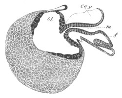
Fig. 106. Longitudinal section through an advanced embryo of Nassa mutabilis. (After Bobretzky.)
f. foot; m. mouth; ce.v. cephalic vesicle; st. stomach.
With the growth of the shell and mantle the foot and the head become sharply separated from the visceral sack (fig. 106). The œsophagus (m) becomes elongated. The eyes and auditory sacks become formed.
With further growth the asymmetry of the embryo becomes more marked. The intestine takes a transverse direction to the right side of the body, and the anus opens on the right side and close to the foot in the mantle cavity which is formed by an epiblastic invagination in this region. The cavity of the stomach (fig. 106, st) increases enormously and passes to the left side of the body, pushing the food-yolk at the same time to the right side, and the point where it communicates with the intestine becomes carried towards the posterior dorsal end of the visceral sack. The walls of the stomach gradually extend so as to narrow the opening to the yolk. The part of it adjoining the œsophagus becomes the true stomach, the remainder the liver; its interior is filled with coagulable fluid.
Paludina. Paludina—Lankester (No. 263) and Bütschli (No. 244)—is a viviparous form characterised by the small amount of food-yolk. The hypoblast and epiblast cells are distinguished very early, but soon become of nearly the same size.
In the later stages of segmentation the epiblast cells differ from the hypoblast cells in the absence of pigment. The segmentation cavity, if developed, is small. A perfectly regular gastrula is formed (fig. 107 A and B), which is preceded by the embryo assuming a flattened form. The blastopore is at first wide, but gradually narrows, and finally assumes a slightly excentric position. It becomes not the mouth, but the anus.
When the blastopore has become fairly narrow, mesoblast cells (B, me.) appear around it, between the epiblast and hypoblast. Whether they are bilaterally arranged or no is not clear; and though coloured like the hypoblast, their actual development from this layer has not been followed.
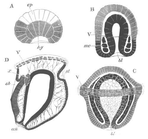
Fig. 107. Four stages in the development of Paludina vivipara. (Copied from Bütschli.)
ep. epiblast; hy. hypoblast; me. mesoblast; bl. blastopore; an. anus; st. stomodæum; sh. shell-gland; V. velum; x. primitive excretory organ.
The velum appears about the same time as the mesoblast, in the form of a double ring of ciliated cells at about the middle of the body (B and C, V). The mesoblast rapidly extends so as to occupy the whole space between the epiblast and hypoblast, and at the same time becomes divided into two layers (C). Shortly afterwards a space—the body cavity—appears between the two layers (D) which then attach themselves respectively to the epiblast and hypoblast, and constitute the somatic and splanchnic layers of mesoblast. The two layers remain connected by transverse strands.
By a change in the relations of the various parts and especially by the growth of the posterior region of the body, the velum now occupies a position at the end of the body opposite the blastopore. Immediately behind it there appear two organs, one on the dorsal and one on the ventral side. That on the dorsal side (sh) is a deep pit—the shell-gland—which is continuous with a layer of columnar epiblast which ends near the anus. The other organ (st), situated on the ventral side, is a simple depression, and is the rudiment of the stomodæum. Between it and the dorsally placed anus is a slight prominence—the rudiment of the foot. On the two sides of the body, between the epiblast and hypoblast on a level with the shell-gland are placed two masses of excretory cells, the provisional kidneys (D, x). These are probably not homologous with the provisional renal organ of Nassa and other marine Prosobranchiata. At a later period a ciliated cavity appears in them, which probably communicates with the exterior at the side of the throat.
In the later stages the foot grows rapidly, and forms a very prominent mass between the mouth and the anus. An operculum is developed somewhat late in a shallow groove lined by thickened epiblast.
A provisional chitinous plug is formed in the shell-gland which soon becomes everted. The shell is formed in the usual way on the everted surface of the shell-gland. The thickened edge of this part becomes the edge of the mantle, and soon projects in the neighbourhood of the anus as a marked fold.
With the rapid growth of the larva the invaginated mesenteron becomes relatively reduced in size. In its central part yolk-spherules become deposited, while the part adjoining the blastopore (anus) becomes elongated to give rise to the intestine. The stomodæum grows greatly in length and joins the dorsal part of the archenteron which then becomes the stomach. The part of the mesenteron with yolk-spherules forms the liver. With the development of the visceral sack the anus shifts its position. It first passes somewhat to the left, and is then carried completely to the right.
The development of Entoconcha mirabilis (Joh. Müller, No. 265), a remarkable Prosobranchiate parasitic in the body cavity of Synapta, which in the adult state is reduced to little more than an hermaphrodite generative sack, deserves a short description. It is viviparous, and the ovum gives rise to a larva which from the hardly sufficient characters of the foot and shell is supposed to be related to Natica.
There is nothing very striking in the development. The food-yolk is scanty. The velum, as might be anticipated from the viviparous development, is small. The tentacles are placed not within, but behind the velar area. There is a nautica-like shell, a large mantle cavity, and a large two-lobed foot.
In Buccinum, and Neritina only one out of the many ova included in each egg-capsule develops. The rest atrophy and are used as food by the one which develops.
Opisthobranchiata. It will be convenient to take a species of Pleurobranchidium (Aplysia), observed by Lankester (No. 239), as a type of Nudibranchiate development. The ovum first divides into two segments, and from these small segments are budded off, which gradually grow round and enclose the two large segments. The small segments now form the epiblast.
At the aboral pole the epiblast becomes thickened and invaginated to form the shell-gland, and shortly afterwards the velum and foot are formed in the normal way, and a stomodæum appears close to the ventral edge of the velum (fig. 101). The two yolk cells (ry) still remain distinct, but a true hypoblastic layer (probably derived from them, though this has not been made out) soon becomes established. Prominent cells early make their appearance at the base of the foot, which become at a later period invaginated to form the anus. Otolithic sacks (ot) become formed in the foot, and the supraœsophageal ganglia from a differentiation of the epiblast (ng).
At a later period the shell-gland becomes everted, and a nautiloid shell developed. The alimentary tract becomes completed, though the two yolk cells long retain their original distinctness. The shell-muscle is developed, and peculiar pigmented bodies are formed below the velum. The foot becomes prominent and acquires an operculum.
The metamorphosis of Tergipes has been more or less completely worked out by Nordmann and by Schultze (No. 271).
In Tergipes Edwardsii worked out by the former author, the larva when hatched is provided with a large velum, eyes, tentacles, an elongated operculated foot, and mantle. In the next stage both shell and operculum are thrown off, and the body becomes elongated and pointed behind. Still later a pair of gill-processes with hepatic diverticula becomes formed.
The velum next becomes reduced, and two small processes, which give rise to the lip tentacles and a second pair of gills, sprout out. An ecdysis now takes place, and leads to further changes which soon result in the attainment of the adult form.
In Tergipes lacinulatus, observed by Schultze, the velum atrophies before the shell and operculum are thrown off.
Pulmonata. The development of the fresh-water Pulmonata appears from Lankester’s observations on the pond-snail (Lymnæus) to be very similar in all important particulars to that of marine Branchiogasteropoda. The velum is however less developed than in most marine forms. The shell-gland, etc. have the normal development. In Lymnæus the blastopore has an elongated form and it is still a matter of dispute whether it closes at the mouth or anus.
In the Helicidæ there is a gastrula by epibole. The shell-gland, as may be gathered from Von Jhering’s figures, has the usual form, and an external shell of the usual larval type is developed. There is a ciliated process above the mouth, which extends into the lumen of the mouth. This process is often regarded as a rudimentary velum, but probably has not this value. There is no other organ which can be homologous with the velum.
The development of Limax presents some peculiarities. The yolk-spheres (hypoblast) form a large mass enclosed by the epiblast cells. A shell-gland is formed in the usual situation, which however, instead of being everted, as in ordinary forms, becomes closed, and in its interior are deposited calcareous plates which give rise to the permanently internal shell. The foot grows out posteriorly, and contains a large provisional contractile vesicle, traversed by muscular strands which contract rhythmically.
Although an external shell is present in Clausilia in the adult, the shell-gland becomes closed in the embryo as in Limax, and an internal plate-like shell is developed. The shell is at first covered by a complete epithelium, which eventually gives way in the centre, leaving covered only the edges of the shell. It thus comes about that the original internal shell becomes an external one. It is very difficult to bring this mode of development of the external shell into relation with that of other forms. Clausilia like Limax develops a large pedal sinus.
In both Limax and Clausilia cilia are early developed and cause a rotation of the embryo, but how far they give rise to a distinct velum is not clear.
Heteropoda. The Heteropod embryos present in their early development the closest resemblance to those of other Gasteropods. The segmentation takes place according to the most usual Gasteropod type; (vide p. 99) and after the yolk cells have ceased to give origin to epiblast cells they divide towards the nutritive pole, become invaginated, and line a spacious archenteron. The epiblast cells at the formative pole gradually envelop the yolk (hypoblast) cells, and the blastopore very early narrows and becomes the permanent mouth.
Simultaneously with the narrowing of the blastopore, the shell-gland is formed at the aboral pole, and the foot on the ventral side. The velum appears as a patch of cilia on the dorsal side, which then gradually extends ventrally so as to form a complete circle just dorsal to the mouth.
The larva, after these changes have been completed, is represented in fig. 102.
In later stages the shell-gland becomes everted, and a shell is developed in all the forms both with and without shells in the adult. The foot grows very rapidly, and an operculum is in all cases formed behind. A bilobed invagination in front gives rise to the mucous gland. The velum enlarges and becomes bilobed.
Though the blastopore remains permanently open as the mouth, the œsophagus is formed as an epiblastic ingrowth. The rudiment of the proctodæum appears as two epiblastic cells symmetrically placed behind the foot, which subsequently pass to the right side, and give rise to a shallow invagination which meets the mesenteric sack. In the latter structure the cells of part of the wall develop a peculiar nutritive material, and form a nutritive sack which eventually becomes the liver. The part of the sack connected with the epiblastic œsophagus becomes constricted off as the stomach. The remainder, which unites with the proctodæum, forms the intestine.
The structural peculiarities of the adult are formed by a post-larval metamorphosis. The caudal appendage of Pterotrachea and Firoloidea is formed as an outgrowth of the upper border of the hind end of the foot. The so-called fin arises as a cylindrical process in front of the base of the foot, which is eventually flattened laterally. In the Atlantidæ it is in some cases at first vermiform, and in other cases attains directly its adult structure. The embryonic foot itself gives rise in Pterotrachea, Firoloidea and Carinaria to the tail, on the dorsal and posterior side of which the operculum may still be seen in young specimens. In Atlanta it forms the posterior part of the foot on which the operculum persists through life.
The embryonic shell is completely lost in Pterotrachea and Firoloidea, and the shell is rudimentary in Carinaria. With its atrophy the mantle region also becomes much reduced.
The velum is enormously developed in many Heteropods. In Atlanta it is six-lobed, each of the two primitive lateral lobes being prolonged into three processes, two in front, and one behind. As in all other cases, it atrophies in the course of the post-larval metamorphosis.
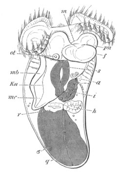
Fig. 108. Embryo of Cavolinia (Hyalea) Tridentata. (After Fol.)
m. mouth; a. anus; s. stomach; i. intestine; σ. nutritive sack; mb. mantle; mc. mantle cavity; Kn. contractile sinus; h. heart; r. renal sack: f. foot; pn. epipodia; q. shell; ot. otolithic sack.
Pteropoda. The early larval form of the Pteropods is closely similar to that of marine Gasteropods. There are usually only three hypoblastic spheres at the close of the segmentation in the Thecosomata, and a somewhat larger number in the Gymnosomata. The blastopore closes at the oral region, on the nutritive side of the ovum, and the shell-gland is placed at the original formative pole. The velum, shell-gland and foot have the usual relations. Although many of the adult forms are symmetrical, there is very early an asymmetry visible in the larva, shewing that the Pteropods are descended from asymmetrical ancestors. In the Gymnosomata there is a second larval stage after the loss of the shell when the larva is provided with three rings of cilia (fig. 109). In most forms of Pteropods the dorsal part of the body, covered by the mantle, is produced into a visceral sack like that of the Cephalopoda (fig. 108).
The velum varies considerably in its development in different forms. In the Hyaleidæ it is comparatively small and atrophies early; while in Cymbulia (fig. 103) and the Gymnosomata it is large and bilobed, and persists till after the foot has attained its full development.
The free edge of the velum is provided with long motor cilia, and its lower border with small cilia which bring the food to the mouth. In Cleodora there is a median bunch of cilia in the centre of the velum like that in the Lamellibranchiata, Nudibranchiata, etc.
The shell-gland forms a pit at the aboral end of the body, and in Cymbulia a chitinous plug appears to be normally formed in this pit. The pit afterwards everts itself. The edge of the everted area becomes thickened and gradually travels towards the anterior end of the body. On this everted area a small plate is developed, which forms the commencement of the embryonic shell with which the larvæ of all Pteropods are provided.
The remainder of the embryonic shell is secreted in successive rings by the thickened edge of the mantle, and grows with this till it reaches the neck (fig. 108). The permanent shell is added subsequently, usually on a very different model to the larval shell. The fate of the embryonic shell is very various in different forms. In the Hyaleidæ the animal withdraws itself from the larval shell, which becomes shut off from the permanent shell by a diaphragm. The larval shell then becomes detached.
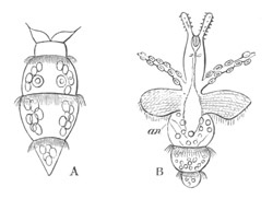
Fig. 109. Free-swimming Pneumodermon Larvæ. (After Gegenbaur, copied from Bronn.)
The velum has atrophied in both larvæ. In A three ciliated bands are present, and the auditory vesicles are visible. In B the tentacles with suckers and the epipodia have become developed.
an. anus.
In the Styliolidæ the permanent shell becomes twice the size of the embryonic shell while the animal is still in an embryonic condition, but the larval shell persists for life. In the Cymbulidæ there is an embryonic and secondary shell, which persist together during larval life. They are eventually cast off at the same time and replaced by a permanent shell.
In the Gymnosomata an embryonic shell is developed, and a secondary shell added to it during embryonic life. Both are cast off before the adult condition is attained. After the shell has been cast off three ciliated rings are developed (fig. 109). The anterior of these is placed between the velum and the foot, and the two hinder ones on the elongated posterior part of the body.
The ciliated rings give to these larvæ a resemblance to Chætopod larvæ; but there can be no doubt that this resemblance is a purely superficial one. The anterior ring atrophies early (fig. 109 B), and the second one soon follows suit. It is probable that the hindermost one does not persist through life, although it has been observed in forms with fully developed sexual organs. Most of these larvæ have not been traced to their adult forms. They have been referred to Pneumodermon, Clio, etc.
The most characteristic organ of the Pteropods is the foot, which is prolonged into two enormous lateral wings, the epipodia. These develop at different periods in different larvæ, but are always distinct lateral outgrowths of the foot.
In the Hyaleidæ the foot is early conspicuous, and soon sends out two lateral prolongations (fig. 108 pn.) which develop with enormous rapidity as compared with the medium portion, and give rise to the epipodia. The whole of the foot becomes ciliated.
In the Cymbulidæ, though not in other forms, an operculum is developed on the hinder surface of the foot (fig. 103 C). The epipodia are late in appearing.
In the Gymnosomata the foot is developed very early, but remains small. The epipodia do not appear till very late in larval life (fig. 109 B).
In Pneumodermon and some other Gymnosomata there appear on the hinder part of the head peculiar tentacles with suckers like those of the Cephalopoda (fig. 109 B). It is not certain that these tentacles are genetically related to the arms of the Cephalopoda.
Cephalopoda. The eggs of the Cephalopoda are usually laid in special capsules formed in the oviduct, which differ considerably in the different members of the group.
In the case of Argonauta each egg is enveloped in an elongated capsule provided with a stalk. By means of the stalk the eggs are attached together in bunches, and these again are connected together and form transparent masses, which are placed in the back of the shell. In octopus the eggs are small and transparent: each of them is enclosed in a stalked capsule. In Loligo the eggs are enveloped in elongated sack-like gelatinous cords, each containing about thirty or forty eggs. The cords are attached in bunches to submarine objects. In Sepia each egg is independently enveloped in a spindle-shaped black capsule, which is attached to a stone or other object.
In a decapod form with pelagic larvæ, described by Grenacher (No. 280), the eggs were enclosed in a somewhat cylindrical gelatinous mass. In each mass there were an immense number of eggs arranged in spirals. Each ovum was enclosed in a structureless membrane, within which it floated in a colourless albumen.
The ovum itself within the capsule is a nearly homogeneous granular mass, without a distinct envelope. Development commences by the segregation, at the narrow pole of the ovum opposite the egg-stalk, of the greater part of the protoplasmic formative material[103]. This material forms a disc equivalent to the germinal disc of meroblastic vertebrate ova. The germinal disc in Sepia and Loligo does not, however, undergo a quite symmetrical segmentation (Bobretzky, No. 279). When eight segments are present, two of them close together are much smaller and narrower than the remainder; and when, in the succeeding stages small segments are formed from the inner ends of the large ones, those derived from the two smaller segments continue to be smaller than the remainder: so that throughout the segmentation one pole of the blastoderm is formed of smaller segments, and the blastoderm exhibits a bilateral symmetry[104]. The partial segmentation results in the formation of a blastoderm covering one pole of the egg, but, unlike the vertebrate blastoderm, formed of a single row of cells. This blastoderm very soon becomes two or three cells deep at its edge, and the cells below the surface constitute the layer from which the mesoblast and hypoblast originate (fig. 110 ms). The origin of the mesoblast at the edge of the blastoderm is a phenomenon equivalent to its origin at the lips of the blastopore in so many other types. The external layer forms the epiblast.
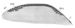
Fig. 110. Section through the blastoderm of a Loligo ovum at the beginning of the fourth day. (After Bobretzky.)
ms. mesoblast; d. cell at the edge of the blastoderm; c. one of the segmentation cells.
The whole blastoderm does not take its origin from the segmentation spheres, but, as was discovered by Lankester (282), a number of nuclei arise spontaneously in the yolk outside the blastoderm, around which cell bodies become subsequently formed. They make their appearance near to, but not at the surface, extending first in a ring-like series in advance of the margin of the blastoderm, but subsequently appearing indiscriminately over all parts of the egg. They take no share in forming the epiblast, but would seem, according to Lankester, to assist in giving rise to the lower layer cells, and also to a layer of flattened cells which eventually completely encloses the yolk, and may be called the yolk membrane. The cells of the yolk membrane first of all appear at the thickened edge of the blastoderm. From this point they spread inwards under the centre of the blastoderm (fig. 115 m´), and, together with the epiblast cells, outwards over the yolk generally; so that before long (on the tenth day in Loligo) the yolk becomes completely invested by a membrane of cells.
In the non-germinal region the blastoderm is formed of two layers, (1) a flattened epiblast, and (2) the yolk membrane. In the region of the original germinal disc the epiblast cells become columnar, and below them is placed a ring of lower layer cells, which gradually extends towards the centre so as finally to form a complete layer. Below this again comes the yolk membrane just spoken of.
Before describing the further fate of the separate layers it is necessary to say a few words as to the external features of the embryo. In the adult Cephalopod it is convenient, for the sake of comparison with other Mollusca, to speak of the narrow space enclosed in the arms, which contains the mouth, as the ventral surface; the aboral apex as the dorsal surface; and what is usually called the upper surface as the anterior and the lower one as the posterior.
Employing this terminology the centre of the original blastoderm is the dorsal apex of the embryo. In the typical forms with a large yolk-sack the whole embryo is formed out of the original germinal disc; the part of the blastoderm which is continued as a thin layer over the remainder of the egg forms a large ventral yolk-sack appended to the head of the embryo. The following description applies especially to two types, which form the extremes of the series in reference to the development of the yolk-sack. The first of these with a large yolk-sack is Sepia, of which Kölliker in his classical memoir (No. 281) has published a series of beautiful figures. The second, with a small yolk-sack, is the pelagic larva of an unknown adult described by Grenadier (No. 280).
In a young blastoderm of Sepia viewed from the dorsal surface, a series of structures appear which are represented in fig. 111 A. In the middle is a somewhat rhomboid prominence which forms the rudiment of the mantle (mt). In its centre is a pit which forms the shell-gland. On each side of the mantle is a somewhat curved fold (f). These folds eventually coalesce to form the funnel. They are divided into two parts by a small body which forms the cartilage of the funnel. The smaller part of the fold behind this body gives rise to the true funnel, the part in front becomes (Kölliker) the strong muscle connecting the funnel with the neck-cartilage. In front and to the sides are two kidney-shaped bodies (oc), the optic pits. Behind the mantle are two buds (br), the rudiments of the gills.
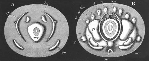
Fig. 111. Two surface views of the germinal disc of Sepia. (After Kölliker.)
mt. mantle; oc. eye; f. folds of funnel; br. branchiæ; an. posterior portion of alimentary tract; m. mouth. 1, 2, 3, 4, 5, arms; p. cephalic lobe.
In the somewhat later stage rudiments of the two posterior pairs of arms make their appearance outside and behind the rudiments of the funnel. The head is indicated by a pair of lateral swellings on each side, the outer of which carries the eyes. The whole embryo now becomes ciliated, though the ciliation does not cause the usual rotation. At a slightly later stage the second, third, and fourth pairs of arms make their appearance slightly in front of those already present. The posterior parts of the funnel rudiments approach each other, and the anterior meet the rudiments of the neck-cartilage. The gills begin to be covered by the mantle-edge, which now projects as a marked fold. At a slightly later period two fresh rudiments may be noted, viz. the oral (fig. 111 B, m) and anal invaginations, the latter of which is extremely shallow and appears at the apex of a small papilla which may be spoken of as the anal papilla. These invaginations appear at the two opposite poles (anterior and posterior) of the blastoderm. Shortly after this the rudiment of the first pair of arms arises considerably in front of the other rudiments, at the sides of the outer pair of cephalic swellings (fig. 111 B, 1).
Fig. 111 B represents a view from the dorsal surface of an embryo at this stage. In the centre is the mantle with the shell-gland which is now very considerably raised beyond the general surface. Concentric with the edge of the mantle are the two halves of the funnel, the anterior half meeting the dorsal or neck-cartilage and the posterior halves approaching each other. The oral invagination is shewn at m and the anal immediately in front of an. The gills, nearly covered by the mantle, are seen at br. At p are the cephalic swellings, and the eye is seen at oc. The arms 1‑5 form a ring outside these parts. The whole of the embryo, with the exception of the gills, the funnel, and the outer border of the blastoderm, is richly ciliated.
The embryo up to this time has had the form of a disc or saucer on the surface of the yolk. After this stage it rapidly assumes its permanent dome-like form, and becomes at the same time folded off from the yolk. The blastoderm is very slow in enveloping the yolk, and the whole yolk is not completely invested till a considerably later stage than that represented in fig. 111 B. As soon as the blastoderm covers the yolk-sack cilia appear upon it. The mantle grows very rapidly, and its free border soon projects over the funnel and gills. After the two halves of the funnel have coalesced into a tube, it comes to project again beyond the edge of the mantle.
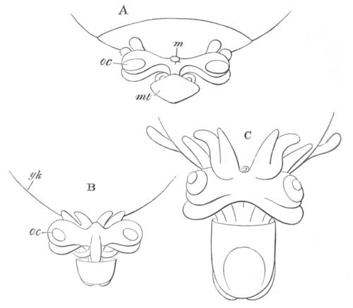
Fig. 112. Side views of three late stages in the development of Sepia. (After Kölliker.)
m. mouth; yk. yolk-sack; oc. eye; mt. mantle.
On the completion of the above changes the resemblance of the embryo to a Cuttle-fish becomes quite obvious. Three of the stages in the accomplishment of these changes are represented in fig. 112.
To the ventral side of the embryo is attached the enormous external yolk-sack (yk), which is continuous with an internal section situated within the body of the embryo. The general relations of the embryo to the yolk will best be understood by reference to the longitudinal section of Loligo, fig. 127.
The arms gradually increase in length, and the second pair passes in front of the first so as eventually to lie completely in front of the mouth. The arms thus come to form a complete ring surrounding the mouth, of which the original second pair, and not, as might be anticipated, the first, completes the circle in front. The second pair develops into the long arms of the adult.
After the embryo has attained more or less completely its definite form (fig. 112 C) it grows rapidly in size as compared with the yolk-sack. The latter structure is at first four or five times as big as the embryo, but, by the time of hatching, the embryo is two to three times as big as the yolk-sack.
Loligo mainly differs from Sepia in the early enclosure of the yolk by the blastoderm, and in the embryo exhibiting the phenomena of rotation within the egg-capsule so characteristic of other Mollusca.
In Argonauta the yolk-sack is still smaller than in Loligo, and the yolk is early completely enclosed by the blastoderm. A well-developed outer yolk-sack is present during early embryonic life, but is completely absorbed within the body before its close. Cilia appear on the blastoderm very early, but vanish again when the yolk is about two-thirds enclosed. There is, during embryonic life, no trace of a shell, but the mantle and other parts of the body become covered by peculiar bunches of fine setæ. The shell-gland develops normally in both Octopus and Argonauta, but disappears again without closing up to form a sack (Lankester).
The pelagic Decapod larva described by Grenacher, which forms my second type, must be placed with reference to the development of the yolk-sack at the opposite pole to Sepia. Segmentation, as in other Cephalopods, is partial, but the blastoderm almost completely envelops the yolk before any organs are developed; and no external yolk-sack is present. At a stage slightly before the closure of the yolk-blastopore the mantle is formed as a slight prominence at the blastodermic pole of the egg, and even at this early stage is marked by the presence of chromatophores. The edge of the blastoderm is ciliated. At a slightly later stage the embryo becomes more cylindrical, the edge of the mantle becomes marked by a fold, which divides the embryo transversely into two unequal parts, a smaller region covered by the mantle, and a larger region beyond this. The yolk is still exposed, but rudiments of the optic pit and of two pairs of arms have appeared. The first-formed arms are apparently the anterior, and not, as in Sepia, the posterior.
At a still later stage, represented in lateral and posterior views in fig. 113 A and B, considerable changes are effected. The yolk-blastopore is nearly though not quite closed. The mantle fold (mt) is much more prominent, and on the posterior side on a level with its edge may be seen the rudiments of the gills (br). The funnel is formed as two independent folds on each side (inf1 and inf2), which apparently correspond with the two divisions of the funnel rudiments in Sepia. The eye has undergone considerable changes. Close to each rudiment of the funnel may be seen a fresh sense organ—the auditory sack (ac). The ventral (upper in the figure) end of the body now forms a marked protuberance, probably equivalent to the foot of other Mollusca (vide p. 225), at the sides of which are seen the rudiments of the arms (1, 2, 3). To the two previously present a third one, on the posterior side, has been added. The blastopore is placed on the anterior side of the ventral protuberance, and immediately dorsal to this is an invagination (os) which gives rise to the stomodæum. The ciliation at the edge of the blastopore still persists, but does not lead to the rotation of the embryo.
In later stages (fig. 113 C) the blastopore becomes closed, and the mantle region increases in length as compared with the remainder of the body. The ventral halves of the funnel, each in the form of a half tube, coalesce together to form a single tube (inf) in the same manner as in Sepia. A shallow proctodæum (an) is formed between the two branchiæ. The eyes (oc) stand out as lateral projections, and the arms become much longer.
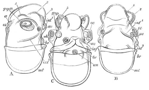
Fig. 113. Three Embryos of a Cephalopod with a very small yolk-sack. (After Grenacher.)
a. blastopore; br. branchiæ; inf.1 and inf.2 posterior and anterior folds of the funnel; g.op. optic ganglion (?); oc. eye; wk. white body; ac. auditory pit; os. stomodæum; an. anus; mt. mantle; 1, 2, 3. 1st, 2nd, and 3rd pairs of arms.
Still later a fourth pair of arms is added as a bud from each of the posterior pair, and with the growth in length of the arms the suckers make their appearance. The mouth is gradually carried up so as to be surrounded by the arms. The ciliation of the surface becomes more extensive.
During the whole of the above development the interior of the embryo is filled with yolk, although no external yolk-sack is present. The internal yolk-sack falls into three sections; a cephalic section, a section in the neck, and an abdominal section. Of these, that in the neck is the first to be absorbed. The cephalic portion fills out the ventral protuberance already spoken of. The hinder section becomes occupied by the liver which exactly fits itself into this space as it absorbs the material previously there.
It will be convenient at this point to complete the account of the Cephalopoda by a short history of their germinal layers, and by a fuller description of the mantle, shell, and funnel than that given in the preceding pages.
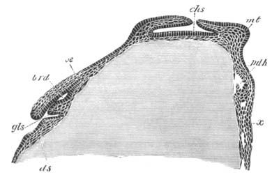
Fig. 114. Longitudinal vertical section through a Loligo ovum when the mesenteric cavity is just commencing to be formed. (After Bobretzky.)
gls. salivary gland; brd. sheath of radula; oe. œsophagus; ds. yolk-sack; chs. shell-gland; mt. mantle; pdh. mesenteron; x. epiblastic thickening between the folds of the funnel.
It has already been shewn that in the region of the germinal disc a thick layer of cells becomes interposed between the epiblast and the yolk membrane. This layer (fig. 115 m) is mainly mesoblastic, but also contains the elements which form the lining of the alimentary tract. Its cells first become differentiated into mesoblast and hypoblast after the shell-gland has become a fairly deep pit. The mode of differentiation is shewn in fig. 114. On the posterior side of the mantle, at the point marked in fig. 111 B, an, a cavity is formed between the yolk membrane and the mesoblast cells (fig. 114, pdh). This cavity is the commencement of the anal extremity of the mesenteron, and the columnar cells lining it constitute the hypoblast. The remainder of the lower layer cells are the mesoblast. The mesenteron gradually extends itself till it meets the stomodæum (fig. 127). The proctodæum is formed as a shallow pit close to the first formed part of the mesenteron.
The mesoblast gives rise not only to the organs usually formed in this layer, but also to the nervous centres, etc.
The mantle and shell. The mantle first arises as a thickening of the epiblast on the dorsal surface of the embryo. The thickened integument, with the subjacent mesoblast, soon forms a definite projection, in the centre of which appears a circular pit (figs. 114 chs and 115 shs). This pit, which has already been spoken of as the shell-gland, resembles very closely the shell-gland of other Mollusca. The fold around the edge of the shell-gland grows inwards so as gradually to circumscribe its opening, which before long becomes completely obliterated; and the gland forms a closed sack lined by epiblast which grows in an anterior direction (figs. 114 and 127 cch).
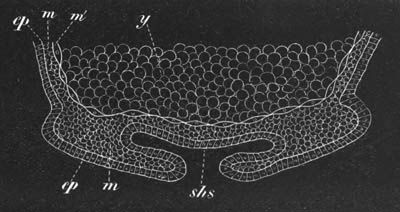
Fig. 115. Diagram of a vertical section through the mantle region of an embryo Loligo. (From Lankester.) [This figure is turned the reverse way up to fig. 114.]
ep. epiblast; y. food-yolk; m. mesoblast; m´. cellular yolk membrane; shs. shell-gland.
The edges of the mantle now begin to project, especially on the posterior side (fig. 127), and within the cavity formed by this projecting lip there are placed the anus (an), gills, etc. The projecting lip of the mantle is formed both of epiblast and mesoblast. The whole of the anterior side of the mantle is filled by the elongated shell-sack (cch), within which the shell or pen soon becomes secreted.
There are certain difficulties in comparing the shell-gland of the Cephalopoda with that of other Mollusca which will best be rendered clear by the following quotation from Lankester[105]:
“The position and mode of development of the shell-gland of the Cephalopoda exactly agree with that of the shell-gland as seen in the other Molluscan embryos figured in this paper. We are therefore fairly entitled to conclude from the embryological evidence that the pen-sack of Cephalopoda is identical with the shell-gland of other Mollusca.
“But here—forming an interesting example of the interaction of the various sources of evidence in genealogical biology—palæontology crosses the path of embryology. I think it is certain that if we possessed no fossil remains of Cephalopoda the conclusion that the pen-sack is a special development of the shell-gland would have to be accepted.
“But the consideration of the nature of the shell of the Belemnites and its relation to the pen of living Cuttle-fish brings a new light to bear on the matter. Reserving anything like a decided opinion as to the question in hand, I may briefly state the hypothesis suggested by the facts ascertained as to the Belemnitidæ. The complete shell of a Belemnite is essentially a straightened nautilus-shell (therefore an external shell inherited from a nautilus-like ancestor), which, like the nautiloid shell of Spirula, has become enclosed by growths of the mantle, and unlike the shell of Spirula, has received large additions of calcareous matter from those enclosing overgrowths. On the lower surface of the enclosed nautilus-shell of the Belemnite—the phragmacone—a series of layers of calcareous matter have been thrown down forming the guard; above, the shell has been continued into the extensive chamber formed by the folds of the mantle, so as to form the flattened pen-like pro-ostracum of Huxley.
“Whether in the Belemnites the folds of the mantle which thus covered in and added to the original chambered shell, were completely closed so as to form a sack or remained partially open with contiguous flaps must be doubtful.
“In Spirula we have an originally external shell enclosed but not added to by the enclosing mantle sack.
“In Spirulirostra, a tertiary fossil, we have a shell very similar to that of Spirula, with a small guard of laminated structure developed as in the Belemnite (see the figures in Bronn Classen u. Ordnungen des Thierreichs).
“In the Belemnites the original nautiloid shell is small as compared with Spirulirostra. It appears to be largest in Huxley’s genus Xiphoteuthis. Hence in the series Spirula, Spirulirostra, Xiphoteuthis, Belemnites, we have evidence of the enclosure of an external shell by growths from the mantle (as in Aplysia), of the addition to that shell of calcareous matter from the walls of its enclosing sack, and of the gradual change of the relative proportions of the original nucleus (the nautiloid phragmacone) and its superadded pro-ostracal and rostral elements tending to the disappearance of the nucleus (the original external shell). If this view be correct as to the nature of these shells, it is clear that the shell-gland and its plug has nothing to do with them. The shell-gland must have preceded the original nautiloid shell, and must be looked for in such a relation whenever the embryology of the pearly Nautilus can be studied. Now, everything points to the close agreement of the Belemnitidæ with the living Dibranchiata. The hooklets on the arms, the ink bag, the horny jaws, and general form of the body, leave no room for doubt on that point; it is more than probable that the living Dibranchiata are modified descendants of the mesozoic Belemnitidæ. If this be so, the pens of Loligo and Sepia must be traced to the more complex shell of the Belemnite. This is not difficult if we suppose the originally external shell the phragmacone, around which as a nucleus the guard and pro-ostracum were developed, to have finally disappeared. The enclosing folds of the mantle remain as a sack and perform their part, producing the chitino-calcareous pen of the living Dibranch, in which parts can be recognised as corresponding to the pro-ostracum, and probably also to the guard of the Belemnite. If this be the case, if the pen of Sepia and Loligo correspond to the entire Belemnite shell minus the phragmacone-nucleus, it is clear that the sack which develops so early in Loligo and which appears to correspond to the shell-gland of the other Molluscs cannot be held to do so. The sack thus formed in Loligo must be held to represent the sack formed by the primæval upgrowth of mantle folds over the young nautiloid shell of its Belemnitoid ancestors, and has accordingly no general significance for the whole Molluscan group, but is a special organ belonging only to the Dibranchiate stem, similar to—but not necessarily genetically connected with—the mantle fold in which the shell of the adult Aplysia and its congeners is concealed. The pen, then, of Cephalopods would not represent the plug of the shell-gland. In regard to this view of the case, it may be remarked that I have found no trace in the embryonic history of the living Dibranchiata of a structure representing the phragmacone; and further, it is possible, though little importance can be attached to this suggestion, that the Dibranchiate pen-sack, as seen in its earliest stage in the embryo Loligo, etc., is fused with the surviving remnants of an embryonic shell-gland. When the embryology of Nautilus pompilius is worked out, we shall probably know with some certainty the fate of the Molluscan shell-gland in the group of the Cephalopoda.”
The funnel. The general development of the funnel has already been sufficiently indicated. The folds of which it is formed are composed both of epiblast and mesoblast. The mesoblast of the anterior part of each half of the funnel would appear to give rise to a muscle passing from the cartilage of the neck to the funnel proper. The posterior parts gradually approximate, but meet in the first instance ventrally. The two folds at first merely form the side of a groove or imperfect tube (fig. 113 C and 124 ff.), but soon the free edges unite and so give rise to a perfect tube, the primitive origin of which by the coalescence of two halves would not be suspected. In Nautilus the two halves remain permanently separate but overlap each other, so as to form a functional tube.
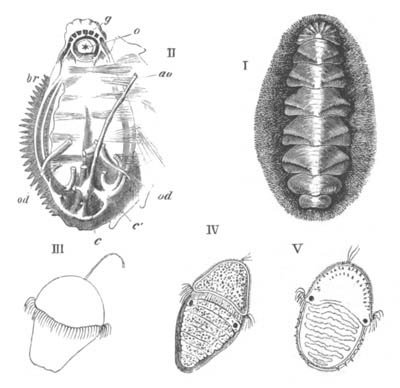
Fig. 116.
I. Chiton Wossnessenskii. (After Middendorf.) II. Chiton dissected to shew o. the mouth; g. the nervous ring; ao. the aorta; c. the ventricle; c´. an auricle; br. the left branchiæ; od. oviducts. (After Cuvier.) III., IV., V. Stages of development of Chiton cinereus. (After Lovén.)
The figure is taken from Huxley.
Polyplacophora. The external characters of the embryo of Chiton have long been known through the classical observations of Lovén (No. 285), while the formation of the layers and the internal phenomena of development have recently been elucidated by Kowalevsky (No. 284). The eggs are laid in April, May, and June, and are enclosed in a kind of chorion with calcareous protuberances. The segmentation remains regular till sixty-four segments are formed. The cells composing the formative half of the ovum then divide more rapidly than the remainder; there is in this way formed an elongated sphere, half of which is composed of small cells and half of larger cells. In the interior is a small segmentation cavity. From its eventual fate the hemisphere of the smaller cells may be called the anterior pole, and that of the larger cells the posterior. An involution of the cells at the apex of the posterior pole (though not of the whole hemisphere of larger cells) now takes place, and gives rise to the archenteron. At the same time an equatorial double ring of large cells appears on the surface between the two poles, which becomes ciliated and forms the velum. At the apex of the anterior pole a tuft of cilia, or at first a single flagellum, is established (fig. 116 III. and IV.).
In the succeeding developmental period the blastopore, which has so far had the form of a circular pore at the posterior extremity of the body, undergoes a series of very remarkable changes. In conjunction with a gradual elongation of the larva it travels to the ventral side, and is prolonged forwards to the velum as a groove. The middle part of the groove is next converted into a tube, which opens externally in front, and posteriorly communicates with the archenteron. The walls of this tube subsequently fuse together, obliterating the lumen, and necessarily causing at the same time the closure of the blastopore. The tube itself becomes thereby converted into a plate of cells on the ventral surface between the epiblast and the hypoblast[106].
While the above changes have been taking place the mesoblast has become established. It is derived from the lateral and ventral cells of the hypoblast.
After the establishment of the germinal layers the further evolution of the larva makes rapid progress. A transverse groove is formed immediately behind the velum, which is especially deep on the ventral surface; and the stomodæum is formed as an invagination of the anterior wall of the deeper section of the groove. Behind the stomodæum the remainder of the ventral surface grows out as a flattened foot.
The dorsal surface behind the velum constitutes the mantle, and becomes divided by six or seven transverse grooves into segment-like areas, which may be called mantle plates (fig. 116 IV.). These areas would seem (?) to correspond to so many flattened out shell-glands. Immediately behind the velum the eyes appear as two black spots (fig. 116 IV.).
While the above external changes take place the archenteron undergoes considerable modifications. Its anterior section gives rise, according to Kowalevsky, to a dorsal (?) sack in which the radula is formed; while the liver arises from it as two lateral diverticula.
From the above statements it would appear that Kowalevsky holds that the œsophagus and radula sack are both derived from the walls of the archenteron and not from the stomodæum. Such an origin for these organs is without parallel amongst Mollusca.
The larva becomes about this time hatched, and after swimming about for some time attaches itself by the foot, throws off its larval organs, cilia, etc., and develops the shell.
The shell appears first of all during larval life in the form of spicula on the middle and sides of the head, and later on the middle and sides of the post-oral mantle plates (fig. 116 V.). The permanent shell arises somewhat later as a series of median and lateral calcareous plates, first of all on the posterior part of the velar area, and subsequently on the mantle plates behind. The three calcareous patches of each plate fuse together and give rise to the permanent shell plates. The original spicula are displaced to the sides, where they partly remain, and are partly replaced by new spicula.
The nervous system is formed during larval life as four longitudinal cords:—two lateral—the branchial cords, and two ventral—the pedal. Paired anterior thickenings of the pedal cords meet in front of the mouth to form the œsophageal ring. The pedal cords and their derivatives are believed by Kowalevsky to be developed from the lateral parts of the plate formed by the metamorphosis of the blastopore. The median part of the plate is still visible after the formation of these parts.
The chief peculiarity of the larva of Chiton (apart from the peculiar ventral plate) consists in the elongation and dorsal segmentation of the posterior part of the body. The velum has the normal situation and relation to its mouth. The position of the eyes behind it is however abnormal.
The elongation and segmentation of the posterior part of the trunk is probably to be regarded as indicating that Chiton has early branched off from the main group of the Odontophora along a special line of its own, and not that the remaining Odontophora are descended from Chiton-like ancestral forms. The shell of Mollusca on this view is not to be derived from one of the plates of Chiton, but the plates of Chiton are to be derived from the segmentation of a primitive simple shell. The segmentation exhibited is of a kind which all the trochosphere larval forms seem to have been capable of acquiring. The bilateral symmetry of Chiton, which is quite as well marked as that of the Lamellibranchiata, indicates that it is a primitive phylum of the Odontophora.
Scaphopoda. The external characters of the peculiar larva of this interesting group have been fully worked out by Lacaze Duthiers (No. 286).
The segmentation is unequal and conforms to the usual molluscan type. At its close the embryo becomes somewhat elongated, and there appears on its surface a series of transverse ciliated rings. As soon as these become formed the larva is hatched, and swims about by means of its cilia. Six ciliated bands are formed in all, and in addition a tuft of cilia is formed in a depression at the anterior extremity.
The larva thus constituted is very different in appearance to the larvæ already described, and its parts very difficult to identify; the next stages in the development shew however that the whole region of the body taken up by the ciliated rings is part of the velar area, while the small papilliform region behind is the post-velar part of the embryo. This latter part grows rapidly, and at the same time the ciliated rings become reduced to four; which gradually approach each other, while the region on which they are placed grows in diameter. The rings finally unite, and form a single ring on a projecting velar ridge. In the centre of this ring is placed the terminal tuft of cilia on a much reduced prominence.
By the time that these changes have been effected in the velum, the post-velar part of the embryo has become by far the largest section of the embryo, so that the velum forms a projecting disc at the front end of an elongated body. The mantle is formed as two lateral outgrowths near the hinder extremity of the body which leave between them a ventral groove lined by cilia; on their dorsal side is formed a delicate shell. The mantle lobes continue to grow, and by the time the above changes in the velum are effected they meet and unite in the ventral line and convert the groove between them into a complete tube open in front and behind. A stream of water is driven through this tube by the action of the cilia. The shell, which is at first disc-shaped like the shell of other molluscan larvæ, moulds itself upon the mantle and is so converted into a tube. At the front end of the mantle tube, which does not at first cover the velum, there is formed the foot. It arises as a protuberance of the ventral wall of the body, which rapidly grows forwards, becomes trilobed as in the adult, and ciliated.
On the completion of these changes the larva mainly differs in appearance from the adult by the projection of the velum beyond the edge of the shell. The velum soon however begins to atrophy; and the larva sinks to the bottom. The mantle tube and shell grow forward and completely envelop the velum, which shortly afterwards disappears. The mouth is formed on the ventral side of the velum at the base of the foot; at its sides arise the peculiar tentacles so characteristic of the adult Dentalium.
Lamellibranchiata.
The larvæ of Lamellibranchiata have in a general way the same characters as those of Gasteropods and Pteropods. A trochosphere stage with a velum but without a shell is succeeded by a veliger stage with a still more developed velum, a dorsal shell, and a ventral foot.
The segmentation is unequal, and in a general way like that of Gasteropoda, but the specially characteristic Gasteropodan type with four large yolk spheres is only known to occur in Pisidium, and a type of segmentation similar to that of Anodon (p. 100) appears to be the most frequent.
There is an epibolic or embolic gastrula, but the further history of the formation of the germinal layers has been worked out so imperfectly, and for so few types, that it is not possible to make general statements about it. What is known on this head is mentioned in connection with the description of the development of special types.
The blastopore in some cases closes at the point where the anus (Pisidium), and probably in other cases where the mouth, is eventually formed. In Anodon it is stated to close at a point corresponding neither with the mouth nor the anus, but on the dorsal surface!
The embryo assumes a somewhat oval form, and in the free marine forms there appears very early in front of the mouth a well-developed velum. This is formed according to Lovén from two papillæ, and takes the form of a circular ridge armed with long cilia. In the centre of the velar area there is usually present a single long flagellum (fig. 117 B and C). The velum never becomes bilobed.
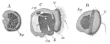
Fig. 117. Three stages in the development of Cardium. (After Lovén.)
hy. hypoblast; b. foot; m. mouth; an. anus; V. velum; cm. anterior adductor muscle.
In the later stages, after the development of the shell, the velum becomes highly retractile and can be nearly completely withdrawn within the mantle by special muscles. It forms the chief organ of locomotion of the free larva.
In some fresh-water forms, which have no free larval existence, the velum is very much reduced (Anodon, Unio, Cyclas) or even aborted (Pisidium). In these forms as well as in Teredo and probably other marine forms (e.g. Ostrea) the central flagellum is absent. It has been suggested by Lovén, though without any direct evidence, that the labial tentacles of adult Lamellibranchiata are the remains of the velum. The velar area is in any case the only representative of the head. In some marine forms a general covering of cilia arises before the formation of the velum; and in Montacuta and other types there is developed, as in many Gasteropoda, a circumanal patch of cilia.
A shell-gland appears at a very early period on the dorsal surface in Pisidium, Cyclas and Ostrea, and probably in most marine forms (fig. 118, shs). It is somewhat saddle-shaped, and formed of elongated non-ciliated cells bounding a groove. It flattens out and on its surface is formed the shell, which appears usually to have the form of an unpaired saddle-shaped cuticle, on the two sides of which the valves are subsequently formed by a deposit of calcareous salts. In Pisidium the two valves are stated by Lankester to be at first quite independent and widely separated, and it has been suggested by Lankester, though not proved, that the ligament of the shell is developed in the median part of the groove of the shell-gland.
The mantle lobes are developed as lateral outgrowths of the body: they usually have a considerable extension before they are covered by the shell. In Anodon and Unio the larval mantle lobes are, however, formed in a somewhat exceptional way, and are from the first completely covered by the valves of the larval shell. The larval mantle lobes and shell in Anodon and Unio are subsequently replaced by the permanent structures.
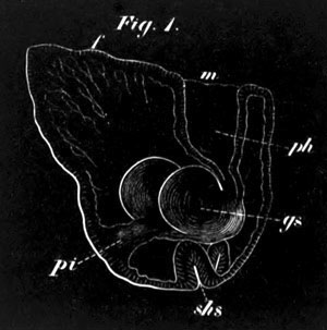
Fig. 118. An embryo of Pisidium pusillum. (From Lankester.)
f. foot; m. mouth; ph. pharynx; gs. bilobed stomach; pi. intestine; shs. shell-gland.
The adductor muscles are formed soon after the appearance of the shell. The posterior sometimes appears first, e.g. Mytilus, and at other times the anterior, e.g. Cardium.
The foot arises in the usual way as a prominence between the mouth and anus. In comparison with Gasteropoda it is late in appearing, and in many cases does not become prominent till the shell has attained a considerable size. In its hinder part a provisional paired byssus-gland is developed from the epidermis in Cyclas and other forms. In other cases, e.g. Mytilus, the byssus-gland is permanent. The byssus-gland occupies very much the position of the Gasteropod operculum, and would appear very probably to correspond with this organ. The anterior part of the foot is usually ciliated.
The gills appear rather late in larval development along the base of the foot on either side, between the mantle and the foot (fig. 120, br). They arise as a linear row of separate ciliated somewhat knobbed papillæ. A second row appears later. The two rows give rise respectively to the two gill lamellæ of each side.
The further history of the development of the gills has been studied by Lacaze Duthiers (No. 297) in Mytilus. The first row of gill papillæ formed becomes the innermost of the two lamellæ of the adult. The number of papillæ goes on increasing from before backwards. When about eleven have been formed, their somewhat swollen free extremities unite together, the basal portions being separated by slits.
The free limb is formed by the free end of the gill lamella bending upon itself towards the inner side and growing towards the line of attachment of the lamella. The free limb is at first not composed of separate bars, but of a continuous membrane. Before this membrane has grown very wide, perforations are formed in it corresponding to the spaces between the bars of the attached limb.
The outer gill lamella develops in precisely the same way as, but somewhat later than, the inner. The rudiments of it appear when about twenty papillæ of the inner lamella are formed. Its first papillæ are formed near the hind border of the inner lamella, and new papillæ are added both in front and behind. Its free limb is on the outer side.
In Mytilus the two limbs (free and attached) of each bar of the gill are joined at wide intervals by extensile processes, the ‘inter-lamellar junctions,’ and the successive bars are attached together by ciliated junctions. In many other types the concrescences between the various parts of the gills are carried much further; the maximum of concrescence being perhaps attained in Anodon and Unio[107].
Large paired auditory sacks seem always to be developed in the foot; and clearly correspond with the auditory sacks in Gasteropoda.
Eyes are frequently present in the larva, though they disappear in the adult. In Montacuta and other types a pair of these organs is formed at the base of the velum on each side of the œsophagus, not far from the auditory sacks. They are provided with a lens.
A row of similar organs is present in the larva of Teredo in front of the foot.
Cardium. As an example of a marine Lamellibranchiate I may take Cardium pygmaeum, the development of which has been studied by Lovén (No. 291). The ova, surrounded by a thickish capsule, are impregnated in the cloaca. The segmentation takes place much as in Nassa (vide p. 101), and the small segments gradually envelop the large hypoblast spheres; so that there would seem to be a gastrula by epibole. After the hypoblast has become enveloped by the epiblast, one side of the embryo is somewhat flattened and marked by a deepish depression (fig. 117 A). From Lovén’s description it appears to me probable that the depression on the flattened side occupies the position of the blastopore, and that the depression itself is the stomodæum. At this stage the embryo becomes covered with short cilia which cause it to rotate within the egg-capsule.
Close above the mouth there appear two small papillæ. These gradually separate and give rise to a circular ridge covered with long cilia, which encircles the embryo anteriorly to the ventrally-placed mouth. This structure is the velum. In its centre is a single long flagellum (fig. 117 B). Shortly after this the shell appears as a saddle-shaped structure on the hinder part of the dorsal surface of the embryo. It is formed at first of two halves which meet behind without the trace of a hinge (fig. 117 C). The two halves rapidly grow and partially cover over the velum, and below them the mantle folds soon sprout out as lateral flaps.
The alimentary tract has by this time become differentiated (fig. 117 C). It consists of a mouth (m) and ciliated œsophagus probably derived from the stomodæum, a stomach and intestine derived from the true hypoblast, and an hepatic organ consisting of two separate lobes opening into the stomach. The anus (an) appears not far behind the mouth, and between the two is a very slightly developed rudiment of the foot (b). The anterior adductor muscle (cm) appears at this stage, though the posterior is not yet differentiated.
The larva is now ready to be hatched, but the further stages of its development were not followed.
Ostrea. The larvæ of Ostrea, figured by Salensky (No. 293), shew a close resemblance to those of Cardium. The velum is however a simple ring of cilia without a central flagellum. The proctodæum would appear to be formed later than the stomodæum, and the earliest stage figured is too far advanced to throw light on the position of the blastopore.
Pisidium. The development of Pisidium has been investigated by Lankester (No. 239). The ovum is invested by a vitelline membrane and undergoes development in a brood-pouch at the base of the inner gill lamella.
The segmentation commences by a division into four equal spheres, each of which, as in so many other Mollusca, then gives rise by budding to a small sphere. The later stages of segmentation have not been followed in detail, but the result of segmentation is a blastosphere. An invagination, presumably at the lower pole, now takes place, and gives rise to an archenteric sack.
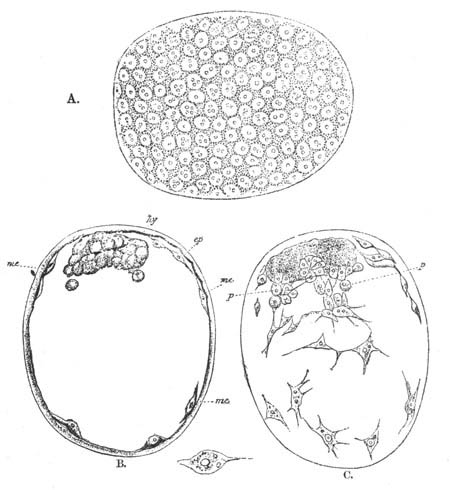
Fig. 119. Three views of an embryo of Pisidium immediately after the closure of the blastopore. (After Lankester.)
A. View from the surface. B. Optical section through the median plane. C. Optical section through a plane a little below the surface.
ep. epiblast; me. mesoblast; hy. hypoblast; p. cells apparently budding from the hypoblast to form mesoblastic elements.
The embryo now rapidly grows in size. The blastopore becomes closed and the archenteric sack forms a small mass attached at one point to the walls of the embryonic vesicle (fig. 119, hy). In the space between the walls of the archenteron and those of the embryonic vesicle stellate mesoblast cells make their appearance, derived in the main from the epiblast, though probably in part also from the hypoblastic vesicle (vide fig. 119 C, p). The cavity between the hypoblast and epiblast, which contains these cells, is the body cavity. Fig. 119 represents three views of the embryo at this stage. A is a surface view shewing the epiblast; B is an optical section through the median plane shewing the hypoblast and some of the mesoblast cells; and C is an optical section shewing the mesoblast cells. A prominence on one side of the embryo now develops which forms the commencement of the foot, and the archenteric sack grows out at its free extremity into two lobes, but remains attached to the epiblast by an imperforate pedicle. The next organ to appear is the stomodæum. It arises as a ciliated epiblastic ingrowth which meets the free end of the archenteric sack, fuses with it, and shortly afterwards opens into it (fig. 118, ph). Between the mouth and the attachment of the enteric pedicle is placed the foot (f), which becomes ciliated. On the dorsal side of the enteric pedicle there appears a saddle-shaped patch of epiblast cells bounding the sides of a groove (shs). This is the rudiment of the shell-gland.
The enteric pedicle, or intestine as it may now be called, soon acquires a lumen, though still imperforate at its termination where the anus is eventually formed. Ventral to the intestine is placed a mass of cells—the rudiment of the organ of Bojanus. It is stated to be developed as an ingrowth of the epiblast.
In a slightly later stage the shell-gland rapidly increases in size and flattens out, and on the two sides of it there appear the rudiments of the two valves, which are at first quite distinct, and separated by a considerable interval (fig. 120). Before the appearance of the valves of the shell, the mantle folds have already grown out from the sides of the body.
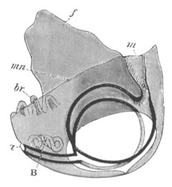
Fig. 120. Diagrammatic view of advanced larva of Pisidium. (Copied from Lankester.)
m. mouth; a. anus; B. organ of Bojanus; mn. mantle; f. foot.
At a somewhat later stage the gills appear as a linear series of small independent buds within the folds of the mantle behind the foot (fig. 120, br). The anterior adductor also becomes differentiated.
The alimentary tract in the meantime has undergone considerable changes. The primitive lateral lobes dilate enormously and become ciliated. At a still later stage their walls undergo peculiar changes, the nature of which is somewhat obscure, but they appear to me to be of the same character as those in many Pteropods and Gasteropods, where the cells of the hepatic diverticula, to which the lobes of Pisidium apparently correspond, become filled with an albuminous material.
The later stages in Pisidium have not been followed.
It is remarkable that in Pisidium a veliger stage does not occur. This is probably due to the development taking place within the brood-pouch. The late development of the otocysts is also remarkable. A byssus-gland was not formed up to the stage observed. In Cyclas calyculata (Schmidt), a byssus-gland also appears to be absent.
Cyclas. The development of Cyclas as described by Von Jhering is very unlike that of Pisidium, and the differences would seem to be too great to be accounted for except by errors of observation.
The segmentation of Cyclas is similar to that of Anodon (vide p. 82), and a mass of large cells enclosed by the smaller cells gives rise to the hypoblast. In the interior of this mass there appears a lumen, and a process from it grows towards and meets the epiblast, and gives rise to the œsophagus and mouth,—a mode of development of these parts without parallel amongst Mollusca. A very rudimentary velum would appear, according to Leydig (No. 290), to be developed at the cephalic extremity. A shell-gland is formed of the same character as in Gasteropods. According to Leydig the shell appears as a single saddle-like structure on the dorsal surface; the lateral parts of this become calcified, and give rise to the two valves, but are united in the middle by the membranous median portion. At the two sides of the body the mantle lobes are formed, as in Pisidium.
Very shortly after the formation of the shell the byssus-gland appears as a pair of small follicles in the hinder part of the foot. It rapidly grows larger and becomes a paired pyriform gland, in which are secreted the byssus threads which serve to attach all the embryos at a common point to the walls of the brood-pouch.
The foot is large, and ciliated anteriorly. Otolithic sacks and peda ganglia are developed in it very early.
Unio. The ovum of Anodonta and Unio is enveloped in a vitelline membrane, the surface of which is raised into a projecting trumpet-like tube perforated at its extremity (fig. 12). This structure is the micropyle. The micropyle disappears in Anodonta piscinalis when the egg is ripe, but in Unio persists during the whole development. The ova are transported, in a manner not certainly made out, into the space between the two limbs of the outer gills of the mother, and there undergo their early development. The animal or upper pole of the egg is placed at the pole opposite to the micropyle.
The segmentation is unequal (vide p. 100) and results in the formation of a blastosphere with a large segmentation cavity. The greater part of the circumference of the egg is formed of small uniform spheres, but the lower (with reference to the segmentation) pole is taken up by a single large cell. The small spheres become the epiblast, and the large cell gives rise to hypoblast and mesoblast[108].
The single large cell next divides into two, and then four, and finally into about ten to fifteen cells. These cells form an especial area of more granular cells than the other cells of the blastosphere. Most of them are nearly of the same size, but two of them (according to Rabl), in contact with each other, but placed on the future right and left sides of the embryo, are considerably larger than the remainder. These two cells soon pass into the cavity of the blastosphere, while at the same time the area of granular cells becomes flattened out, and then becomes involuted as a small sack with a transversely elongated opening, which does not nearly fill up the cavity of the blastosphere. This involuted sack is the archenteron.
The two large cells, which lie in immediate contact with what, following Rabl, I shall call the anterior lip of the blastopore, next bud off small cells, which first form a layer covering the walls of the archenteron, but subsequently develop into a network filling up the whole cavity of the primitive blastosphere. The space between these cells is the primitive body cavity. For a long time the two primitive mesoblast cells retain their preponderating size[109]. At the hinder end of the body, and at the end opposite therefore to the two mesoblast cells, are placed three especially large epiblast cells.
In Anodonta and Unio tumidus there appears at this period a patch of long cilia at the anterior end of the body. These cilia cause a rotation of the embryo and would appear to be the velum. In Unio pictorum they do not appear till much later.
Immediately following this stage the changes in the embryo take place with great rapidity. In the first place a special mass of mesoblast cells appears at the hinder end of the archenteric sack; and becoming elongated transversely gives rise to the single adductor muscle. On the subsequent formation of the shell the muscle becomes inserted in its two valves. The blastopore next becomes closed, and the small archenteron grows forwards till it meets the epiblast anteriorly, and at the same time detaches itself from the epiblast in the region where the blastopore was placed. Where it comes in contact with the wall of the body in front a small epiblastic invagination arises, which meets and opens into the archenteric sack and forms the permanent mouth.
While these changes have been taking place the shell is formed as a continuous saddle-shaped plate on the dorsal surface. From this plate the two valves are subsequently differentiated. On the dorsal surface they meet with a straight hinge-line. Each valve is at first rounded, but subsequently becomes triangular with the hinge-line as base. The valves are not quite equi-sided, but the anterior side is less convex than the posterior. At a later period a beak-shaped organ is formed at the apex of each valve in the same manner as the remainder of the shell. This organ is placed at about a right angle with the main portion of the valve. It is pointed at its extremity and bears numerous sharp spines on its outer side, which are especially large in the median line (vide fig. 121 A). It is employed in fixing the larva, after it is hatched, on to the fish on which it is for some time parasitic. The shell is perforated by numerous pores.
After the shell has become formed a new structure makes its appearance which is known as the byssus-gland. It is developed as an invagination of the epiblast at the hinder end of the body: Rabl was unable to determine whether it was formed from the three large epiblastic cells present there or no. It subsequently forms an elongated gland with three coils or so round the adductor muscle on the left side of the body, but opening in the median ventral line. It secretes an elongated cord by which the larva becomes suspended after hatching.
For some time the ventral portion of the body projects behind the ends of the valves of the shell, but before these are completely formed a median invagination of the body wall takes place, which obliterates to a large extent the body cavity, and gives rise to two great lateral lobes, one for each valve. These lobes are the mantle lobes.
Before the mantle lobes are fully formed peculiar sense organs, usually four in number, make their appearance on each lobe. Each of them consists of a columnar cell, bearing at its free end a cuticle from which numerous fine bristles proceed. Covering the cell and the parts adjoining it is a delicate membrane perforated for the passage of the bristles. The largest and first formed of these organs is placed near the anterior and dorsal part of the mantle. The three others are placed near the free end of the mantle (vide fig. 121 A). These organs probably have the function of enabling the larva to detect the passage of a fish in its vicinity, and to assist it therefore in attaching itself. When the embryo is nearly ripe there appears immediately ventral to and behind the velum a shallow pit on each side of the middle line, and the two pits appear to be connected by a median transverse bridge. These structures have been the cause of great perplexity to different investigators, and their meaning is not yet clear. According to Rabl the median structure is the somewhat bilobed archenteron, and according to his view it is not really connected with the laterally placed pits. The cilia of the velum overlie these latter structures and make them appear as if their edges were ciliated. They are regarded by Rabl as the rudiments of the nervous system.
With the development of the shell, the mantle, and the sense organs, the young mussel reaches its full larval development, and is now known as a Glochidium (fig. 121 A).
If the parent, with Glochidia in its gills, is placed in a tank with fish, it very soon (as I have found from numerous experiments) ejects the larvæ from its gills, and as soon as this occurs the larvæ become free from the egg membrane, attach themselves by the byssus-cord, and when suspended in this position continually close and open their shells by the contraction of the adductor muscle. If the mussels are not placed in a tank with fish the larvæ may remain for a long time in the gills.
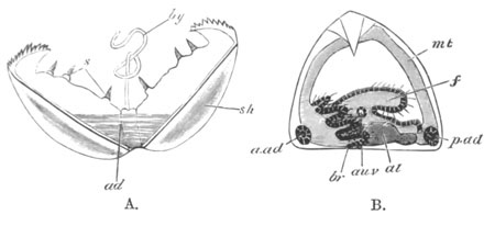
Fig. 121.
A. Glochidium Immediately After It Is Hatched. ad. adductor; sh. shell; by. byssus cord; s. sense organs.
B. Glochidium After It Has Been on the Fish for Some Weeks. br. branchiæ; au.v. auditory sack; f. foot; a.ad. and p.ad. anterior and posterior adductors; al. mesenteron; mt. mantle.
Before passing on to state what is known with reference to the larval metamorphosis, it may be well to call attention to certain, and to my mind not inconsiderable, difficulties in the way of accepting in all particulars Rabl’s account of the development.