Volume 1 / Part 1 — CHAPTER XVII.
Reading edition
TRACHEATA.
Prototracheata.
The remarkable researches of Moseley (No. 396) on Peripatus capensis have brought clearly to light the affinities of this form with the tracheate Arthropoda; and its numerous primitive characters, such as the generally distributed tracheal apertures, the imperfectly segmented limbs, the diverging ventral nerve cords with imperfectly marked ganglia, and the nephridia (segmental organs[160]), would render its embryology of peculiar interest. Unfortunately Moseley was unable, from want of material, to make so complete a study of its development as of its anatomy. The youngest embryo observed was in part distinctly segmented, and coiled up within the egg (fig. 168 A). The procephalic lobes resemble those of the Arthropoda generally, and are unlike the præ-oral lobe of Chætopods or Discophora. They are not marked off by a transverse constriction from the succeeding segments. The three embryonic layers are differentiated, and the interior is filled with a brownish mass—the remnant of the yolk—which is probably enclosed in a distinct intestinal wall, and is lobed in correspondence with the segmentation of the body. The mouth invagination is not present, and but two pairs of slight prominences mark the rudiments of the two anterior post-oral appendages.
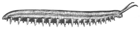
Fig. 167. Adult example of Peripatus capensis, natural size. (From Moseley.)
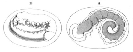
Fig. 168. Two stages in the development of Peripatus capensis. (After Moseley.)
A. Youngest stage hitherto observed before the appearance of the legs. B. Later stage after the legs and antennæ have become developed. Both figures represent the larva as it appears within the egg. 1 and 2. First and second post-oral appendages.
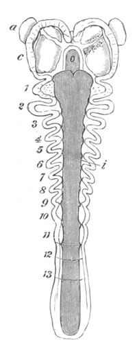
Fig. 169. Embryo of Peripatus capensis. Slightly older than A in fig. 168; unrolled. (After Moseley.)
a. antennæ; o. mouth; i. intestine; c. procephalic lobe. 1, 2, 3, etc., post-oral appendages.
The single pair of antennæ is formed in the next stage, and is followed by the remaining post-oral appendages, which arise in succession from before backwards somewhat later than the segments to which they appertain.
The posterior part of the embryo becomes uncoiled, and the whole embryo bent double in the egg (fig. 168 B).
The mouth appears as a slit-like opening between and below the procephalic lobes. On each side and somewhat behind it there grows out an appendage—the first post-oral pair (fig. 169, 1)—while in front and behind it are formed the upper and lower lips. These two appendages next turn inwards towards the mouth, and their bases become gradually closed over by two processes of the procephalic region (fig. 170, m). The whole of these structures assist in forming a kind of secondary mouth cavity, which is at a later period further completed by the processes of the procephalic region meeting above the mouth, covering over the labrum, and growing backwards to near the origin of the second pair of post-oral appendages.
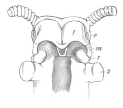
Fig. 170. Ventral view of the head of an embryo of Peripatus capensis at a late stage of development.
l. thickening of epiblast of procephalic lobe to form supra-œsophageal ganglion; m. process from procephalic lobe growing over the first post-oral appendage; o. mouth; e. eye; 1 and 2, first and second pair of post-oral appendages.
The antennæ early become jointed, and fresh joints continue to be added throughout embryonic life; in the adult there are at present fully thirty joints. It appears to me probable (though Mr Moseley takes the contrary view) from the late development of the paired processes of the procephalic lobes, which give rise to the circular lip of the adult, that they are not true appendages. The next pair therefore to the antennæ is the first post-oral pair. It is the only pair connected with the mouth. At their extremities there is formed a pair of claws similar to those of the ambulatory legs (fig. 171). The next and largest pair of appendages in the embryo are the oral papillæ. They are chiefly remarkable for containing the ducts of the slime glands which open at their bases. They are without claws. The succeeding appendages become eventually imperfectly five-jointed; two claws are formed as cuticular investments of papillæ in pockets of the skin at the ends of their terminal joints.
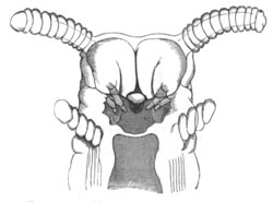
Fig. 171. Head of an embryo Peripatus. (From Moseley.)
The figure shews the jaws (mandibles), and close to them epiblastic involutions, which grow into the supra-œsophageal ganglia. The antennæ, oral cavity, and oral papillæ are also shewn.
I have been able to make a few observations on the internal structure of the embryos from specimens supplied to me by Moseley. These are so far confined to a few stages, one slightly earlier, the others slightly later, than the embryo represented in fig. 168 B. The epiblast is formed of a layer of columnar cells, two deep on the ventral surface, except along the median line where there is a well-marked groove and the epiblast is much thinner (fig. 172).
The ventral cords of the trunk are formed as two independent epiblastic thickenings. In my earlier stage these are barely separated from the epiblast, but in the later ones are quite independent (fig. 172, v.n), and partly surrounded by mesoblast.
The supra-œsophageal ganglia are formed as thickenings of the epiblast of the ventral side of the procephalic lobes in front of the stomodæum. They are shewn at l in fig. 170. The thickenings of the two sides are at first independent. At a somewhat later period an invagination of the epiblast grows into each of these lobes. The openings of these invaginations extend from the oral cavity forwards; and they are shewn in fig. 171[161]. Their openings become closed, and the walls of the invaginations constitute a large part of the embryonic supra-œsophageal ganglia.
Similar epiblastic invaginations assist in forming the supra-œsophageal ganglia of other Tracheata. They are described in the sequel for Insects, Spiders and Scorpions. The position of the supra-œsophageal ganglia on the ventral side of the procephalic lobes is the same as that in other Tracheata.
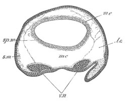
Fig. 172. Section through the trunk of an embryo of Peripatus. The embryo from which the section is taken was somewhat younger than fig. 171.
sp.m. splanchnic mesoblast. s.m. somatic mesoblast. mc. median section of body cavity. lc. lateral section of body cavity. v.n. ventral nerve cord. me. mesenteron.
The mesoblast is formed, in the earliest of my embryos, of scattered cells in the fairly wide space between the mesenteron and the epiblast. There are two distinct bands of mesoblast on the outer sides of the nervous cords. In the later stage the mesoblast is divided into distinct somatic and splanchnic layers, both very thin; but the two layers are connected by transverse strands (fig. 172). There are two special longitudinal septa dividing the body cavity into three compartments, a median (mc), containing the mesenteron, and two lateral (lc) containing the nerve cords. This division of the body cavity persists, as I have elsewhere shewn, in the adult. A similar division is found in some Chætopoda, e.g. Polygordius.
I failed to make out that the mesoblast was divided into somites, and feel fairly confident that it is not so in the stages I have investigated.
There is a section of the body cavity in the limbs as in embryo Myriapods, Spiders, etc.
In the procephalic lobe there is a well-developed section of the body cavity, which lies dorsal to and in front of the rudiment of the supra-œsophageal ganglia.
The alimentary tract is formed of a mesenteron (fig. 172), a stomodæum, and proctodæum. The wall of the mesenteron is formed, in the stages investigated by me, of a single layer of cells with yolk particles, and encloses a lumen free from yolk. The forward extension of the mesenteron is remarkable.
The stomodæum in the earlier stage is a simple pit, which meets but does not open into the mesenteron. In the later stage the external opening of the pit is complicated by the structures already described. The proctodæum is a moderately deep pit near the hinder end of the body.
The existence of a tracheal system[162] is in itself almost sufficient to demonstrate the affinities of Peripatus with the Tracheata, in spite of the presence of nephridia. The embryological characters of the procephalic lobes, of the limbs and claws, place however this conclusion beyond the reach of scepticism. If the reader will compare the figure of Peripatus with that of an embryo Scorpion (fig. 196 A) or Spider (fig. 200 C) or better still with Metschnikoff’s figure of Geophilus (No. 399) Pl. XXI. fig. II, he will be satisfied on this point.
The homologies of the anterior appendages are not very easy to determine; but since there does not appear to me to be sufficient evidence to shew that any of the anterior appendages have become aborted, the first post-oral appendages embedded in the lips may provisionally be regarded as equivalent to the mandibles, and the oral papillæ to the first pair of maxillæ, etc. Moseley is somewhat doubtful about the homologies of the appendages, and hesitates between considering the oral papillæ as equivalent to the second pair of maxillæ (on account of their containing the openings of the mucous glands, which he compares with the spinning glands of caterpillars), or to the poison claws (fourth post-oral appendages) of the Chilopoda (on account of the poison glands which he thinks may be homologous with the mucous glands).
The arguments for either of these views do not appear to me conclusive. There are glands opening into various anterior appendages in the Tracheata, such as the poison glands in the Cheliceræ (mandibles) of Spiders, and there is some evidence in Insects for the existence of a gland belonging to the first pair of maxillæ, which might be compared with the mucous gland of Peripatus. For reasons already stated I do not regard the processes of the cephalic lobes, which form the lips, as a pair of true appendages.
Bibliography.
(396) H. N. Moseley. “On the Structure and Development of Peripatus capensis.” Phil. Trans. Vol. 164, 1874.
Myriapoda[163].
Chilognatha. The first stages in the development of the Chilognatha have been investigated by Metschnikoff and Stecker, but their accounts are so contradictory as hardly to admit of reconciliation.
According to Metschnikoff, by whom the following four species have been investigated, viz., Strongylosoma Guerinii, Polydesmus complanatus, Polyxenus lagurus, and Julus Moneletei, the segmentation is at first regular and complete, but, when the segments are still fairly large, the regular segmentation is supplemented by the appearance of a number of small cells at various points on the surface, which in time give rise to a continuous blastoderm.
The blastoderm becomes thickened on the ventral surface, and so forms a ventral plate[164].
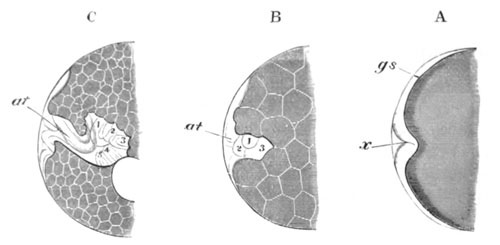
Fig. 173. Three stages in the development of Strongylosoma Guerinii. (After Metschnikoff.)
A. Embryo on eleventh day with commencing ventral flexure (x) B. Embryo with three pairs of post-oral appendages. C. Embryo with five pairs of post-oral appendages.
gs. ventral plate; at. antennæ; 1‑5 post-oral appendages; x. point of flexure of the ventral plate.
The most important sources of information for the general embryology of the Chilognatha are the papers of Newport (No. 397) and Metschnikoff (No. 398). The development of Strongylosoma may be taken as fairly typical for the group; and the subsequent statements, unless the reverse is stated, apply to the species of Strongylosoma investigated by Metschnikoff.
After the segmentation and formation of the layers the first observable structure is a transverse furrow in the thickening of the epiblast on the ventral surface of the embryo. This furrow rapidly deepens, and gives rise to a ventral flexure of the embryo (fig. 173 A, x), which is much later in making its appearance in Julus than in Strongylosoma and Polyxenus. A pair of appendages, which become the antennæ, makes its appearance shortly after the formation of the transverse furrow, and there soon follow in order the next three pairs of appendages. All these parts are formed in the infolded portion of the ventral thickening of the blastoderm (fig. 173 B). The ventral thickening has in the meantime become marked by a longitudinal furrow, but whether this is connected with the formation of the nervous system, or is equivalent to the mesoblastic furrow in Insects, and connected with the formation of the mesoblast, has not been made out. Shortly after the appearance of the three pairs of appendages behind the antennæ two further pairs become added, and at the same time oral and anal invaginations become formed (fig. 173 C). In front of the oral opening an unpaired upper lip is developed. The præ-oral part of the ventral plate develops into the bilobed procephalic lobes, the epiblast of which is mainly employed in the formation of the supra-œsophageal ganglia. The next important change which takes place is the segmentation of the body of the embryo (fig. 174 A), the most essential feature in which is the division of the mesoblast into somites. Segments are formed in order from before backwards, and soon extend to the region behind the appendages. On the appearance of segmentation the appendages commence to assume their permanent form. The two anterior pairs of post-oral appendages become jaws; and the part of the embryo which carries them and the antennæ is marked off from the trunk as the head. The three following pairs of appendages grow in length and assume a form suited for locomotion. Behind the three existing pairs of limbs there are developed three fresh pairs, of which the two anterior belong to a single primitive segment. While the above changes take place in the appendages the embryo undergoes an ecdysis, which gives rise to a cuticular membrane within the single egg membrane (chorion, Metschnikoff). On this cuticle a tooth-like process is developed, the function of which is to assist in the hatching of the embryo (fig. 174 A).
In Polyxenus a cuticular membrane is present as in Strongylosoma, but it is not provided with a tooth-like process. In the same form amœboid cells separate themselves from the blastoderm at an early period. These cells have been compared to the embryonic envelopes of Insects described below.
In Julus two cuticular membranes are present at the time of hatching: the inner one is very strongly developed and encloses the embryo after hatching. After leaving the chorion the embryo Julus remains connected with it by a structureless membrane which is probably the outer of the two cuticular membranes.
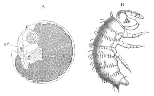
Fig. 174. Two stages in the development of Strongylosoma Guerinii. (After Metschnikoff.)
A. A seventeen days’ embryo, already segmented. B. A just hatched larva.
At the time when the embryo of Strongylosoma is hatched (fig. 174 B) nine post-cephalic segments appear to be present. Of these segments the second is apparently (from Metschnikoff’s figure, 174 B) without a pair of appendages; the third and fourth are each provided with a single functional pair of limbs; the fifth segment is provided with two pairs of rudimentary limbs, which are involuted in a single sack and not visible without preparation, and therefore not shewn in the figure. The sixth segment is provided with but a single pair of appendages, though a second pair is subsequently developed on it[165].
Julus, at the time it leaves the chorion, is imperfectly segmented, but is provided with antennæ, mandibles, and maxillæ, and seven pairs of limbs, of which the first three are much more developed than the remainder. Segmentation soon makes its appearance, and the head becomes distinct from the trunk, and on each of the three anterior trunk segments a single pair of limbs is very conspicuous (Metschnikoff)[166]. Each of the succeeding segments bears eventually two pairs of appendages. At the time when the inner embryonic cuticle is cast off, the larva appears to be hexapodous, like the young Strongylosoma, but there are in reality four pairs of rudimentary appendages behind the three functional pairs. The latter only appear on the surface after the first post-embryonic ecdysis. Pauropus (Lubbock) is hexapodous in a young stage. At the next moult two pairs of appendages are added, and subsequently one pair at each moult.
There appear to be eight post-oral segments in Julus at the time of hatching. According to Newport fresh segments are added in post-embryonic life by successive budding from a blastema between the penultimate segment and that in front of it. They arise in batches of six at the successive ecdyses, till the full number is completed. A functional, though not a real hexapodous condition, appears to be characteristic of Chilognatha generally at the time of hatching.
The most interesting anatomical feature of the Chilognatha is the double character of their segments, the feet (except the first three or four, or more), the circulatory, the respiratory, and the nervous systems shewing this peculiarity. Newport’s and Metschnikoff’s observations have not thrown as much light on the nature of the double segments as might have been hoped, but it appears probable that they have not originated from a fusion of two primitively distinct segments, but from a later imperfect division of each of the primitive segments into two, and the supply to each of the divisions of a primitive segment of a complete set of organs.
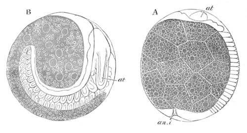
Fig. 175. Two stages in the development of Geophilus. (After Metschnikoff.)
A. Side view of embryo at the stage when the segments are beginning to be formed. B. Later stage after the appendages have become established.
at. antennæ; an.i. proctodæum.
Chilopoda. Up to the present time the development of only one type of Chilopoda, viz. that of Geophilus, has been worked out. Most forms lay their eggs, but Scolopendra is viviparous. The segmentation appears to resemble that in the Chilognatha, and at its close there is present a blastoderm surrounding a central mass of yolk cells. A ventral thickening of the blastoderm is soon formed. It becomes divided into numerous segments, which continue to be formed successively from the posterior unsegmented part. The antennæ are the first appendages to appear, and are well developed when eighteen segments have become visible (fig. 175 A). The post-oral appendages are formed slightly later, and in order from before backwards. As the embryo grows in length, and fresh segments continue to be formed, the posterior part of it becomes bent over so as to face the ventral surface of the anterior, and it acquires an appearance something like that of many embryo Crustaceans (fig. 175 B). Between forty and fifty segments are formed while the embryo is still in the egg. The appendages long remain unjointed. The fourth post-oral appendage, which becomes the poison claw, is early marked out by its greater size: on the third post-oral there is formed a temporary spine to open the egg membrane.
It does not appear, from Metschnikoff’s figures of Geophilus, that any of the anterior segments are without appendages, and it is very probable that Newport is mistaken in supposing that the embryo has a segment without appendages behind that with the poison claws, which coalesces with the segment of the latter. It also appears to me rather doubtful whether the third pair of post-oral appendages, i.e. those in front of the poison claws, can fairly be considered as forming part of the basilar plate. The basilar plate is really the segment of the poison claws, and may fuse more or less completely with the segment in front and behind it, and the latter is sometimes without a pair of appendages (Lithobius, Scutigera).
Geophilus, at the time of birth, has a rounded form like that of the Chilognatha.
The young of Lithobius is born with only six pairs of limbs.
General observation on the homologies of the appendages of Myriapoda.
The chief difficulty in this connection is the homology of the third pair of post-oral appendages.
In adult Chilognatha there is present behind the mandibles a four-lobed plate, which is usually regarded as representing two pairs of appendages, viz. the first and second pairs of maxillæ of Insects. Metschnikoff’s observations seem however to shew that this plate represents but a single pair of appendages, which clearly corresponds with the first pair of maxillæ in Insects. The pair of appendages behind this plate is ambulatory, but turned towards the head; it is in the embryo the foremost of the three functional pairs of legs with which the larva is born. Is it equivalent to the second pair of maxillæ of Insects or to the first pair of limbs of Insects? In favour of the former view is the fact (1) that in embryo Insects the second pair of maxillæ sometimes resembles the limbs rather than the jaws, so that it might be supposed that in Chilognatha a primitive ambulatory condition of the third pair of appendages has been retained; (2) that the disappearance of a pair of appendages would have to be postulated if the second alternative is adopted, and that if Insects are descended from forms related to the Myriapods it is surprising to find a pair of appendages always present in the former, absent in the latter. The arguments which can be urged for the opposite view do not appear to me to have much weight, so that the homology of the appendages in question with the second pair of maxillæ may be provisionally assumed.
The third pair of post-oral appendages of the Chilopoda may probably also be assumed to be equivalent to the second pair of maxillæ; though they are limb-like and not connected with the head. The subjoined table shews the probable homologies of the appendages.
Chilognatha (Strongylosoma at time of birth). Chilopoda (Scolopendra adult).
Pre-oral region. Antennæ. Antennæ.
1st Post-oral segment. Mandibles. Mandibles.
2nd Post-oral segment. Maxillæ 1. (Four-lobed plate in adult, but a simple pair of appendages in embryo). Maxillæ 1. (Palp and bilobed median process).
3rd Post-oral segment. (probably equivalent to segment bearing 2nd pair of maxillæ in Insects). 1st pair of ambulatory limbs. Limb-like appendages with basal parts in contact.
4th Post-oral segment. (?) Apodous. Poison claws.
5th Post-oral segment. 2nd pair of ambulatory limbs. 1st pair of ambulatory limbs.
6th Post-oral segment. 3rd pair of ambulatory limbs. 2nd pair of ambulatory limbs.
7th Post-oral segment. 4th and 5th pair of ambulatory limbs. 3rd pair of ambulatory limbs.
8th Post-oral segment. 6th pair of ambulatory limbs. 4th pair of ambulatory limbs.
9th Post-oral segment. Apodous. 5th pair of ambulatory limbs.
10th Post-oral segment. " (last segment in embryo). 6th pair of ambulatory limbs.
The germinal layers and formation of organs.
The development of the organs of the Myriapoda, and the origin of the germinal layers, are very imperfectly known: Myriapoda appear however to be closely similar to Insects in this part of their development, and the general question of the layers will be treated more fully in connection with that group.
The greater part of the blastoderm gives rise to the epiblast, which furnishes the skin, nervous system, tracheal system, and the stomodæum and proctodæum.
The mesoblast arises in connection with the ventral thickening of the blastoderm, but the details of its formation are not known. Metschnikoff describes a longitudinal furrow which appears very early in Strongylosoma, which is perhaps equivalent to the mesoblastic furrows of Insects, and so connected with the formation of the mesoblast.
The mesoblast is divided up into a series of protovertebra-like bodies—the mesoblastic somites—the cavities of which become the body cavity and the walls the muscles and probably the heart. They are (Metschnikoff) prolonged into the legs, though the prolongations become subsequently segmented off from the main masses. The splanchnic mesoblast is, according to Metschnikoff, formed independently of the somites, but this point requires further observation.
The origin of the hypoblast remains uncertain, but it appears probable that it originates, in a large measure at least, from the yolk segments. In the Chilognatha the mesenteron is formed in the interior of the yolk segments, so that those yolk segments which are not employed in the formation of the alimentary canal lie freely in the body cavity. In the relation of the yolk segments to the alimentary canal the Chilopoda present a strong contrast to the Chilognatha, in that the greater part of the yolk lies within their mesenteron. The mesenteron is at first a closed sack, but is eventually placed in communication with the stomodæum and the proctodæum. The Malpighian bodies arise as outgrowths from the blind extremity of the latter.
Bibliography.
(397) G. Newport. “On the Organs of Reproduction and Development of the Myriapoda.” Philosophical Transactions, 1841. (398) E. Metschnikoff. “Embryologie der doppeltfüssigen Myriapoden (Chilognatha).” Zeit. f. wiss. Zool., Vol. XXIV. 1874. (399) —— “Embryologisches über Geophilus.” Zeit. f. wiss. Zool., Vol. XXV. 1875. (400) Anton Stecker. “Die Anlage d. Keimblatter bei den Diplopoden.” Archiv f. mik. Anatomie, Bd. XIV. 1877.
Insecta[167].
The formation of the embryonic layers in Insects has not been followed out in detail in a large number of types; but, as in so many other instances, some of the most complete histories we have are due to Kowalevsky (No. 416). The development of Hydrophilus has been worked out by him more fully than that of any other form, and will serve as a type for comparison with other forms.
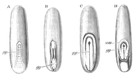
Fig. 176. Four embryos of Hydrophilus piceus viewed from the ventral surface. (After Kowalevsky.)
The upper end is the anterior. gg. germinal groove; am. amnion.
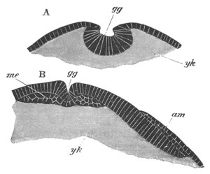
Fig. 177. Two transverse sections through embryos of Hydrophilus piceus. (After Kowalevsky.)
A. Section through an embryo of the stage represented in fig. 176 B, at the point where the two germinal folds most approximate. B. Section through an embryo somewhat later than the stage fig. 176 D, through the anterior region where the amnion has not completely closed over the embryo.
gg. germinal groove; me. mesoblast; am. amnion; yk. yolk.
The segmentation has not been studied, but no doubt belongs to the centrolecithal type (vide pp. 110‑120). At its close there is an uniform layer of cells enclosing a central mass of yolk. These cells, in the earliest observed stage, were flat on the dorsal, but columnar on part of the ventral surface of the egg, where they form a thickening which will be called the ventral plate. At the posterior part of the ventral plate two folds, with a furrow between them, make their appearance. They form a structure which may be spoken of as the germinal groove (fig. 176 A, gg). The cells which form the floor of the groove are far more columnar than those of other parts of the blastoderm (fig. 177 A). The two folds on each side of it gradually approach each other. They do so at first behind, and then in the middle; from the latter point the approximation gradually extends backwards and forwards (fig. 176 B and C). In the middle and hinder parts of the ventral plate the groove becomes, by the coalescence of the folds, converted into a canal (fig. 178 A, gg), the central cavity of which soon disappears, while at the same time the cells of the wall undergo division, become more rounded, and form a definite layer (me)—the mesoblast—beneath the columnar cells of the surface. Anteriorly the process is slightly different, though it leads to the similar formation of mesoblast (fig. 177 B). The flat floor of the groove becomes in front bodily converted into the mesoblast, but the groove itself is never converted into a canal. The two folds simply meet above, and form a continuous superficial layer.
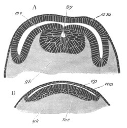
Fig. 178. Sections through two embryos of Hydrophilus piceus. (After Kowalevsky.)
A. Section through the posterior part of the embryo fig. 176 D, shewing the completely closed amnion and the germinal groove. B. Section through an older embryo in which the mesoblast has grown out into a continuous plate beneath the epiblast.
gg. germinal groove; am. amnion; yk. yolk; ep. epiblast.
During the later stages of the process last described remarkable structures, eminently characteristic of the Insecta, have made their first appearance. These structures are certain embryonic membranes or coverings, which present in their mode of formation and arrangement a startling similarity to the true and false amnion of the Vertebrata. They appear as a double fold of the blastoderm round the edge of the germinal area, which spreads over the ventral plate, from behind forwards, in a general way in the same manner as the amnion in, for instance, the chick. The folds at their origin are shewn in surface view in fig. 176 D, am, and in section in fig. 177 B, am. The folds eventually meet, coalesce (fig. 178, am) and give rise to two membranes covering the ventral plate, viz. an inner one, which is continuous with the edge of the ventral plate; and an outer, continuous with the remainder of the blastoderm. The vertebrate nomenclature may be conveniently employed for these membranes. The inner limb of the fold will therefore be spoken of as the amnion, and the outer one, including the dorsal part of the blastoderm, as the serous envelope[168]. A slight consideration of the mode of formation of the membranes, or an inspection of the figures illustrating their formation, makes it at once clear that the yolk can pass in freely between the amnion and serous envelope (vide fig. 181). At the hind end of the embryo this actually takes place, so that the ventral plate covered by the amnion appears to become completely imbedded in the yolk: elsewhere the two membranes are in contact. At first (fig. 176) the ventral plate occupies but a small portion of the ventral surface of the egg, but during the changes above described it extends over the whole ventral surface, and even slightly on the dorsal surface both in front and behind. It becomes at the same time (fig. 179) divided by a series of transverse lines into segments, which increase in number and finally amount in all to seventeen, not including the most anterior section, which gives off as lateral outgrowths the two procephalic lobes (pc.l). The changes so far described are included within what Kowalevsky calls his first embryonic period; at its close the parts contained within the chorion have the arrangement shewn in fig. 178 B. The whole of the body of the embryo is formed from the ventral plate, and no part from the amnion or serous envelope.
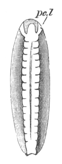
Fig. 179. Embryo Of Hydrophilus piceus viewed from the ventral surface. (After Kowalevsky.)
pc.l. procephalic lobe.
The general history of the succeeding stages may be briefly told.
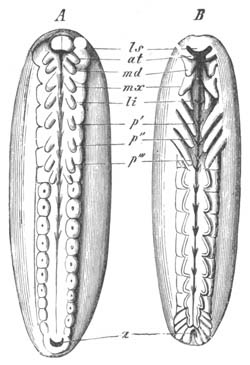
Fig. 180. Two stages in the development of Hydrophilus piceus. (From Gegenbaur, after Kowalevsky.)
ls. labrum; at. antenna; md. mandible; mx. maxilla I.; li. maxilla II.; p´ p´´ p´´´. feet; a. anus.
The appendages appear as very small rudiments at the close of the last stage, but soon become much more prominent (fig. 180 A). They are formed as outgrowths of both layers, and arise nearly simultaneously. There are in all eight pairs of appendages. The anterior or antennæ (at) spring from the procephalic lobes, and the succeeding appendages from the segments following. The last pair of embryonic appendages, which disappears very early, is formed behind the third pair of the future thoracic limbs. Paired epiblastic involutions, shewn as pits in the posterior segments in fig. 180 A, give rise to the tracheæ; and the nervous system is formed as two lateral epiblastic thickenings, one on each side of the mid-ventral line. These eventually become split off from the skin; while between them there passes in a median invagination of the skin (fig. 189 C). The two nervous strands are continuous in front with the supra-œsophageal ganglia, which are formed of the epiblast of the procephalic lobes. These plates gradually grow round the dorsal side of the embryo, and there is formed immediately behind them an oral invagination, in front of which there appears an upper lip (fig. 180, ls). A proctodæum is formed at the hind end of the body slightly later than the stomodæum. The mesoblast cells become divided into two bands, one on each side of the middle line (fig. 189 A), and split into splanchnic and somatic layers. The central yolk mass at about the stage represented in fig. 179 begins to break up into yolk spheres. The hypoblast is formed first on the ventral side at the junction of the mesoblast and the yolk, and gradually extends and forms a complete sack-like mesenteron, enveloping the yolk (fig. 185 al). The amnion and serous membrane retain their primitive constitution for some time, but gradually become thinner on the ventral surface, where a rupture appears eventually to take place. The greater part of them disappears, but in the closure of the dorsal parietes the serous envelope plays a peculiar part, which is not yet understood. It is described on p. 404. The heart is formed from the mesoblastic layers, where they meet in the middle dorsal line (fig. 185 C, ht). The somatic mesoblast gives rise to the muscles and connective tissue, and the splanchnic mesoblast to the muscular part of the wall of the alimentary tract, which accompanies the hypoblast in its growth round the yolk. The proctodæum forms the rectum and Malpighian bodies[169], and the stomodæum the œsophagus and proventriculus. The two epiblastic sections of the alimentary tract are eventually placed in communication with the mesenteron.
The development of Hydrophilus is a fair type of that of Insects generally, but it is necessary to follow with somewhat greater detail the comparative history of the various parts which have been briefly described for this type.
The embryonic membranes and the formation of the layers.
All Insects have at the close of segmentation a blastoderm formed of a single row of cells enclosing a central yolk mass, which usually contains nuclei, and in the Poduridæ is divided up in the ordinary segmentation into distinct yolk cells. The first definite structure formed is a thickening of the blastoderm, which forms a ventral plate.
The ventral plate is very differently situated in relation to the yolk in different types. In most Diptera, Hymenoptera and (?) Neuroptera (Phryganea) it forms from the first a thickening extending over nearly the whole ventral surface of the ovum, and in many cases extends in its subsequent growth not only over the whole ventral surface, but over a considerable part of the apparent dorsal surface as well (Chironomus, Simulia, Gryllotalpa, etc.). In Coleoptera, so far as is known, it commences as a less extended thickening either of the central part (Donacia) or posterior part (Hydrophilus) of the ventral surface, and gradually grows in both directions, passing over to the dorsal surface behind.
Embryonic membranes. In the majority of Insects there are developed enveloping membranes like those of Hydrophilus.
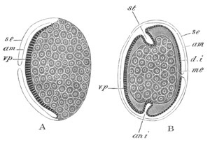
Fig. 181. Diagrammatic longitudinal sections of an Insect embryo at two stages to shew the development of the embryonic envelopes.
In A the amniotic folds have not quite met so as to cover the ventral plate. The yolk is represented as divided into yolk cells. In B the sides of the ventral plate have extended so as nearly to complete the dorsal integument. The mesenteron is represented as a closed sack filled with yolk cells. am. amnion; se. serous envelope; v.p. ventral plate; d.i. dorsal integument; me. mesenteron; st. stomodæum; an i. proctodæum.
The typical mode of formation of these membranes is represented diagrammatically in fig. 181 A and B. A fold of the blastoderm arises round the edge of the ventral plate. This fold, like the amniotic fold of the higher Vertebrata, is formed of two limbs, an outer, the serous membrane (se), and an inner, the true amnion (am). Both limbs extend so as to cover over the ventral plate, and finally meet and coalesce, so that a double membrane is present over the ventral plate. At the same time (fig. 181 B) the point where the fold originates is carried dorsalwards by the dorsal extension of the edges of the ventral plate, which give rise to the dorsal integument (d.i). This process continues till the whole dorsal surface is covered by the integument. The amnion then separates from the dorsal integument, and the embryo becomes enveloped in two membranes—an inner, the amnion, and an outer, the serous membrane. In fig. 181 B the embryo is represented at the stage immediately preceding the closure of the dorsal surface.
By the time that these changes are effected, the serous membrane and amnion are both very thin and not easily separable. The amnion appears to be usually absorbed before hatching; but in hatching both membranes, if present, are either absorbed, or else ruptured and thrown off.
The above mode of development of the embryonic membranes has been especially established by the researches of Kowalevsky (No. 416) and Graber (No. 412) for various Hymenoptera (Apis), Diptera (Chironomus), Lepidoptera and Coleoptera (Melolontha, Lina).
Considerable variations in the development of the enveloping membranes are known.
When the fold which gives rise to the membranes is first formed, there is, as is obvious in fig. 181 A, a perfectly free passage by which the yolk can pass in between the amnion and serous membrane. Such a passage of the yolk between the two membranes takes place posteriorly in Hydrophilus and Donacia: in Lepidoptera the yolk passes in everywhere, so that in this form the ventral plate becomes first of all imbedded in the yolk, and finally, on the completion of the dorsal integument, the embryo is enclosed in a complete envelope of yolk contained between the amnion and the serous membrane. During the formation of the dorsal integument the external yolk-sack communicates by a dorsally situated umbilical canal with the yolk cavity within the body. On the rupture of the amnion the embryo is nourished at the expense of the yolk contained in the external yolk-sack.
In the Hemiptera and the Libellulidæ the ventral plate also becomes imbedded in the yolk, but in a somewhat different fashion to the Lepidoptera, which more resembles on an exaggerated scale what takes place in Hydrophilus.
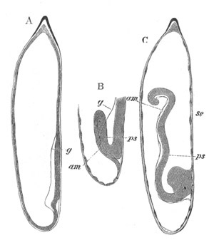
Fig. 182. Three stages in the development of the embryo of Calopteryx. (After Brandt.)
The embryo is represented in the egg-shell. A. Embryo with ventral plate. B. Commencing involution of ventral plate. C. Involution of ventral plate completed.
ps. ventral plate; g. edge of ventral plate; am. amnion; se. serous envelope.
In the Libellulidæ (Calopteryx) there is first of all formed (Brandt, No. 403) a small ventral and posterior thickening of the blastoderm (fig. 182 A). The hinder part of this becomes infolded into the yolk as a projection (fig. 182 B), which consists of two laminæ, an anterior and a posterior, continuous at the apex of the invagination. The whole structure, which is completely imbedded within the yolk, rapidly grows in length, and turns towards the front end of the egg (fig. 182 C). Its anterior lamina remains thick and gives rise to the ventral plate (ps), the posterior (am) on the other hand becomes very thin, and forms a covering corresponding with the amnion of the more ordinary types. The remainder of the blastoderm covering the yolk (se) forms the homologue of the serous membrane of other types. The ventral surface of the ventral plate is turned towards the dorsal side (retaining the same nomenclature as in ordinary cases) of the egg, and the cephalic extremity is situated at the point of origin of the infolding.
The further history is however somewhat peculiar. The amnion is at first (fig. 182 C) continuous with the serous envelope on the posterior side only, so that the serous envelope does not form a continuous sack, but has an opening close to the head of the embryo. In the Hemiptera parasita this opening (Melnikow, No. 422) remains permanent, and the embryo, after it has reached a certain stage of development, becomes everted through it, while the yolk, enclosed in the continuous membrane formed by the amnion and serous envelope, forms a yolk-sack on the dorsal surface. In the Libellulidæ however and most Hemiptera, a fusion of the two limbs of the serous membrane takes place in the usual way, so as to convert it into a completely closed sack (fig. 183 A). After the formation of the appendages a fusion takes place between the amnion and serous envelope over a small area close to the head of the embryo. In the middle of this area a rupture is then effected, and the head of the embryo followed by the body is gradually pushed through the opening (fig. 183 B and C). The embryo becomes in the process completely rotated, and carried into a position in the egg-shell identical with that of the embryos of other orders of Insects (fig. 183 C).
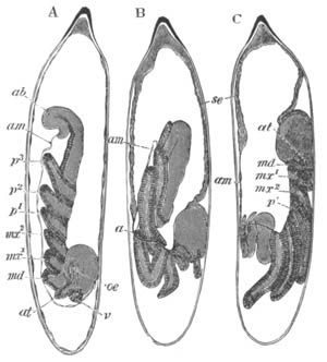
Fig. 183. Three stages in the development of Calopteryx. (After Brandt.)
The embryo is represented in the egg-shell; B. and C. shew the inversion of the embryo.
se. serous envelope; am. amnion; ab. abdomen; v. anterior end of head; at. antennæ; md. mandible; mx1. maxilla 1; mx2. maxilla 2; p1-p3. three pairs of legs; oe. œsophagus.
Owing to the rupture of the embryonic envelopes taking place at the point where they are fused into one, the yolk does not escape in the above process, but is carried into a kind of yolk-sack, on the dorsal surface of the embryo, formed of the remains of the amnion and serous envelope. The walls of the yolk-sack either assist in forming the dorsal parietes of the body, or are more probably enclosed within the body by the growth of the dorsal parietes from the edge of the ventral plate.
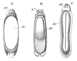
Fig. 184. Three larval stages of Hydrophilus from the dorsal side, shewing the gradual closing in of the dorsal region with the formation of the peculiar dorsal organ do. (After Kowalevsky.)
do. dorsal organ; at. antennæ.
In Hydrophilus and apparently in the Phryganidæ also, there are certain remarkable peculiarities in the closure of the dorsal surface. The fullest observations on the subject have been made by Kowalevsky (No. 416), but Dohrn (No. 408) has with some probability thrown doubts on Kowalevsky’s interpretations. According to Dohrn the part of the serous envelope which covers the dorsal surface becomes thickened, and gives rise to a peculiar dorsal plate which is shewn in surface view in fig. 184 A, do, and in section in fig. 185 A, do. The ventral parts of the amnion and serous membrane have either been ruptured or have disappeared. While the dorsal plate is being formed, the mesoblast, and somewhat later the lateral parts of the epiblast of the ventral plate gradually grow towards the dorsal side and enclose the dorsal plate, the wall of which in the process appears to be folded over so as first of all to form a groove and finally a canal. The stages in this growth are shewn from the surface in fig. 184 B and C and in section in fig. 185 B, do. The canal is buried on the dorsal part of the yolk, but for some time remains open by a round aperture in front (fig. 184 C). The whole structure is known as the dorsal canal. It appears to atrophy without leaving a trace. The heart when formed lies immediately dorsal to it[170].
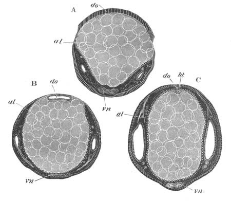
Fig. 185. Three transverse sections through advanced embryos of Hydrophilus.
A. Section through the posterior part of the body of the same age as fig. 184 A. B. Section through the embryo of the same age as fig. 184 C. C. Section through a still older embryo.
do. dorsal plate; vn. ventral nerve cord; al. mesenteron; ht. heart.
The large spaces at the sides are parts of the body cavity.
In the Poduridæ the embryonic membranes appear to be at any rate imperfect. Metschnikoff states in his paper on Geophilus that in some ants no true embryonic membranes are found, but merely scattered cells which take their place. In the Ichneumonidæ the existence of two embryonic membranes is very doubtful.
Formation of the embryonic layers. The formation of the layers has been studied in sections by Kowalevsky (No. 416), Hatschek (No. 414), and Graber (No. 412), etc. From their researches it would appear that the formation of the mesoblast always takes place in a manner closely resembling that in Hydrophilus. The essential features of the process (figs. 177 and 178) appear to be that a groove is formed along the median line of the ventral plate, and that the sides of this groove either (1) simply close over like the walls of the medullary groove in Vertebrates, and so convert the groove into a tube, which soon becomes solid and forms a mass or plate of cells internal to the epiblast; or (2) that the cells on each side of the groove grow over it and meet in the middle line, forming a layer external to the cells which lined the groove. The former of these processes is the most usual; and in the Muscidæ the dimensions of the groove are very considerable (Graber, No. 411). In both cases the process is fundamentally the same, and causes the ventral plate to become divided into two layers[171]. The external layer or epiblast is an uniform sheet forming the main part of the ventral plate (fig. 178 B, ep). It is continuous at its edge with the amnion. The inner layer or mesoblast constitutes an independent plate of cells internal to the epiblast (fig. 178 B, me). The mesoblast soon becomes divided into two lateral bands.
The origin of the hypoblast is still in dispute. It will be remembered (vide pp. 114 and 116) that after the segmentation a number of nuclei remain in the yolk; and that eventually a secondary segmentation of the yolk takes place around these nuclei, and gives rise to a mass of yolk cells, which fill up the interior of the embryo. These cells are diagrammatically shewn in figs. 181 and 189, and it is probable that they constitute the true hypoblast. Their further history is given below.
Formation of the organs and their relation to the germinal layers.
The segments and appendages. One of the earliest phenomena in the development is the appearance of transverse lines indicating segmentation (fig. 186). The transverse lines are apparently caused by shallow superficial grooves, and also in many cases by the division of the mesoblastic bands into separate somites. The most anterior line marks off a præ-oral segment, which soon sends out two lateral wings—the procephalic lobes. The remaining segments are at first fairly uniform. Their number does not, however, appear to be very constant. So far as is known they never exceed seventeen, and this number is probably the typical one (figs. 186 and 187).
In Diptera the number appears to be usually fifteen though it may be only fourteen. In Lepidoptera and in Apis there appear to be sixteen segments. These and other variations affect only the number of the segments which form the abdomen of the adult.
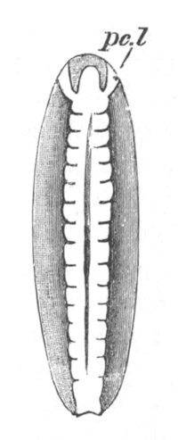
Fig. 186. Embryo of Hydrophilus piceus viewed from the ventral surface. (After Kowalevsky.)
pc.l. procephalic lobe.
The appendages arise as paired pouch-like outgrowths of the epiblast and mesoblast; and their number and the order of their appearance are subject to considerable variation, the meaning of which is not yet clear. As a rule they arise subsequently to the segmentation of the parts of the body to which they belong. There is always formed one pair of appendages which spring from the lateral lobes of the procephalic region, or from the boundary line between these and the median ventral part of this region. These appendages are the antennæ. They have in the embryo a distinctly ventral position as compared to that which they have in the adult.
In the median ventral part of the procephalic region there arises the labrum (fig. 187, ls). It is formed by the coalescence of a pair of prominences very similar to true appendages, though it is probable that they have not this value[172].
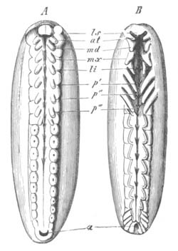
Fig. 187. Two stages in the development of Hydrophilus piceus. (From Gegenbaur, after Kowalevsky.)
ls. labrum; at. antenna; md. mandible; mx. maxilla I.; li. maxilla II.; p´ p´´ p´´´. feet; a. anus.
The antennæ themselves can hardly be considered to have the same morphological value as the succeeding appendages. They are rather equivalent to paired processes of the præ-oral lobes of the Chætopoda.
From the first three post-oral segments there grow out the mandibles and two pairs of maxillæ, and from the three following segments the three pairs of thoracic appendages. In many Insects (cf. Hydrophilus) a certain number of appendages of the same nature as the anterior ones are visible in the embryo on the abdominal segments, a fact which shews that Insects are descended from ancestors with more than three pairs of ambulatory appendages.
In Apis according to Bütschli (No. 405) all the abdominal segments are provided with appendages, which always remain in a very rudimentary condition. All trace of them as well as of the thoracic appendages is lost by the time the embryo is hatched. In the phytophagous Hymenoptera the larva is provided with 9‑11 pairs of legs.
In the embryo of Lepidoptera there would appear from Kowalevsky’s figures to be rudiments of ten pairs of post-thoracic appendages. In the caterpillar of this group there are at the maximum five pairs of such rudimentary feet, viz. a pair on the 3rd, 4th, 5th, and 6th, and on the last abdominal segment. The embryos of Hydrophilus (fig. 187), Mantis, etc. are also provided with additional appendages. In various Thysanura small prominences are present on more or fewer of the abdominal segments (fig. 192), which may probably be regarded as rudimentary feet.
Whether all or any of the appendages of various kinds connected with the hindermost segments belong to the same category as the legs is very doubtful. Their usual absence in the embryo or in any case their late appearance appears to me against so regarding them; but Bütschli is of opinion that in the Bee the parts of the sting are related genetically to the appendages of the penultimate and antepenultimate abdominal segments, and this view is to some extent supported by more recent observations (Kraepelin, etc.), and if it holds true for the Bee must be regarded as correct for other cases also.
As to the order of the appearance of the appendages observations are as yet too scanty to form any complete scheme. In many cases all the appendages appear approximately at the same moment, e.g. Hydrophilus, but whether this holds good for all Coleoptera is by no means certain. In Apis the appendages are stated by Bütschli to arise simultaneously, but according to Kowalevsky the two mouth appendages first appear, then the antennæ, and still later the thoracic appendages. In the Diptera the mouth appendages are first formed, and either simultaneously with these, or slightly later, the antennæ. In the Hemiptera and Libellulidæ the thoracic appendages are the first to be formed, and the second pair of maxillæ makes its appearance before the other cephalic appendages.
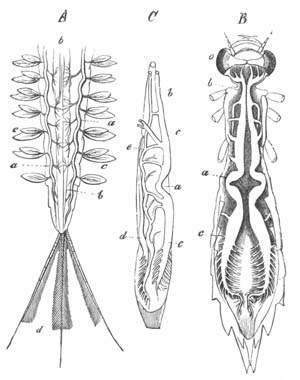
Fig. 188. Figures illustrating aquatic respiration in Insects. (After Gegenbaur.)
A. Hinder portion of the body of Ephemera vulgata. a. longitudinal tracheal trunks; b. alimentary canal; c. tracheal gills. B. Larva of Æschna grandis. a. superior longitudinal tracheal trunks; b. their anterior end; c. portion branching on proctodæum; o. eyes. C. Alimentary canal of the same larva from the side. a, b, and c. as in B; d. inferior tracheal trunk; e. transverse branches between upper and lower tracheal trunks.
The history of the changes in the embryonic appendages during the attainment of the adult condition is beyond the scope of this treatise, but it may be noted that the second pair of maxillæ are relatively very large in the embryo, and not infrequently (Libellula, etc.) have more resemblance to the ambulatory than to the masticatory appendages.
The exact nature of the wings and their relation to the other segments is still very obscure. They appear as dorsal leaf-like appendages on the 2nd and 3rd thoracic segments, and are in many respects similar to the tracheal gills of the larvæ of Ephemeridæ and Phryganidæ (fig. 188 A), of which they are supposed by Gegenbaur and Lubbock to be modifications. The undoubtedly secondary character of the closed tracheal system of larvæ with tracheal gills tells against this view. Fritz Müller finds in the larvæ of Calotermes rugosus (one of the Termites) that peculiar and similar dorsal appendages are present on the two anterior of the thoracic segments. They are without tracheæ. The anterior atrophies, and the posterior acquires tracheæ and gives rise to the first pair of wings. The second pair of wings is formed from small processes on the third thoracic segment like those on the other two. Fritz Müller concludes from these facts that the wings of Insects are developed from dorsal processes of the body, not equivalent to the ventral appendages. What the primitive function of these appendages was is not clear. Fritz Müller suggests that they may have been employed as respiratory organs in the passage from an aqueous to a terrestrial existence, when the Termite ancestors lived in moist habitations—a function for which processes supplied with blood-channels would be well adapted. The undoubted affinity of Insects to Myriapods, coupled with the discovery by Moseley of a tracheal system in Peripatus, is however nearly fatal to the view that Insects can have sprung directly from aquatic ancestors not provided with tracheæ. But although this suggestion of Fritz Müller cannot be accepted, it is still possible that the processes discovered by him may have been the earliest rudiments of wings, which were employed first as organs of propulsion by a water-inhabiting Insect ancestor which had not yet acquired the power of flying.
The nervous system. The nervous system arises entirely from the epiblast; but the development of the præ-oral and post-oral sections may be best considered separately.
The post-oral section, or ventral cord of the adult, arises as two longitudinal thickenings of the epiblast, one on each side of the median line (fig. 189 B, vn), which are subsequently split off from the superficial skin and give rise to the two lateral strands of the ventral cord. At a later period they undergo a differentiation into ganglia and connecting cords.
Between these two embryonic nerve cords there is at first a shallow furrow, which soon becomes a deep groove (fig. 189 C). At this stage the differentiation of the lateral elements into ganglia and commissures takes place, and, according to Hatschek (No. 414), the median groove becomes in the region of the ganglia converted into a canal, the walls of which soon fuse with those of the ganglionic enlargements of the lateral cords, and connect them across the middle line. Between the ganglia on the other hand the median groove undergoes atrophy, becoming first a solid cord interposed between the lateral strands of the nervous system, and finally disappearing without giving rise to any part of the nervous system. It is probable that Hatschek is entirely mistaken about the entrance of a median element into the ventral cord, and that the appearances he has described are due to shrinkage. In Spiders the absence of a median element can be shewn with great certainty, and, as already stated, this element is not present in Peripatus. Hatschek states that in the mandibular segment the median element is absorbed, and that the two lateral cords of that part give rise to the œsophageal commissures, while the sub-œsophageal ganglion is formed from the fusion of the ganglia of the two maxillary segments.
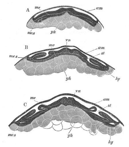
Fig. 189. Three transverse sections through the embryo of Hydrophilus. (After Kowalevsky.)
A. Transverse section through the larva represented in fig. 187 A. B. Transverse section through a somewhat older embryo in the region of one of the stigmata. C. Transverse section through the larva represented in fig. 187 B.
vn. ventral nerve cord; am. amnion and serous membrane; me. mesoblast; me.s. somatic mesoblast; hy. hypoblast(?); yk. yolk cells (true hypoblast); st. stigma of trachea.
The præ-oral portion of the nervous system consists entirely of the supra-œsophageal ganglion. It is formed, according to Hatschek, of three parts. Firstly and mainly, of a layer separated from the thickened inner part of the cephalic lobe on each side; secondly, of an anterior continuation of the lateral cords; and thirdly, of a pit of skin invaginated on each side close to the dorsal border of the antennæ. This pit is at first provided with a lumen, which is subsequently obliterated; while the walls of the pit become converted into true ganglion cells. The two supra-œsophageal ganglia remain disconnected on the dorsal side till quite the close of embryonic life.
The tracheæ and salivary glands. The tracheæ, as was first shewn by Bütschli (No. 405), arise as independent segmentally arranged paired invaginations of the epiblast (fig. 189 B and C, st). Their openings are always placed on the outer sides of the appendages of their segments, where such are present.
Although in the adult stigmata are never found in the space between the prothorax and head[173], in the embryo and the larva tracheal invaginations may be developed in all the thoracic (and possibly in the three jaw-bearing segments) and in all the abdominal segments except the two posterior.
In the embryo of the Lepidoptera, according to Hatschek (No. 414), there are 14 pairs of stigmata, belonging to the 14 segments of the body behind the mouth; but Tichomiroff states that Hatschek is in error in making this statement for the foremost post-oral segments. The last two segments are without stigmata. In the larvæ of Lepidoptera as well as those of many Hymenoptera, Coleoptera and Diptera, stigmata are present on all the postcephalic segments except the 2nd and 3rd thoracic and the two last abdominal. In Apis there are eleven pairs of tracheal invaginations according to Kowalevsky (No. 416), but according to Bütschli (No. 405) only ten, the prothorax being without one. In the Bee they appear simultaneously, and before the appendages.
The blind ends of the tracheal invaginations frequently (e.g. Apis) unite together into a common longitudinal canal, which forms a longitudinal tracheal stem. In other cases (e.g. Gryllotalpa, Dohrn, No. 408) they remain distinct, and each tracheal stem has a system of branches of its own.
The development of the tracheæ strongly supports the view, arrived at by Moseley from his investigations on Peripatus, that they are modifications of cutaneous glands.
The salivary and spinning glands are epiblastic structures, which in their mode of development are very similar to the tracheæ, and perhaps have a similar origin. The salivary glands arise as paired epiblastic invaginations, not, as might be expected, of the Stomodæum, but of the ventral plate behind the mouth on the inner side of the mandibles. At first independent, they eventually unite in a common duct, which falls into the mouth. The spinning glands arise on the inner side of the second pair of maxillæ in Apis and Lepidoptera, and form elongated glands extending through nearly the whole length of the body. They are very similar in their structure and development to salivary glands, and are only employed during larval life. They no doubt resemble the mucous glands of the oral papillæ of Peripatus, with which they have been compared by Moseley. The mucous glands of Peripatus may perhaps be the homologous organs of the first pair of maxillæ, for the existence of which there appears to be some evidence amongst Insects.
Mesoblast. It has been stated that the mesoblast becomes divided in the region of the body into two lateral bands (fig. 189 A). These bands in many, if not all forms, become divided into a series of somites corresponding with the segments of the body. In each of them a cavity appears—the commencing perivisceral cavity—which divides them into a somatic plate in contact with the epiblast, and a splanchnic plate in contact with the hypoblast (fig. 189). In the interspaces between the segments the mesoblast is continuous across the median ventral line. The mesoblast is prolonged into each of the appendages as these are formed, and in the appendages there is present a central cavity. By Metschnikoff these cavities are stated to be continuous, as in Myriapods and Arachnida, with those of the somites; but by Hatschek (No. 414) they are stated to be independent of those in the somites and to be open to the yolk.
The further details of the history of the mesoblast are very imperfectly known, and the fullest account we have is that by Dohrn (No. 408) for Gryllotalpa. It would appear that the mesoblast grows round and encloses the dorsal side of the yolk earlier than the epiblast. In Gryllotalpa it forms a pulsating membrane. As the epiblast extends dorsalwards the median dorsal part of the membrane is constricted off as a tube which forms the heart. At the same time the free space between the pulsating membrane and the yolk is obliterated, but transverse passages are left at the lines between the somites, through which the blood passes from the ventral part of the body to corresponding openings in the wall of the heart. The greater part of the membrane gives rise to the muscles of the trunk.
Ventrally the mesoblastic bands soon meet across the median line. The cavities in the appendages become obliterated and their mesoblastic walls form the muscles, etc. The cavities in the separate mesoblastic somites also cease to be distinctly circumscribed.
The splanchnic mesoblast follows the hypoblast in its growth, and gives rise to the connective tissue and muscular parts of the walls of the alimentary tract. The mesoblastic wall of the proctodæum is probably formed independently of the mesoblastic somites. In the head the mesoblast is stated to form at first a median ventral mass, which does not pass into the procephalic lobe; though it assists in forming both the antennæ and upper lip.
The alimentary canal. The alimentary tract of Insects is formed of three distinct sections (fig. 181)—a mesenteron or middle section (me), a stomodæum (st) and a proctodæum (an). The stomodæum and proctodæum are invaginations of the epiblast, while the mesenteron is lined by the hypoblast. The distinction between the three is usually well marked in the adult by the epiblastic derivatives being lined by chitin. The stomodæum consists of mouth, œsophagus, crop, and proventriculus or gizzard, when such are present. The mesenteron includes the stomach, and is sometimes (Orthoptera, etc.) provided at its front end with pyloric diverticula—posteriorly it terminates just in front of the Malpighian bodies. These latter fall into the proctodæum, which includes the whole of the region from their insertion to the anus.
The oral invagination appears nearly coincidently with the first formation of segments at the front end of the groove between the lateral nerve cords, and the anal invagination appears slightly later at the hindermost end of the ventral plate.
The Malpighian bodies arise as two pairs of outgrowths of the epiblast of the proctodæum, whether solid at first is not certain. The subsequent increase which usually takes place in their number is due to sproutings (at first solid) of the two original vessels.
The glandular walls of the mesenteron are formed from the hypoblast; but the exact origin of the layer has not been thoroughly worked out in all cases. In Hydrophilus it is stated by Kowalevsky (No. 416) to appear as two sheets split off from the lateral masses of mesoblast, which gradually grow round the yolk, and a similar mode of formation would seem to hold good for Apis. Tichomiroff (No. 420) confirms Kowalevsky on this point, and further states that these two masses meet first ventrally and much later on the dorsal side. In Lepidoptera, on the other hand, Hatschek finds that the hypoblast arises as a median mass of polygonal cells in the anterior part of the ventral plate. These cells increase by absorbing material from the yolk, and then gradually extend themselves and grow round the yolk.
Dohrn (No. 408) believes that the yolk cells, the origin of which has already been spoken of, give rise to the hypoblastic walls of the mesenteron, and this view appears to be shared by Graber (No. 412), though the latter author holds that some of the yolk cells are derived by budding from the blastoderm[174].
From the analogy of Spiders I am inclined to accept Dohrn’s and Graber’s view. It appears to me probable that Kowalevsky’s observations are to be explained by supposing that the hypoblast plates which he believes to be split off from the mesoblast are really separated from the yolk.
It will be convenient to add here a few details to what has already been stated as to the origin of the yolk cells. As mentioned above, the central yolk breaks up at a period, which is not constant in the different forms, into polygonal or rounded masses, in each of which a nucleus has in many instances been clearly demonstrated although in others such nuclei have not been made out. It is probable however that nuclei are in all cases really present, and that these masses must be therefore regarded as cells. They constitute in fact the yolk cells. The periphery of the yolk breaks up into cells while the centre is still quite homogeneous.
The hypoblastic walls of the mesenteron appear to be formed in the first instance laterally (fig. 189 B and C, hy). They then meet ventrally (fig. 185 A and B), and finally close in the mesenteron on the dorsal side.
The mesenteron is at first a closed sack, independent of both stomodæum and proctodæum; and in the case of the Bee it so remains even after the close of embryonic life. The only glandular organs of the mesenteron are the not unfrequent pyloric tubes, which are simple outgrowths of its anterior end. It is possible that in some instances they may be formed in situ around the lateral parts of the yolk.
In many instances the whole of the yolk is enclosed in the walls of the mesenteron, but in other cases, as in Chironomus and Simulia (Weismann, No. 430; Metschnikoff, No. 423), part of the yolk may be left between the ventral wall of the mesenteron and the ventral plate. In Chironomus the mass of yolk external to the mesenteron takes the form of a median and two lateral streaks. Some of the yolk cells either prior to the establishment of the mesenteron, or derived from the unenclosed portions of the yolk, pass into the developing organs (Dohrn, 408) and serve as a kind of nutritive cell. They also form blood corpuscles and connective-tissue elements. Such yolk cells may be compared to the peculiar bodies described by Reichenbach in Astacus, which form the secondary mesoblast. Similar cells play a very important part in the development of Spiders.
Generative organs. The observations on the development of the generative organs are somewhat scanty. In Diptera certain cells—known as the pole cells—are stated by both Metschnikoff (No. 423) and Leuckart to give rise to the generative organs. The cells in question (in Chironomus and Musca vomitoria, Weismann, No. 430) appear at the hinder end of the ovum before any other cells of the blastoderm. They soon separate from the blastoderm and increase by division. In the embryo, produced by the viviparous larva of Cecidomyia, there is at first a single pole cell, which eventually divides into four, and the resulting cells become enclosed within the blastoderm. They next divide into two masses, which are stated by Metschnikoff (No. 423) to become surrounded by indifferent embryonic cells[175]. Their protoplasm then fuses, and their nuclei divide, and they give rise to the larval ovaries, for which the enclosing cells form the tunics.
In Aphis Metschnikoff (No. 423) detected at a very early stage a mass of cells which give rise to the generative organs. These cells are situated at the hind end of the ventral plate; and, except in the case of one of the cells which gives rise by division to a green mass adjoining the fat body, the protoplasm of the separate cells fuses into a syncytium. Towards the close of embryonic life the syncytium assumes a horse-shoe form. The mass is next divided into two, and the peripheral layer of each part gives rise to the tunic, while from the hinder extremity of each part an at first solid duct—the egg-tube—grows out. The masses themselves form the germogens. The oviduct is formed by a coalescence of the ducts from each germogen.
Ganin derives the generative organs in Platygaster (vide p. 347) from the hind end of the ventral plate close to the proctodæum; while Suckow states that the generative organs are outgrowths of the proctodæum. According to these two sets of observations the generative organs would appear to have an epiblastic origin—an origin which is not incompatible with that from the pole cells.
In Lepidoptera the genital organs are present in the later periods of embryonic life as distinct paired organs, one on each side of the heart, in the eighth postcephalic segment. They are elliptical bodies with a duct passing off from the posterior end in the female or from the middle in the male. The egg-tubes or seminal tubes are outgrowths of the elliptical bodies.
In other Insects the later stages in the development of the generative organs closely resemble those in the Lepidoptera, and the organs are usually distinctly visible in the later stages of embryonic life.
It may probably be laid down, in spite of some of Metschnikoff’s observations above quoted, that the original generative mass gives rise to both the true genital glands and their ducts. It appears also to be fairly clear that the genital glands of both sexes have an identical origin.
Special types of larvæ.
Certain of the Hymenopterous forms, which deposit their eggs in the eggs or larvæ of other Insects, present very peculiar modifications in their development. Platygaster, which lays its egg in the larvæ of Cecidomyia, undergoes perhaps the most remarkable development amongst these forms. It has been studied especially by Ganin (No. 410), from whom the following account is taken.
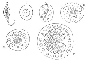
Fig. 190. A series of stages in the development of Platygaster. (From Lubbock; after Ganin.)
The very first stages are unfortunately but imperfectly known, and the interpretations offered by Ganin do not in all cases appear quite satisfactory. In the earliest stage after being laid the egg is enclosed in a capsule produced into a stalk (fig. 190 A). In the interior of the egg there soon appears a single spherical body, regarded by Ganin as a cell (fig. 190 B). In the next stage three similar bodies appear in the vitellus, no doubt derived from the first one (fig. 190 C). The central one presents somewhat different characters to the two others, and, according to Ganin, gives rise to the whole embryo. The two peripheral bodies increase by division, and soon appear as nuclei imbedded in a layer of protoplasm (fig. 190 D, E, F). The layer so formed serves as a covering for the embryo, regarded by Ganin as equivalent to the amnion (? serous membrane) of other Insect embryos. In the embryo cell new cells are stated to be formed by a process of endogenous cell formation (fig. 190 D, E). It appears probable that Ganin has mistaken nuclei for cells in the earlier stages, and that a blastoderm is formed as in other Insects, and that this becomes divided in a way not explained into a superficial layer which gives rise to the serous envelope, and a deeper layer which forms the embryo. However this may be, a differentiation into an epiblastic layer of columnar cells and a hypoblastic layer of more rounded cells soon becomes apparent in the body of the embryo. Subsequently to this the embryo grows rapidly, till by a deep transverse constriction on the ventral surface it becomes divided into an anterior cephalothoracic portion and a posterior caudal portion (fig. 190 F). The cephalothorax grows in breadth, and near its anterior end an invagination appears, which gives rise to the mouth and œsophagus. On the ventral side of the cephalothorax there is first formed a pair of claw-like appendages on each side of the mouth, then a posterior pair of appendages near the junction of the cephalothorax and abdomen, and lastly a pair of short conical antennæ in front.
At the same time the hind end of the abdomen becomes bifid, and gives rise to a fork-like caudal appendage; and at a slightly later period four grooves make their appearance in the caudal region, and divide this part of the embryo into successive segments. While these changes have been taking place in the general form of the embryo, the epiblast has given rise to a cuticle, and the hypoblastic cells have become differentiated into a central hypoblastic axis—the mesenteron—and a surrounding layer of mesoblast, some of the cells of which form longitudinal muscles.
With this stage closes what may be regarded as the embryonic development of Platygaster. The embryo becomes free from the amnion, and presents itself as a larva, which from its very remarkable characters has been spoken of as the Cyclops larva by Ganin.
The larvæ of three species have been described by Ganin, which are represented in fig. 191 A, B, C. These larvæ are strangely dissimilar to the ordinary Hexapod type, whether larval or adult. They are formed of a cephalothoracic shield with the three pairs of appendages (a, kf, lfg), the development of which has already been described, and of an abdomen formed of five segments, the last of which bears the somewhat varying caudal appendages. The nervous system is as yet undeveloped.
The larvæ move about in the tissues of their hosts by means of their claws.
The first larval condition is succeeded by a second with very different characters, and the passage from the first to the second is accompanied by an ecdysis.
The ecdysis commences at the caudal extremity, and the whole of the last segment is completely thrown off. As the ecdysis extends forwards the tail loses its segmentation and becomes strongly compressed, the appendages of the cephalothorax are thrown off, and the whole embryo assumes an oval form without any sharp distinction into different regions and without the slightest indication of segmentation (fig. 191 D). Of the internal changes which take place during the shedding of the cuticle, the first is the formation of a proctodæum (gh) by an invagination, which ends blindly in contact with the mesenteron. Shortly after this a thickening of the epiblast (bsm) appears along the ventral surface, which gives rise mainly to the ventral nerve cord; this thickening is continuous behind with the epiblast which is invaginated to form the proctodæum, and in front is prolonged on each side into two procephalic lobes, in which there are also thickenings of the epiblast (gsae), which become converted into supra-œsophageal ganglia, and possibly other parts.
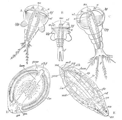
Fig. 191. A series of stages in the development of Platygaster. (From Lubbock; after Ganin.)
A. B. C. Cyclops larvæ of three species of Platygaster. D. Second larval stage. E. Third larval stage.
mo. mouth; a. antenna; hf. hooked feet; lfg. lateral feet; f. branches of tail; ul. lower lip; slkf. œsophagus; gsae. supra-œsophageal ganglion; bsm. ventral epiblastic plate; lm. lateral muscles (the letters also point in D to the salivary glands); gh. proctodæum; ga. generative organs; md. mandibles; ag. ducts of salivary glands; sp. (in E) salivary glands; mls. stomach; ed. intestine; ew. rectum; ao. anus; tr. tracheæ; fk. fat body.
Towards the close of the second larval period the muscles (lm) become segmentally arranged, and give indications of the segmentation which becomes apparent in the third larval period. The third and last larval stage (fig. 191 E) of Platygaster, during which it still remains in the tissues of its host, presents no very peculiar features. The passage from the second to the third form is accompanied by an ecdysis.
Remarkable as are the larvæ just described, there can I think be no reason, considering their parasitic habits, for regarding them as ancestral.
Metamorphosis and heterogamy.
Metamorphosis. The majority of Insects are born in a condition in which they obviously differ from their parents. The extent of this difference is subject to very great variations, but as a rule the larvæ pass through a very marked metamorphosis before reaching the adult state. The complete history of this metamorphosis in the different orders of Insects involves a far too considerable amount of zoological detail to be dealt with in this work; and I shall confine myself to a few observations on the general characters and origin of the metamorphosis, and of the histological processes which take place during its occurrence[176].
In the Aptera the larva differs from the adult only in the number of facets in the cornea and joints in the antennæ.
In most Orthoptera and Hemiptera the larvæ differ from the adult in the absence of wings and in other points. The wings, etc., are gradually acquired in the course of a series of successive moultings. In the Ephemeridæ and Libellulidæ, however, the metamorphosis is more complicated, in that the larvæ have provisional tracheal gills which are exuviated before the final moult. In the Ephemeridæ there are usually a great number of moultings; the tracheal gills appear after the second moult, and the rudiments of the wings when the larva is about half grown. Larval life may last for a very long period.
In all the other groups of Insects, viz. the Diptera, Neuroptera, Coleoptera, Lepidoptera, and Hymenoptera, the larva passes—with a few exceptions—through a quiescent stage, in which it is known as a pupa, before it attains the adult stage. These forms are known as the Holometabola.
In the Diptera the larvæ are apodous. In the true flies (Muscidæ) they are without a distinct head and have the jaws replaced by hooks. In the Tipulidæ there is on the other hand a well-developed head with the normal appendages. The pupæ of the Muscidæ are quiescent, and are enclosed in the skin of the larva which shrinks and forms a firm oval case. In the Tipulidæ the larval skin is thrown off at the pupa stage, and in some cases the pupæ continue to move about.
The larvæ of the Neuroptera are hexapodous voracious forms. When the larva becomes a pupa all the external organs of the imago are already established. The pupa is often invested in a cocoon. It is usually quiescent, though sometimes it begins to move about shortly before the imago emerges.
In the Coleoptera there is considerable variety in the larval forms. As a rule the larvæ are hexapodous and resemble wingless Insects. But some herbivorous larvæ (e.g. the larva of Melolontha) closely resemble true caterpillars, and there are also grub-like larvæ without feet (Curculio) which resemble the larvæ of Hymenoptera. The pupa is quiescent, but has all the parts of the future beetle plainly visible. The most interesting larvæ among the Coleoptera are those of Sitaris, one of the Meloidæ (Fabre, No. 409). They leave the egg as active hexapodous larvæ which attach themselves to the bodies of Hymenoptera, and are thence transported to a cell filled with honey. Here they eat the ovum of the Hymenopterous form. They then undergo an ecdysis, in which they functionally lose their appendages, retaining however small rudiments of them, and become grubs. They feed on the honey and after a further ecdysis become pupæ.
In the Lepidoptera the larva has the well-known form of a caterpillar. The caterpillars have strong jaws, adapted for biting vegetable tissues, which are quite unlike the oral appendages of the adult. They have three pairs of jointed thoracic legs, and a variable number (usually five) of pairs of rudimentary abdominal legs—the so-called pro-legs. The larva undergoes numerous ecdyses, and the external parts of the adult such as the wings, etc., are formed underneath the chitinous exoskeleton before the pupa stage. The pupa is known as a chrysalis and in some Lepidoptera is enveloped in a cocoon.
The Hymenoptera present considerable variations in the character of the larvæ. In the Aculeata, many Entomophaga, the Cynipidæ, etc., the larvæ are apodous grubs, incapable of going in search of their food; but in the Siricidæ they are hexapodous forms like caterpillars, which are sometimes even provided with pro-legs. In some of the Entomophaga the larvæ have very remarkable characters which have already been described in a special section, vide pp. 418, 419.
Before proceeding to the consideration of the value of the various larval forms thus shortly enumerated, it is necessary to say a few words as to the internal changes which take place during the occurrence of the above metamorphosis. In the simplest cases, such as those of the Orthoptera and Hemiptera, where the metamorphosis is confined to the gradual formation of the wings, etc. in a series of moults, the wings first appear as two folds of the epidermis beneath the cuticle on the two posterior thoracic segments. At the next moult these processes become covered by the freshly formed cuticle, and appear as small projections. At every successive moult these projections become more prominent owing to a growth in the epidermis which has taken place in the preceding interval. Accompanying the formation of such organs as the wings, internal changes necessarily take place in the arrangement of the muscles, etc. of the thorax, which proceed pari passu with the formation of the organs to which they belong. The characters of the metamorphosis in such forms as the Ephemeridæ only differ from the above in the fact that provisional organs are thrown off at the same time that the new ones are formed.
In the case of the Holometabola the internal phenomena of the metamorphosis are of a very much more remarkable character. The details of our knowledge are largely due to Weismann (Nos. 430 and 431). The larvæ of the Holometabola have for the most part a very different mode of life to the adults. A simple series of transitions between the two is impossible, because intermediate forms would be for the most part incapable of existing. The transition from the larval to the adult state is therefore necessarily a more or less sudden one, and takes place during the quiescent pupa condition. Many of the external adult organs are however formed prior to the pupa stage, but do not become visible on the surface. The simplest mode of Holometabolic metamorphosis may be illustrated by the development of Corethra plumicornis, one of the Tipulidæ. This larva, like that of other Tipulidæ, is without thoracic appendages, but before the last larval moult, and therefore shortly before the pupa stage, certain structures are formed, which Weismann has called imaginal discs. These imaginal discs are in Corethra simply invaginations of the epidermis. There are in the thorax six pairs of such structures, three dorsal and three ventral. The three ventral are attached to the terminations of the sensory nerves, and the limbs of the imago are formed as simple outgrowths of them, which as they grow in length take a spiral form. In the interior of these outgrowths are formed the muscles, tracheæ, etc., of the limbs; which are believed by Weismann (it appears to me without sufficient ground) to be derived from a proliferation of the cells of the neurilemma. The wings are formed from the two posterior dorsal imaginal discs. The hypodermis of the larva passes directly into that of the imago.
The pupa stage of Corethra is relatively very short, and the changes in the internal parts which take place during it are not considerable. The larval abdominal muscles pass for the most part unchanged into those of the imago, while the special thoracic muscles connected with the wings, etc., develop directly during the latest larval period from cords of cells already formed in the embryo.
In the Lepidoptera the changes in the passage from the larval to the adult state are not very much more considerable than those in Corethra. Similar imaginal discs give rise during the later larval periods to the wings, etc. The internal changes during the longer pupa period are somewhat more considerable. Important modifications and new formations arise in connection with the alimentary tract, the nervous and muscular systems.
The changes which take place in the true flies (Muscidæ) are far more complicated than either those in Corethra or in the Lepidoptera. The abdomen of the larva of Musca becomes bodily converted into the abdomen of the imago as in the above types, but the whole epidermis and appendages of the head and thorax are derived from imaginal discs which are formed within and (so far as is known) independently of the epidermis of the larva or embryo. These imaginal discs are simple masses of apparently indifferent cells, which for the most part appear at the close of embryonic life, and are attached to nerves or tracheæ. They grow in size during larval life, but during the relatively long pupa stage they unite together to give rise to a continuous epidermis, from which the appendages grow out as processes. The epidermis of the anterior part of the larva is simply thrown off, and has no share in forming the epidermis of the adult.
There are a pair of cephalic imaginal discs and six pairs of thoracic discs. Two pairs, a dorsal and a ventral, give rise to each thoracic ring, and the appendages attached to it.
Though, as mentioned above, no evidence has yet been produced to shew that the imaginal discs of Musca are derived from the embryonic epiblast, yet their mode of growth and eventual fate proves beyond the shadow of a doubt that they are homologous with the imaginal discs of Corethra. Their earliest origin is well worth further investigation.
The metamorphosis of the internal organs is still more striking than that of the external. There is a disruption, total or partial, of all the internal organs except the generative organs. In the case of the alimentary tract, the Malpighian vessels, the heart and the central nervous system, the disruption is of a partial kind, which has been called by Weismann histolysis. The cells of these organs undergo a fatty degeneration, the nuclei alone in some cases remaining. The kind of plasma resulting from this degeneration retains the shape of the organs, and finally becomes built up again into the corresponding organs of the imago. The tracheæ, muscles and peripheral nerves, and an anterior part of the alimentary tract, are entirely disrupted. They seem to be formed again from granular cells derived from the enormous fat body.
The phenomena of the development of the Muscidæ are undoubtedly of rather a surprising character. Leaving for the moment the question of the origin of the pupa stage to which I return below, it will be admitted on all hands that during the pupa stage the larva undergoes a series of changes which, had they taken place by slow degrees, would have involved, in such a case as Musca, a complete though gradual renewal of the tissues. Such being the case, the cells of the organs common to the larva and the imago would, in the natural course of things, not be the same cells as those of the larva but descendants of them. We might therefore expect to find in the rapid conversion of the larval organs into those of the adult some condensation, so to speak, of the process of ordinary cell division. Such condensations are probably represented in the histolysis in the case of the internal organs, and in the formation of imaginal discs in the case of the external ones, and I think it probable that further investigation will shew that the imaginal discs of the Muscidæ are derivatives of the embryonic epiblast. The above considerations by no means explain the whole of Weismann’s interesting observations, but an explanation is I believe to be found by following up these lines.
More or less parallel phenomena to those in Insects are found in the development of the Platyelminthes and Echinoderms. The four disc-like invaginations of the skin in many larval Nemertines (vide p. 198), which give rise to the permanent body wall of the Nemertine, may be compared to the imaginal discs. The subsequent throwing off of the skin of Pilidium or larva of Desor is a phenomenon like the absorption of part of the larval skin of Musca. The formation of an independent skin within the first larval form in the Distomeæ and in the Cestoda may be compared to the apparently independent formation of the imaginal discs in Musca.
The fact that in a majority of instances it is possible to trace an intimate connection between the surroundings of a larva and its organization proves in the clearest way that the characters of the majority of existing larval forms of Insects have owed their origin to secondary adaptations. A few instances will illustrate this point.
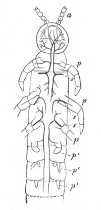
Fig. 192. Anterior half of Campodea Fragilis. (From Gegenbaur; after Palmen.)
a. antennæ; p. feet; p´. post-thoracic rudimentary feet; s. stigma.
In the simplest types of metamorphosis, e.g. those of the Orthoptera genuina, the larva has precisely the same habits as the adult. We find that a caterpillar form is assumed by phytophagous larvæ amongst the Lepidoptera, Hymenoptera and Coleoptera. Where the larva has not to go in search of its nutriment the grub-like apodous form is assumed. The existence of such an apodous larva is especially striking in the Hymenoptera, in that rudiments of thoracic and abdominal appendages are present in the embryo and disappear again in the larva. The case of the larva of Sitaris, already described (p. 421), affords another very striking proof that the organization of the larva is adapted to its habits.
It follows from the above that the development of such forms as the Orthoptera genuina is more primitive than that of the holometabolous forms; a conclusion which tallies with the fact that both palæontological and anatomical evidence shew the Orthoptera to be a very primitive group of Insects.
The above argument probably applies with still greater force to the case of the Thysanura; and it seems to be probable that this group is more nearly related than any other to the primitive wingless ancestors of Insects[177]. The characters of the oral appendages in this group, the simplicity of their metamorphosis, and the presence of abdominal appendages (fig. 192), all tell in favour of this view, while the resemblance of the adult to the larvæ of the Pseudoneuroptera, etc., points in the same direction. The Thysanura and Collembola are not however to be regarded as belonging to the true stock of the ancestors of Insects, but as degenerated relations of this stock; much as Amphioxus and the Ascidians are degenerate relations of the ancestral stock of Vertebrates, and Peripatus of that of the Tracheata. It is probable that all these forms have succeeded in retaining their primitive characters from their degenerate habits, which prevented them from entering into competition in the struggle for existence with their more highly endowed relatives. While in a general way it is clear that the larval forms of Insects cannot be expected to throw much light on the nature of Insect ancestors, it does nevertheless appear to me probable that such forms as the caterpillars of the Lepidoptera are not without a meaning in this respect. It is easy to conceive that even a secondary larval form may have been produced by the prolongation of one of the embryonic stages; and the general similarity of a caterpillar to Peripatus, and the retention by it of post-thoracic appendages, are facts which appear to favour this view of the origin of the caterpillar form.
The two most obscure points which still remain to be dealt with in the metamorphosis of Insects are (1) the origin of the quiescent pupa stage; (2) the frequent dissimilarity between the masticatory apparatus of the larva and adult.
These two points may be conveniently dealt with together, and some valuable remarks about them will be found in Lubbock (No. 420).
On grounds already indicated it may be considered certain that the groups of Insects without a pupa stage, and with a larva very similarly organised to the adult, preceded the existing holometabolic groups. The starting point in the metamorphosis of the latter groups was therefore something like that of the Orthoptera. Suppose it became an advantage to a species that the larva and adult should feed in a somewhat different way, a difference in the character of their mouth parts would soon make itself manifest; and, since an intermediate type of mouth parts would probably be disadvantageous, there would be a tendency to concentrate into a single moult the transition from the larval to the adult form of mouth parts. At each ordinary moult there is a short period of quiescence, and this period of quiescence would naturally become longer in the important moult at which the change in the mouth parts was effected. In this way a rudimentary pupa stage might be started. The pupa stage, once started, might easily become a more important factor in the metamorphosis. If the larva and imago diverged still more from each other, a continually increasing amount of change would have to be effected at the pupa stage. It would probably be advantageous to the species that the larva should not have rudimentary functionless wings; and the establishment of the wings as external organs would therefore become deferred to the pupa stage. The same would probably apply to other organs.
Insects usually pass through the pupa stage in winter in cold climates and during the dry season in the tropics, this stage serving therefore apparently for the protection of the species during the inclement season of the year. These facts are easily explained on the supposition that the pupa stage has become secondarily adapted to play a part in the economy of the species quite different from that to which it owes its origin.
Heterogamy. The cases of alternations of generations amongst Insects all fall under the heading already defined in the introduction as Heterogamy. Heterogamy amongst Insects has been rendered possible by the existence of parthenogenesis, which, as stated in the introduction, has been taken hold of by natural selection, and has led to the production of generations of parthenogenetic forms, by which a clear economy in reproduction is effected. Parthenogenesis without heterogamy occurs in a large number of forms. In Bees, Wasps, and a Sawfly (Nematus ventricosus) the unfertilized ova give rise to males. In two Lepidopterous genera (Psyche and Solenobia) the unfertilized ova give rise mainly, if not entirely, to females. Heterogamy occurs in none of the above types, but in Psyche and Solenobia males are only occasionally found, so that a series of generations producing female young from unfertilized ova are followed by a generation producing young of both sexes from fertilized ova. It would be interesting to know if the unimpregnated female would not after a certain number of generations give rise to both males and females; such an occurrence might be anticipated on grounds of analogy. In the cases of true heterogamy parthenogenesis has become confined to special generations, which differ in their character from the generations which reproduce themselves sexually. The parthenogenetic generations generally flourish during the season when food is abundant; while the sexual generations occur at intervals which are often secondarily regulated by the season, supply of food, etc.