Volume 1 / Part 1 — CHAPTER IX. (continued 2)
Reading edition
In all Gasteropod Molluscs the lower or vegetative pole of the ovum is ventral, not dorsal as Rabl would make it in Unio. The blastopore in other Molluscs always coincides either with the mouth or anus, or extends between the two. The surface on which the foot is formed is the ventral surface. On the dorsal surface are placed, (1) the velum near the mouth, (2) the shell-gland near the anus. In Anodon the velum is placed just dorsal to the mouth, then according to Rabl follows the blastopore, and in the region of the blastopore is formed the shell. The blastopore is therefore dorsal in position. It occupies in fact the ordinary place of the shell-gland, and looks very much like this organ (which is not otherwise present in Anodon and Unio). Without necessarily considering Rabl’s interpretations false, I think that the above difficulties should have been at any rate discussed in his paper. More especially is this the case when there is no doubt that Rabl has made in his paper on Lymnæus a confusion between the mouth and the shell-gland.
Investigations on the post-embryonic metamorphosis of Glochidium have been made by Braun (No. 287), and several years ago I made a series of observations on this subject, the results of which agree in most points with those of Braun. I was however unsuccessful in carrying on my observations till the young mussel left its host.
The free Glochidia very soon attach themselves to the gills, fins, or other parts of fish which are placed in the tank containing them; after attachment they become covered by a growth of the epidermic cells of their host, and undergo their metamorphosis.
The first change that takes place is the disappearance of the byssus and the byssus organ. This occurs very soon; shortly afterwards all traces of the velum and sense organs also become lost.
At the time of the disappearance of these bodies, at the point of the projection from which the byssus cord arose, and very possibly from this very projection, the foot arises as a rounded process which rapidly grows and soon becomes ciliated (fig. 121 B, f).
The single adductor muscle begins to atrophy very early, but before its entire disappearance rudiments are formed at the two ends of the body, which at a later period can be distinctly recognised as the anterior and posterior adductor muscles (fig. 1211 B, a.ad and p.ad).
After the formation of these parts the gills arise as solid and at first somewhat knobbed papillæ covered with a ciliated epidermis, on each side of, but somewhat in front of (!) the foot (fig. 121 B, br). In the foot there soon appear the auditory sacks (au.v), and the foot itself becomes a long tongue-like ciliated organ projecting backwards[110].
The mantle lobes undergo great changes, and indeed by Braun the mantle lobes are stated to be formed almost entirely de novo. The permanent shell is (Braun) formed on the dorsal surface of the still parasitic larva in the form of two small independent plates. I have not followed the changes of the alimentary canal, etc., but at an early stage there is visible, dorsal to the foot, a simple enteric sack.
By the time the larva leaves its host all the organs of the adult, except the generative organs, have become established.
The post-embryonic development of the organs of Glochidium is similar in the main to that of other Lamellibranchiata. This fact is of some importance on account of the peculiarities of the earlier developmental stages.
The byssus organ, the toothed processes of the shell, and the sense organs of the Glochidium can hardly be ancestral rudiments, but must be organs which have been specially developed for the peculiar mode of life of the Glochidium. Whether the single muscle is to be counted amongst such provisional organs is perhaps a more doubtful point, but I am inclined to think that it ought to be so.
If however the single muscle is an ancestral organ, it is important to observe that it entirely disappears as development goes on and the two adductor muscles in the adult are developed independently of it.
General review of the characters of the Molluscan larvæ.
The typical larva of a Mollusc, as has been more especially pointed out by Lankester, is essentially similar to the larva of a number of invertebrate types, and especially the Chætopoda, with the addition of certain special organs characteristic of the Mollusca.
It has a bent alimentary tract, with a mouth on the ventral surface and a terminal or ventral anus. The alimentary tract is divided into three regions: œsophagus, stomach, and intestine. There is a variously developed præoral lobe with a ring of cilia—the velum, and a peri-anal lobe, often with a patch of cilia (Paludina, etc.). In all these characters it is essentially similar to a Chætopod larva. The two characteristic molluscan organs are (1) a foot between the mouth and anus, and (2) an invagination of the epiblast on the dorsal side at the hinder end of the body, which is connected with the formation of the shell.
The larvæ of most Gasteropoda, Pteropoda, and Lamellibranchiata present no features which call for special remark; but the larvæ of the Gymnosomata amongst the Pteropoda, and of the Scaphopoda, Polyplacophora and Cephalopoda present interesting peculiarities.
The larvæ of the Gymnosomata are peculiar in the presence of three transverse ciliated rings, situated behind the velum (Fig. 109). These rings might be regarded as indications of a rudimentary segmentation; but, as already indicated, this view is not satisfactory. There is every reason for thinking that these rings have been specially acquired by these larvæ.
At first sight the larvæ of the Gymnosomata might be supposed to resemble those of the Scaphopoda, which are also provided with transverse ciliated rings; but, as shewn above, the rings of the Scaphopoda are merely parts of the extended velar ring.
Thus, the ciliated rings of the two larvæ—so similar in appearance—are in reality structures of entirely different values, being in the one case parts of the velum, and in the other special developments of cilia behind the velum.
The great peculiarity of the early larva of the Scaphopoda is the enormous development of the præoral lobe, which gives room for the development of the ciliated rings. In the presence of a central tuft of cilia, at the anterior extremity, the larva of the Scaphopoda resembles that of the Lamellibranchiata, etc.
The larva of the Polyplacophora resembles that of Lamellibranchiata in its anterior flagellum, and that of the Scaphopoda in the large development of the præoral lobe; but is quite peculiar amongst Mollusca in the transverse segmentation of the mantle area.
The embryo of the Cephalopoda agrees very closely with that of normal Odontophora in the formation of the mantle and (?) of the shell-gland, but is quite exceptional (1) in the almost invariable presence of a more or less developed external yolk-sack, (2) in the absence of a velum, (3) in the absence of a median foot, and in the presence of the arms.
The presence of a yolk-sack may most conveniently be spoken of in connection with the foot, and we may therefore pass on to the question of the velum.
The velum is one of the most characteristic embryonic appendages of the Mollusca, and its absence in the Cephalopoda is certainly very striking. By some investigators the arms have been regarded as representing the velum, but considering that they are primitively placed on the posterior and ventral side of the mouth, and that the velum is essentially an organ on the dorsal side of the mouth, this view cannot, in my opinion, be maintained with any plausibility.
Various views have been put forward with reference to the Cephalopod foot. Huxley’s view, which is the one most generally adopted, is given in the following quotation[111].
“But that which particularly distinguishes the Cephalopoda is the form and disposition of the foot. The margins of this organ are, in fact, produced into eight or more processes termed arms, or brachia; and its anterolateral portions have grown over and united in front of the mouth, which thus comes, apparently, to be placed in the centre of the pedal disk. Moreover, two muscular lobes which correspond with the epipodia of the Pteropods and Branchiogasteropods, developed from the sides of the foot, unite posteriorly, and, folding over, give rise to a more or less completely tubular organ—the funnel or infundibulum.”
Grenacher, from his observations on the development of Cephalopoda, argues strongly against this view, and maintains that no median structure comparable with the foot is present in this group: and that the arms cannot be regarded as taking the place of the foot, but are more probably representatives of the velum.
The difficulty of arriving at a decision on this subject is mainly due to the presence of the yolk-sack, which, amongst the Cephalopoda as amongst the Vertebrata, is the cause of considerable modifications in the course of the development. The foot is essentially a protuberance on the ventral surface, between the mouth and the anus. In Gasteropods it is usually not filled with yolk, but contains a cavity, traversed by contractile mesoblastic cells. In this group the blastopore is a slit-like opening (vide p. 187) extending over the region of the foot, from the mouth to the anus, the final point of the closure of which is usually at the oral but sometimes at the anal extremity. In Cephalopods the position of the Gasteropod foot is occupied by the external yolk-sack. In normal forms the blastopore closes at the apex of the yolk-sack, and at the two sides of the yolk-sack the arms grow out. These considerations seem to point to the conclusion that the normal Gasteropod foot is represented in the Cephalopod embryo by the yolk-sack, which has, owing to the immense bulk of food-yolk present in the ovum, become filled with food-yolk and enormously dilated. The closure of the blastopore at the apex of the yolk-sack, and not at its oral or anal side, is what might naturally be anticipated from the great extension of this part.
Grenacher’s type of larva, where the external yolk-sack is practically absent, appears to me to lend confirmation to this view. If the reader will turn to fig. 113, he will observe a prominence between the mouth and anus, which exactly resembles the ordinary Gasteropod foot. At the sides of this prominence are placed the rudiments of the arms. This prominence is filled with yolk, and represents the rudiment of the external yolk-sack of the typical Cephalopod embryo. The blastopore, owing to the smaller bulk of the food-yolk, reverts more nearly to its normal position on the oral side of this prominence.
If the above considerations have the weight which I attribute to them, the unpaired part of the Cephalopod foot has been overlooked in the embryo on account of the enormous dilatation it has undergone from being filled with food-yolk; and also owing to the fact that in the adult the median part of the foot is unrepresented. The arms are clearly, as Huxley states, processes of the margin of the foot.
Both Grenacher and Huxley agree in regarding the funnel as representing the coalesced epipodia; but Grenacher points out that the anterior folds which assist in forming the funnel (vide p. 253) represent the great lateral epipodia of the Pteropod foot, and the posterior folds the so-called horse-shoe shaped portion of the Pteropod foot.
Development of Organs.
The epiblast. With reference to the general structure of the epiblast there is nothing very specially deserving of notice. It gives rise to the whole of the general epidermis and to the epithelium of the organs of sense. The most remarkable feature about it is a negative one, viz. that it does not, in all cases at any rate, give rise to the nervous system.
The epiblast of the mantle has the special capacity of secreting a shell, and the integument of the foot has also a more or less similar property in that it forms the operculum, and a byssus in some Lamellibranchiata, other parts of the integument form the radula, setæ in Chiton, and other similar structures.
Nervous system. The origin of the nervous system in Mollusca is still involved in some obscurity. It is the general opinion amongst the majority of investigators that the nervous ganglia in Gasteropods and Pteropods are formed from detached thickenings of the epiblast. Both Lankester (No. 239) and Fol (No. 249‑251) have arrived at this conclusion, and Rabl has shewn by sections that in Planorbis there are two lateral thickenings of the epiblast in the velar area; from which the supra-œsophageal ganglia become subsequently separated off. The observations on the pedal ganglia are less precise: they very probably arise as thickenings of the epiblast of the side of the foot.
According to Fol, the nervous system in the Hyaleacea amongst the Pteropoda originates in a somewhat different way. A disc-like area appears in the centre of the velum, which soon becomes nearly divided into two halves. From each of these there is formed by invagination a small sack. The axes of invagination of the two sacks meet at an angle on the surface. The cavities of the sacks become obliterated; the sacks themselves become detached from the surface, fuse in the middle line, and come to lie astride of the œsophagus. Fol has detected a similar process in Limax. The exact origin of the pedal ganglia was not observed, but Fol is inclined to believe that they develop from the mesoblast of the foot.
A very different view is held by Bobretzky (No. 242), whose observations were made by means of sections.
The supra-œsophageal and pedal ganglia are formed according to this author as independent and ill-defined local thickenings of cells which are apparently mesoblastic. The two sets of ganglia appear nearly simultaneously, and later than the rudiments of the auditory and optic organs.
In the Cephalopoda there seems to be but little doubt, as first pointed out by Lankester, that the various ganglia originate in what is apparently mesoblastic tissue.
There is still very much requiring to be made out with reference to their origin, unless details on this subject are given in Bobretzky’s Russian memoir. It would seem however that each ganglion develops as an independent differentiation of the mesoblast (unless the optic and cerebral ganglia are from the first continuous)[112]. The corresponding ganglia of the two sides become subsequently united and the various ganglia become connected by their proper commissural cords. The ganglia are shewn in figures 124, 126, and 127.
In Lamellibranchiata the development of the nervous system has not been worked out.
The two points which are most striking in the development of the nervous system of Mollusca are (1) the fact that in the Cephalopoda at any rate it is developed from tissue apparently mesoblastic; and (2) the fact that the several ganglia frequently originate quite independently, and subsequently become connected.
With reference to the first of these points it should be noticed that the supra-œsophageal and pedal ganglia are at first respectively connected with the optic and auditory organs, and that these sense organs are in some cases at any rate developed anteriorly in point of time to the ganglia. It seems perhaps not impossible that primitively the ganglia may have been simply differentiations of the walls of the sense organ, and perhaps their apparent derivation from the mesoblast is really a derivation from cells which primitively belonged to the walls of these sense organs. Bobretzky’s observations on Fusus fit in well with this view.
In the Hyaleacea and in other Pteropods, where the eyes are absent in the adult, Fol finds the supra-œsophageal ganglia resulting from a pair of epiblastic invaginations. May not these invaginations be really rudiments of the eyes as well as of the ganglia? Fol also, it is true, describes a similar mode of origin for these ganglia in Limax. It would be interesting to have further observations on this subject. The independent origin of the pedal and supra-œsophageal ganglia finds its parallel amongst the Chætopoda.
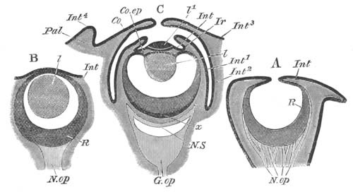
Fig. 122. Three diagrammatic sections of the eyes of Mollusca. (After Grenacher.)
A. Nautilus. B. Gasteropod (Limax or Helix). C. Dibranchiate Cephalopod.
Pal. eyelid; Co. cornea; Co.ep. epithelium of ciliary body; Ir. iris; Int. Int1 ... Int4. different parts of the integument; l. lens; l1. outer segment of lens; R. retina; N.op. optic nerve; G.op. optic ganglion; x. inner layer of retina; N.S. nervous stratum of retina.
The supra-œsophageal ganglia appear always to develop within the region of the velar area. This area corresponds with the præ-oral lobe of the Chætopod larva, at the apex of which is developed the supra-œsophageal ganglion. Embryology thus confirms the results of Comparative Anatomy in reference to the homology of these ganglia in the two groups.
Optic organs[113]. An eye is present in most Gasteropods and in many larval Pteropods. Although its development has not been fully worked out, yet it has clearly been shewn by Bobretzky and other investigators that it originates as an involution of the epidermis, which first forms a cup and eventually a closed vesicle. The posterior wall of the vesicle gives rise to the retina, the anterior to the inner epithelium of the cornea. The external epidermis becomes continued over the outer surface of the vesicle.
The lens is formed in the interior of the vesicle, probably as a cuticular deposit, which increases by the addition of concentric layers. Pigment becomes deposited between the cells of the retina. Fig. 122 B is a diagrammatic representation of the adult eye of a Gasteropod.
The Cephalopod eye is formed, as first shewn by Lankester, as a pit in the epiblast round which a fold arises (fig. 123 A) and gradually grows over the mouth of the pit so as to shut it off from communication with the exterior (fig. 123 B).
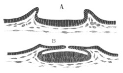
Fig. 123. Two sections through the developing eye of a Cephalopod to shew the formation of the optic cup. (After Lankester.)
The epiblast lining the posterior region of the vesicle gives rise to the retina, that lining the anterior region to the ciliary body and processes. It is important to notice that the condition of the eye just before the above pit becomes closed is exactly that which is permanent in Nautilus (vide fig. 122 A). After the pit has become closed a mesoblastic layer grows in between its wall and the external epiblast.
The lens becomes formed in two independent segments. The inner and larger of these arises as a rod-like process (fig. 124) projecting from the front wall of the optic vesicle into the cavity of the vesicle. It is a cuticular structure and therefore without cells. By the deposition of a series of concentric layers it soon assumes a spherical form (fig. 125, hl). The condition of the eye, with a closed optic vesicle and the lens projecting into it, is that which is permanent in the majority of Gasteropods (vide fig. 122 B). At about the time when the lens first becomes formed a fold composed of epiblast and mesoblast appears round the edge of the optic cup (fig. 124, cc), and gives rise to a structure known in the adult as the iris. Shortly afterwards this becomes more prominent (fig. 125, if), and at the same time the layers of cells of the ciliary region in front of the inner segment of the lens become reduced to the condition of mere membranes (fig. 125 B); and in front of them the anterior or outer segment of the lens becomes formed as a cuticular deposit (fig. 125 B, vl). At a still later period a fresh fold of epiblast and mesoblast appears round the eye and gradually constitutes the anterior optic chamber (vide fig. 122 C, Co). In most forms this chamber communicates with the exterior by a small aperture, but in some it is completely closed. The fold itself gives rise to the cornea in front and to the sclerotic at the sides. At a later period another fold may appear forming the eyelids (fig. 122 C, Pal).
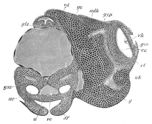
Fig. 124. Transverse section through the head of an advanced embryo of Loligo. (After Bobretzky.)
vd. œsophagus; gls. salivary gland; g.vs. visceral ganglion; gc. cerebral ganglion; g.op. optic ganglion; adk. optic cartilage; ak. and y. lateral cartilage or (?) white body; rt. retina; gm. limiting membrane; vk. ciliary region of eye; cc. iris; ac. auditory sack (the epithelium lining the auditory sacks is not represented); vc. vena cava; ff. folds of funnel.
Auditory organs. A pair of auditory sacks is found in the larvæ of almost all Gasteropods and Pteropods, and usually originates very early. They are placed in the front part of the foot, and on the formation of the pedal ganglia come into close connection with it, though they receive their nervous supply in the adult from the supra-œsophageal ganglia.
In a very considerable number of cases amongst Gasteropods and Pteropods the auditory organs have been observed to develop as invaginations of the epiblast, which give rise to closed vesicles lying in the foot, e.g. Paludina, Nassa, Heteropods, Limax, some Pteropods (Clio).
This is no doubt the primitive mode of origin, but in other cases, which perhaps require confirmation, the sacks are stated to originate from a differentiation of solid thickenings of the epidermis or of the tissues subjacent to it.
The auditory sacks are provided with an otolith, which according to Fol’s observations is first formed in the wall of the sack.
In Cephalopods the auditory organs are formed as epiblastic pits on the posterior surface of the embryo, and are at first widely separated (fig. 113, ac). The openings of the pits become narrowed, and finally the original pits form small sacks lined by an epithelium, and communicating with the exterior by narrow ducts, equivalent to the recessus vestibuli of Vertebrates, and named, after their discoverer, Kölliker’s ducts. The external openings of these ducts become completely closed at about the same time as the shell-gland, and the ducts remain as ciliated diverticula of the auditory pits. The widely separated auditory sacks gradually approach in the middle ventral line, and are immediately invested by the visceral ganglia (fig. 124, ac). They finally come to lie in contact on the inner side of the funnel.
On the side opposite Kölliker’s duct, an epithelial ridge is formed—the crista acustica—the cells of which give rise to an otolith connected with the crista by a granular material. At a later period of development three regions of the epithelium of the sack become especially differentiated. Each of these regions is provided with two rows of cells, bearing on their free edges numerous very short auditory hairs. The cells of each row are placed nearly at right angles to those of the adjoining row.
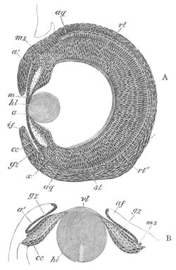
Fig. 125. Sections through the developing eye of Loligo at two stages. (After Bobretzky.)
hl. inner segment of lens; vl. outer segment of lens; a and a´. epithelium lining the anterior optic chamber; gz. large epiblast cells of ciliary body; cc. small epiblast cells of ciliary body; ms. layer of mesoblast between the two epiblastic layers of the ciliary body; af. and if. fold of iris; rt. retina; rt´´. inner layer of retina; st. rods; aq. equatorial cartilage.
Muscular system. The muscular system in all groups of Molluscs is derived entirely from the mesoblast.
The greater part of the system takes its origin from the somatic mesoblast. In almost all Gasteropod and Pteropod larvæ there is present a well-developed spindle muscle attaching the embryo to the shell. This muscle appears to be absent in the Cephalopoda.
Body cavity and vascular system. The body cavity in Gasteropods and Pteropods originates either by a definite splitting of the mesoblast, or by the appearance of intercellular spaces. It becomes divided into numerous sinuses which freely communicate with the vascular system.
Very different accounts have been given by different investigators of the development of the heart in the Gasteropoda and Pteropoda.
It would seem however in most cases to arise as a solid mass of mesoblast cells at the hind end of the pallial cavity, which subsequently becomes hollowed out and divided into an auricle and ventricle. Bobretzky’s careful observations have fully established this mode of development for Nassa.
In Pteropods the heart is formed (Fol) close to the anus, but slightly dorsal to it (fig. 108, h). The pericardium is formed from the mesoblast at a considerably later period than the heart.
A very different account of the formation of the heart is given by Bütschli for Paludina. He states that there appears an immense contractile sack on the left side of the body. This becomes subsequently reduced in size, and in the middle of it appears the heart, probably from a fold of its wall. The original sack would appear to give rise to the pericardium.
In connection with the vascular system mention may be made of certain contractile sinuses frequently found in the larvæ of Gasteropoda and Pteropoda. One of these is placed at the base of the foot, and the other on the dorsal surface within the mantle cavity immediately below the velum[114]. The completeness of the differentiation of these sinuses varies considerably; in some forms they are true sacks with definite walls, in other cases mere spaces traversed by muscular strands. They are found in the majority of marine Gasteropods, Heteropods and Pteropods. In Limax a large posteriorly placed pedal sinus is well developed, and there is also a sinus in the visceral sack. The rhythmical contraction of the yolk-sack of Cephalopods appears to be a phenomenon of the same nature as the contraction of the foot sinus of Limax.
In Calyptræa (Salensky) there is an enormous provisional cephalic dilatation within the velum which does not appear to be contractile. Similar though less marked cephalic vesicles are found in Fusus, Buccinum and most marine Gasteropods.
In Cephalopods the vascular system is formed by a series of independent (?) spaces originating in the mesoblast, the cells around which give rise to the walls of the vessels. The branchial hearts are formed at about the time at which the shell-gland becomes closed. The aortic heart (fig. 127, c) is formed of two independent halves which subsequently coalesce (Bobretzky).
The true body cavity arises as a space in the mesoblast subsequently to the formation of the main vascular trunks.
Renal organs. Amongst the Gasteropods and Pteropods there are present provisional renal organs, which may be of two kinds, and a permanent renal organ.
The provisional organs consist of either (1) an external paired mass of excretory cells or (2) an internal organ provided with a duct, which is not in all cases certainly known to open externally. The former structure is found especially in the marine Prosobranchiates (Nassa, etc.) where it has been fully studied by Bobretzky. It consists of a mass of cells on each side of the body, close to the base of the foot, and not far behind the velum. This mass grows very large, and below it may be seen a continuous layer of epiblast. The cells forming it fuse together, their nuclei disappear, and numerous vacuoles containing concretions arise in them. At a later stage all the vacuoles unite together and form a cavity filled with a brown granular mass.
The provisional internal renal organ is found in many pulmonate Gasteropods—Lymnæus, Planorbis, etc. It consists of a paired V-shaped ciliated tube with a pedal and cephalic limb. The former has an external opening, but the termination of the latter is still in doubt.
It consists, according to Büschli’s description (No. 244), in the freshwater Pulmonata (Lymnæus, Planorbis) of a round sack, close to the head, opening by an elongated and richly ciliated tube in the neighbourhood of the eye. From the sack a second shorter tube passes off towards the foot, which seems however to end blindly. The cells lining the sack contain concretions, and there is one especially large cell in the lumen of the sack attached on the side turned towards the eye. It coexists in Lymnæus with provisional renal organs of the type of those in marine Prosobranchiata.
A somewhat different description of the structure and development of this organ in Planorbis has recently been given by Rabl (No. 268). It consists of a V-shaped tube on each side with both extremities opening into the body cavity. The one limb is directed towards the velar area, the other towards the foot. It is developed from the mesoblast cells of the anterior part of the mesoblastic band. The large mesoblast (p. 227) of each side grows into two processes, the two limbs of the future organ. A lumen in the cell is continued into each limb, while continuations of the two limbs of the V are formed from the hollowing out of the central parts of the adjoining mesoblast cells.
In Limax embryos Gegenbaur found a pair of elongated provisional branched renal sacks, the walls of which contained concretions. These sacks are provided with anteriorly directed ducts opening on the dorsal side of the mouth. This organ is probably of the same nature as the provisional renal organ in other Pulmonata.
Permanent renal organ. According to the most recent observer (Rabl, No. 268), whose statements are supported by the sections figured, the permanent renal organ in Gasteropods is developed from a mass of mesoblast cells close to the end of the intestine. This is first carried somewhat to the left side, and then becomes elongated and hollow, and attaches itself to the epiblast on the left side of the anus (fig. 108, r). After the formation of the heart the inner end opens into the pericardium and becomes ciliated, the median part becomes glandular and concrements appear in its lining cells, and the terminal part forms the duct.
Previous observers have usually derived this organ from the epiblast; according to Rabl this is owing to their having studied too late a stage in the development.
In Cephalopoda the excretory sacks or organ of Bojanus are apparently differentiations of the mesoblast[115]. At an early stage part of their walls envelops the branchial veins. From this part of the wall the true glandular section of the organ would seem to be formed. The epithelium forming the inner wall of each sack is at an early age very columnar.
The development of the organ of Bojanus in Lamellibranchiata has been studied by Lankester. He finds that it develops as a paired invagination of the epiblast immediately ventral to the anus.
Generative glands. The generative glands in Mollusca would appear to be usually developed in the post-larval period, but our knowledge on this subject is extremely scanty.
In Pteropods Fol believes that he has proved that the hermaphrodite gland originates from two independent formations, one (the testicular) epiblastic in origin, and the other (the ovarian) hypoblastic.
These views of Fol do not appear to me nearly sufficiently substantiated to be at present accepted.
The generative glands in Cephalopoda appear to be simple differentiations of the mesoblast. They are at first very closely connected with the aortic heart (fig. 127, kd), but soon become completely separated from it.
Alimentary tract. The formation of the archenteron, and the relation of its opening to the permanent mouth and anus, has already been described and needs no further elucidation. It will be convenient to treat the subject of this section under three headings for each group—viz. (1) the mesenteron, (2) the stomodæum, and (3) the proctodæum.
The mesenteron. In the Gasteropoda and Pteropoda the mesenteron, as has already been mentioned, forms a simple sack, which may however, owing to the presence of food-yolk, be at first without a lumen. Of this sack an anterior portion gives rise to the stomach and liver, and a posterior to the intestine. This latter portion is the first to be distinctly differentiated as such, and forms a narrowish tube connecting the anterior dilatation with the anus. In the meantime the cells of a great part of the anterior portion of the mesenteron undergo peculiar changes. They enlarge, and in each of them a deposit of food material appears, which is often at any rate derived from the absorption of the albumen in which the embryo floats. The cells on the dorsal side, adjoining the œsophageal invagination, and the whole of the cells on the ventral side do not however undergo these changes. There thus arises an anterior and ventral region adjoining the œsophagus, which becomes completely enclosed by small cells and forms the true stomach. The part behind and dorsal to the stomach is lined by the large nutritive cells and forms the liver. It opens into the stomach at the junction of the latter with the intestine, which in the later stages becomes bent somewhat forwards and to the right. Still later the hepatic region becomes branched, the albuminous contents of its cells are replaced by a coloured secretion, and it becomes bodily converted into the liver. The stomach is usually richly ciliated.
The various modifications of the above type of development of the alimentary tract are to be regarded as due to the disturbing influence of food-yolk. Where primitively the hypoblast cells are very bulky, though invaginated in a normal way, the wall of the hepatic region becomes immensely swollen with food-yolk, e.g. Nautica. In other cases amongst certain Pteropods (Fol, No. 249) where the hypoblast is still more bulky, part of the archenteric walls becomes converted into a bilobed sack opening into the pyloric region, in the walls of which a large deposit of food material is stored, which gradually passes into the remainder of the alimentary tract and is there digested. The bilobed nutritive sack, as it is called by Fol, is eventually completely absorbed, though the liver in some, if not all cases, grows out as a fresh sack from its duct.
The formation of the permanent alimentary tract, when the hypoblast is so bulky that there is no true archenteric cavity, has been especially investigated by Bobretzky (No. 242).
In the case of a species of Fusus the hypoblast, when enclosed by the epiblast, is composed of four cells only. The blastopore remains permanently open at the oral region, and around it the œsophagus grows in a wall-like fashion. The protoplasmic portions of the four hypoblast cells are turned towards the œsophageal opening, and from them are budded off small cells which are continuous at the blastopore with the epiblast of the œsophagus. These cells give rise posteriorly to the intestine and anteriorly to the sack, which becomes the stomach and liver. This sack always remains open towards the four primitive yolk cells. The cells of the posterior part of it become larger and larger and form the hepatic sack, which fills up the left and posterior part of the visceral sack, pushing the yolk cells to the right. The cells lining the hepatic sack become pyramidal in shape, and each of them is filled with a peculiar mass of albuminous material. The cells adjoining the opening of the œsophagus remain small, become ciliated, and form the stomach. They are not sharply separated off from the cells of the hepatic sack. The yolk cells remain distinct on the right side of the body during larval life, and their food material is gradually absorbed for the nutrition of the embryo.
A modification of the above mode of development, where the food material is still more bulky and the blastopore closed, is found in Nassa, and has already been described (vide p. 233).
The stomodæum. The stomodæum in most cases is formed as a simple epiblastic invagination which meets and opens into the mesenteron. When the blastopore remains permanently open at the oral region the stomodæum is formed as an epiblastic wall round its opening. In all cases the stomodæum gives rise to the mouth and œsophagus. At a subsequent period there are developed in the oral region of the stomodæum the radula in a special ventral pit, and the salivary glands—the latter as simple outgrowths.
The œsophagus is usually ciliated.
The proctodæum. Except where the blastopore remains as the permanent anus (Paludina) the proctodæum is always formed subsequently to the mouth. Its formation is usually preluded by the appearance of two projecting epiblast cells, but it is always developed as a very shallow epiblastic invagination, which does not give rise to any part of the true intestine.
In the Cephalopods the alimentary tract is formed, as in other cephalophorous Mollusca, of three sections. (1) A stomodæum, formed by an epiblastic invagination, which gives rise to the mouth, œsophagus and salivary glands. (2) A proctodæum, which is an extremely small epiblastic invagination. (3) A mesenteron, lined by true hypoblast, which forms the main section of the alimentary tract, viz. the stomach, intestine, the liver, and ink sack[116].
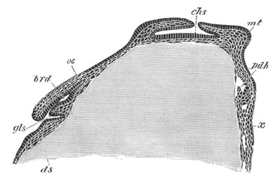
Fig. 126. Longitudinal vertical section through a Loligo ovum when the mesenteric cavity is just commencing to be formed. (After Bobretzky.)
gls. salivary gland; brd. sheath of radula; oe. œsophagus; ds. yolk-sack; chs. shell-gland; mt. mantle; pdh. mesenteron; x. epiblastic thickening between the folds of the funnel.
The mesenteron. The mesenteron is first visible from the surface as a small tubercle on the posterior side of the mantle between the rudiments of the two gills (fig. 111 B, an). Within this, as was first shewn by Lankester, a cavity appears.
This cavity is as in Gasteropods open to the yolk-sack, and only separated from the yolk itself by the yolk membrane already spoken of. It is at first lined by indifferent cells of the lower layer of the blastoderm, which however soon become columnar and form a definite hypoblastic layer (fig. 126, pdh). Between the hypoblast and epiblast there is a very well marked layer of mesoblast. As the mesenteric cavity extends, its walls meet the epiblast, and at the point of contact of the two layers the epiblast becomes slightly pitted in. At this point the anus is formed at a considerably later period (fig. 127, an).
On the ventral side of the primitive mesenteron an outgrowth appears very early, which becomes the ink sack (fig. 127, bi).
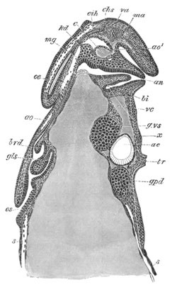
Fig. 127. Longitudinal section through an advanced embryo of Loligo. (After Bobretzky.)
os. mouth; gls. salivary gland; brd. sheath of radula; ao. anterior aorta; ao1. posterior aorta; va. branch of posterior aorta to shell sack; ma. branch of posterior aorta to mantle; c. aortic heart; oe. œsophagus; mg. stomach; an. anus; bi. ink sack; kd. germinal tissue; eih. shell sack; vc. vena cava; g.vs. visceral ganglion; g.pd. pedal ganglion; ac. auditory sack; tr. funnel.
The mesenteric cavity, still open to the yolk, gradually extends itself in a dorsal direction over the yolk-sack, but remains for some time completely open to it ventrally, and only separated from the actual yolk by the yolk membrane. There early grow out from the walls of the mesenteron a pair of hepatic diverticula.
As the mesenteric cavity extends it dilates at its distal extremity into a chamber destined to form the stomach (fig. 127, mg). At about this time the anus becomes perforated. Shortly afterwards the mesenteron meets and opens into the œsophagus at the dorsal extremity of the yolk-sack, but at the time when this takes place the hypoblast has extended round the entire cavity, and has shut it off from the yolk. The yolk membrane throughout the whole of this period is quite passive, and has no share in forming the walls of the alimentary tract.
The stomodæeum. The stomodæum appears as an epiblastic invagination at the anterior side of the blastoderm, before any trace of the mesenteron is present. It rapidly grows deeper, and, shortly after the mesenteric cavity becomes formed, an outgrowth arises from its wall adjoining the yolk-sack, which gives rise to the salivary glands (figs. 126 and 127, gls). Immediately behind the opening of the salivary glands there appears on its floor a swelling which becomes the odontophore, and behind this a pocket of the stomodæal wall forms the sheath of the radula (figs. 126 and 127, brd). Behind this again the œsophagus is continued dorsalwards as a very narrow tube, which eventually opens into the stomach (fig. 127).
The terminal portion of the rudiment of the salivary gland divides into two parts, each of which sends out numerous diverticula which constitute the permanent glands. The greater part of the original outgrowth remains as the unpaired duct of the two glands[117].
In the larva observed by Grenacher the anterior pair of salivary glands originated from independent lateral outgrowths of the floor of the mouth, close to the opening of the posterior salivary glands.
The yolk-sack of the Cephalopoda. The yolk, as has already been stated, becomes at an early period completely enclosed in a membrane formed of flattened cells, which constitutes a definite yolk-sack. It is, in the more typical forms of Cephalopoda, divided into an external and an internal section, of which the former is probably a special differentiation of the median part of the foot of other cephalophorous Mollusca (vide p. 272). At no period does the yolk-sack communicate with the alimentary tract. The two sections of the yolk-sack are at first not separated by a constriction. In the second half of embryonic life the condition of the yolk-sack undergoes considerable changes. The internal part grows greatly in size at the expense of the external, and the latter diminishes very rapidly and becomes constricted off from the internal part of the sack, with which it remains connected by a narrow vitelline duct.
The internal yolk-sack becomes divided into three sections: a dilated section in the head, a narrow section in the neck, and an enormously developed portion in the mantle region. It is the latter part which mainly grows at the expense of the external yolk-sack. It gives off at its dorsal end two lobes, which pass round and embrace the lower part of the œsophagus. The passage of the yolk from the external to the internal yolk-sack is probably largely due to the contractions of the former.
The external yolk-sack is not vascular, and probably the absorption of the yolk for the nutrition of the embryo can only take place in the internal yolk-sack. The most remarkable feature of the Cephalopod yolk-sack is the fact that it lies on the opposite side of the alimentary tract to the yolk cells, which form a rudimentary yolk-sack in such Gasteropoda as Nassa and Fusus. In these forms, the yolk-sack is at first dorsal, but subsequently is carried by the growth of the liver to the right side. In Cephalopoda on the contrary, the yolk-sack is placed on the ventral side of the body.
What is known of the development of the alimentary tract in the Polyplacophora has already been mentioned.
In the Lamellibranchiata (Lankester, No. 239), the mesenteron early grows out into two lateral lobes which form the liver, while the part between them forms the stomach.
In Pisidium the intestine is formed from the original pedicle of invagination, which remains permanently attached to the epiblast. The stomodæum is formed by the usual epiblastic invagination, and becomes the mouth and œsophagus. The development of the crystalline rod and its sack do not appear to be known. In the adult the sack of the crystalline rod opens into a part of the alimentary tract which appears to belong to the mesenteron. Were however the development to shew them to be really derived from the stomodæum they might be interpreted as rudiments of the organ which constitutes the odontophore and its sack in cephalophorous Mollusca—an interpretation which would be of considerable phylogenetic interest.
Bibliography.
General.
(238) T. H. Huxley. “On the Morphol. of the Cephal. Mollusca.” Phil. Trans. 1853. (239) E. R. Lankester. “On the developmental history of the Mollusca.” Phil. Trans. 1875. (240) H. G. Bronn and W. Keferstein. Die Klassen u. Ordnungen d. Thierreichs, Vol. III. 1862‑1866.
Gasteropoda and Pteropoda.
(241) J. Alder and A. Hancock. “Devel. of Nudibr.” Ann. and Magaz. Nat. Hist., Vol. XII. 1843. (242) N. Bobretzky. “Studien über die embryonale Entwicklung d. Gasteropoden.” Archiv f. mikr. Anat., Vol. XIII. (243) W. K. Brooks. “Preliminary Observations on the Development of Marine Gasteropods.” Chesapeake Zoological Laboratory, Session of 1878. Baltimore, 1879. (244) O. Bütschli. “Entwicklungsgeschichtliche Beiträge (Paludina vivipara).” Zeit. f. wiss. Zool., Vol. XXIX. 1877. (245) W. Carpenter. “On the devel. of the embr. of Purpura lapillus.” Trans. Micros. Soc., 2d series, Vol. III. 1855. (246) W. Carpenter. “On the devel. of the Purpura.” Ann. and Mag. of Nat. Hist., 2d series, Vol. XX. 1857. (247) E. Claparède. “Anatomie u. Entwickl. der Neritina fluviatilis.” Müller’s Archiv, 1857. (248) H. Eisig. “Beitr. z. Anat. u. Entwickl. der Geschlechtsorg. von Lymnæus.” Zeitschr. f. wiss. Zool., Vol. XIX. 1869. (249) H. Fol. “Sur le développement des Ptéropodes.” Archiv. de Zool. expérim. et générale, Vol. IV. 1875. (250) H. Fol. “Sur le développement des Gastéropodes pulmonés.” Compt. rend., 1875, pp. 523‑526. (251) H. Fol. “Sur le développement des Hétéropodes.” Archiv. de Zool. expérim. et générale, Vol. V. 1876. (252) C. Gegenbaur. “Beit. z. Entwicklungsgesch. der Landgasteropoden.” Zeitschr. f. w. Zool., Vol. III. 1851. (253) C. Gegenbaur. Untersuch. üb. Pteropoden u. Heteropoden, Leipzig, 1855. (254) H. von Jhering. “Entwicklungsgeschichte von Helix.” Jenaische Zeitschrift, Vol. IX. 1875. (255) W. Keferstein and E. Ehlers. “Beob. üb. d. Entwick. v. Æolis peregr.” Zool. Beitr., 1861. (256) J. Koren and D. C. Danielssen. “Bemärk. til Mollusk Udvikling.” Nyt Mag. f. Naturvidensk., Vol. V. 1847. Isis, p. 202. 1848. (257) J. Koren and D. C. Danielssen. Bidrag til Pectinibr. Udvikl. Bergen, 1851 (supplement, 1852). Ann. and Mag. Nat. Hist., 1857. (258) A. Krohn. “Beobacht. aus d. Entwickl. der Pteropoden u. Heterop.” Müller’s Archiv, 1856 and 1857. (259) A. Krohn. Beitr. zur Entwickl. der Pteropoden u. Heteropoden. Leipzig, 1860. (260) H. de Lacaze-Duthiers. “Mém. sur l’anat. et l’embryog. des Vermets.” 2e partie. Ann. sc. nat., 4e série, T. XIII. 1860. (261) P. Langerhans. “Zur Entwickl. der Gasterop. Opisthobr.” Zeitschr. f. w. Zool., Vol. XXIII. 1873. (262) E. R. Lankester. “On the development of the Pond-Snail.” Quart. J. of Micr. Scie., Vol. XIV. 1874. (263) E. R. Lankester. “On the coincidence of the blastopore and anus in Paludina vivipara.” Quart. J. of Micr. Scie., Vol. XVI. 1876. (264) F. Leydig. “Ueber Paludina vivipara.” Zeitschr. f. w. Zool., Vol. II. 1850. (265) J. Müller. Ueber Synapta dig. u. üb. d. Erzeug. v. Schnecken in Holoth., 1852. (266) J. Müller. “Bemerk. aus d. Entwickl. der Pteropoden.” Monatsber. Berl. Akad., 1857. (267) C. Rabl. “Die Ontogenie d. Süsswasser-Pulmonaten.” Jenaische Zeitschrift, Vol. IX. 1875. (268) C. Rabl. “Ueb. d. Entwick. d. Tellerschnecke (Planorbis).” Morph. Jahrbuch, Vol. V. 1879. (269) W. Salensky. “Beitr. zur Entwickl. d. Prosobr.” Zeitschr. f. w. Zool., Vol. XXII. 1872. (270) O. Schmidt. “Ueb. Entwick. von Limax agrestis.” Müller’s Archiv, 1851. (271) Max S. Schultze. “Ueber d. Entwick. des Tergipes lacinulatus.” Arch. f. Naturg., Jahrg. XV. 1849. (272) E. Selenka. “Entwick. von Tergipes claviger.” Niederl. Arch. f Zool., Vol. I. 1871. (273) E. Selenka. “Die Anlage d. Keimbl. bei Purpura lapillus.” Niederl. Arch. f. Zool., Vol. I. 1872. (274) C. Semper. “Entwickl. der Ampullaria polita, etc.” Natuurk. Verhandl. Utrechts Genootsch., 1862. (275) An. Stecker. “Furchung u. Keimblatterbildung bei Calyptræa.” Morphol. Jahrbuch, Vol. II. 1876.
(276) A. Stuart. “Ueb. d. Entwickl. einiger Opisthobr.” Zeitschr. f. w. Zool., Vol. XV. 1865. (277) N. A. Warneck. “Ueber d. Bild. u. Entwick. d. Embryos bei Gasterop.” Bullet. Soc. natural. de Moscou, T. XXIII. 1850.
Cephalopoda.
(278) P. J. van Beneden. “Recherches sur l’Embryogénie des Sépioles.” Nouv. Mém. Acad. Roy. de Bruxelles, Vol. XIV. 1841. (279) N. Bobretzky. Observation on the development of the Cephalopoda (Russian). Nachrichten d. kaiserlichen Gesell. d. Freunde der Naturwiss. Anthropolog. Ethnogr. bei d. Universität Moskau. (280) H. Grenacher. “Zur Entwicklungsgeschichte d. Cephalopoden.” Zeit. f. wiss. Zool., Bd. XXIV. 1874. (281) A. Kölliker. Entwicklungsgeschichte d. Cephalopoden. Zurich, 1844. (282) E. R. Lankester. “Observations on the development of the Cephalopoda.” Quart. J. of Micr. Science, Vol. XV. 1875. (283) E. Metschnikoff. “Le développement des Sépioles.” Archiv d. Sc. phys. et nat., Vol. XXX. Genève, 1867.
Polyplacophora.
(284) A. Kowalevsky. “Ueb. d. Entwick. d. Chitonen.” Zoologischer Anzeiger, No. 37. 1879. (285) S. L. Lovén. “Om utvecklingen hos slägtet Chiton.” Stockholm öfversigt, XII. 1855. [Vide also Ann. and Mag. of Nat. Hist., Vol. XVII. 1856, and Archiv f. Naturgeschichte, 1856.]
Scaphopoda.
(286) H. Lacaze-Duthiers. “Développement du Dentale.” Ann. d. Sci. Nat., Series IV. Vol. VII. 1857.
Lamellibranchiata.
(287) M. Braun. “Postembryonale Entwicklung d. Süsswasser-Muscheln.” Zoologischer Garten. (288) C. G. Carus. “Neue Untersuch. üb. d. Entwickl. unserer Flussmuschel.” Verh. Leop.-Car. Akad., Vol. XVI. 1832. (289) W. Flemming. “Studien in d. Entwicklungsgeschichte der Najaden.” Sitz. d. k. Akad. Wiss. Wien, Vol. LXXI. 1875. (290) F. Leydig. “Ueber Cyclas Cornea.” Müller’s Archiv, 1855. (291) S. L. Lovén. “Bidrag til Känned. om Utweckl. af Moll. Acephala Lamellibr.” Vetensk. Akad. Handl., 1848. [Vide also Arch. f. Naturg., 1849.] (292) C. Rabl. “Ueber d. Entwicklungsgeschichte d. Malermuschel.” Jenaische Zeitschrift, Vol. X. 1876. (293) W. Salensky. “Bemerkungen über Haeckels Gastræa-Theorie (Ostrea).” Arch. f. Naturg., 1874. (294) O. Schmidt. “Ueb. d. Entwick. von Cyclas calyculata.” Müller’s Arch., 1854. (295) O. Schmidt. “Zur Entwickl. der Najaden.” Wien, Sitzungsber. math.-nat. Cl., Vol. XIX. 1856. (296) P. Stepanoff. “Ueber die Geschlechtsorgane u. die Entwicklung von Cyclas.” Archiv f. Naturgeschichte, 1865. (297) H. Lacaze-Duthiers. “Développement d. branchies d. Mollusques Acéphales.” An. Sc. Nat., Ser. IV. Vol. V. 1856.