Volume 1 / Part 1 — CHAPTER XVIII.
Reading edition
CRUSTACEA[185].
History of the larval forms[186].
The larval forms of the Crustacea appear to have more faithfully preserved their primitive characters than those of almost any other group.
Branchiopoda.
The Branchiopoda, comprising under that term the Phyllopoda and Cladocera, contain the Crustacea with the maximum number of segments and the least differentiation of the separate appendages. This and other considerations render it probable that they are to be regarded as the most central group of the Crustaceans, and as in many respects least modified from the ancestral type from which all the groups have originated.
The free larval stages when such exist commence with a larval form known as the Nauplius.
The term Nauplius was applied by O. F. Müller to certain larval forms of the Copepoda (fig. 229) in the belief that they were adult.
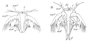
Fig. 208. Two stages in the development of Apus cancriformis. (After Claus.)
A. Nauplius stage at the time of hatching. B. Stage after first ecdysis.
an1. and an2. First and second antennæ; md. mandible; mx. maxilla; l. labrum; fr. frontal sense organ; f. caudal fork; s. segments.
The term has now been extended to a very large number of larvæ which have certain definite characters in common. They are provided (fig. 208 A) with three pairs of appendages, the future two pairs of antennæ and mandibles. The first pair of antennæ (an1) is uniramous and mainly sensory in function, the second pair of antennæ (an2) and mandibles (md) are biramous swimming appendages, and the mandibles are without the future cutting blade. The Nauplius mandibles represent in fact the palp. The two posterior appendages are both provided with hook-like prominences on their basal joints, used in mastication. The body in most cases is unsegmented, and bears anteriorly a single median eye. There is a large upper lip, and an alimentary canal formed of œsophagus, stomach and rectum. The anus opens near the hind end of the body. On the dorsal surface small folds of skin frequently represent the commencement of a dorsal shield. One very striking peculiarity of the Nauplius according to Claus and Dohrn is the fact that the second pair of antennæ is innervated from a sub-œsophageal ganglion. A larval form with the above characters occurs with more or less frequency in all the Crustacean groups. In most instances it does not exactly conform to the above type, and the divergences are more considerable in the Phyllopods than in most other groups. Its characters in each case are described in the sequel.
Phyllopoda..For the Phyllopoda the development of Apus cancriformis may conveniently be taken as type (Claus, No. 454). The embryo at the time it leaves the egg (fig. 208 A) is somewhat oval in outline, and narrowed posteriorly. There is a slight V-shaped indentation behind, at the apex of which is situated the anus. The body, unlike that of the typical Nauplius, is already divided into two regions, a cephalic and post-cephalic. On the ventral side of the cephalic region there are present the three normal pairs of appendages. Foremost there are the small anterior antennæ (an1), which are simple unjointed rod-like bodies with two moveable hairs at their extremities. They are inserted at the sides of the large upper lip or labrum (l). Behind these are the posterior antennæ, which are enormously developed and serve as the most important larval organs of locomotion. They are biramous, being formed of a basal portion with a strong hook-like bristle projecting from its inner side, an inner unjointed branch with three bristles, and an outer large imperfectly five-jointed branch with five long lateral bristles. The hook-like organ attached to this pair of appendages would seem to imply that it served in some ancestral form as jaws (Claus). This character is apparently universal in the embryos of true Phyllopods, and constantly occurs in the Copepoda, etc.
The third pair of appendages or mandibles (md) is attached close below the upper lip. They are as yet unprovided with cutting blades, and terminate in two short branches, the inner with two and the outer with three bristles.
At the front of the head there is the typical unpaired eye. On the dorsal surface there is already present a rudiment of the cephalic shield, continuous anteriorly with the labrum (l) or upper lip, the extraordinary size of which is characteristic of the larvæ of Phyllopods. The post-cephalic region, which afterwards becomes the thorax and abdomen, contains underneath the skin rudiments of the five anterior thoracic segments and their appendages, and presents in this respect an important variation from the typical Nauplius form. After the first ecdysis the larva (fig. 208 B) loses its oval form, mainly owing to the elongation of the hinder part of the body and the lateral extension of the cephalic shield, which moreover now completely covers over the head and has begun to grow backwards so as to cover over the thoracic region. At the second ecdysis there appears at its side a rudimentary shell-gland. In the cephalic region two small papillæ (fr) are now present at the front of the head close to the unpaired eye. They are of the nature of sense organs, and may be called the frontal sense papillæ. They have been shewn by Claus to be of some phylogenetic importance. The three pairs of Nauplius appendages have not altered much, but a rudimentary cutting blade has grown out from the basal joint of the mandible. A gland opening at the base of the antennæ is now present, which is probably equivalent to the green gland often present in the Malacostraca. Behind the mandibles a pair of simple processes has appeared, which forms the rudiment of the first pair of maxillæ (mx).
In the thoracic region more segments have been added posteriorly, and the appendages of the three anterior segments are very distinctly formed. The tail is distinctly forked. The heart is formed at the second ecdysis, and then extends to the sixth thoracic segment: the posterior chambers are successively added from before backwards.
At the successive ecdyses which the larva undergoes new segments continue to be formed at the posterior end of the body, and limbs arise on the segments already formed. These limbs probably represent the primitive form of an important type of Crustacean appendage, which is of value for interpreting the parts of the various malacostracan appendages. They consist (fig. 209) of a basal portion (protopodite of Huxley) bearing two rami. The basal portion has two projections on the inner side. To the outer side of the basal portion there is attached a dorsally directed branchial sack (br) (epipodite of Huxley). The outer ramus (ex) (exopodite of Huxley) is formed of a single plate with marginal setæ. The inner one (en) (endopodite of Huxley) is four-jointed, and a process similar to those of the basal joint is given off from the inner side of the three proximal joints.
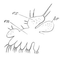
Fig. 209. Typical phyllopod appendage. (Copied from Claus.)
ex. exopodite; en. endopodite; br. branchial appendage (epipodite). The basal portion bearing the two proximal projections is not sharply separated from the endopodite.
At the third ecdysis several new features appear in the cephalic region, which becomes more prominent in the succeeding stages. In the first place the paired eyes are formed at each side of and behind the unpaired eye, secondly the posterior pair of maxillæ is formed though it always remains very rudimentary. The shell-gland becomes fully developed opening at the base of the first pair of maxillæ. The dorsal shield gradually grows backwards till it covers its full complement of segments.
After the fifth ecdysis the Nauplius appendages undergo a rapid atrophy. The second pair of antennæ especially becomes reduced in size, and the mandibular palp—the primitive Nauplius portion of the mandible—is contracted to a mere rudiment, which eventually completely disappears, while the blade is correspondingly enlarged and also becomes toothed. The adult condition is only gradually attained after a very large number of successive changes of skin.
The chief point of interest in the above development is the fact of the primitive Nauplius form becoming gradually converted without any special metamorphosis into the adult condition[187].
Branchipus like Apus is hatched as a somewhat modified Nauplius, which however differs from that of Apus in the hinder region of the body having no indications of segments. It goes through a very similar metamorphosis, but is at no period of its metamorphosis provided with a dorsal shield: the second pair of antennæ does not abort, and in the male is provided with clasping organs, which are perhaps remnants of the embryonic hooks so characteristic of this pair of antennæ.
The larva of Estheria when hatched has a Nauplius form, a large upper lip, caudal fork and single eye. There are two functional pairs of swimming appendages—the second pair of antennæ and mandibles. The first pair of antennæ has not been detected, and a dorsal mantle to form the shell is not developed. At the first moult the anterior pair of antennæ arises as small stump-like structures, and a small dorsal shield is also formed. Rudiments of six or seven pairs of appendages sprout out in the usual way, and continue to increase in number at successive moults: the shell is rapidly developed. The chief point of interest in the development of this form is the close resemblance of the young larva to a typical adult Cladocera (Claus). This is shewn in the form of the shell, which has not reached its full anterior extension, the rudimentary anterior antennæ, the large locomotor second pair of antennæ, which differ however from the corresponding organs in the Cladocera in the presence of typical larval hooks. Even the abdomen resembles that of Daphnia. These features perhaps indicate that the Cladocera are to be derived from some Phyllopod form like Estheria by a process of retrogressive metamorphosis. The posterior antennæ in the adult Estheria are large biramous appendages, and are used for swimming; and though they have lost the embryonic hook, they still retain to a larger extent than in other Phyllopod families their Nauplius characteristics.
The Nauplius form of the Phyllopods is marked by several definite peculiarities. Its body is distinctly divided into a cephalic and post-cephalic region. The upper lip is extraordinarily large, relatively very much more so than at the later stages. The first pair of antennæ is usually rudimentary and sometimes even absent; while the second pair is exceptionally large, and would seem to be capable of functioning not only as a swimming organ, but even as a masticating organ. A dorsal shield is nearly or quite absent.
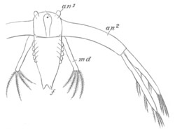
Fig. 209 A. Nauplius larva of Leptodora hyalina from winter egg. (Copied from Bronn; after Sars.)
an1. antenna of first pair; an2. antenna of second pair; md. mandible; f. caudal fork.
Cladocera. The probable derivation of the Cladocera from a form similar to Estheria has already been mentioned, and it might have been anticipated that the development would be similar to that of the Phyllopods. The development of the majority of the Cladocera takes place however in the egg, and the young when hatched closely resembles their parents, though in the egg they pass through a Nauplius stage (Dohrn). An exception to the general rule is however offered by the case of the winter eggs of Leptodora, one of the most primitive of the Cladoceran families. The summer eggs develop without metamorphosis, but Sars (No. 461) has discovered that the larva leaves the winter eggs in the form of a Nauplius (fig. 209). This Nauplius closely resembles that of the Phyllopods. The body is elongated and in addition to normal Nauplius appendages is marked by six pairs of ridges—the indications of the future feet. The anterior antennæ are as usual small; the second large and biramous, but the masticatory bristle characteristic of the Phyllopods is not present. The mandibles are without a cutting blade. A large upper lip and unpaired eye are present.
The adult form is attained in the same manner as amongst the Phyllopods after the third moult.
Malacostraca.
Owing to the size and importance of the various forms included in the Malacostraca, greater attention has been paid to their embryology than to that of any other division of the Crustacea; and the proper interpretation of their larval forms involves some of the most interesting problems in the whole range of Embryology.
The majority of Malacostraca pass through a more or less complicated metamorphosis, though in the Nebaliadæ, the Cumaceæ, some of the Schizopoda, a few Decapoda (Astacus, Gecarcinus, etc.), and in the Edriophthalmata, the larva on leaving the egg has nearly the form of the adult. In contradistinction to the lower groups of Crustacea the Nauplius form of larva is rare, though it occurs in the case of one of the Schizopods (Euphausia, fig. 212), in some of the lower forms of the Decapods (Penæus, fig. 214), and perhaps also, though this has not been made out, in some of the Stomatopoda.
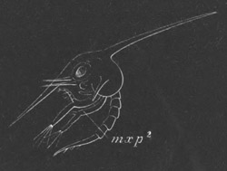
Fig. 210. Zoæa of Thia polita. (After Claus.) mxp2. second maxillipede.
In the majority of the Decapoda the larva leaves the egg in a form known as the Zoæa (fig. 210). This larval form is characterised by the presence of a large cephalo-thoracic shield usually armed with lateral, anterior, and dorsal spines. The caudal segments are well developed, though without appendages, and the tail, which functions in swimming, is usually forked. The six posterior thoracic segments are, on the other hand, rudimentary or non-existent. There are seven anterior pairs of appendages shewn in detail in fig. 211, viz. the two pairs of antennæ (At. I. and At. II.), neither of them used as swimming organs, the mandibles without a palp (md), well-developed maxillæ (two pairs, mx 1 and mx 2), and two or sometimes (Macrura) three pairs of biramous natatory maxillipeds (mxp 1 and mxp 2). Two lateral compound stalked eyes are present, together with a median Nauplius eye. The heart has in the majority of cases only one or two (Brachyura) pairs of ostia.
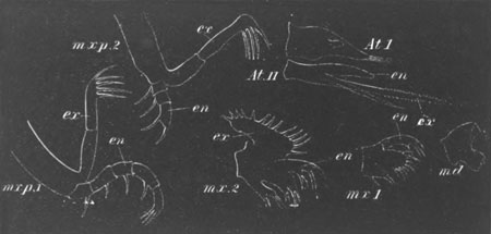
Fig. 211. The Appendages of a Crab Zoæa.
At. I. first antenna; At. II. second antenna; md. mandible (without a palp); mx. 1. first maxilla; mx. 2. second maxilla; mxp. 1. first maxilliped; mxp. 2. second maxilliped.
ex. exopodite; en. endopodite.
The Zoæa larva, though typically developed in the Decapoda, is not always present (e.g. Astacus and Homarus), and sometimes occurs in a very modified form. It makes its appearance in an altered garb in the ontogeny of some of the other groups.
The two Malacostracan forms, amongst those so far studied, in which the phylogenetic record is most fully preserved in the ontogeny, are Euphausia amongst the Schizopods and Penæus amongst the Decapods.
Schizopoda. Euphausia leaves the egg (Metschnikoff, No. 468‑9) as a true Nauplius with only three pairs of appendages, the two hinder biramous, and an unsegmented body. The second pair of antennæ has not however the colossal dimensions so common in the lower types. A mouth is present, but the anus is undeveloped.
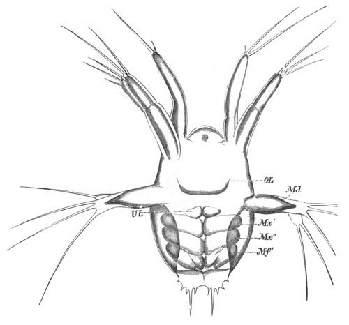
Fig. 212. Nauplius of Euphausia. (From Claus; after Metschnikoff.)
The Nauplius is represented shortly before an ecdysis, and in addition to the proper appendages rudiments of the three following pairs are present. OL. upper lip; UL. lower lip; Md. mandible; Mx´. and Mx´´. two pairs of maxillæ; mf´. maxilliped 1.
After the first moult three pairs of prominences—the rudiments of the two maxillæ and 1st maxillipeds arise behind the Nauplius appendages (fig. 212). At the same time an anus appears between the two limbs of a rudimentary caudal fork; and an unpaired eye and upper lip appear in front. After another moult (fig. 212) a lower lip is formed (UL) as a pair of prominences very similar to true appendages; and a delicate cephalo-thoracic shield also becomes developed. Still later the cutting blade of the mandible is formed, and the palp (Nauplius appendage) is greatly reduced. The cephalo-thoracic shield grows over the front part of the embryo, and becomes characteristically toothed at its edge. There are also two frontal papillæ very similar to those already described in the Phyllopod larvæ. Rudiments of the compound eyes make their appearance, and though no new appendages are added, those already present undergo further differentiations. They remain however very simple; the maxillipeds especially are very short and resemble somewhat Phyllopod appendages.
Up to this stage the tail has remained rudimentary and short, but after a further ecdysis (Claus) it grows greatly in length. At the same time the cephalo-thoracic shield acquires a short spine directed backwards. The larva is now in a condition to which Claus has given the name of Protozoæa (fig. 213 A).
Very shortly afterwards the region immediately following the segments already formed becomes indistinctly segmented, while the tail is still without a trace of segmentation. The region of the thorax proper soon becomes distinctly divided into seven very short segments, while at the same time the now elongated caudal region has become divided into its normal number of segments (fig. 213 B). By this stage the larva has become a true Zoæa—though differing from the normal Zoæa in the fact that the thoracic region is segmented, and in the absence of a second pair of maxillipeds.
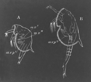
Fig. 213. Larvæ of Euphausia. (After Claus.) From the side.
A. Protozoæa larva. B. Zoæa larva. mx.´ and mx´´. maxillæ 1 and 2; mxp1. maxilliped 1.
The adult characters are very gradually acquired in a series of successive moults; the later development of Euphausia resembling in this respect that of the Phyllopods. On the other hand Euphausia differs from that group in the fact that the abdominal (caudal) and thoracic appendages develop as two independent series from before backwards, of which the abdominal series is the earliest to attain maturity.
This is shewn in the following table compiled from Claus’ observations.
Length of Larva. Appendages of thoracic region; viz. the 2nd and 3rd maxilliped and 5 ambulatory appendages. Appendages of abdomen.
3-3½ mm. 2nd maxilliped, rudimentary. 1st abdominal appendage.
3½-4 mm. 2nd maxilliped, biramous. 3rd rudimentary. 1st and 2nd ambulatory appendages, rudimentary. 2nd and 3rd abdominal appendages. 4th and 5th rudimentary.
4½-5 mm. 3rd maxilliped, biramous. 4th, 5th, and 6th fully developed.
5-5½ mm. 3rd and 4th ambulatory appendages.
6 mm. 5th ambulatory appendage.
All the appendages following the second pair of maxillæ are biramous, and the first eight of these bear branched gills as their epipodites. It is remarkable that the epipodite is developed on all the appendages anteriorly in point of time to the outer ramus (exopodite).
Although in Mysis there is no free larval stage, and the development takes place in a maternal incubatory pouch, yet a stage may be detected which clearly corresponds with the Nauplius stage of Euphausia (E. van Beneden, No. 465). At this stage, in which only the three Nauplius appendages are developed, the Mysis embryo is hatched. An ecdysis takes place, but the Nauplius skin is not completely thrown off, and remains as an envelope surrounding the larva during its later development.
Decapoda. Amongst the Decapoda the larva usually leaves the egg in the Zoæa form, but a remarkable exception to this general rule is afforded by the case of one or more species of Penæus. Fritz Müller was the first to shew that the larva of these forms leaves the egg as a typical Nauplius, and it is probable that in the successive larval stages of these forms the ancestral history of the Decapoda is most fully preserved[188].
The youngest known larva of Penæus (fig. 214) has a somewhat oval unsegmented body. There spring from it the three typical pairs of Nauplius appendages. The first is uniramous, the second and third are biramous, and both of them adapted for swimming, and the third of them (mandibles) is without a trace of the future blade. The body has no carapace, and bears anteriorly a single median simple eye. Posteriorly it is produced into two bristles.
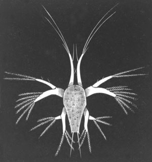
Fig. 214. Nauplius stage of Penæus. (After Fritz Müller.)
After the first moult the larva has a rudiment of a forked tail, while a dorsal fold of skin indicates the commencement of the cephalo-thoracic shield. A large provisional helmet-shaped upper lip like that in Phyllopods has also appeared. Behind the appendages already formed there are stump-like rudiments of the four succeeding pairs (two pairs of maxillæ and two pairs of maxillipeds); and in a slightly older larva the formation of the mandibular blade has commenced, together with the atrophy of the palp or Nauplius appendage.
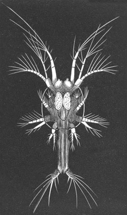
Fig. 215. Protozoæa stage of Penæus. (After Fritz Müller.)
Between this and the next observed stage there is possibly a slight lacuna. The next stage (fig. 215) at any rate represents the commencement of the Zoæa series. The cephalo-thoracic shield has greatly grown, and eventually acquires the usual dorsal spine. The posterior region of the body is prolonged into a tail, which is quite as long as the whole of the remainder of the body. The four appendages which were quite functionless at the last stage have now sprouted into full activity. The region immediately behind them is divided (fig. 215) into six segments (the six thoracic segments) without appendages, while somewhat later the five anterior abdominal segments become indicated, but are equally with the thoracic segments without feet. The mode of appearance of these segments shews that the thoracic and abdominal segments develop in regular succession from before backwards (Claus). Of the palp of the mandibles, as is usual amongst Zoæa forms, not a trace remains, though in the youngest Zoæa caught by Fritz Müller a very small rudiment of the palp was present. The first pair of antennæ is unusually long, and the second pair continues to function as a biramous swimming organ; the outer ramus is multiarticulate. The other appendages are fully jointed, and the two maxillipeds biramous. On the dorsal surface of the body the unpaired eye is still present, but on each side of it traces of the stalked eyes have appeared. Frontal sense organs like those of Phyllopods are also present.
From the Protozoæa form the larva passes into that of a true Zoæa with the usual appendages and spines, characterised however by certain remarkable peculiarities. Of these the most important are (1) the large size of the two pairs of antennæ and the retention of its Nauplius function by the second of them; (2) the fact that the appendages of the six thoracic segments appear as small biramous Schizopod legs, while the abdominal appendages, with the exception of the sixth, are still without their swimming feet. The early appearance of the appendages of the sixth abdominal segment is probably correlated with their natatory function in connection with the tail. As a point of smaller importance which may be mentioned is the fact that both pairs of maxillæ are provided with small respiratory plates (exopodites) for regulating the flow of water under the dorsal shield. From the Zoæa form the larva passes into a Mysis or Schizopod stage (fig. 216), characterised by the thoracic feet and maxillipeds resembling in form and function the biramous feet of Mysis, the outer ramus being at first in many cases much larger than the inner. The gill pouches appear at the base of these feet nearly at the same time as the endopodites become functional. At the same time the antennæ become profoundly modified. The anterior antennæ shed their long hairs, and from the inner side of the fourth joint there springs a new process, which eventually elongates and becomes the inner flagellum. The outer ramus of the posterior antennæ is reduced to a scale, while the flagellum is developed from a stump-like rudiment of the inner ramus (Claus). A palp sprouts on the mandible and the median eye disappears.
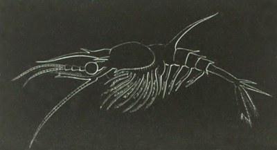
Fig. 216. Penæus larva in the Mysis stage. (After Claus.)
The abdominal feet do not appear till the commencement of the Mysis stage, and hardly become functional till its close.
From the Mysis stage the larva passes quite simply into the adult form. The outer ramus of the thoracic feet is more or less completely lost. The maxillipeds, or the two anterior pairs at any rate, lose their ambulatory function, cutting plates develop on the inner side of their basal joints, and the two rami persist as small appendages on their outer side. Gill pouches also sprout from their outer side.
The respiratory plate of the second maxilla attains its full development and that on the first maxilla disappears[189]. The Nauplius, so far as is known, does not occur in any other Decapod form except Penæus.
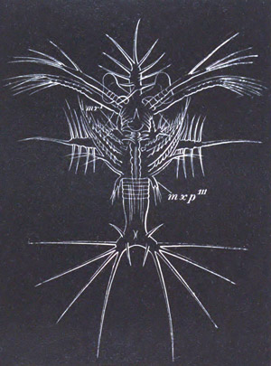
Fig. 217. Latest Protozoæa stage of Sergestes larva (Elaphocaris). (After Claus.)
mxp´´´. third pair of maxillipeds.
The next most primitive larval history known is that which appears in the Sergestidæ. The larval history, which has been fully elucidated by Claus, commences with a Protozoæa form (fig. 217), which develops into a remarkable Zoæa first described by Dohrn as Elaphocaris. This develops into a form originally described by Claus as Acanthosoma, and this into a form known as Mastigopus (fig. 218) from which it is easy to pass to the adult.
The remarkable Protozoæa (fig. 217) is characterised by the presence on the dorsal shield of a frontal, dorsal and two lateral spikes, each richly armed with long side spines. The normal Zoæa appendages are present, and in addition to them a small third pair of maxillipeds. The thoracic region is divided into five short rings, but the abdomen is unsegmented. The tail is forked and provided with long spines. The antennæ, like those of Penæus, are long—the second pair biramous; the mandibles unpalped. Both pairs of maxillæ are provided with respiratory plates; the second pair is footlike, and has at its base a glandular mass believed by Claus to be the equivalent of the Entomostracan shell-gland. The maxillipeds have the usual biramous characters. A helmet-shaped upper lip like that of a typical Nauplius is present, and the eyes are situated on very long stalks.
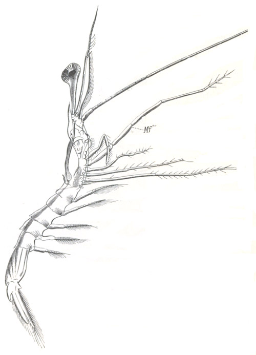
Fig. 218. Mastigopus Stage Of Sergestes. (From Claus.)
Mf´´´. maxilliped 3.
In the true Zoæa stage there appear on the five thoracic segments pouch-like biramous rudiments of the limbs. The tail becomes segmented; but the segments, with the exception of the sixth, remain without appendages. On the sixth a very long bilobed pouch appears as the commencement of the swimming feet of this segment. The segments of the abdomen are armed with lateral spines.
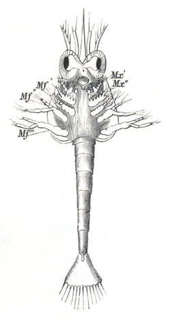
Fig. 219. Larva of Hippolyte In Zoæa Stage. (From Claus.)
Mx´. and Mx´´. maxillæ 1 and 2; Mf´. Mf´´. Mf´´´. maxillipeds.
From the Zoæa stage the larva passes into the form known as Acanthosoma, which represents the Mysis stage of Penæus. The complex spikes on the dorsal shield of the Zoæa stage are reduced to simple spines, but the spines of the tail still retain their full size. In the appendages the chief changes consist (1) in the reduction of the jointed outer ramus of the second pair of antennæ to a stump representing the scale, and the elongation of the inner one to the flagellum; (2) in the elongation of the five ambulatory thoracic appendages into biramous feet, like the maxillipeds, and in the sprouting forth of rudimentary abdominal feet.
The most obvious external indications of the passage from the Acanthosoma to the Mastigopus stage (fig. 218) are to be found in the elongation of the abdomen, the reduction and flattening of the cephalo-thoracic shield, and the nearly complete obliteration of all the spines but the anterior. The eyes on their elongated stalks are still very characteristic, and the elongation of the flagellum of the second pair of antennæ is very striking.
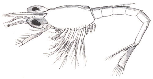
Fig. 220. Older Larva of Hippolyte after the thoracic appendages have become formed. (From Claus.)
The maxillæ and maxillipeds undergo considerable metamorphosis, the abdominal feet attain their adult form, and the three anterior thoracic ambulatory legs lose their outer rami. The most remarkable change of all concerns the two last pairs of thoracic appendages, which, instead of being metamorphosed like the preceding ones, are completely or nearly completely thrown off in the moult which inaugurates the Mastigopus stage, and are subsequently redeveloped. With the reappearance of these appendages, and the changes in the other appendages already indicated, the adult form is practically attained.
With reference to the development of the majority of the Carabidæ, Penæinæ, Palæmoninæ, Crangoninæ, it may be stated generally that they leave the egg in the Zoæa stage (fig. 219) with anterior appendages up to the third pair of maxillipeds. The thorax is unsegmented and indeed almost unrepresented, but the abdomen is long and divided into distinct segments. Both thoracic and abdominal appendages are absent, and the tail is formed by a simple plate with numerous bristles, not forked, as in the case of the Zoæa of Fritz Müller’s Penæus and Sergestes. A dorsal spine is frequently found on the second abdominal segment. From the Zoæa form the embryo passes into a Mysis stage (fig. 220), during which the thoracic appendages gradually appear as biramous swimming feet; they are all developed before any of the abdominal appendages, except the last. In some cases the development is still further abbreviated. Thus the larvæ of Crangon and Palæmonetes (Faxon, No. 476) possess at hatching the rudiments of the two anterior pairs of thoracic feet, and Palæmon of three pairs[190].
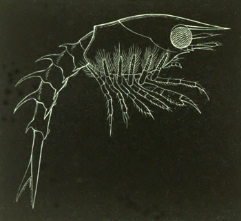
Fig. 221. Newly-hatched Larva of the American Lobster. (After Smith.)
Amongst the other Macrura the larva generally leaves the egg as a Zoæa similar to that of the prawns. In the case of the Thalassinidæ and Paguridæ a Mysis stage has disappeared. The most remarkable abbreviations of the typical development are presented on the one hand by Homarus and Astacus, and on the other by the Loricata.
The development of Homarus has been fully worked out by S. J. Smith (No. 491) for the American lobster (Homarus americanus). The larva (fig. 221) leaves the egg in an advanced Mysis stage. The cephalo-thoracic shield is fully developed, and armed with a rostrum in front. The first pair of antennæ is unjointed but the second is biramous, the outer ramus forming a large Mysis-like scale. The mandibles, which are palped, the maxillæ, and the two anterior maxillipeds differ only in minor details from the same appendages of the adult. The third pair of maxillipeds is Mysis-like and biramous, and the five ambulatory legs closely resemble them, the endopodite of the first being imperfectly chelate. The abdomen is well developed but without appendages. The second, third, fourth and fifth segments are armed with dorsal and lateral spines.
In the next stage swimming feet have appeared on the second, third, fourth and fifth abdominal segments, and the appendages already present have approached their adult form. Still later, when the larva is about half an inch in length, the approach to the adult form is more marked, and the exopodites of the ambulatory legs though present are relatively much reduced in size. The swimmerets of the sixth abdominal segment are formed. In the next stage observed the larva has entirely lost its Schizopod characters, and though still retaining its free-swimming habits differs from the adult form only in generic characters.
As has been already stated, no free larval stages occur in the development of Astacus, but the young is hatched in a form in which it differs only in unimportant details from the adult.
The peculiar larval form of the Loricata (Scyllarus, Palinurus) has long been known under the name Phyllosoma (fig. 222 C), but its true nature was first shewn by Couch (No. 474) [Couch did not however recognise the identity of his larva with Phyllosoma; this was first done by Gerstäcker] and shortly afterwards by Gerbe and Coste. These observations were however for a long time not generally accepted, till Dohrn (No. 477) published his valuable memoir giving an account of how he succeeded in actually rearing Phyllosoma from the eggs of Scyllarus and Palinurus, and shewing that some of the most remarkable features of the metamorphosis of the Loricata occur before the larva is hatched.
The embryo of Scyllarus in the egg first of all passes through the usual Nauplius stage, and then after the formation of a cuticle develops an elongated thoracico-abdominal region bent completely over the anterior part of the body. There appear moreover a number of appendages and the rudiments of various organs; and the embryo passes into a form which may be described as the embryonic Phyllosoma stage. In this stage there are present on the anterior part of the body, in front of the ventral flexure, two pairs of antennæ, mandibles, two pairs of maxillæ, the second commencing to be biramous, and a small stump representing the first pair of maxillipeds. The part of the body bent over consists of a small quadrate caudal plate, and an appendage-bearing region to which are attached anteriorly three pairs of biramous appendages—the second and third maxillipeds, and the anterior pair of ambulatory legs—and two pairs of undivided appendages—the second and third pairs of ambulatory legs. In a slightly later stage the first pair of maxillæ becomes biramous, as also does the first pair of maxillipeds in a very rudimentary fashion. The second and third pairs of ambulatory legs become biramous, while the second and third maxilliped nearly completely lose their outer ramus. Very small rudiments of the two hinder ambulatory legs become formed. If the embryo is taken at this stage (vide fig. 222 A, which represents a nearly similar larva of Palinurus) out of the egg, it is seen to consist of (1) an anterior enlargement with a vaulted dorsal shield enclosing the yolk, two stalked eyes, and a median eye; (2) a thoracic region in which the indications of segmentation are visible with the two posterior pairs of maxillipeds (mxp2 and mxp3) and the ambulatory legs (p1); (3) an abdominal region distinctly divided into segments and ending in a fork.
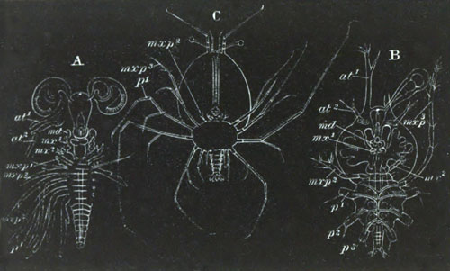
Fig. 222. Larvæ of the Loricata. (After Claus.)
A. Embryo of Palinurus shortly before hatching. B. Young Phyllosoma larva of Scyllarus, without the first maxilliped, the two last thoracic appendages, or the abdominal appendages. C. Fully-grown Phyllosoma with all the Decapod appendages.
at1. antenna of first pair; at2. antenna of second pair; md. mandible; mx1. first maxilla; mx2. second maxilla; mxp1-mxp3. maxillipeds; p1-p3. thoracic appendages.
Before the embryo becomes hatched the first pair of maxillipeds becomes reduced in size and finally vanishes. The second pair of maxillæ becomes reduced to simple stumps with a few bristles, the second pair of antennæ also appears to undergo a retrogressive change, while the two last thoracic segments cease to be distinguishable. It thus appears that during embryonic life the second pair of antennæ, the second pair of maxillæ, and the second and third pair of maxillipeds and the two hinder ambulatory appendages undergo retrogressive changes, while the first pair of maxillipeds is completely obliterated!
The general form of the larva when hatched (fig. 222 B) is not very different from that which it has during the later stages within the egg. The body is divided into three regions: (1) an anterior cephalic, (2) a middle thoracic, and (3) a small posterior abdominal portion; and all of them are characterised by their extreme dorso-ventral compression, so that the whole animal has the form of a three-lobed disc, the strange appearance of which is much increased by its glass-like transparency.
The cephalic portion is oval and projects slightly behind so as to overlap the thorax. Its upper surface constitutes the dorsal shield, from which there spring anteriorly the two compound eyes on long stalks, between which is a median Nauplius eye. The mouth is situated about the middle of the under surface of the anterior disc. It leads into a stomach from which an anterior and a lateral hepatic diverticulum springs out on each side. The former remains as a simple diverticulum through larval life, but the latter becomes an extremely complicated glandular structure.
At the front border of the disc is placed the unjointed but elongated first pair of antennæ (at1). Externally to and behind these there spring the short posterior antennæ (at2), at the base of which the green gland is already formed. Surrounding the mouth are the mandibles (md) and anterior pair of maxillæ (mx1), and some distance behind the second pair of maxillæ (mx2), consisting of a cylindrical basal joint and short terminal joint armed with bristles. The first pair of maxillipeds is absent.
The thoracic region is formed of an oval segmented disc attached to the under surface of the cephalic disc. From its front segment arises the second pair of maxillipeds (mxp2) as single five-jointed appendages, and from the next segment springs the five-jointed elongated but uniramous third pair of maxillipeds (mxp3), and behind this there arise three pairs of six-jointed ambulatory appendages (p1, p2, p3, of which only the basal joint is represented in the figure) with an exopodite springing from their second joint. The two posterior thoracic rings and their appendages cannot be made out.
The abdomen is reduced to a short imperfectly segmented stump, ending in a fork, between the prongs of which the anus opens. Even the youngest larval Phyllosoma, such as has just been described, cannot be compared with a Zoæa, but belongs rather, in the possession of biramous thoracic feet, to a Mysis stage. In the forked tail and Nauplius eye there appear however to be certain very primitive characters carried on to this stage.
The passage of this young larva to the fully formed Phyllosoma (fig. 222 C) is very simple. It consists essentially in the fresh development of the first pair of maxillipeds and the two last ambulatory appendages, the growth and segmentation of the abdomen, and the sprouting on it of biramous swimming feet. In the course of these changes the larva becomes a true Decapod in the arrangement and number of its appendages; and indeed it was united with this group before its larval character was made out. In addition to the appearance of new appendages certain changes take place in those already present. The two posterior maxillipeds, in the Palinurus Phyllosoma at any rate, acquire again an exopodite, and together with the biramous ambulatory feet develop epipodites in the form of gill pouches.
The mode of passage of the Phyllosoma to the adult is not known, but it can easily be seen from the oldest Phyllosoma forms that the dorsal cephalic plate grows over the thorax, and gives rise to the cephalo-thoracic shield of the adult.
There are slight structural differences, especially in the antennæ, between the Phyllosoma of Scyllarus and that of Palinurus, but the chief difference in development is that the first pair of maxillipeds of the Palinurus embryo, though reduced in the embryonic state, does not completely vanish, at any rate till after the free larval state has commenced; and it is doubtful if it does so even then. The freshly hatched Palinurus Phyllosoma is very considerably more developed than that of Scyllarus.
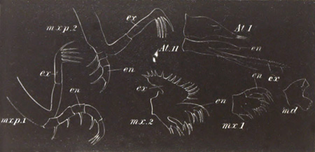
Fig. 223. The Appendages of a Crab Zoæa.
At. I. first antenna; At. II. second antenna; md. mandible (without a palp); mx. 1. first maxilla; mx. 2. second maxilla; mx. 3. third maxilla; mxp. 1. first maxilliped; mxp. 2. second maxilliped.
ex. exopodite; en. endopodite.
Brachyura. All the Brachyura, with the exception of one or more species of land crabs[191], leave the egg in the Zoæa condition, and though there are slight variations of structure, yet on the whole the Crab Zoæa is a very well marked form. Immediately after leaving the egg (fig. 210) it has a somewhat oval shape with a long distinctly segmented abdomen bent underneath the thorax. The cephalo-thoracic shield covers over the front part of the body, and is prolonged into a long frontal spine pointing forwards, and springing from the region between the two eyes; a long dorsal spine pointing backwards; and two lateral spines.
To the under surface of the body are attached the anterior appendages up to the second maxilliped, while the six following pairs of thoracic appendages are either absent or represented only in a very rudimentary form. The abdomen is without appendages.
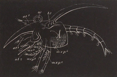
Fig. 224. Crab Zoæa after the third pair of maxillipeds and the thoracic and abdominal appendages have become developed.
at1. antenna of first pair; at2. antenna of second pair; mx1. first maxilla; mx2. second maxilla; mxp1. first maxilliped; mxp2. second maxilliped; mxp3. third maxilliped; oc. eye; ht. heart.
The anterior antennæ are single and unjointed, but provided at their extremity with a few olfactory hairs (only two in Carcinus Mœnas) and one or two bristles. The rudiment of the secondary flagellum appears in very young Zoææ on the inner side of the antennules (fig. 223 At. I.). The posterior antennæ are without the flagellum, but are provided with a scale representing the exopodite (fig. 223 At. II. ex) and usually a spinous process. The flagellum is very early developed and is represented in fig. 223, At. II. en. The mandibles (md) are large but without a palp. The anterior maxillæ (mx 1) have a short two-jointed endopodite (palp) with a few hairs, and a basal portion with two blades, of which the distal is the largest, both armed with stiff bristles. The posterior maxillæ have a small respiratory plate (exopodite), an endopodite (palp) shaped like a double blade, and two basal joints each continued into a double blade. The two maxillipeds (mxp 1 and mxp 2) have the form and function of biramous swimming feet. The exopodite of both is two-jointed and bears long bristles at its extremity; the endopodite of the anterior is five-jointed and long, that of the second is three-jointed and comparatively short.
In the six-jointed tail the second segment has usually two dorsally directed spines, and the three succeeding segments each of them two posteriorly directed. The telson or swimming plate is not at first separated from the sixth segment; on each side it is prolonged into two well-marked prongs; and to each prong three bristles are usually attached (fig. 224). The heart (fig. 224 ht) lies under the dorsal spine and is prolonged into an anterior, posterior, and dorsal aorta. It has only two pairs of venous ostia.
During the Zoæa stage the larva rapidly grows in size, and undergoes considerable changes in its appendages which reach the full Decapod number (fig. 224). On both pairs of antennæ a flagellum becomes developed and grows considerably in length. Before the close of the Zoæa condition a small and unjointed palp appears on the mandible. Behind the second maxilliped the third maxilliped (mxp3) early appears as a small biramous appendage, and the five ambulatory feet become distinctly formed as uniramous appendages—the exopodites not being present. The third pair of maxillipeds and three following ambulatory appendages develop gill pouches. The abdominal feet are formed on the second to the sixth segments of the tail as simple pouches.
The oldest Zoæa is transmuted at its moult into a form known as Megalopa, which is really almost identical with an anomurous Decapod. No Schizopod stage is intercalated, which shews that the development is in many respects greatly abbreviated. The essential characters of the Megalopa are to be found in (1) the reduction of the two anterior maxillipeds, which cease to function as swimming feet, and together with the appendages in front of them assume the adult form; (2) the full functional development of the five ambulatory appendages; (3) the reduction of the forked telson to an oval swimming plate, and the growth in size of the abdominal feet, which become large swimming plates and are at the same time provided with short endopodites which serve to lock the feet of the two sides.
With these essential characters the form of the Megalopa differs considerably in different cases. In some instances (e.g. Carcinus mœnas) the Zoæa spines of the youngest Megalopa are so large that the larva appears almost more like a Zoæa than a Megalopa (Spence Bate, No. 470). In other cases, e.g. that represented on fig. 225, the Zoæa spines are still present but much reduced; and the cephalo-thoracic shield has very much the adult form. In other cases again (e.g. Portunus) the Zoæa spines are completely thrown off at the youngest Megalopa stage.
There is a gradual passage from the youngest Megalopa to the adult form by a series of moults.
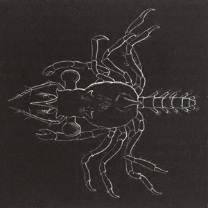
Fig. 225. Megalopa stage of Crab larva.
Some of the brachyurous Zoæa forms exhibit considerable divergences from the described type, more especially in the armature of the shield. In some forms the spines are altogether absent, e.g. Maja (Couch, No. 474) and Eurynome. In other forms the frontal spine may be much reduced or absent (Inachus and Achæus). The dorsal spine may also be absent, and in one form described by Dohrn (No. 478) there is a long frontal spine and two pairs of lateral spines, but no dorsal spine. Both dorsal and frontal spines may attain enormous dimensions and be swollen at their extremities (Dohrn). A form has been described by Claus as Pterocaris in which the cephalo-thoracic shield is laterally expanded into two wing-like processes.
The Zoæa of Porcellana presents on the whole the most remarkable peculiarities and, as might be anticipated from the systematic position of the adult, is in some respects intermediate between the macrurous Zoæa and that of the Brachyura. It is characterized by the oval form of the body, and by the presence of one enormously long frontal spine and two posterior spines. The usual dorsal spine is absent. The tail plate is rounded and has the character of the tail of a macrurous Zoæa, but in the young Zoæa the third pair of maxillipeds is absent and the appendages generally have a brachyurous character. A Megalopa stage is hardly represented, since the adult may almost be regarded as a permanent Megalopa.
Stomatopoda. The history of the larval forms of the Stomatopoda (Squilla etc.) has not unfortunately been thoroughly worked out, but what is known from the researches of Fritz Müller (No. 495) and Claus (No. 494) is of very great importance. There are it appears two types, both of which used to be described as adult forms under the respective names Erichthus and Alima.
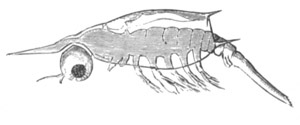
Fig. 226. Second stage of Erichthus larva of Squilla with five maxillipeds and the first pair of abdominal appendages. (From Claus.)
The youngest known Erichthus form is about two millimetres in length, and has the characters of a modified Zoæa (fig. 226). The body is divided into three regions, an anterior unsegmented region to which are attached two pairs of antennæ, mandibles, and maxillæ (two pairs). This portion has a dorsal shield covering the next or middle region, which consists of five segments each with a pair of biramous appendages. These appendages represent the five maxillipeds of the adult[192]. The portion of the body behind this is without appendages. It consists of three short anterior segments,—the three posterior thoracic segments of the adult,—and a long unsegmented tail. The three footless thoracic segments are covered by the dorsal shield. Both pairs of antennæ are uniramous and comparatively short. The mandibles, like those of Phyllopods, are without palps, and the two following pairs of maxillæ are small. The five maxillipeds have the characters of normal biramous Zoæa feet. From the front of the head spring a pair of compound eyes with short stalks, which grow longer in the succeeding stages; between them is a median eye. The dorsal shield is attached just behind this eye, and is provided, as in the typical Zoæa, with a frontal spike—while its hinder border is produced into two lateral spikes and one median. In a larva of about three millimetres a pair of biramous appendages arises behind the three footless thoracic segments. It is the anterior pair of abdominal feet (fig. 226). The inner ramus of the second pair of maxillipeds soon grows greatly in length, indicating its subsequent larger size and prehensile form (fig. 227 g). When the larva after one or two moults attains a length of six millimetres (fig. 227) the abdomen has six segments (the sixth hardly differentiated), each with a pair of appendages (the two hindermost still rudimentary) which have become gradually developed from before backwards. The three hindermost thoracic segments are still without appendages.
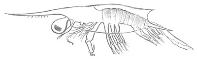
Fig. 227. Advanced Erichthus larva of Squilla with five pairs of abdominal appendages. (From Claus.)
f. first maxilliped; g. second maxilliped.
Some changes of importance have occurred in the other parts. Both antennæ have acquired a second flagellum, but the mandible is still without a palp. The first and second pair of maxillipeds have both undergone important modifications. Their outer ramus (exopodite) has been thrown off, and a gill plate (epipodite) has appeared as an outgrowth from their basal joint. Each of them is composed of six joints. The three following biramous appendages have retained their earlier characters but have become much reduced in size. In the subsequent moults the most remarkable new features concern the three posterior maxillipeds, which undergo atrophy, and are either completely lost or reduced to mere unjointed sacks (fig. 228). In the stage where the complete Erichthus type has been reached, these three appendages have again sprouted forth in their permanent form and each of them is provided with a gill sack on its coxal joint. Behind them the three ambulatory appendages of the thorax have also appeared, first as simple buds, which subsequently however become biramous. On their development the full number of adult appendages is acquired.
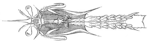
Fig. 228. Advanced Erichthus larva of Squilla when the three posterior maxillipeds have become reduced to minute pouches. (From Claus.)
The most noteworthy points in the developmental history detailed above are the following:
(1) The thoracic and abdominal segments (apart from their appendages) develop successively from before backwards.
(2) The three last maxillipeds develop before the abdominal feet, as biramous appendages, but subsequently completely atrophy, and then sprout out again in their permanent form.
(3) The abdominal feet develop in succession from before backwards, and the whole series of them is fully formed before a trace of the appendages of the three hindermost thoracic segments has appeared. It may be mentioned as a point of some importance that the Zoæa of Squilla has an elongated many-chambered heart, and not the short compact heart usually found in the Zoæa.
The younger stages of the Alima larva are not known[193], but the earliest stage observed is remarkable for presenting no trace of the three posterior pairs of maxillipeds, or of the three following pairs of thoracic appendages. The segments belonging to these appendages are however well developed. The tail has its full complement of segments with the normal number of well-developed swimming feet. The larva represents in fact the stage of the Erichthus larva when the three posterior pairs of maxillipeds have undergone atrophy; but it is probable that these appendages never become developed in this form of larva.
Apart from the above peculiarities the Alima form of larva closely resembles the Erichthus form.
Nebaliadæ. The development of Nebalia is abbreviated, but from Metschnikoff’s figures[194] may be seen to resemble closely that of Mysis. The abdomen has comparatively little yolk, and is bent over the ventral surface of the thorax. There is in the egg a Nauplius stage with three appendages, and subsequently a stage with the Zoæa appendages.
The larva when it leaves the egg has the majority of its appendages formed, but is still enveloped in a larval skin, and like Mysis bends its abdomen towards the dorsal side. When the larva is finally hatched it does not differ greatly from the adult.
Cumaceæ. The development of the Cumaceæ takes place for the most part within the egg, and has been shewn by Dohrn (No. 496) to resemble in many points that of the Isopods. A dorsal organ is present, and a fold is formed immediately behind this which gives to the embryo a dorsal flexure. Both of these features are eminently characteristic of the Isopoda.
The formation of the two pairs of antennæ, mandibles, and two pairs of maxillæ and the following seven pairs of appendages takes place very early. The pair of appendages behind the second maxillæ assumes an ambulatory form, and exhibits a Schizopod character very early, differing in both these respects from the homologous appendages in the Isopoda. The cephalo-thoracic shield commences to be formed when the appendages are still quite rudimentary as a pair of folds in the maxillary region. The eyes are formed slightly later on each side of the head, and only coalesce at a subsequent period to form the peculiar median sessile eye of the adult.
The two pairs of appendages behind the second maxillæ become converted into maxillipeds, and the exopodite of the first of them becomes the main ramus, while in the externally similar second maxilliped the exopodite atrophies and the endopodite alone remains.
The larva is hatched without the last pair of thoracic limbs or the abdominal appendages (which are never developed in the female), but in other respects closely resembles the adult. Before hatching the dorsal flexure is exchanged for a ventral one, and the larva acquires a character more like that of a Decapod.
Copepoda.
Natantia. The free Copepoda are undoubtedly amongst the lowest forms of those Crustacea which are free or do not lead a parasitic existence. Although some features of their anatomy, such for instance as the frequent absence of a heart, may be put down to a retrogressive development, yet, from their retention of the median frontal eye of the Nauplius as the sole organ of vision[195], their simple biramous swimming legs, and other characters, they may claim to be very primitive forms, which have diverged to no great extent from the main line of Crustacean development. They supply a long series of transitional steps from the Nauplius stage to the adult condition.
While still within the egg-shell the embryo is divided by two transverse constrictions into three segments, on which the three Nauplius appendages are developed, viz. the two pairs of antennæ and the mandibles. When the embryo is hatched the indication of a division into segments has vanished, but the larva is in the fullest sense a typical Nauplius[196]. There are slight variations in the shape of the Nauplius in different genera, but its general form and character are very constant. It has (fig. 229 A) an oval unsegmented body with three pairs of appendages springing from the ventral surface. The anterior of these (at 1) is uniramous, and usually formed of three joints which bear bristles on their under surface. The two posterior pairs of appendages are both biramous. The second pair of antennæ (at 2) is the largest. Its basal portion (protopodite) bears on its inner side a powerful hook-like bristle. The outer ramus is the longest and many-jointed; the inner ramus has only two joints. The mandibles (md), though smaller than the second pair of antennæ, have a nearly identical structure. No blade-like projection is as yet developed on their protopodite. Between the points of insertion of the first pair of antennæ is the median eye (oc), which originates by the coalescence of two distinct parts. The mouth is ventral, and placed in the middle line between the second pair of antennæ and the mandibles: it is provided with an unpaired upper lip. There are two bristles at the hind end of the embryo between which the anus is placed; and in some cases there is at this part a slight indication of the future caudal fork.
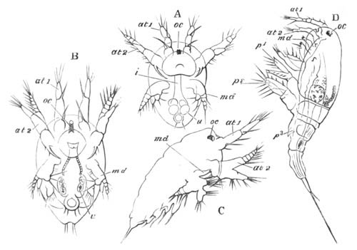
Fig. 229. Successive stages in the development of Cyclops tenuicornis. (Copied from Bronn; after Claus.)
A. B. and C. Nauplius stages. D. Youngest Copepod stage. In this figure maxillæ and the two rami of the maxilliped are seen immediately behind the mandible md. oc. eye; at1. first pair of antennæ; at2. second pair of antennæ; md. mandible; p1. first pair of feet; p2. second pair of feet; p3. third pair of feet; u. excretory concretions in the intestine.
The larva undergoes a number of successive ecdyses, at each of which the body becomes more elongated, and certain other changes take place. First of all a pair of appendages arises behind the mandibles, which form the maxillæ (fig. 229 B); at the same time the basal joint of the maxillæ develops a cutting blade. Three successive pairs of appendages (fig. 229 C) next become formed—the so-called maxillipeds (the homologues of the second pair of maxillæ), and the two first thoracic limbs. Each of these though very rudimentary is nevertheless bifid. The body becomes greatly elongated, and the caudal fork more developed.
Up to this stage of development the Nauplius appendages have retained their primitive character almost unaltered; but after a few more ecdyses a sudden change takes place; a cephalo-thoracic shield becomes fully developed, and the larva comes to resemble in character an adult Copepod, from which it mainly differs in the smaller number of segments and appendages. In the earliest ‘Cyclops’ stage the same number of appendages are present as in the last Nauplius stage. There (fig. 229 D) is a well-developed cephalo-thorax, and four free segments behind it. To the cephalo-thoracic region the antennæ, mandibles, maxillæ, the now double pair of maxillipeds (derived from the original single pair of appendages), and first pair of thoracic appendages (p1) are attached. The second pair of thoracic appendages (p2) is fixed to the first free segment, and the rudiment of a third pair (p3) projects from the second free segment. The first pair of antennæ has grown longer by the addition of new joints, and continues to increase in length in the following ecdyses till it attains its full adult development, and then forms the chief organ of locomotion. The second pair of antennæ is much reduced and has lost one of its rami. The two rami of the mandibles are reduced to a simple palp, while the blade has assumed its full importance. The maxillæ and following appendages have greatly increased in size. They are all biramous, though the two rami are not as yet jointed. The adult state is gradually attained after a number of successive ecdyses, at which new segments and appendages are added, while new joints are formed for those already present.
Parasita. The earliest developmental stages of the parasitic types of Copepoda closely resemble those of the free forms, but, as might be expected from the peculiarly modified forms of the adult, they present a large number of secondary characters. So far as is known a more or less modified Nauplius larva is usually preserved.
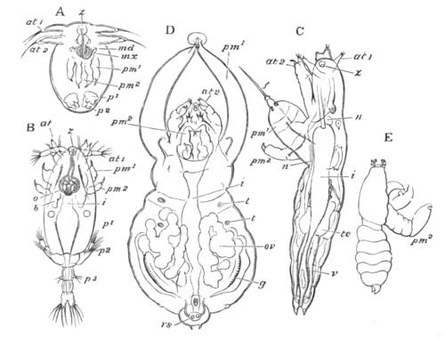
Fig. 230. Successive stages in the development of Achtheres percarum. (Copied from Bronn; after Claus.)
A. Modified Nauplius stage. B. Cyclops stage. C. Late stage of male embryo. D. Sexually mature female. E. Sexually mature male. at1. first pair of antennæ; at2. second pair of antennæ; md. mandible; mx. maxillæ; pm1. outer pair of maxillipeds; pm2. inner pair of maxillipeds; p1. first pair of legs; p2. second pair of legs; z. frontal organ; i. intestine; o. larval eye; b. glandular body; t. organ of touch; ov. ovary; f. rod projecting from coalesced maxillipeds; g. cement gland; rs. receptaculum seminis; n. nervous system; te. testis; v. vas deferens.
The development of Achtheres percarum, one of the Lernæopoda parasitic in the mouth, etc. of the common Perch, may be selected to illustrate the mode of development of these forms. The larva leaves the egg as a much simplified Nauplius (fig. 230 A). It has an oval body with only the two anterior pairs of Nauplius appendages; both of them in the rudimentary condition of unjointed rods. The usual median eye is present, and there is also found a peculiar sternal papilla, on which opens a spiral canal filled with a glutinous material, which is probably derived from a gland which disappears on the completion of the duct. The probable function of this organ is to assist at a later period in the attachment of the parasite to its host. Underneath the Nauplius skin a number of appendages are visible, which become functional after the first ecdysis. This takes place within a few hours after the hatching of the Nauplius, and the larva then passes from this rudimentary Nauplius stage into a stage corresponding with the Cyclops stage of the free forms (fig. 230 B). In the Cyclops stage the larva has an elongated body with a large cephalo-thoracic shield, and four free posterior segments, the last of which bears a forked tail.
There are now present eight pairs of appendages, viz. antennæ (two pairs), mandibles, maxillæ, maxillipeds, and three pairs of swimming feet. The Nauplius appendages are greatly modified. The first pair of antennæ is three-jointed, and the second biramous. The outer ramus is the longest, and bears a claw-like bristle at its extremity. This pair of appendages is used by the larva for fixing itself. The mandibles are small and connected with the proboscidiform mouth; and the single pair of maxillæ is small and palped. The maxillipeds (pm1 and pm2) are believed by Claus to be primitively a single biramous appendage, but early appear as two distinct structures[197], the outer and larger of which becomes the main organ by which the larva is fixed. Both are at this stage simple two-jointed appendages. The two anterior pairs of swimming feet have the typical structure, and consist of a protopodite bearing an unjointed exopodite and endopodite. The first pair is attached to the cephalo-thorax and the second (p2) to the first free thoracic segment. The third pair is very small and attached to the second free segment. The mouth is situated at the end of a kind of proboscis formed by prolongations of the upper and lower lips. The alimentary tract is fairly simple, and the anus opens between the caudal forks.
Between this and the next known stage it is quite possible that one or more may intervene. However this may be the larva in the next stage observed (fig. 230 C) has already become parasitic in the mouth of the Perch, and has acquired an elongated vermiform aspect. The body is divided into two sections, an anterior unsegmented, and a posterior formed of five segments, of which the foremost is the first thoracic segment which in the earlier stage was fused with the cephalo-thorax. The tail bears a rudimentary fork between the prongs of which the anus opens. The swimming feet have disappeared, so also has the eye and the spiral duct of the embryonic frontal organ. The outer of the two divisions of the maxilliped have undergone the most important modification, in that they have become united at their ends, where they form an organ from which an elongated rod (f) projects, and attaches the larva to the mouth or gills of its host. The antennæ and jaws have nearly acquired their adult form. The nervous system consists of supra- and infra-œsophageal ganglia and two lateral trunks given off from the latter. At this stage the males and females can already be distinguished, not only by certain differences in the rudimentary generative organs, but also by the fact that the outer branch of the maxillipeds is much longer in the female than in the male, and projects beyond the head.
In the next ecdysis the adult condition is reached. The outer maxillipeds of the male (fig. 230 E, pm2) separate again; while in the female (fig. 230 D) they remain fused and develop a sucker. The male is only about one-fifth the length of the female. In both sexes the abdomen is much reduced.
In the genera Anchorella, Lernæopoda, Brachiella and Hessia, Ed. van Beneden (No. 506) has shewn that the embryo, although it passes through a crypto-Nauplius stage in the egg, is when hatched already in the Cyclops stage.
Branchiura. The peculiar parasite Argulus, the affinities of which with the Copepoda have been demonstrated by Claus (No. 511), is hatched in a Cyclops stage, and has no Nauplius stage. At the time of hatching it closely resembles the adult in general form. Its appendages are however very nearly those of a typical larval Copepod. The body is composed of a cephalo-thorax and free region behind this. The cephalo-thorax bears on its under surface antennæ (two pairs), mandibles, maxillipeds, and the first pair of thoracic feet.
The first pair of antennæ is three-jointed, but the basal joint bears a hook. The second pair is biramous, the inner ramus terminating in a hook. The mandible is palped, but the palp is completely separated from the cutting blade[198]. The maxilla would, according to Claus, appear to be absent.
The two typical divisions of the Copepod maxillipeds are present, viz. an outer and anterior larger division, and an inner and posterior smaller one. The first pair of thoracic feet, as is usual amongst Copepoda, is attached to the cephalo-thorax. It has not the typical biramous Copepod character. There are four free segments behind the cephalo-thorax, the last of which ends in a fork. Three of them bear appendages, which are rudimentary in this early larval stage. On the dorsal surface are present paired eyes as well as an unpaired median eye.
Between the larval condition and that of the adult a number of ecdyses intervene.
Cirripedia.
The larvæ of all the Cirripedia, with one or two exceptions, leave the egg in the Nauplius condition. The Nauplii differ somewhat in the separate groups, and the post-nauplial stages vary not inconsiderably.
It will be most convenient to treat successively the larval history of the four sub-orders, viz. Thoracica, Abdominalia, Apoda, and Rhizocephala.
Thoracica. The just hatched larvæ at once leave the egg lamellæ of their parent. They pass out through an opening in the mantle near the mouth, and during this passage the shell of the parent is opened and the movements of the cirriform feet cease.
The larval stages commence with a Nauplius[199] which, though regarded by Claus as closely resembling the Copepod Nauplius (figs. 231 and 232 A), certainly has very marked peculiarities of its own, and in some respects approaches the Phyllopod Nauplius. It is in the youngest stage somewhat triangular in form, and covered on the dorsal side by a very delicate and hardly perceptible dorsal shield, which is prolonged laterally into two very peculiar conical horns (fig. 231 lh), which are the most characteristic structures of the Cirriped Nauplius. They are connected with a glandular mass, the secretion from which passes out at their apex. Anteriorly the dorsal shield has the same extension as the body, but posteriorly it projects slightly.
An unpaired eye is situated on the ventral surface of the head, and immediately behind it there springs a more or less considerable upper lip (lb), which resembles the Phyllopod labrum rather than that of the Copepoda. Both mouth and anus are present, and the hind end of the body is slightly forked in some forms, but ends in others, e.g. Lepas fascicularis, in an elongated spine. The anterior of the three pairs of Nauplius appendages (At1) is uniramous, and the two posterior (At2, and md) are biramous. From the protopodites of both the latter spring strong hooks like those of the Copepod and Phyllopod Nauplii. In some Nauplii, e.g. that of Balanus, the appendages are at first not jointed, but in other Nauplii, e.g. that of Lepas fascicularis, the jointing is well marked. In Lepas fascicularis the earliest free Nauplius is enveloped in a larval skin, which is thrown off after a few hours. The Nauplii of all the Thoracica undergo a considerable number of moults before their appendages increase in number or segmentation of the body appears. During these moults they grow larger, and the posterior part of the body—the future thoracic and abdominal region—grows relatively in length. There also appear close to the sides of the unpaired eye two conical bodies, which correspond with the frontal sense organs of the Phyllopods. During their growth the different larvæ undergo changes varying greatly in degree.
In Balanus the changes consist for the most part in the full segmentation of the appendages and the growth and distinctness of the dorsal shield, which forms a somewhat blunt triangular plate, broadest in front, with the anterior horns very long, and two short posterior spines. The tail also becomes produced into a long spine.
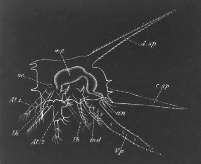
Fig. 231. Nauplius larva of Lepas fascicularis viewed from the side.
oc. eye; At. 1. antenna of first pair; At. 2. antenna of second pair; md. mandible; lb. labrum; an. anus; me. mesenteron; d.sp. dorsal spine; c.sp. caudal spine; Vp. ventral spine; lh. lateral horns.
In Lepas fascicularis the changes in appearance of the Nauplius, owing to a great spinous development on its shield, are very considerable; and, together with its enormous size, render it a very remarkable form. Dohrn (No. 520), who was the first to describe it, named it Archizoæa gigas.
The dorsal shield of the Nauplius of Lepas fascicularis (fig. 231) becomes somewhat hexagonal, and there springs from the middle of the dorsal surface an enormously long spine (d.sp), like the dorsal spine of a Zoæa. The hind end of the shield is also produced into a long caudal spine (c.sp) between which and the dorsal spine are some feather-like processes. From its edge there spring in addition to the primitive frontal horns three main pairs of horns, one pair anterior, one lateral, and one posterior, and smaller ones in addition. All these processes (with the exception of the dorsal and posterior spines) are hollow and open at their extremities, and like the primitive frontal horns contain the ducts of glands situated under the shield. On the under surface of the larva is situated the unpaired eye (oc) on each side of which spring the two-jointed frontal sense organs. Immediately behind these is the enormous upper lip (lb) which covers the mouth[200]. At the sides of the lip lie the three pairs of Nauplius appendages, which are very characteristic but present no special peculiarities. Posteriorly the body is produced into a long ventral spine-like process (Vp) homologous with that of other more normal Nauplii. At the base of this process large moveable paired spines appear at successive moults, six pairs being eventually formed. These spines give to the region in which they are situated a segmented appearance, and perhaps similar structures have given rise to the appearance of segmentation in Spence Bate’s figures. The anus is situated on the dorsal side of this ventral process, and between it and the caudal spine of the shield above. The fact that the anus occupies this position appears to indicate that the ventral process is homologous with the caudal fork of the Copepoda, on the dorsal side of which the anus so often opens[201].
From the Nauplius condition the larvæ pass at a single moult into an entirely different condition known as the Cypris stage. In preparation for this condition there appear, during the last Nauplius moults, the rudiments of several fresh organs, which are more or less developed in different types. In the first place a compound eye is formed on each side of the median eye. Secondly there appears behind the mandibles a fourth pair of appendages—the first pair of maxillæ—and internal to these a pair of small prominences, which are perhaps equivalent to the second pair of maxillæ, and give rise to the third pair of jaws in the adult (sometimes spoken of as the lower lip).
Behind these appendages there are moreover formed the rudiments of six pairs of feet. Under the cuticle of the first pair of antennæ there may be seen just before the final moult the four-jointed antennæ of the Cypris stage with the rudiment of a disc on the second joint by which the larvæ eventually become attached.
By the free Cypris stage, into which the larva next passes, a very complete metamorphosis has been effected. The median and paired eyes are present as before, but the dorsal shield has become a bivalve shell, the two valves of which are united along their dorsal, anterior, and posterior margins. The two valves are further kept in place by an adductor muscle situated close below the mouth. Remains of the lateral horns still persist. The anterior antennæ have undergone the metamorphosis already indicated. They are four-jointed, the two basal joints being long, and the second provided with a suctorial disc, in the centre of which is the opening of the duct of the so-called antennary or cement gland, which is a granular mass lying on the ventral side of the anterior region of the body. The gland arises (Willemoes Suhm) during the Nauplius stage in the large upper lip. The two distal joints of the antennæ are short, and the last of them is provided with olfactory hairs. The great upper lip and second pair of antennæ and mandibles have disappeared, but a small papilla, forming the commencement of the adult mandibles, is perhaps developed in the base of the Nauplius mandibles. The first pair of maxillæ have become small papillæ and the second pair probably remain. The six posterior pairs of appendages have grown out as functional biramous swimming feet, which can project beyond the shell and are used in the locomotion of the larva. They are composed of two basal joints, and two rami with swimming hairs, each two-jointed. These feet resemble Copepod feet, and form the main ground for the views of Claus and others that the Copepoda and Cirripedia are closely related. They are regarded by Claus as representing the five pairs of natatory feet of Copepoda, and the generative appendages of the segment behind these. Between the natatory feet are delicate chitinous lamellæ, in the spaces between which the cirriform feet of the adult become developed. The ventral spinous process of the Nauplius stage is much reduced, though usually three-jointed. It becomes completely aborted after the larva is fixed.
In addition to the antennary gland there is present, near the dorsal side of the body above the natatory feet, a peculiar paired glandular mass, the origin of which has not been clearly made out, but which is perhaps equivalent to the entomostracan shell-gland. It probably supplies the material for the shell in succeeding stages[202].
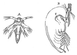
Fig. 232. Larval forms of the Thoracica. (From Huxley.)
A. Nauplius of Balanus balanoides. (After Sp. Bate.) B. Pupa stage of Lepas australis. (After Darwin.) n. antennary apodemes; t. cement gland with duct to antenna.
The free Cypris stage is not of long duration; and during it the larva does not take food. It is succeeded by a stage known as the pupa stage (fig. 232 B), in which the larva becomes fixed, while underneath the larval skin the adult structures are developed. This stage fully deserves its name, since it is a quiescent stage during which no nutriment is taken. The attachment takes place by the sucker of the antennæ, and the cement gland (t) supplies the cementing material for effecting it. A retrogressive metamorphosis of a large number of the organs sets in, while at the same time the formation of new adult structures is proceeded with. The eyes become gradually lost, but the Nauplius eye is retained, though in a rudimentary state, and the terminal joints of the antennæ with their olfactory hairs are thrown off. The bivalve shell is moulted about the same time as the eyes, the skin below it remaining as the mantle. The caudal process becomes aborted. Underneath the natatory feet, and between the above-mentioned chitinous lamellæ, the cirriform feet are formed; and on their completion the natatory feet become thrown off and replaced by the permanent feet. In the Lepadidæ, in which the metamorphosis of the pupa stages has been most fully studied, the anterior part of the body with the antennæ gradually grows out into an elongated stalk, into which pass the ovaries, which are formed during the Cypris stage. At the base of the stalk is the protuberant mouth, the appendages of which soon attain a higher development than in the Cypris stage. At the front part of it a large upper lip becomes formed. Above the mantle and between it and the shell there are formed in the Lepadidæ the provisional valves of the shell. These valves are chitinous, and have a fenestrated structure, owing to the chitin being deposited round the margin of the separate epidermis (hypodermis) cells. These valves in the Lepadidæ “prefigure in shape, size, and direction of growth, the shelly valves to be formed under and around them” (Darwin, No. 519, p. 129).
Whatever may be the number of valves in the adult the provisional valves never exceed five, viz. the two scuta, the two terga and the carina. They are relatively far smaller than the permanent valves and are therefore separated by considerable membranous intervals. They are often preserved for a long time on the permanent calcareous valves. In the Balanidæ the embryonic valves are membranous and do not overlap, but do not present the peculiar fenestrated structure of the primordial valves of the Lepadidæ.
In connection with the moult of the pupa skin, and the conversion of the pupa into the adult form, a remarkable change in the position takes place. The pupa lies with the ventral side parallel to and adjoining the surface of attachment, while the long axis of the body of the young Cirriped is placed nearly at right angles to the surface of attachment. This change is connected with the ecdyses of the antennary apodemes (n), which leave a deep bay on the ventral surface behind the peduncle. The chitinous skin of the Cirriped passes round the head of this bay, but on the moult of the pupa skin taking place becomes stretched out, owing to the posterior part of the larva bending dorsalwards. It is this flexure which causes the change in the position of the larva.
In addition to the remarkable external metamorphosis undergone during the pupa stage, a series of hardly less considerable internal changes take place, such as the atrophy of the muscles of the antennæ, a change in the position of the stomach, etc.
Abdominalia. In the Alcippidæ the larva leaves the egg as a Nauplius, and this stage is eventually followed by a pupa stage closely resembling that of the Thoracica. There are six pairs of thoracic natatory legs (Darwin, No. 519). Of these only the first and the last three are preserved in the adult, the first being bent forward in connection with the mouth. The body moreover partially preserves its segmentation, and the mantle does not secrete calcareous valves.
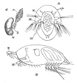
Fig. 233. Stages in the development of the Rhizocephala. (From Huxley, after Fritz Müller.)
A. Nauplius of Sacculina purpurea. B. Cypris stage of Lernæodiscus porcellanæ. C. Adult of Peltogaster paguri. II. III. IV. Two pairs of antennæ and mandibles; cp. carapace; a. anterior end of body; b. generative aperture; c. root-like processes.
The very remarkable genus Cryptophialus, the development of which is described by Darwin (No. 519) in his classical memoir, is without a free Nauplius stage. The embryo is at first oval but soon acquires two anterior processes, apparently the first pair of antennæ, and a posterior prominence, the abdomen. In a later stage the abdominal prominence disappears, and the antennary processes, within which the true antennæ are now visible, are carried more towards the ventral surface. The larva next passes into the free Cypris stage, during which it creeps about the mantle cavity of its parent. It is enveloped in a bivalve shell, and the antennæ have the normal cirriped structure. There are no other true appendages, but posteriorly three pairs of bristles are attached to a rudimentary abdomen. Paired compound eyes are present. During the succeeding pupa stage the metamorphosis into the adult form takes place, but this has not been followed out in detail.
In Kochlorine, a form discovered by Noll (No. 526) and closely related to Cryptophialus, the larvæ found within the mantle represent apparently two larval stages, similar to two of the larval stages described by Darwin.
Rhizocephala. The Rhizocephala, as might have been anticipated from their close relationship to Anelasma squalicola amongst the Thoracica, undergo a development differing much less from the type of the Thoracica than that of Cryptophialus and Kochlorine.
Sacculina leaves the egg as a Nauplius (fig. 233 A), which differs from the ordinary type mainly (1) in the large development of an oval dorsal shield (cp) which projects far beyond the edge of the body, but is provided with the typical sternal horns, etc.; and (2) in the absence of a mouth. The Cypris and pupa stages of Sacculina and other Rhizocephala (fig. 233 B) are closely similar to those of the Thoracica, but the paired eye is absent. The attachment takes place in the usual way, but the subsequent metamorphosis leads to the loss of the thoracic feet and generally to retrogressive changes.
Ostracoda.
Our knowledge of the development of this remarkable group is entirely due to the investigations of Claus.
Some forms of Cythere are viviparous, and in the marine form Cypridina the embryo develops within the valves of the shell. Cypris attaches its eggs to water plants. The larvæ of Cypris are free, and their development is somewhat complicated. The whole development is completed in nine ecdyses, each of them accompanied by more or less important changes in the constitution of the larva.
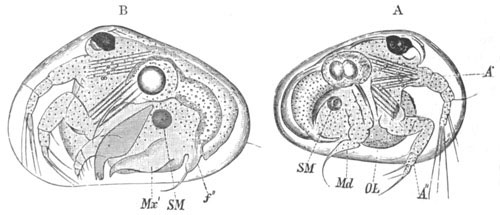
Fig. 234. Two stages in the development of Cypris. (From Claus.) A. Earliest (Nauplius) stage. B. Second stage.
A´. A´´. First and second pairs of antennæ; Md. mandibles; OL. labrum; Mx´. first pair of maxillæ; f´´. first pair of feet.
In the earliest free stage the larva has the characters of a true Nauplius with three pairs of appendages (fig. 234 A). The Nauplius presents however one or two very marked secondary characters. In the first place it is completely enveloped in a fully formed bivalve shell, differing in unessential points from the shell of the adult. An adductor muscle (SM) for the shell is present. Again the second and third appendages, though locomotive in function are neither of them biramous, and the third one already contains a rudiment of the future mandibular blade, and terminates in an anteriorly directed hook-like bristle. The first pair of antennæ is moreover very similar to the second and is used in progression. Neither of the pairs of antennæ become much modified in the subsequent metamorphosis. The Nauplius has a single median eye, as in the adult Cypris, and a fully developed alimentary tract.
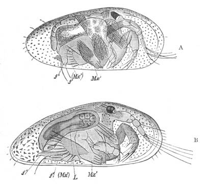
Fig. 235. Stages in the development of Cypris. (From Claus.) A. Fourth stage. B. Fifth stage.
Mx´. first maxilla; Mx´´. and f´. second maxilla; f´´. first pair of feet; L. liver.
The second stage (fig. 234 B), inaugurated by the first moult, is mainly characterized by the appearance of two fresh pairs of appendages, viz. the first pair of maxillæ and the first pair of feet; the second pair of maxillæ not being developed till later. The first pair appear as leaf-like curved plates (Mx´) more or less like Phyllopod appendages (Claus) though at this stage without an exopodite. The first pair of feet (f´´) terminates in a curved claw and is used for adhering. The mandibles have by this stage fully developed blades, and have practically attained their adult form, consisting of a powerful toothed blade and a four-jointed palp.
During the third and fourth stages the first pair of maxillæ acquire their pectinated gill plate (epipodite) and four blades; and in the fourth stage (fig. 235 A) the second pair of maxillæ (Mx´´) arises, as a pair of curved plates, similar to the first pair of maxillæ at their first appearance. The forked tail is indicated during the fourth stage by two bristles. During the fifth stage (fig. 235 B) the number of joints of the first pair of antennæ becomes increased, and the posterior maxillæ develop a blade and become four-jointed ambulatory appendages terminating in a hook. The caudal fork becomes more distinct.
In the sixth stage (fig. 236) the second and hindermost pair of feet becomes formed (f´´´) and the maxillæ of the second pair lose their ambulatory function, and begin to be converted into definite masticatory appendages by the reduced jointing of their palp, and the increase of their cutting blades. By the seventh stage all the appendages have practically attained their permanent form; the second pair of maxillæ has acquired small branchial plates, and the two following feet have become jointed. In the eighth and ninth stages the generative organs attain their mature form.
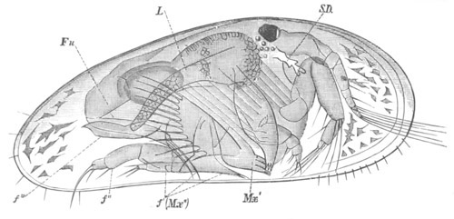
Fig. 236. Sixth stage in the development of Cypris. (From Claus.)
Mx´. first maxilla; Mx´´. f´. second maxilla; f´´. and f´´´. first and second pair of feet; Fu. caudal fork; L. liver; S.D. shell-gland.
The larva of Cythere at the time of birth has rudiments of all the limbs, but the mandibular palp still functions as a limb, and the three feet (2nd pair of maxillæ and two following appendages) are very rudimentary.
The larvæ of Cypridina are hatched in a condition which to all intents and purposes resembles the adult.
Phylogeny of the Crustacea.
The classical work of Fritz Müller (No. 452) on the phylogeny of the Crustacea has given a great impetus to the study of their larval forms, and the interpretations of these forms which he has offered have been the subject of a very large amount of criticism and discussion. A great step forward in this discussion has been recently made by Claus in his memoir (No. 448).
The most fundamental question concerns the meaning of the Nauplius. Is the Nauplius the ancestral form of the Crustacea, as is believed by Fritz Müller and Claus, or are its peculiarities and constant occurrence due to some other cause? The most plausible explanation on the second hypothesis would seem to be the following. The segments with their appendages of Arthropoda and Annelida are normally formed from before backwards, therefore every member of these two groups with more than three segments must necessarily pass through a stage with only three segments, and the fact that in a particular group this stage is often reached on the larva being hatched is in itself no proof that the ancestor of the group had only three segments with their appendages. This explanation appears to me, so far as it goes, quite valid; but though it relieves us from the necessity of supposing that the primitive Crustacea had only three pairs of appendages, it does not explain several other peculiarities of the Nauplius[203]. The more important of these are the following.
1. That the mandibles have the form of biramous swimming feet and are not provided with a cutting blade.
2. That the second pair of antennæ are biramous swimming feet with a hook used in mastication, and are innervated (?) from the subœsophageal ganglion.
3. The absence of segmentation in the Nauplius body. An absence which is the more striking in that before the Nauplius stage is fully reached the body of the embryo is frequently divided into three segments, e.g. Copepoda and Cirripedia.
4. The absence of a heart.
5. The presence of a median single eye as the sole organ of vision.
Of these points the first, second, and fifth appear only to be capable of being explained phylogenetically, while with reference to the absence of a heart it appears very improbable that the ancestral Crustacea were without a central organ of circulation. If the above positions are accepted the conclusion would seem to follow that in a certain sense the Nauplius is an ancestral form—but that, while it no doubt had its three anterior pairs of appendages similar to those of existing Nauplii, it may perhaps have been provided with a segmented body behind provided with simple biramous appendages. A heart and cephalo-thoracic shield may also have been present, though the existence of the latter is perhaps doubtful. There was no doubt a median single eye, but it is difficult to decide whether or no paired compound eyes were also present. The tail ended in a fork between the prongs of which the anus opened; and the mouth was protected by a large upper lip. In fact, it may very probably turn out that the most primitive Crustacea more resembled an Apus larva at the moult immediately before the appendages lose their Nauplius characters (fig. 208 B), or a Cyclops larva just before the Cyclops stage (fig. 229), than the earliest Nauplius of either of these forms.
If the Nauplius ancestor thus reconstructed is admitted to have existed, the next question in the phylogeny of the Crustacea concerns the relations of the various phyla to the Nauplius. Are the different phyla descended from the Nauplius direct, or have they branched at a later period from some central stem? It is perhaps hardly possible as yet to give a full and satisfactory answer to this question, which requires to be dealt with for each separate phylum; but it may probably be safely maintained that the existing Phyllopods are members of a group which was previously much larger, and the most central of all the Crustacean groups; and which more nearly retains in the characters of the second pair of antennæ etc. the Nauplius peculiarities. This view is shared both by Claus and Dohrn, and appears to be in accordance with all the evidence we have both palæontological and morphological. Claus indeed carries this view still further, and believes that the later Nauplius stages of the different Entomostracan groups and the Malacostraca (Penæus larva) exhibit undoubted Phyllopod affinities. He therefore postulates the earlier existence of a Protophyllopod form, which would correspond very closely with the Nauplius as reconstructed above, from which he believes all the Crustacean groups to have diverged.
It is beyond the scope of this work to attempt to grapple with all the difficulties which arise in connection with the origin and relationships of the various phyla, but I confine myself to a few suggestions arising out of the developmental histories recorded above.
Malacostraca. In attempting to reconstitute from the evidence in our possession the ancestral history of the Malacostraca we may omit from consideration the larval history of all those types which leave the egg in nearly the adult form, and confine our attention to those types in which the larval history is most completely preserved.
There are three forms which are of special value in this respect, viz. Euphausia, Penæus and Squilla. From the history of these which has already been given it appears that in the case of the Decapoda four stages (Claus) may be traced in the best preserved larval histories.
1. A Nauplius stage with the usual Nauplius characters.
2. A Protozoæa stage in which the maxillæ and first pair of maxillipeds are formed behind the Nauplius appendages; but in which the tail is still unsegmented. This stage is comparatively rarely preserved and usually not very well marked.
3. A Zoæa stage the chief features of which have already been fully characterised (vide p. 465). Three more or less distinct types of Zoæa are distinguished by Claus. (a) That of Penæus, in which the appendages up to the third pair of maxillipeds are formed, and the thorax and abdomen are segmented, the former being however very short. The heart is oval, with one pair of ostia. From this type the Zoæa forms of the other Decapoda are believed by Claus to be derived. (b) That of Euphausia, with but one pair of maxillipeds and those short and Phyllopod-like. The heart oval with one pair of ostia. (c) That of Squilla, with an elongated many-chambered heart, two pairs of maxillipeds and the abdominal appendages in full activity.
4. A Mysis stage, which is only found in the macrurous Decapod larvæ.
The embryological questions requiring to be settled concern the value of the above stages. Do they represent stages in the actual evolution of the present types, or have their characters been secondarily acquired in larval life?
With reference to the first stage this question has already been discussed, and the conclusion arrived at, that the Nauplius does in a much modified form represent an ancestral type. As to the fourth stage there can be no doubt that it is also ancestral, considering that it is almost the repetition of an actually existing form.
The second stage can clearly only be regarded as an embryonic preparation for the third; and the great difficulty concerns the third stage.
The natural view is that this stage like the others has an ancestral value, and this view was originally put forward by Fritz Müller and has been argued for also by Dohrn. On the other hand the opposite side has been taken by Claus, who has dealt with the question very ably and at great length, and has clearly shewn that some of Fritz Müller’s positions are untenable. Though Claus’ opinion is entitled to very great weight, an answer can perhaps be given to some of his objections. The view adopted in this section can best be explained by setting forth the chief points which Claus urges against Fritz Müller’s view.
The primary question which needs to be settled is whether the Malacostraca have diverged very early from the Nauplius root, or later in the history of the Crustacea from the Phyllopod stem. On this question Claus[204] brings arguments, which appear to me very conclusive, to shew that the Malacostraca are derived from a late Protophyllopod type, and Claus’ view on this point is shared also by Dohrn. The Phyllopoda present so many characters (not possessed by the Nauplius) in common with the Malacostraca or their larval forms, that it is incredible that the whole of these should have originated independently in the two groups. The more important of these characters are the following.
1. The compound eyes, so often stalked in both groups.
2. The absence of a palp on the mandible, a very marked character of the Zoæa as well as of the Phyllopoda.
3. The presence of a pair of frontal sense knobs.
4. The Phyllopod character of many of the appendages. Cf. first pair of maxillipeds of the Euphausia Zoæa.
5. The presence of gill pouches (epipodites) on many of the appendages[205].
In addition to these points, to which others might be added, Claus attempts to shew that Nebalia must be regarded as a type intermediate between the Phyllopods and Malacostraca. This view seems fairly established, and if true is conclusive in favour of the Phyllopod origin of the Malacostraca. If the Protophyllopod origin of the Malacostraca is admitted, it seems clear that the ancestral forms of the Malacostraca must have developed their segments regularly from before backwards, and been provided with nearly similar appendages on all the segments. This however is far from the case in existing Malacostraca, and Fritz Müller commences his summary of the characters of the Zoæa in the following words[206]. “The middle body with its appendages, those five pairs of feet to which these animals owe their name, is either entirely wanting or scarcely indicated.” This he regards as an ancestral character of the Malacostraca, and is of opinion that their thorax is to be regarded as a later acquirement than the head or abdomen. Claus’ answer on this point is that in the most primitive Zoææ, viz. those already spoken of as types, the thoracic and abdominal segments actually develop in regular succession from before backwards, and he therefore concludes that the late development of the thorax in the majority of Zoæa forms is secondary and not an ancestral Phyllopod peculiarity.