THE INTERNAL PARASITES OF RATS AND MICE IN THEIR RELATION TO DISEASES OF MAN.
Reading edition
By Ch. Wardell Stiles, Chief, and Charles G. Crane, B. S., Assistant, Division of Zoology, Hygienic Laboratory, United States Public Health and Marine-Hospital Service.
SUMMARY.
Rats and mice may harbor 11 species of internal parasites which come into consideration as possible or established parasites of man. From this point of view, 7 of the parasites are of more academic interest than practical importance. The rat may, however, be viewed as the practical, theoretical, and permanent reservoir for one zooparasitic disease (trichinosis) of considerable importance, and of at least one, perhaps two, other zooparasitic infections (“Lamblia duodenalis” and Hymenolepis diminuta) of much less importance. Its possible future rôle in connection with sleeping sickness should not be entirely ignored.
From the standpoint of internal zooparasitism, therefore, the present public health interest in rats and mice centers in trichinosis. This disease will probably never be eradicated from man until rats and mice are practically eradicated, and any rational public health campaign directed against trichinosis must take the rat into serious consideration.
The eradication of rats and mice would be a very substantial contribution toward a reduction and eradication of trichinosis.
INTRODUCTION.
From the habits of rats, it is to be expected that they harbor many species of parasites, and on account of their presence in our houses the question naturally arises as to whether any of these parasites are transmissible, either directly or indirectly, to man.
The species of internal parasites which come especially into consideration in this connection are the following:
Protozoa: Chlamydophrys enchelys (p. 88), Lamblia duodenalis (p. 89), Trypanosoma gambiense (p. 94).
Trematoda: None.
Cestoda: Cysticercus cellulosæ (p. 95), C. fasciolaris (p. 96), C. pisiformis (p. 95), H. murina Duj. [= fraterna] (p. 96), Hymenolepis diminuta (p. 98).
Nematoda: Trichinella spiralis (p. 101).
Acanthocephala: Gigantorhynchus moniliformis (p. 108).
Arachnoidea: Linguatula denticulata (p. 110).
Of these 11 species, the trichina worm (sometimes called the flesh worm) exceeds all the others combined, both in frequency and importance, as a cause of disease in man.
88
PROTOZOA.
Genus CHLAMYDOPHRYS[U] Cienkowski, 1876.
Species CHLAMYDOPHRYS ENCHELYS[V] (Ehrenberg.)
A very peculiar organism has been described under the name of Leydenia gemmipara Schaudinn, 1896. This was found in fluid, obtained by puncture, from two ascites patients in Berlin, Germany. More recently Schaudinn has concluded that Leydenia gemmipara represents an abnormal condition of a protozoon known as Chlamydophrys. The latter passes through the intestinal tract of various animals (as man, mice, squirrels, rabbits, cattle), and thus is occasionally found in fresh human stools. According to Schaudinn, if pathological conditions in the colon cause an alkaline reaction of its entire content, the usual shell formation in Chlamydophrys fails to take place, the organisms then multiply in an atypical manner by division and budding, and the result is the structure described as Leydenia gemmipara.
Genus LAMBLIA[W] R. Blanchard, 1888.
Generic diagnosis.—Polymastigidæ: Body bilaterally symmetrical, pyriform, excavate antero-ventrally to form a sucker; flagella directed posteriorly; 3 pairs inserted on margin of the sucker, 1 pair at posterior end of body. Parasitic in intestine of mammals.
Type species.—Lamblia duodenalis s. l. (“L. intestinalis” of man).
Flagellate protozoa belonging to this genus are reported as parasitic in the intestinal canal of various species of mammals. At present the forms in question are usually looked upon as belonging to the species L. duodenalis. Evidence is, however, accumulating (p. 92) to the effect that there are at least three distinct species of Lamblia (“L. intestinalis” of man, L. muris of mice, and L. cuniculi (or duodenalis?) of rabbits). Admitting that there may be three species, the intertransmissibility of these forms from one host to another remains to be investigated to some extent. It seems thus far definitely proved that the form which occurs in man is transmissible to mice, rabbits, and guinea pigs, hence mice still remain a source of danger in respect to the infection in man. To exactly what extent this fact is of academic interest or of practical significance is at present sub judice.
89
Species LAMBLIA DUODENALIS[X] (Davaine, 1875) Stiles, 1902, s. l.
[Figs. 9 to 15.]
Specific diagnosis.—Lamblia (p. 88): Body pyriform, 5 to 16μ (21μ Lambl) long, 4 to 12.5μ (8.6 to 11μ Lambl) broad; flagella 9 to 14μ long; anterior end bluntly rounded, posterior end sharply pointed, dorsum convex, antero-ventrally concave, venter flat to convex; antero-ventral concavity forms a sucker, the margins of which project from the surface and are contractile. Four pairs of ventral posteriorly directed flagella, arranged as follows: 1 pair insert on anterior margin of sucker; 2 pairs on posterior margin of sucker, near median line; 1 pair on posterior extremity. Body membrane (“cuticula”) very delicate, permitting some change of body form; protoplasm finely granular; nucleus dumb-bell shaped, pre-equatorial. Vacuoles not observed. Copulation sucker-to-sucker, followed by an encystation, in which stage complicated nuclear changes occur; cysts 10 by 7μ.
Habitat.—Upper portion of small intestine of man (Homo); also of the common house mouse (Mus musculus), the brown rat (M. decumanus), the black rat (M. rattus), “Mus sylvestris” [=? M. decumanus], field mouse (Microtus arvalis), water mole (Arvicola amphibius), rabbits, guinea pigs, domesticated cats, dogs, and sheep.
Geographical distribution.—Europe, Egypt, and United States.
This parasite is very common in animals in certain parts of Europe, and cases of its presence in man have been reported by a number of authors (Lambl, 1859; Grassi, 1881; Perroncito, 1888; Moritz, 1891; Moritz & Holzl, 1892; Roos, 1893; Kruse & Pasquale, 1894; Piccardi, 1895; Sievers; Mueller; Frshezjesski & Ucke; Stiles, 1902; Braun, 1908; etc.). The indications are that it is more common in man than is generally assumed.
Possibly man becomes infected through eating food (as bread, etc.) which has been soiled by the excrements (containing the encysted stage) of mice and rats. Grassi infected himself, Perroncito infected mice and rabbits, and Stiles infected guinea pigs by feeding to them human feces containing the encysted stage.
The parasite may be present in large numbers. Moritz estimated a discharge of 18 milliards within twenty-four hours from one of his patients. It has been observed in healthy persons and also in cases of various diseases, but especially in children and in cases of tuberculosis. It is an inhabitant chiefly of the duodenum and jejunum, where it attaches itself (fig. 13) by means of the sucker to the epithelial cells. It is rarer in the ileum. In case the stomach is alkaline (carcinoma) the parasite may occur in this organ (Cohnheim, Zabel). In P. Schmidt’s case the hydrochloric acid was 1 per cent. In case the intestinal peristalsis is normal the parasite 90becomes encysted in the colon, so that usually only the encysted stage is found in the feces; but in case of increased peristalsis and diarrhea the organisms have not time to encyst, so that the free stages are observed in the stools. As the parasites become cool motion decreases; when raised to high temperature, as 50° C., motion becomes slow, and the organisms die at 52° C. or below 0° C.
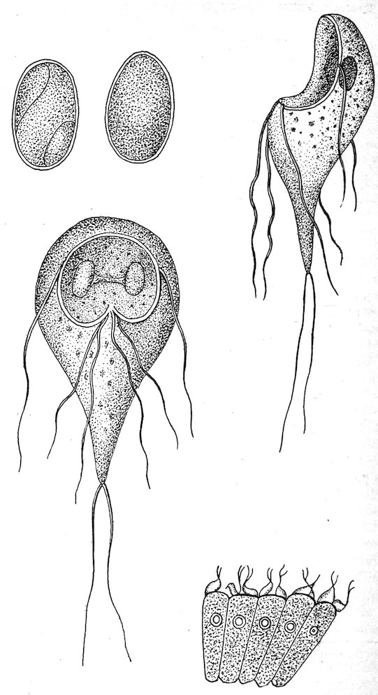
Fig. 9.—Lateral view of encysted Lamblia duodenalis. Fig. 10.—Cyst from large intestine. Fig. 11.—Ventral view of Lamblia. Fig. 12.—Lateral view. Fig. 13.—Epithelial cells of the villous coating of the small intestine infested with Lamblia. (After Grassi & Schewiakoff, 1888, pl. 15, figs. 1, 2, 5, 11, 12.)
91
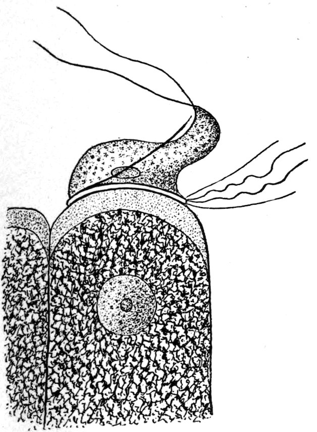
Fig. 14.—An epithelial cell with parasitic Lamblia. Greatly enlarged. (After Grassi & Schewiakoff, 1888, pl. 15, fig. 6.)
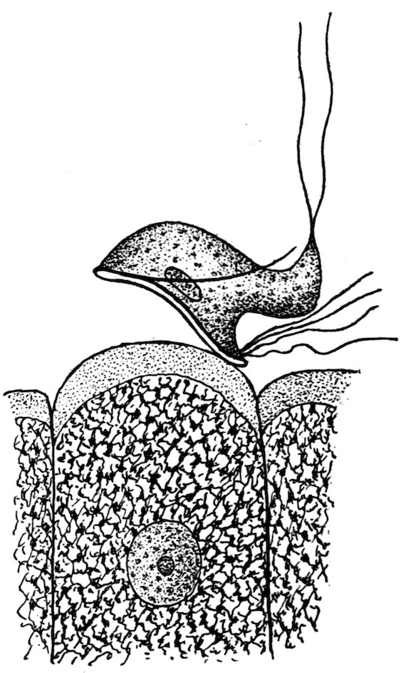
Fig. 15.—An individual in the act of joining an epithelial cell. (After Grassi & Schewiakoff, 1888, pl. 15, fig. 7.)
Pathogenicity.—Opinion differs as to the pathogenicity of this organism. Perroncito (1902b) reports it as causing a fatal disease in rabbits. Braun (1908) is inclined to consider it harmless. From conversation with Doctor Hemmeter, we are persuaded that in his case in a child in Baltimore the parasite was not without effect. Possibly the question as to its pathogenicity is a relative one in that light infections may produce no recognizable disturbance, while heavy infections may produce recognizable effects. Doctor Hemmeter’s original letter regarding his case contained the following notes:
Patient, male, white child, 3 years old, born in Maryland. Had recurrent attacks of colitis all its life; three acute attacks within the last three weeks, accompanied by fever, distended abdomen, sensitive, etc. Stools have always been like putty, containing large amount of mucus, and some blood streaks; fever lasting three days, no pronounced diarrhea, 2 to 3 passages per day; intervals between attacks variable, stools at such times like putty, also with mucus.
92Later information from Doctor Hemmeter states that the disappearance of the parasites from the stools coincided with improvement in the child’s condition.
Clinical diagnosis.—The fresh unstained stools should be examined microscopically; or the diluted stool may be stained with methylene blue, by which nearly all objects become promptly stained, except Lamblia, which remains grayish white (Roos, 1893) for several hours.
Treatment.—Attempts to expel Lamblia have not always met with marked success. Among the drugs used are male fern, sulphate of quinine, naphthol, calomel, hydrochloric acid, and arsenic. Grassi appears to have had success with calcined magnesia.
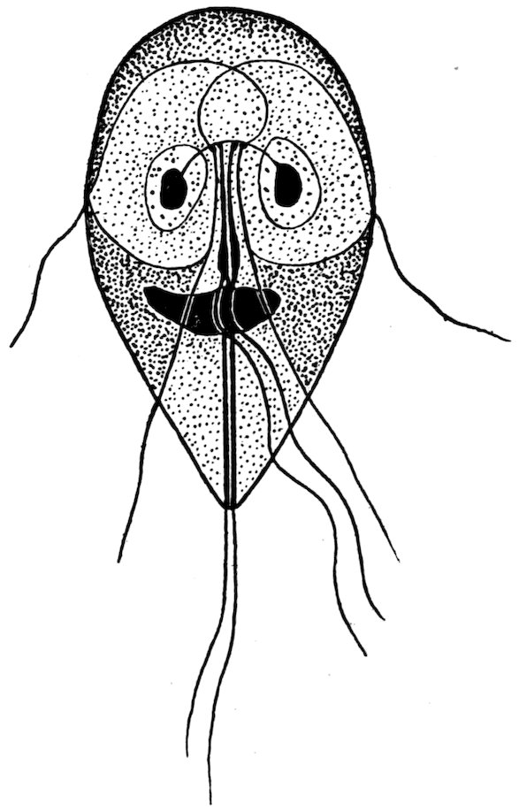
Fig. 16.—“Lamblia intestinalis” of man. (After Bensen, 1908, fig. 5.)
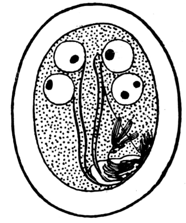
Fig. 17.—Copulation cyst of “Lamblia intestinalis” of man. (After Bensen, 1908, fig. 5.)
The division of Lamblia duodenalis INTO SEPARATE SPECIES.—Bensen (1908) has recently divided Lamblia duodenalis s. l. into three species: L. “intestinalis,” L. muris, and L. cuniculi. His preliminary paper seems to offer fairly convincing data for the correctness of his interpretation, but it may be well to await the publication of his more complete paper, in which he promises fuller details, before the species are definitely accepted. Several nomenclatural points will come up for consideration in this connection.
Lamblia intestinalis, which Bensen accepts as name for the species (fig. 16) occurring in man, can not be accepted, as this name is based on Cercomonas intestinalis Lambl, 1859, which is invalidated by 93Cercomonas intestinalis (Ehrenberg, 1838) Perty, 1852, found in frogs.
Lamblia muris (fig. 18) will probably stand, based on Dimorphus muris Grassi, 1879.
Lamblia cuniculi.—There is some doubt as to the status of this name. Davaine (1875a, 128–129) has described from rabbits a protozoon, which he designated as Hexamita duodenalis and which Railliet (1893a, 169) identifies as a synonym of Lamblia intestinalis (Lambl). On basis of the principle that identifications are to be accepted as correct until shown to be incorrect, Stiles has accepted duodenalis as name (1902) for the form in question. The question now arises as to the relation of cuniculi to duodenalis. If they are accepted as identical, duodenalis will supplant cuniculi, and a new name must be given to the form found in man. If duodenalis is taken as identical with intestinalis Lambl, duodenalis remains as name for the form in man, and cuniculi Bensen will stand for the species in rabbits. If Railliet’s interpretation of synonymy be shown to be incorrect by proving that duodenalis Davaine is not to be considered in connection with either form, a new name must be given to intestinalis Lambl.
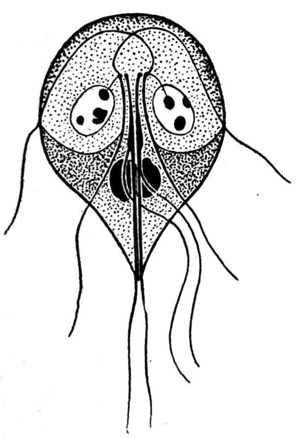
Fig. 18.—Lamblia muris of mice. (After Bensen, 1908, fig. 1.)
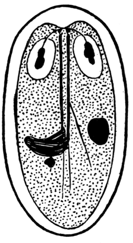
Fig. 19.—Autogametocyte of Lamblia muris. (After Bensen, 1908, fig. 6.)
Genus TRYPANOSOMA s. l.
An extensive group of parasitic protozoa, known as “trypanosomes,” has recently been the basis of considerable literature. The genus Trypanosoma was originally based upon a species (T. rotatorium) found in frogs, and while most trypanosomes have been 94described as members of this genus several authors have separated out certain forms into separate genera.
Luehe (1906) has recently placed the trypanosomes of mammals in the
Genus TRYPANOZOON Luehe, 1906.
One of these species (Trypanozoon gambiense, usually known as Trypanosoma gambiense) is the cause of “sleeping sickness” in man, and has been transmitted in laboratory experiments to rats and mice.
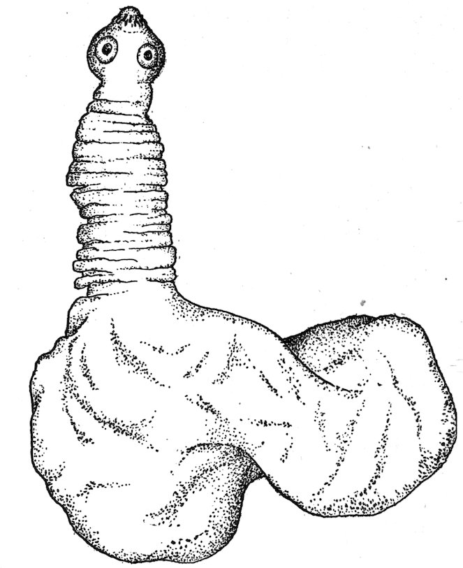
Fig. 20.—An isolated pork-measle bladder worm (Cysticercus cellulosæ), with extended head. Greatly enlarged. (After Stiles, 1898a, 90, fig. 76.)
Just what practical importance there may be in the ability of the parasite to live in rats and mice remains to be seen, but theoretically this biological factor may possibly become one of considerable magnitude. At present the least conclusion to be drawn is that it adds one more to the many arguments in favor of a world-wide destruction of rats and mice.
Several trypanosomes, other than Trypanozoon gambiense, are transmissible by experiment to rats and mice, while one species (Trypanozoon lewisi) has rats for its normal host, and two other species (Trypanozoon duttoni and Trypanosoma musculi Kendall, 1896) are reported originally from the mouse.
95
CESTODA—TAPEWORMS.
Of the five cestodes mentioned as coming into consideration in the subject under discussion, only one (Hymenolepis diminuta) need be considered seriously.
CYSTICERCUS CELLULOSÆ—TÆNIA SOLIUM.
[Fig. 20.]
The larval cestode known as Cysticercus cellulosæ (which causes “measles” in swine) develops (when eaten by man) into a tapeworm which is known as Tænia solium. This larva is also reported as encysted in the peritoneum of Mus rattus.
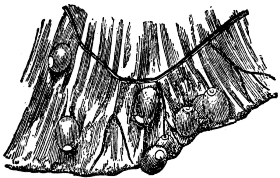
Fig. 21.—Portion of mesentery of rabbit infected with Cysticercus pisiformis. Natural size. (After Railliet, 1893a, 216, fig. 114.)
Even if it be granted that the specific determination of the specimen in question as Cysticercus cellulosæ is correct, the occasional infection of rats with this parasite would be of very little practical significance in this country from a public health point of view, as we do not use rats for food for man. Theoretically it is possible to conceive of combinations of circumstances in which such infection in the rat might under certain conditions eventually affect man, but the chances are so remote as to be negligible, especially when compared with the much greater questions which demand attention.
CYSTICERCUS PISIFORMIS—TÆNIA PISIFORMIS.
[Fig. 21.]
The larval stage of this parasite occurs in rabbits, the adult stage in canines. Parona (1901) reports the occurrence of the larval stage in Mus brasiliensis, and Vital has recorded the presence of the adult stage in man.
In view of the fact that Galli-Valerio was unable to infect himself experimentally with this species, the specific determination made by Vital is open to some question. Even assuming that this tapeworm may develop in man, the presence of the larval stage in rats is of such little importance as to be negligible.
96
CYSTICERCUS FASCIOLARIS—TÆNIA TENIÆFORMIS.
[Fig. 22.]
This encysted larval tapeworm is exceedingly common in the liver of rats and mice, and when swallowed by cats it develops into an adult tapeworm.

Fig. 22.—Larval stage of Tænia teniæformis. Natural size. (After Leuckart, 1880, 450, fig. 202.)
There are two possible points of view in connection with which this parasite is of indirect interest in public health matters: (1) Occasionally these encysted parasites are mistaken for lesions of tuberculosis; (2) Krabbe (1880) relates that in Jutland there exists a folk custom of eating chopped raw mice in case of retention of urine, and in this connection the point has been raised that the possibility is not excluded that such action might eventually give rise to infection of man by the parasite in question. No case of such infection in man is as yet established.
[Figs. 23 and 24.]
Under the name Tænia murina, Dujardin (1845) described for rats a tapeworm which has been identified by a number of authors (including Stiles) as identical with the dwarf tapeworm (Hymenolepis nana) of man. If this identification be correct, the rats must be considered as the great disseminators of this tapeworm. Serious doubts have been raised, however, as to whether the tapeworm in man is not in reality distinct from that of the rat, and the evidence in favor of such conclusion is accumulating. Some slight differences between the two forms have been noticed, but by some authors these differences have 97been considered insufficient to hold the two worms apart. Looss has tried to infect rats with the dwarf tapeworm found in man in Egypt, but his results have been negative. Here in Washington Stiles has repeatedly attempted to infect rats with the dwarf tapeworm found in man in the United States, but thus far no positive infection has occurred in the rodents. In Italy, Grassi attempted to transmit the rat form to six persons, and in one case he found tapeworms, but in view of the frequency of H. nana in Italy the significance of this one instance has been questioned; Grassi was not successful in trying to infect rats with H. nana of man.
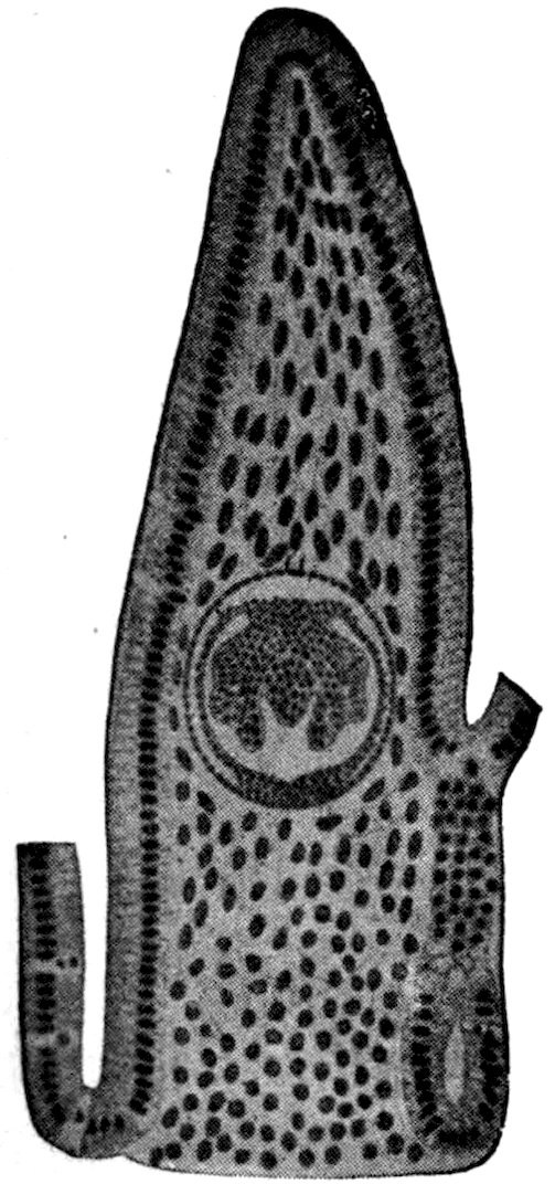
Fig. 23.—Longitudinal section of the intestinal villus of a rat, containing cystic stage of dwarf tapeworm. Enlarged. (After Grassi & Rovelli, 1892a, pl. 3, fig. 25.)
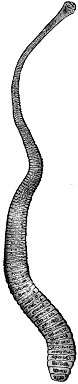
Fig. 24.—Adult dwarf tapeworm (Hymenolepis nana) of man. Enlarged. (After Leuckart, 1863, p. 393, fig. 112.)
Thus at present the evidence is to the effect that rats and mice are not to be viewed as the source or reservoir for the dwarf tapeworm (H. nana) of man.
98
HYMENOLEPIS DIMINUTA[Z] (Rudolphi, 1819) R. Blanchard, 1891.
[Figs. 25 to 30.]
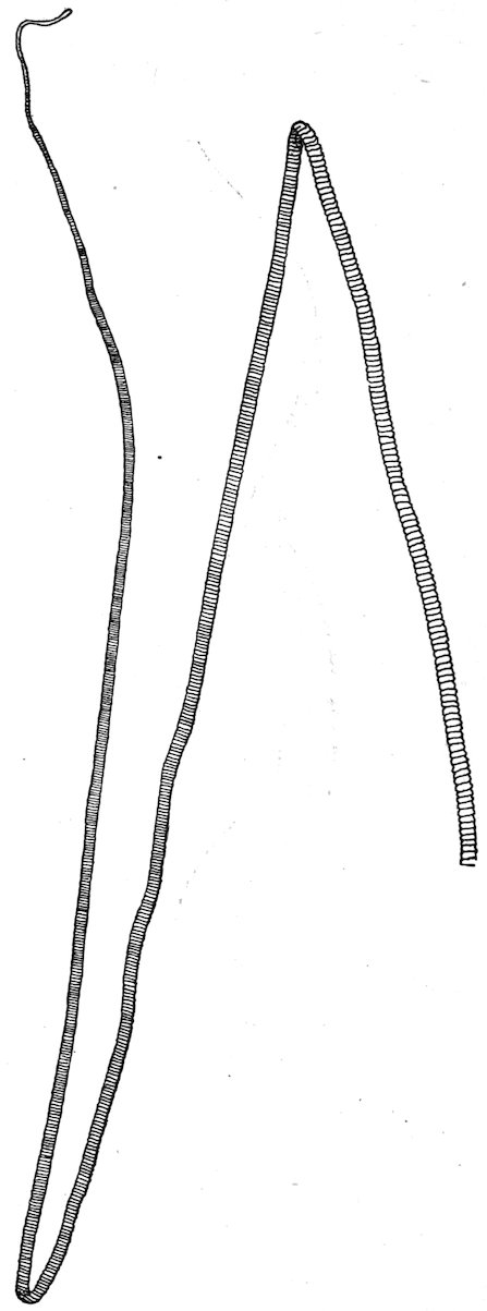
Fig. 25.—Strobila of Hymenolepis diminuta. Natural size. (After Grassi, 1881, pl. 11, fig. 1.)
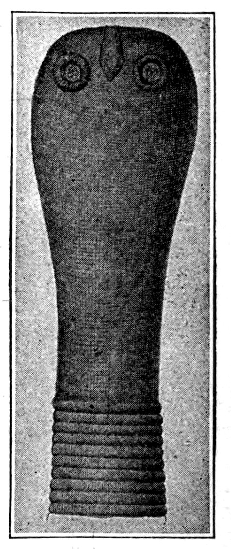
Fig. 26.—Head and anterior portion of H. diminuta from the rat. Enlarged. (After Zschokke, 1889, pl. 1, fig. 21.)
Specific diagnosis.—Hymenolepis: Strobila 10 to 60 millimeters in length, 2.5 to 4 millimeters in maximum breadth; composed of 800 to 1,300 segments. Head small, almost globular; 200 to 600μ in width; rostellum rudimentary, pyriform, only slightly 99protractile; hooks absent; suckers globular, near the apical portion of the head, 80 to 160μ in diameter. Neck usually short. Segments throughout strobila broader than long. Genital pores on left margin, near the junction of the anterior and middle thirds of each segment. Three testes in each segment; vas deferens dilates into a prominent seminal vesicle before entering the cirrus pouch, within which also is a vesicle. Gravid uterus occupies most of the proglottids; its cavity is subdivided into a large number of incompletely separated compartments filled with eggs. Eggs round or slightly oval; outer membrane 54 to 86μ in diameter, yellowish in color, may be radially striated; inner membrane 24 by 20μ to 40 by 35μ in diameter, with mammillate projection at each pole often not apparent; between outer and inner membranes a prominent third layer of albuminous substance, often appearing as two delicate smooth membranes, with intervening space filled by a granular coagulum; embryonal hooks 11 to 16μ in length.
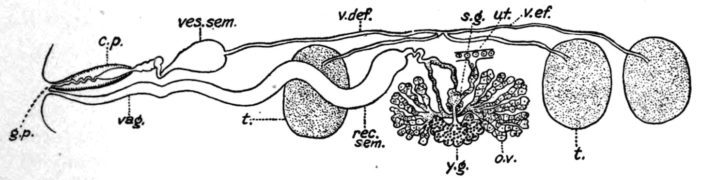
Fig. 27.—Male and female organs of H. diminuta: c. p., cirrus pouch; g. p., genital pore; ov., ovary; rec. sem., receptaculum seminis; s. g., shell gland; t., testiculæ; ut., uterus; vag., vagina; v. def., vas deferens; v. ef., vas efferens; ves. sem., vesicula seminalis; y. g., yolk gland. Enlarged. (After Zschokke, 1889, pl. 2, fig. 22.)
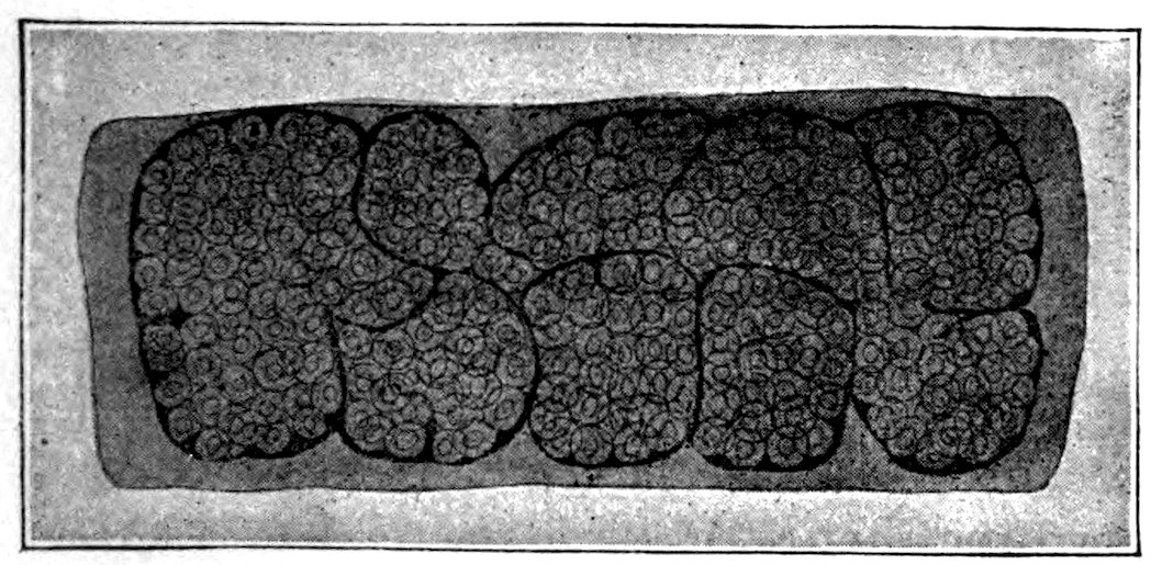
Fig. 28.—Gravid segment of H. diminuta. Enlarged. (After Grassi, 1881, pl. 11, fig. 15.)
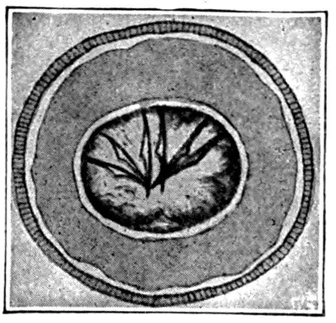
Fig. 29.—Egg of H. diminuta from man. Greatly enlarged. (After Bizzozero, 1889a, pl. 4, fig. g″.)
Habitat.—Adults in small intestine of brown or Norway rat (Mus decumanus), black rat (M. rattus), house mouse (M. musculus), Egyptian or roof rat (M. rattus alexandrinus), wood or field mouse (M. sylvaticus), Rhipidomys pyrrhorhinus [according to Linstow, 1878a, 23], and man (Homo sapiens).
100Development.—The larval stage (Cercocystis H. diminutæ) occurs in larval and adult meal moths (Asopia farinalis); in young and adult earwigs (Anisolabis annulipes); and in adult beetles (Acis spinosa and Scaurus striatus).
Geographic distribution.—Massachusetts, Pennsylvania, Nebraska, Iowa, District of Columbia, Maryland, Brazil, Italy, Germany, France, Austria.
This parasite is certainly more common in man in this country than has heretofore been assumed, but fortunately it seems to be one of the most harmless and most easily expelled tapeworms occurring in man.
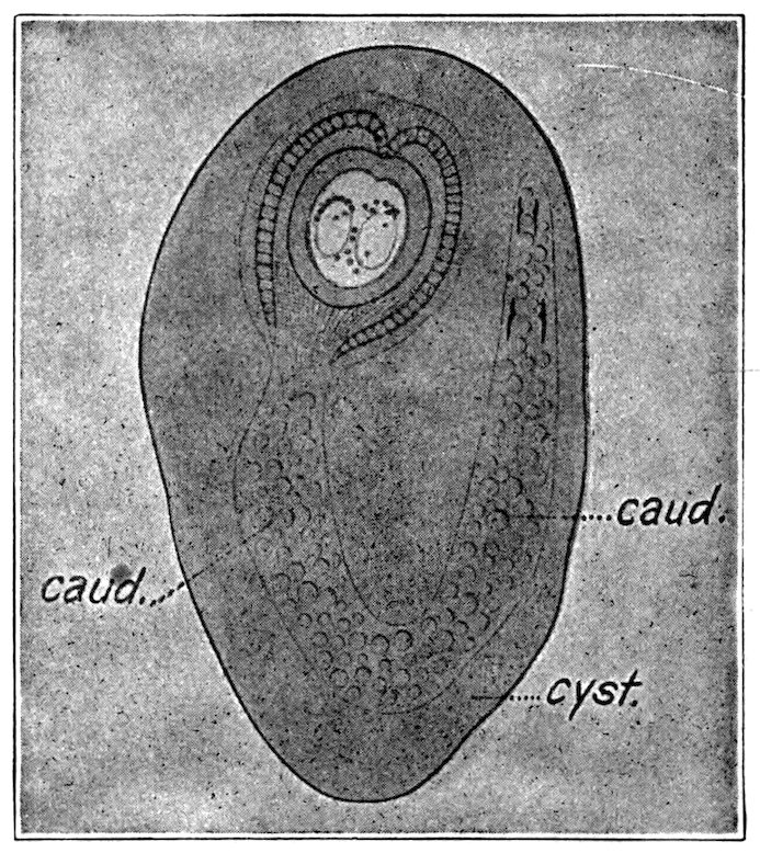
Fig. 30.—Encysted cystic stage of H. diminuta: caud., caudal appendage; cyst., adventitious capsule inclosing the cercocystis. Enlarged. (After Grassi & Rovelli, 1892a, pl. 4, fig. 1.)
From present evidence, the rats and mice are looked upon as the regular hosts for this worm, and hence as the natural reservoir of the infection. The intermediate host becomes infected from the rodents and then transmits the infection to man.
It might be mentioned that as yet no extensive study has been conducted in the United States to differentiate clearly the various species of Hymenolepis found in our rats and mice. The possibility is therefore not entirely excluded that some of our cases of Hymenolepis diminuta may eventually be shown to be referable to other species of the same genus.
101
NEMATODA—TRUE ROUND WORMS.
Family TRICHINELLIDÆ.[AA]
Family diagnosis.—Nematoda: Elongate cylindrical worms; cephalic portion long and very slender, caudal portion more or less swollen. Mouth rounded, without lips. Esophagus relatively very long, composed of a single row of large cells, forming the so-called “cell body” and supporting a narrow esophageal tube; anus terminal or nearly so.
Male: With a single spicule or without spicule.
Female: With one ovary; vulva near caudal end of cell body, close to point where body increases in diameter; oviparous or viviparous.
Eggs: Oviparous species, with thick shell, with opening at each pole, closed by a transparent plug.
Type genus.—Trichinella Railliet, 1895.
This family furnishes two parasites to man: The whipworm (Trichuris trichiura) of the colon, and the trichina or flesh worm (Trichinella spiralis, see p. 101).
Genus TRICHINELLA[AB] Railliet, 1895.
Generic diagnosis.—Trichinellidæ: Very minute worms, of nearly uniform diameter. Adults in intestine of mammals, larvæ encysted in muscles.
Male: Without spicules, but with 2 conical appendages on the tail, at side of terminal cloacal opening.
Female: Vulva about one-fifth the length from anterior end; viviparous.
Type species.—Trichinella spiralis (Owen, 1835) Railliet, 1895.
TRICHINELLA SPIRALIS (Owen, 1835) Railliet, 1895.
[Figs. 31 to 51.]
Specific diagnosis.—Trichinella: Body thread-like, visible to naked eye.
Male: Length, 1.4 to 1.6 millimeters; diameter, 40μ; distal of cloacal opening, 2 pairs of papillæ, the anterior pair hemispherical, posterior pair conical.
Female: Length, 3 to 4 millimeters; diameter, 60μ; anus terminal; vulva one-fifth of length of body from the mouth; viviparous.
Habitat.—Adults in lumen and wall of small intestine, encysted larvæ in muscles of various mammals, particularly in rats, mice, swine, and man.
Geographic distribution.—More or less cosmopolitan.
Source of infection.—From the life cycle of this parasite it is clear that the permanent reservoir of infection must be some animal with cannibalistic tendencies. Of the three most important hosts (man, swine, and rats), the rats present ideal conditions in this respect. It is true that there are some tribes of man which are cannibalistic, but their distribution is restricted. Likewise swine are in so far cannibalistic that they eat uncooked swine offal and swill, but this is due to the shortsightedness of man rather than to 102the habits of the swine. Accordingly, neither man nor the hog presents the proper theoretical conditions for the perpetuation of the parasites and hence to serve as reservoir for the disease it causes.
Rats, on the contrary, are cannibalistic, and trichinosis is a common disease among them. Hence they may be viewed as the natural reservoir for the parasites and for the disease it causes; hence, also, any well-directed public health campaign against trichinosis should consider the eradication of rats.
Rats become infected by eating each other; by eating scraps of pork found on the offal pile of slaughterhouses, or in swill; and by eating scraps of human flesh in dissecting rooms of medical schools.
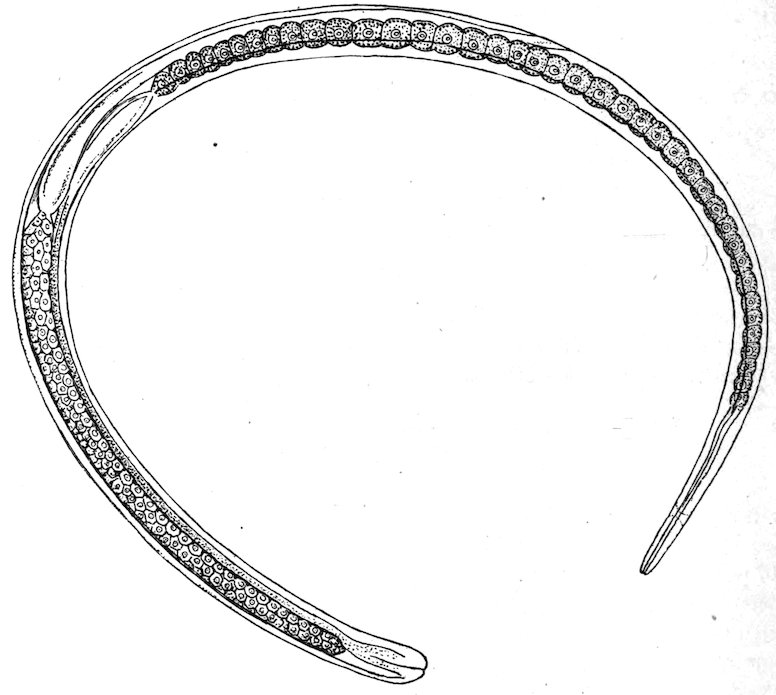
Fig. 31.—Female trichina from the intestine; 24 hours after infection. Enlarged. (After Leuckart, 1866a, pl. 1, fig. 1.)
Swine become infected by eating rats, and by eating scraps of pork on the offal pile of slaughterhouses, or in swill.
Man becomes infected almost exclusively by eating pork and boar meat. The rare infections which occur from eating other meat are almost negligible.
Medical significance.—Trichiniasis or trichinosis refers to infection with the trichina or flesh worm. Normally it occurs only in mammals, chiefly carnivorous and omnivorous species, and it is transmissible from any infected mammal to any other mammal susceptible to it, in case the latter eats the uncooked flesh of the former.
103Symptoms.—In heavy infections there may be three more or less distinct periods of the disease, corresponding to the three stages in the life cycle of the parasite; but these stages are obscure in light or in repeated infections. Profuse sweating may last during the entire attack.
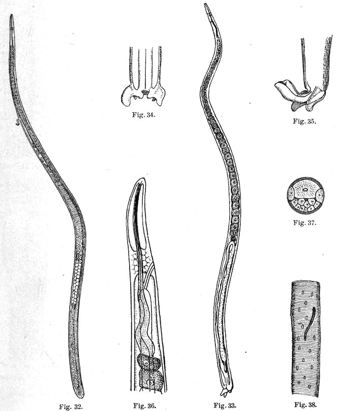
Fig. 32.—Gravid adult female trichina. Enlarged. (After Leuckart, 1866a, pl. 1, fig. 2.) Fig. 33.—Adult male trichina from the intestine. Enlarged. (After Leuckart, 1866a, pl. 1, fig. 5.) Fig. 34.—External genitalia of same. Enlarged. (After Leuckart, 1866a, pl. 1, fig. 7.) Fig. 35.—The same with extruded cloaca. Enlarged. (After Leuckart, 1866a, pl. 1, fig. 8.) Fig. 36.—Cephalic portion of a trichina showing central nervous system and anterior portion of intestinal canal. Greatly enlarged. (After Leuckart, 1866a, pl. 1, fig. 13.) Fig. 37.—Transverse section of a female trichina. Greatly enlarged. (After Leuckart, 1866a, pl. 1, fig. 16.) Fig. 38.—Young trichina embryo in a muscle fibre. Greatly enlarged. (After Leuckart, 1866a, pl. 2, fig. 1.)
Period of ingression: The adult parasites are in the intestine, hence gastro-intestinal symptoms develop; irregular appetite, nausea, diarrhea 104or constipation, colicky pains; a temporary edema around eyes about the eighth day; muscular pains begin.
Period of digression: This begins about the eighth to the fifteenth day, sometimes later; young embryos are wandering to and attacking the muscles, hence muscular symptoms (myositis) develop; painful tension of muscles, especially biceps; members assume semiflexed position; movements, chewing, swallowing, breathing, and speech become difficult; eyes become fixed; fever.
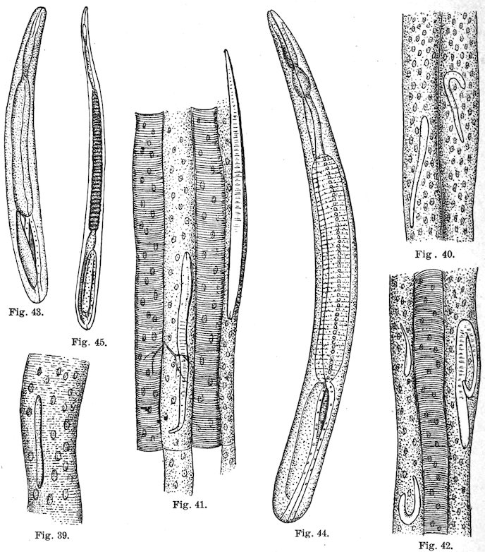
Figs. 39–42.—Later stages of same; the muscular structure is undergoing changes. (After Leuckart, 1866a, pl. 2, figs. 3, 6, 7, 8.) Figs. 43–45.—Muscle trichinæ, 0.3 mm., 0.4 mm., and 0.6 mm. long. (After Leuckart, 1866a, pl. 2, figs. 10–12.)
Period of regression: The parasites become encysted in muscles. All symptoms may increase, then gradually decrease; cachexia and anemia resulting from malnutrition; pruritis, miliary cutaneous eruptions; desquamation; about twenty-fourth day, a “second” 105edema develops, especially about the face; lungs may become edematous; bronchial catarrh, pneumonia, or pleurisy may appear; gradual recovery.
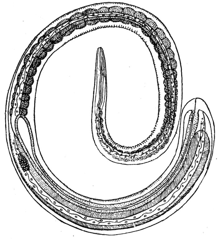
Fig. 46.—A female trichina from the muscle. Greatly enlarged. (After Leuckart, 1866a, pl. 1, fig. 12.)
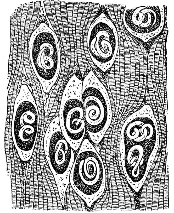
Fig. 47.—A piece of pork with encysted trichinæ. Enlarged. (After Braun, 1903, p. 251, fig. 195.)
Lethality.—The lethality varies from 0 to 100 per cent; it averaged 5.6 per cent in 14,820 cases collected from literature; it is dependent upon amount of infection which remains in the body; low before second and after seventh week, highest from fourth to sixth week.
Prognosis.—Better in cases having severe diarrhea in first stage.
106Complications and sequelæ.—Abortion, menstrual disturbances, pneumonia, pleurisy, peritonitis.
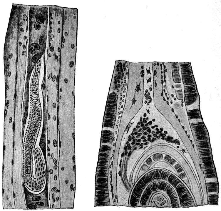
Fig. 48.—Section through a rat’s muscle; the infected muscle fiber has lost its striation, its nuclei are enlarged and increased in number. Greatly enlarged. (After Hertwig-Graham, see Braun, 1903, p. 284, fig. 212B.) Fig. 49.—Portion of an isolated trichina cyst, at the pole of which connective tissue cells have wandered into the thickened sarcolemma. Greatly enlarged. (After Hertwig-Graham, see Braun, 1903, p. 284, fig. 212C.)
Clinical diagnosis.—Make microscopic examination:
(1) Of pork, if any has been left, to find encysted larvæ; if larvæ are found, feed pork immediately to two or three guinea pigs or white rats, to determine if the encysted larvæ are alive; kill one rat after three days and examine intestinal content for adult; kill the second rat after two weeks, the third rat after three weeks, and hunt for larvæ in muscular portion of diaphragm. Even if live trichinæ are found in intestine, an examination of the muscles may show that the worms were too weak to reproduce, hence prognosis is favorable.
(2) Of patient’s blood, for increased proportion of eosinophiles.
(3) Of patient’s stools, for discharged adult worms, especially if diarrhea is severe; dilute the fecal matter with warm water and pour off whatever floats; place remainder in a shallow glass dish so that it will not be over one-twelfth of an inch deep; move the dish gently around over a dark background (such as dark paper), and hunt for small hair-like objects; place these, if found, in a drop of water on a 107slide, cover with a cover slip, and examine under low power. Or, if necessary, make a microscopic examination.
(4) Of small excised portion of patient’s deltoid, about three to four weeks after infection, for encysted larvæ; cut a small piece parallel to muscle fibers, tease this on a slide, add a drop of pure water, or water and glycerine, cover with another slide, flatten gently by pressure while examining under low power.
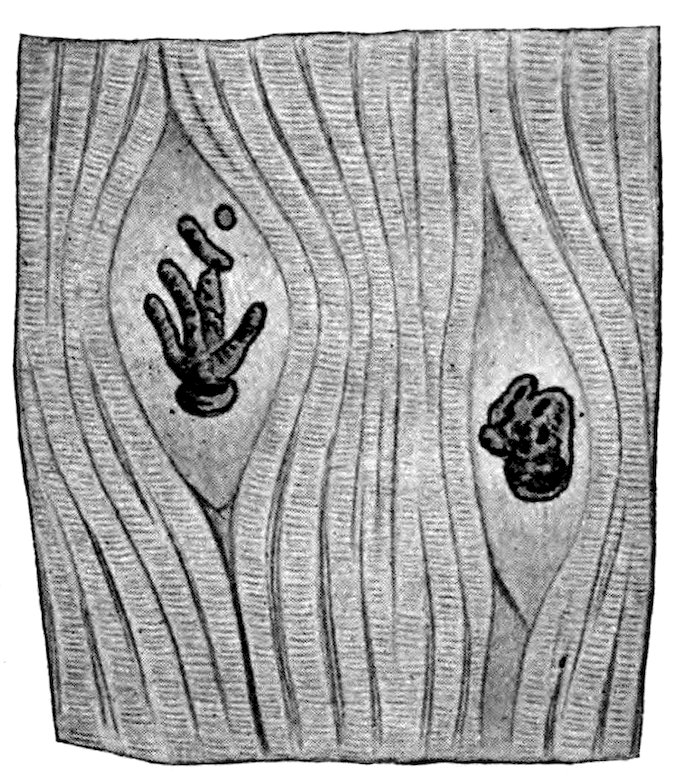
Fig. 50.—Calcified trichinæ in uncalcified cysts, from pork. Enlarged. (After Ostertag, see Braun, 1903, p. 285, fig. 213.)
Suspect trichinosis especially under following circumstances: Several patients in same family or in same neighborhood, usually of North German descent, show typhoid-like symptoms shortly after a celebration (wedding, birthday party, etc.) at which pork was served.
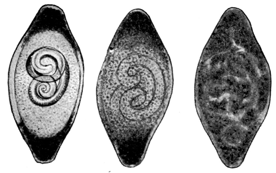
Fig. 51.—Three phases of calcification of trichinæ and their cysts, the changes starting from the poles of the cysts. Enlarged. (After Ostertag, see Braun, 1903, p. 285, fig. 214.)
Differential diagnosis.—Consider especially typhoid fever and rheumatism.
Treatment.—Purge in early stage to carry away the adult worms and thus eventually decrease the amount of muscular infection. No treatment is known which can be relied upon to kill the larvæ in the 108muscles; benzine has been suggested. Stimulants may be given to carry patients through until the larvæ encyst.
Prophylaxis.—Kill off rats and mice.—Educate public to eat pork only when thoroughly cooked or thoroughly cured. A practical test of cooking is the white color of the meat on being cut; if the cut surface is reddish and serous, the pork is not sufficiently cooked to kill trichinæ.
As a matter of practical experience, the microscopic inspection of pork has not given the protection it is generally supposed to give. Of 6,329 cases with 318 deaths reported for Germany during the years 1881–1898, 3,388 cases with 132 deaths are directly attributable to faults in the inspection. This system directly increases the tendency to eat raw pork, gives the public a false sense of security, and does not give practical results commensurate with its expense.
ACANTHOCEPHALA—THORN-HEADED WORMS.
Genus GIGANTORHYNCHUS Hamann, 1892.
Generic diagnosis.—Acanthocephala, Gigantorhynchidæ: Large worms with annulate round to flat, tape-like body. Hooks with 2 roots and completely covered with transparent chitin. Proboscis sheath a muscular apparatus, without cavity. Central nervous system caudad of equator of proboscis sheath and eccentric. Lemnisci long, cylindrical, with central canal.
Type species.—Gigantorhynchus echinodiscus (Diesing, 1851).
The Moniliform Thorn-headed Worm—GIGANTORHYNCHUS MONILIFORMIS
(Bremser, 1819).
[Figs. 52 to 58.]
Specific diagnosis.—Gigantorhynchus (p. 108): Body attenuated anteriorly, with fine transverse striæ or rings, or even constrictions which give the appearance of a series of beads, except in the caudal fourth of body, which is nearly smooth and cylindrical. Proboscis 425 to 450μ long, 176 to 190μ in diameter, armed with feeble, very curved, 26μ long, hooks arranged more or less in quincunx and forming at most 15 transverse and about 12 longitudinal rows. Lemnisci more than a centimeter in length, cylindrical, undulated posteriorly.
Male: Length 4 to 4.5 centimeters long; bursa campaniform.
Female: Length 7 to 8 centimeters (to 27 centimeters after Westrumb).
Eggs: Ellipsoidal, 85 by 45μ; external envelope thin, yellowish; middle envelope very thick, colorless, homogeneous; inner envelope less thick, colorless, and quite pliant. Embryo striated transversely in posterior two-thirds, and covered with spines which increase in size toward anterior end of embryo, the anterior spines being transformed into hooklets with prong and base.
Development: With beetles (Blaps mucronata) as intermediate host.
Habitat.—Small intestine of various small mammals; brown rat (Mus decumanus); white rat (Mus norvegicus albus); M. fuscirostris; hamster (Cricetus frumentarius); dormice (Myoxus quercinus or glis); field mole (Arvicola arvalis or agrestris?); Lemnus arvalis; and Mustela putorius. It can also develop in man, as has been shown experimentally by Grassi and Calandruccio (1888, 521–525).
Medical significance.—Grassi and Calandruccio report a doubtful case of infection in a girl near Catania. Calandruccio infected 109himself experimentally by swallowing the young worms taken from a Blaps. Twenty days later he was seized with severe pains which increased on pressure; diarrhea followed, with ringing in the ears, fatigue, and somnolence. Seventeen days later the characteristic eggs were found in his stools, and twelve days later the symptoms became so severe that he took 8 grams of extract of male fern; one to two hours later he passed 53 of the parasites. For two days the symptoms continued, on the second day fever developed, but all symptoms disappeared on the third day.
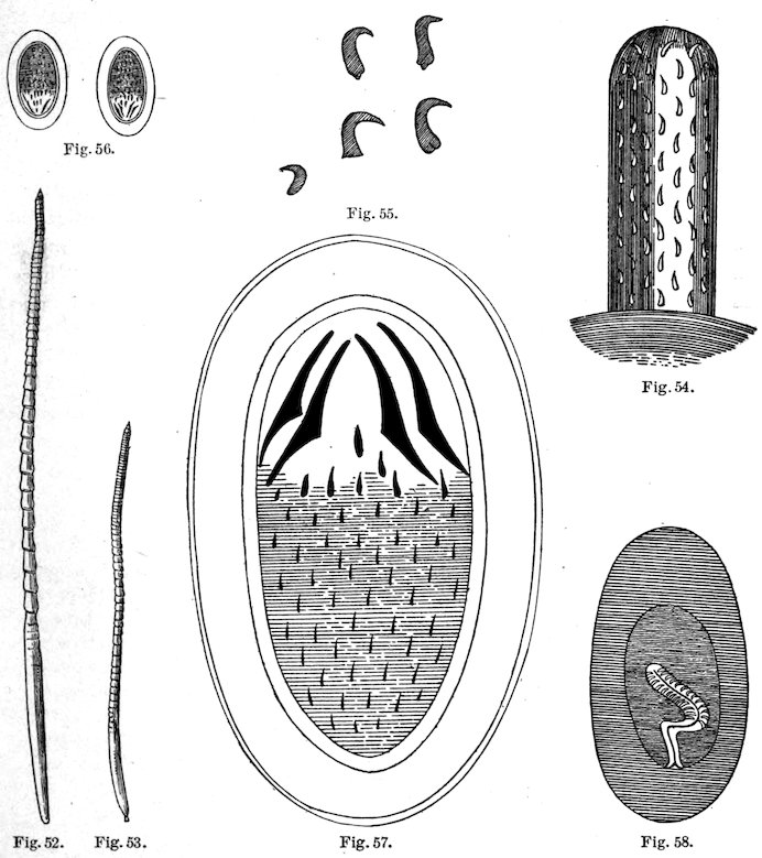
Fig. 52.—Gigantorhynchus moniliformis, female. ×2. (After Grassi & Calandruccio, 1888, p. 523, fig. 1.) Fig. 53.—G. moniliformis, male. ×2. (After Grassi & Calandruccio, 1888, p. 523, fig. 2.) Fig. 54.—Rostellum of G. moniliformis. Greatly enlarged. (After Grassi & Calandruccio, 1888, p. 523, fig. 3.) Fig. 55.—Hooks from same. Greatly enlarged. (After Grassi & Calandruccio, 1888, p. 523, fig. 4.) Fig. 56.—Eggs of G. moniliformis, with embryo. Greatly enlarged. (After Grassi & Calandruccio, 1888, p. 523, fig. 5.) Fig. 57.—Egg very greatly enlarged. (After Grassi & Calandruccio, 1888, p. 524, fig. 6.) Fig. 58.—A young larva of G. moniliformis in a Blaps; the rostellum is invaginated and the larva is surrounded by a thick inner jelly-like and thin outer cuticular covering. Enlarged. (After Grassi & Calandruccio, 1888, p. 524, fig. 7.)
110
ARACHNOIDEA.
Genus LINGUATULA Frœhlich, 1789.—Tongue worms.
Species LINGUATULA SERRATA Frœhlich, 1789.
The larva of this parasite is found encysted in the entrails of rabbits, cattle, and certain other animals, and it becomes mature in the nasal cavities of canines.
Both the larva and the adult have been reported for man, and the larva has been reported as occurring in Mus decumanus.
As canines are not fond of eating rats, the presence of the larval tongue worm in the latter is of more academic interest than practical importance, and although the theoretical possibility must be admitted that a dog by eating rats might become infected with tongue worms and eventually might transmit the infection to man, these possibilities seem somewhat remote. Remote possibilities must also be admitted to the effect that if a person ate a rat infected with tongue worms this person might become infected.
111