THE FLEA AND ITS RELATION TO PLAGUE.
Reading edition
By Passed Assistant Surgeon Carroll Fox,
United States Public Health and Marine-Hospital Service.
THEORIES AS TO TRANSMISSION OF PLAGUE.
1. Direct contagion from man to man.
2. Through slight abrasions of the skin, mucous membranes of mouth, tonsils, nose, and conjunctiva receiving contaminated material.
3. Through the respiratory tract, from air contaminated with dried infectious sputum or dejecta. (Possibly the cause of primary pneumonic plague.)
4. Through the alimentary tract from food contaminated with saliva or excretions from plague patients, or dejecta or the feet of insects that have fed on plague material. In the case of rats, from eating the carcasses of infected rats.
5. Infected clothes, soil, or houses.
6. Through the bites of insects, especially the flea.
It has been noticed for many years that an epidemic of plague in man was associated with an epizootic of high mortality among rats, but it was not until Yersin discovered the Bacillus pestis in 1894 that the disease in man and rats was shown to be identical. The first five theories are not satisfactory in explaining the epidemiology of plague, and in 1897 Simond advanced the theory that plague was carried by means of fleas. Hankin in 1898 also suggested an insect as an intermediate host. This theory has been developed by Ashburton Thompson, Gauthier and Raybaud, Liston, Verjbitski, and others, and finally by the last Indian Plague Commission, whose work makes a distinct advance in our knowledge of this subject. The reader is referred to the work of this commission for a review of the subject, which has been liberally used in the preparation of this paper.[AD]
124
INSECTS THAT HAVE BEEN SUSPECTED IN THE TRANSMISSION OF PLAGUE.
It is probable that all insects capable of sucking blood will take the Bacillus pestis into their alimentary canal if they feed on a septicæmic plague animal. Ogata suggested that not only the flea but the mosquito also may be responsible for the transmission of plague. Yersin, Hankin, and Nuttall have each demonstrated the presence of Bacillus pestis in the dejecta of flies and ants; and Nuttall and Verjbitski in the stomach and dejecta of the bedbug. Hertzog found the bacilli in the Pediculus capitis taken from a child which died of plague, and McCoy[201] has found the organism in lice, Hæmatopinus columbianus, taken from a plague-infected squirrel. The plague bacilli have been frequently demonstrated in rat fleas taken from plague rats, and McCoy has shown its presence in the flea (Ceratophyllus acutus) of the California ground squirrel (Citellus beecheyi). The cockroach has also been thought to be instrumental in spreading the infection by contaminating food. The presence of bacilli in the stomach and dejecta of insects has not only been proven microscopically but by animal inoculation as well.
Assuming that the relation between rat plague and human plague has been proven without a doubt—that is, that an outbreak of human plague is associated with an infection in rats, or, in other words, that plague is primarily a disease of rats and secondarily a disease of man—the theory that it is conveyed through an intermediate parasitic host is the only one which will fulfill all the requirements, and after a study of their habits we are able to exclude all of the parasites but the flea as the active agent in its transmission.
Plague is rarely or never contracted either in rat or in man by eating contaminated food. Therefore those insects like flies and cockroaches, which are supposed to spread the infection by contaminating food with their dejecta, need not be considered.
The habits of the domestic mosquitoes are such that while they occasionally do bite animals they usually feed on the blood of man, and are not known to feed where there is much hair, as there is on the rat. This also applies to the bedbug. Verjbitski has shown experimentally that bedbugs would not feed on rats until the animals were shaved.
Pediculi are degenerate insects, their powers of locomotion being limited. Their eggs are laid on and are attached to the hair of the host. They are born, live, and die on the same host, and rarely pass from one animal to another of a different species. It can not be denied, however, that this parasite occasionally may be instrumental in spreading plague from rat to rat. The Pediculus capitis, if placed on a rat, will feed with avidity, but these insects are rarely found upon rats in nature.
125We have no record of plague bacilli having been demonstrated in mites commonly found on rats, but no doubt if search be made they could be found after feeding on a septicæmic plague rat. These mites, however, always confine themselves closely to their particular host and are not known to bite man. The tiny itch mite (Notoedres alepis, Railliet and Lucet) producing rat scabies has, according to Schumann,[202] been known to cause a cutaneous lesion in man, but this mite need not be considered from a plague standpoint.
The flea, on the other hand, lives but part of the time on its host, its eggs developing in the nests or runs of the animal. Again, this insect does not confine itself to one particular species of host only, as frequently the flea of one animal is found on an animal of an entirely different species. Unlike the lice, they are very active and can readily move from place to place. Not only that, but it has been frequently demonstrated that the fleas of rats and of other animals would readily take to man, especially if their natural host was scarce. That rat fleas will bite man has been demonstrated by Gauthier and Raybaud, working with the Leomopsylla cheopis; Tidswell, Lœmopsylla cheopis and Ceratophyllus fasciatus; Liston, Lœmopsylla cheopis; Tiraboschi, Lœmopsylla cheopis; Indian Plague Commission, Lœmopsylla cheopis; and McCoy and Mitzmain[203], Lœmopsylla cheopis, Ceratophyllus fasciatus, and Ctenopsyllus musculi. It has generally been considered that the Ctenopsyllus musculi, above all others, would not bite man, but the last-named observers showed that it would occasionally feed, although it would not live long, in captivity. One of the fleas, a Ceratophyllus fasciatus, was kept alive by Mitzmain for over four months on man’s blood alone.
EXPERIMENTS PROVING THAT FLEAS CAN TRANSMIT PLAGUE.
By a series of experiments carried out in specially constructed cages and go-downs where healthy rats in the absence of fleas were brought in contact with plague-infected rats, the Indian Plague Commission showed that the healthy rats would not contract the disease, notwithstanding the fact that they were not only in intimate contact with the sick rats, but also with the contaminated food and excreta of the sick rats. They then showed that if fleas were introduced the healthy rats would contract plague, the rate of progress of the epizootic being in direct proportion to the number of fleas present. By hanging cages containing healthy rats in cages holding infected rats, but above the jumping distance of a flea, it was shown that the healthy rats would remain well, while those in cages hung within 2 inches from the ground would contract plague. Thus they excluded aerial infection. They also found that if fleas were excluded young rats could suckle a plague-infected mother without contracting the disease.
126Guinea pigs were allowed to run in houses where cases of human and of rat plague were known to have occurred and where many fleas were present. These rodents served as good traps for the fleas and 29 per cent of them contracted plague.
Most of the experiments of the Indian Plague Commission were done with the Indian rat flea, the Lœmopsylla cheopis, but they also performed 27 experiments with the cat flea, Ctenocephalus felis, with negative results; 35 experiments with the human flea, Pulex irritans, 3 of which were successful; and 2 experiments with the Ceratophyllus fasciatus, the common rat flea of Europe and North America, both of which were successful.
In San Francisco a few experiments under purely experimental conditions have been carried on by McCoy to determine the ability of the squirrel flea, the Ceratophyllus acutus, to transmit plague. Fleas that had been previously fed on the blood of a septicæmic plague-infected squirrel were then allowed to feed from test tubes on healthy guinea pigs. While the feces of some of these fleas up to four days, when inoculated into guinea pigs, were proven to be infective, none of those guinea pigs on which the fleas were allowed to feed contracted plague. It might be said, however, that in no case were they seen to eject feces while feeding, the significance of which will be apparent later.
THE BACILLUS IN THE FLEA.
The Indian Plague Commission found that the average capacity of the rat flea’s stomach (Leomopsylla cheopis) was 0.5 cubic millimeter, and that it might receive as many as 5,000 germs while imbibing blood from a plague rat. They further found that the bacillus would multiply in the stomach of a flea and that the percentage of fleas with bacilli in the stomach varied with the season of the year. In the epidemic season the percentage was greatest for the first four days, and on one occasion the stomach was found filled with Bacillus pestis on the twentieth day. In the nonepidemic season no plague bacilli were found in the stomach after the seventh day. They also found that in the epidemic season fleas might remain infective up to fifteen days, while in the nonepidemic season but seven days, and in the latter case the percentage of infection in animals was much less than in the epidemic season. They showed that while one flea was occasionally able to carry the infection this was not usual. It was found that both the males and the females were capable of transmitting the disease.
After a number of dissections they were unable to demonstrate the presence of bacilli anywhere but in the stomach and rectum. At no time was anything found in the body cavity or salivary glands and 127but rarely in the œsophagus, and then only when the flea was killed immediately after feeding.
We have in San Francisco examined quite a number of serial sections of plague-infected fleas with the same result as obtained by the Indian Plague Commission. The bacilli are readily demonstrated, sometimes in enormous numbers, in the gizzard, stomach, and in the rectum, but at no time have they been found in the body cavity, the salivary glands, or the ovary. In fact, as we are dealing with a vegetable organism and not an animal organism, like the Plasmodium malariæ, we could hardly expect to find any biologic change, except simple multiplication, occurring in the intermediate host.
HOW THE FLEA CLEARS ITSELF OF BACILLI.
Some explanation is necessary as to why the bacilli eventually disappear from the flea, although they seem to multiply during the first few days. It is evident that the peristaltic action of the stomach during the course of digestion forcing the blood at the proper time into the rectum, finally to be ejected from the body, would in itself cause many bacilli to be discharged, but naturally a few would remain to multiply indefinitely. The bacteriacidal action of the blood is soon lost after entering the flea’s stomach, but it has been shown by proper staining that the leucocytes after the first feeding with healthy blood contain numbers of Bacillus pestis, and it seems probable that this phagocytic action is important in the cleansing process. It has been shown that after successive feedings on the blood of noninfected animals the power of phagocytosis is increased, and that successive feedings on the fresh blood of animals that have been immunized against plague still further assists and hastens the process. When there is a frequent introduction of fresh normal or immunized blood its bactericidal action is also instrumental in the cleansing process.
REGIONAL DISTRIBUTION OF FLEAS ON RATS.
The location of the primary bubo in a case of plague, human or rodent, depends upon the site of inoculation, for that group of glands will first enlarge which has direct lymphatic connection with the area through which the Bacillus pestis enters the animal organism. The British Indian Plague Commission found that 72 per cent of their naturally infected rats and 61 per cent of the rats experimentally infected by fleas had cervical buboes, while in no instance in over 5,000 plague rats was a mesenteric bubo encountered. On the other hand, where plague was induced through feeding healthy rats with the carcasses of plague rats a mesenteric bubo was found in 74.5 per cent of those infected and a cervical bubo in 36 per cent. In San Francisco in naturally infected rats a primary mesenteric bubo has never been seen, and a cervical bubo has been seen but once. These 128figures show conclusively that naturally infected rats are not infected by feeding. It is curious, as has been pointed out by McCoy[204], that such a large percentage of cervical buboes should be found in India, while a cervical bubo has been seen but once in naturally infected animals in San Francisco. Here the axillary and inguinal buboes are the rule. The Indian Commission found that the commonest situation to find fleas on guinea pigs was the head and neck. They combed 53 guinea pigs to determine the regional distribution of fleas, and found that 65.3 per cent were taken from the neck and head. This would account for the preponderance of cervical buboes in guinea pigs observed in their work, and inferentially for the preponderance of cervical buboes found in naturally infected rats. Thinking that the predominating rat flea in San Francisco, the Ceratophyllus fasciatus, might be the carrier of the infection and that it might prefer a different part of the body than the Lœmopsylla cheopis, McCoy and Mitzmain carried on a series of investigations to determine the regional distribution of fleas on the rat’s body, but this has shown that while the Ctenopsyllus musculi seems to be generally confined to the head and neck, the Ceratophyllus fasciatus and Lœmopsylla cheopis are almost invariably taken from the body, especially from the pelvic region.
ANATOMY OF THE MOUTH PARTS OF THE CERATOPHYLLUS FASCIATUS.
The following description differs somewhat from that given by Wagner[205] and the description found in the Journal of Hygiene, both of which, however, refer to different species of Siphonaptera.
The mouth parts may be divided into those inside and those outside of the head.
OUTSIDE THE HEAD.
The epipharynx, or pricker, is a long, slender, hollow organ. Its cavity is closed distally, and proximally connects with the hoemocoel. It is made up of a dorsal and a ventral portion. Its dorsal portion ends just within the head. Its ventral portion is grooved and is continuous with the posterior wall of the aspiratory pharynx. Its distal extremity is slightly expanded, forming a stylet for piercing, while the little papillæ seen along the anterior surface in many species are absent in this one. Laterally there is a membranous expansion which interlocks with a similar expansion on the mandibles, forming a tube, through which the blood is sucked.
The mandibles are two in number, articulating just within the head, so that they are capable, of independent movement. They are serrated at their distal extremities. Above, within the head, the anterior portion of the mandibles ends just behind the beginning of the hypopharynx, to which it is connected, becoming practically continuous with that organ. The posterior portion is attached to its 129basal element. Each mandible contains a groove, forming practically a closed canal, which becomes continuous with the exit duct of the salivary pump.
The rostrum (labial palpi) forms a protection and guide to the mandibles and epipharynx. Its first portion is unpaired and articulates within the head, with its basal element. At the apex of its first portion it bifurcates, forming a paired organ, which is divided into a varying number of pseudojoints, depending on the species of the flea. As it is a chitinous structure, these pseudojoints, areas in which there is little chitin, enable it to double up as the mandibles and epipharynx are inserted into the skin. At the apex of the rostrum are some tactile hairs.
The maxillæ are triangular chitinous plates situated on either side of that portion of the head where the biting organs emerge. These structures serve to protect the origin of the epipharynx and mandibles, rest upon the cutaneous surface in the act of biting, thereby steadying the head and serving as a fulcrum when the flea withdraws its biting apparatus when through feeding. The maxillæ have their palpi, which are four jointed, paired organs coming out at the anterior lower angle of the head. Their function is sensory.
INSIDE THE HEAD.
The hypopharynx is a chitinous plate forming part of the floor of the aspiratory canal. To its under surface are attached the muscles which operate the salivary pump. Its lower portion is connected to the mandibles, while its upper portion is connected to the posterior portion of the floor of the aspiratory pharynx by a membranous ligament.
The aspiratory pharynx extends from the connection of the hypopharynx with the mandibles to the œsophageal commissure. In a general way it first passes upward and then turns, passing backward. Its roof is formed by the continuation of the ventral surface of the epipharynx, while its floor is formed by the hypopharynx below and above by the chitinous layer which is continuous with the œsophagus. The anterior end of this particular portion curves strongly downward, where it is attached to the upper portion of the hypopharynx by a membranous ligament. In a general way it may be divided into a vertical and longitudinal portion. The longitudinal portion expands laterally, so that its capacity is greatly increased when dilated. Into the floor of this longitudinal portion empties the vertical part of the aspiratory pharynx, and at the junction of the two there seems to be a valvular arrangement, preventing blood from escaping after it has entered the upper part of the aspiratory canal. The œsophagus starts at the œsophageal commissure and ends in the gizzard. It is 130not expanded as in some insects, forming a gullet, but is practically the same diameter throughout its entire extent. It is lined with chitin, surrounded by a delicate basement membrane.
The gizzard is a mushroom-shaped organ, opening into the stomach and receiving the contents of the œsophagus and the aspiratory pharynx. From its anterior concave inner surface project a number of finger-like processes that arise from a basement membrane. They are lined with chitin, and each one near its base contains an elongated nucleus. These processes reach to the center of the gizzard and in a general way point towards the opening into the stomach. The gizzard is surrounded by circular bands of muscle fibers. Its function is not entirely understood. Wagner[205] has pointed out that these processes may act as whips to defibrinate the blood. It is more probable that their action is mainly valvular, preventing regurgitation of blood from the stomach.
The stomach of a flea is large and is capable of great distention. It is composed of a layer of secretory cells, resting on a basement membrane, the organ being surrounded by muscle fibers passing in different directions. The epithelial surface is thrown into little projections like villi. As absorption occurs in the stomach, these villi, or projections of the epithelial cells, may serve to increase the absorptive surface as well as serving a glandular function. At the anterior end of the stomach are the cecal glands.
The intestine is short, receives the excretion from the Malpighian tubules, and ends in the rectum. In the rectum may be seen the so-called “rectal glands.” All of the alimentary canal, with the exception of the stomach, is lined with chitin. The stomach and the rectum are capable of peristaltic movement.
The salivary glands, four in number, two on each side of the anterior part of the stomach, are simple acinous glands, lined with a single layer of secreting cells. The lumen of the glands is large and acts as a reservoir for the salivary secretion. The ducts from these glands unite to form a single duct which passes beneath the subœsophagal ganglion and empties into the salivary pump. This duct is lined on its inner surface by a spiral arrangement of chitin, giving it a very characteristic appearance.
DESCRIPTION OF FIGURE SHOWING MOUTH PARTS.
1.
Epipharynx.
2.
Mandibles.
3.
Rostrum, paired portion.
4.
Rostrum, unpaired portion.
5.
Maxilla.
6.
Maxillary palpus.
7.
Salivary grooves.
8.
Basal element of rostrum.
9.
Basal element of mandibles.
10.
Salivary pump.
11.
Salivary duct.
12.
Vertical portion, aspiratory canal.
13.
Longitudinal portion, aspiratory canal.
14.
Œsophagus.
15.
Œsophageal ganglia.
16.
Muscles operating aspiratory canal.
17.
Hypopharynx.
18.
Muscles operating salivary pump.
19.
Ligament connecting hypopharynx with floor of aspiratory canal.
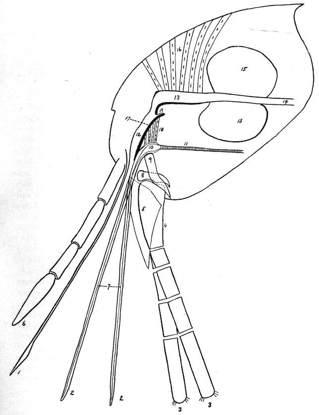
Plate I. MOUTH PARTS OF CERATOPHYLLUS FASCIATUS.
131
THE ACT OF BITING.
The epipharynx, or pricker, makes an opening into the skin, through which the mandibles are inserted. These organs, by means of their serrations and independent movement, then enlarge the opening as with a saw, permitting them, with the epipharynx, to pass deeper and deeper until the points of the maxilla rest upon the cutaneous surface. The labial palpi serve as a protective case when the organs are not in action. When in action they serve as a guide to the piercing organs, but are not inserted into the skin. They double up like a bow, on each side, the bend of the bow becoming greater and greater as the biting apparatus passes deeper and deeper. Mitzmain[206] has pointed out that the spring-like action of this bow may assist the flea to withdraw the mandibles and epipharynx.
During the process of penetration the salivary pump receives saliva from the salivary glands and pumps it down, through the channel in the mandibles, into the wound. It will be seen that the hypopharynx, being attached above by a membranous ligament and connected intimately with the mandibles below, moves downward with these organs as they pass through the skin. At the same time the muscles attached to its under surface and the salivary pump contract, enlarging the lumen of the pump. When the mandibles are retracted the salivary pump collapses, thereby forcing the saliva out with the movement upward of the mandibles. At the proper time the muscles operating the aspiratory pharynx contract, drawing the canal open and aspirating blood through the canal made by the approximation of the epipharynx and mandibles and into the aspiratory pharynx. When full, the muscles relax from before backward and the pharynx, by means of the elastic reaction of its chitinous lining, contracts and forces the blood backward through the gizzard and into the stomach. It has already been pointed out that the finger-like processes in the gizzard probably act as valves to prevent regurgitation from the stomach.
132
HOW THE FLEA INFECTS ITS HOST.
The exact method by which the flea can transmit plague from animal to animal has, in our opinion, never been satisfactorily explained. There have been several explanations offered: First, that the rat may eat the flea. Miller[207] has found that the Hepatazoon perniciosum is transmitted from rat to rat through the rat eating the mite, Lelaps echidninus, which acts as the intermediate host. We know, however, that when a rat is fed on plague material a mesenteric bubo is the rule, while in naturally infected rats a mesenteric bubo is a rare condition. This, then, negatives the possibility of plague being contracted through eating the flea.
Another explanation is that the infection comes from the saliva injected at the time of biting. We have already stated that after repeated examinations, both by dissecting out the salivary glands and by serial sections of the entire flea, plague bacilli have never been demonstrated in these glands or anywhere outside of the alimentary tract.
Another explanation has been advanced, that the bacillus is introduced by the contaminated mandibles. It is not possible to exclude this as a means of infection, although the Indian Plague Commission made numerous investigations and was unable to demonstrate the bacillus on the mandibles.
The possibility of infection taking place by regurgitation from the stomach has also been considered. As the stomach is guarded by the finger-like processes in the gizzard which seem to act as competent valves, and as the movement of the blood aspirated by reason of the mechanism already explained is in a backward direction, it would seem improbable that there is any regurgitation from the stomach.
The most plausible explanation that has been advanced has been based on an observation that blood-sucking insects at the time of biting frequently eject a drop of blood from the rectum. We know that the rectum may contain numerous plague bacilli, and it is supposed that this blood ejected in the vicinity of the bite is either brought in contact with the slight wound by the feet or mandibles of the flea itself or is rubbed in as a result of scratching. Verjbitski has shown that an emulsion of the feces of fleas or any plague material when placed upon the bitten part before the expiration of twenty-four hours is sufficient to give the animal plague. After twenty-four hours the animals did not develop plague, it being supposed that the slight wound in the skin made by the biting apparatus had healed. It is probable that this ejection of blood is purely accidental and does not necessarily occur at the time of biting, but it is likely that the insect had just previously had a full meal, which had been digested and passed into the rectum. In the many biting experiments done 133by McCoy and Mitzmain they report never having seen this ejection of rectal contents taking place. It might also be stated that where they used plague-infected fleas none of the animals developed plague after being bitten.
ENUMERATION OF FLEAS THAT HAVE BEEN FOUND ON RATS.
Various writers have reported the following fleas taken off rats:
Family SARCOPSYLLIDÆ Taschenberg.
Genus DERMATOPHILUS.
1. Dermatophilus cæcata Enderlein.—Seventeen specimens (females) were found by Doctor Enderlein on the skin behind the ears of a specimen of Mus rattus from Saopaulo, Brazil.
Genus ECHIDNOPHAGA Olliff.
2. Echidnophaga gallinacea Westwood.—Tiraboschi has found this flea on the Mus rattus in Italy.
3. Echidnophaga rhynchopsylla Tiraboschi.—This flea has been taken in Italy from Mus rattus and Mus alexandrinus. It has been described by Rothschild under the name of Echidnophaga murina.
Family PULICIDÆ Taschenberg.
Genus CERATOPHYLLUS Curtis.
4. Ceratophyllus fasciatus Bosc.—This is the common rat flea of Europe and the United States. It has also been found in Cape Town, Australia, and is occasionally found on rats in India.
5. Ceratophyllus londiniensis Rothschild.—This flea has been taken off mice in England; off rats in Italy (Ceratophyllus italicus Tiraboschi) and has been found once on Mus rattus in San Francisco, Cal.
6. Ceratophyllus acutus Baker.—This is the common flea of the California ground squirrel; and has been taken off Mus norvegicus in San Francisco, Cal.
7. Ceratophyllus anisus Rothschild.—This flea has been described by Rothschild from Yokohama, Japan, taken off Felis sp. One specimen was found in San Francisco, Cal., taken off Mus norvegicus.
8. Ceratophyllus niger Fox.—This flea is commonly found in San Francisco, Cal., in chicken yards and sparrows’ nests and has also been found on rats, Mus norvegicus, and on man.
9. Ceratophyllus consimilis Wagner.
10. Ceratophyllus lagomys Wagner.
11. Ceratophyllus mustelæ Wagner.
12. Ceratophyllus penicilliger Grube.
These fleas have been taken off Mus norvegicus in Europe.
134
Genus PULEX Linn.
13. Pulex irritans Linn.—This flea is widely distributed throughout the world, and while essentially the human flea has been found on many different species of animals and has frequently been encountered on rats. A very large number of specimens have been taken off rats in San Francisco, Cal.
Genus LŒMOPSYLLA Rothschild.
14. Lœmopsylla cheopis Rothschild.—This is the common rat flea in tropical and subtropical countries. It has also been found in seaports of the temperate zone, where it has been brought by ship rats. Ninety-eight per cent of the rat fleas in India are of this species. It has been found in Australia, where it was described by Tidswell under the name of Pulex pallidus. In the Philippine Islands, where it was described by Hertzog as the Pulex philippinensis. It has been found in Brazil, where it was described by Baker as Pulex brasiliensis, and Tiraboschi has found it in Italy, where it has been described as the Pulex murinus. This flea has been frequently found on man in India.
Genus CTENOCEPHALUS Kolenati.
15. Ctenocephalus canis Curtis.—This is the common dog flea found in many parts of the world and is frequently taken off rats.
16. Ctenocephalus felis Bouché.—This is the common cat flea and is also a widely distributed species. Frequently taken off rats.
Genus CTENOPSYLLUS Kolenati.
17. Ctenopsyllus musculi Dugés.—In England this flea is commonly found on the domestic mouse. It has a wide distribution and has been found on rats and mice in Europe, South Africa, India, Australia, Mexico, and other places, and has been taken off Mus norvegicus, Mus rattus, and Mus musculus in San Francisco, Cal.
Genus NEOPSYLLA Wagner.
18. Neopsylla bidentatiformis Wagner.—This flea has been taken off Mus norvegicus in the Crimea.
Genus HOPLOPSYLLUS Baker.
19. Hoplopsyllus anomalus Baker.—This is one of the common groundsquirrel fleas of California and has been found on Mus norvegicus in San Francisco and Palo Alto, Cal. That these squirrel fleas are occasionally found on rats is interesting from the fact that plague has been demonstrated both in rats and the ground squirrel in California.
135
Genus HYSTRICHOPSYLLA Taschenberg.
20. Hystrichopsylla tripectinata Tiraboschi.—Reported by Tiraboschi from Mus musculus in Italy.
Genus CTENOPTHALMUS Kolenati.
21. Ctenopthalmus agyrtes Heller.—Taken off Mus Norvegicus in England.
The results of the identification of 19,768 fleas in San Francisco and Oakland, Cal.
SAN FRANCISCO, 1908.
Host: Mus norvegicus.
Month. C. fasciatus. L. cheopis. P. irritans. Cten. musculi. Cten. felis, Cten. canis.
Male. Female. Male. Female. Male. Female. Male. Female. Male. Female.
April to July 31 1,343 2,510 485 837 31 76 78 211 16 31
August 489 883 145 228 156 206 27 90 17 22
September 543 1,180 655 930 339 387 33 109 46 119
October 254 435 509 652 59 64 9 45 6 18
November 129 252 256 288 52 69 20 54 6 6
2,758 5,260 2,050 2,935 637 802 167 509 91 196
Host: Mus rattus.
23 43 3 3 0 0 17 16 1 0
4 7 1 0 9 16 3 3 0 0
27 50 4 3 9 16 20 19 1 0
Host: Mus musculus.
4 10 1 0 0 0 2 10 0 0
11 10 1 6 4 4 0 3 1 1
15 20 2 6 4 4 2 13 1 1
OAKLAND, CAL., 1909.
Host: Mus norvegicus.
February 135 304 166 178 1 1 229 506 1 3
March 253 456 167 215 2 5 125 243 1 2
April 227 479 105 129 0 1 62 143 0 1
615 1,239 438 522 3 7 416 892 2 6
Host: Mus alexandrinus.
April 1 5 0 0 0 0 0 0 0 0
136This does not include a few other specimens of different species taken from Mus rattus and Mus norvegicus, which have been included under the heading of “Enumeration of fleas which have been found on rats.”
SYNOPSIS OF FLEAS COMMONLY FOUND ON RATS.
A. WITHOUT A COMB OF SPINES ON THE PROTHORAX OR THE HEAD.
1. Two bristles on the gena, an ocular bristle placed below the eye, an oral bristle placed just above root of maxilla. Mesothorax not divided by an internal incrassation. Claspers in male forming prominent hump, claw like and covered by hairy flap. An irregular row of about 10 teeth on inner side of hind coxa Pulex irritans.
2. Two bristles on gena, an ocular placed in front of and just above middle of eye, an oral bristle placed just above root of maxilla. Mesothorax divided by internal incrassation, claspers not forming prominent hump, not claw like, not covered by hairy flap. A regular row of about six teeth on inner side of hind coxa, Lœmopsylla cheopis.
AA. WITH A COMB OF SPINES ON PROTHORAX BUT NOT ON HEAD.
3. Three bristles on lower genal row, upper genal row represented by three or four small bristles running along anterior margin of antennal groove. Eye present, about five hairs on second joint of antenna, not as long as third joint. Maxillary palpi not as long as labial palpi. Labial palpi reach to apex of fore coxa. Spines on posterior tibia in pairs of about five groups. Head of male flattened on top, Ceratophyllus fasciatus.
AAA. WITH A COMB OF SPINES ON THE PROTHORAX AND ON THE HEAD.
4. Eye present, seven spines on lower margin of gena. Spines on posterior border of tibia in pairs Ctenocephalus canis or felis.
5. Eye absent, four spines on hind margin of gena. Spines on posterior tibia single and in a close set row Ctenopsyllus musculi.
Description of Plate II.
Fig. 1. Clasping organs of male. P Process.
F Finger.
M Manubrium.
IX St Ninth Sternite.
Fig. 2. Head of female.
Fig. 3. Terminal abdominal segments, female. 8 T Eighth Tergite.
8 St Eighth Sternite.
10 T Tenth Tergite.
10 St Tenth Sternite.
Sp Spermatheca.
Sty Stylet.
Fig. 4. Hind tibia.
CERATOPHYLLUS FASCIATUS Bosc.
[Plate II.]
Head.—Evenly and gently rounded in the female, flattened on top in the male. Frontal notch distinct. Eye present, placed low down in head. Gena acutely pointed posteriorly. Maxilla triangular. Maxillary palpi not as long as the labial palpi. Labial palpi reach to apex of anterior coxa, 5-jointed. Antennal groove in the male reaches to top of head, in the female to within one-third. There are 3 bristles on the lower genal row, the middle of which is the smallest, while the upper genal row is represented by 3 small bristles, extending along the edge of the antennal groove. In the male the lowermost bristle is frequently paired. There are several fine hairs above the eye. The occiput contains the normal row of apical bristles, the lowest of which is the largest. There is one bristle back of the middle of the antennal groove and a number of fine hairs along the posterior margin of the antennal groove. The antenna is 3-jointed, the first joint contains a row of about 5 very short fine hairs, while the second joint contains about 5 not as long as the third joint.
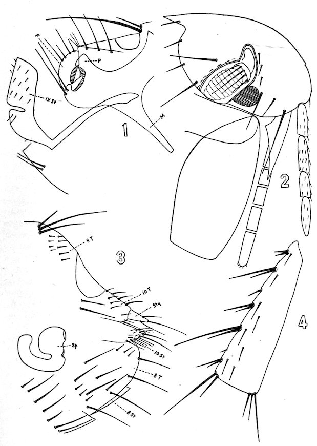
Plate II. CERATOPHVLLUS FASCIATUS, BOSC.
137Thorax.—The pronotum has one row of about 10 bristles, and a ctenidium composed of about 16 or 18 spines. The mesonotum has a posterior row of about 10 long bristles and there is an anterior row of more numerous smaller ones. The metanotum has also a posterior row of about 10 large bristles, with an anterior row of more numerous smaller ones, while still anterior to this there are 5 or 6 still smaller bristles. The metathorax contains 8 or 10 bristles which are small anteriorly, larger posteriorly. On the sternum of the metathorax there are 2 large bristles, while the episternum has 3 smaller ones. On the epimerum are 2 bristles placed anteriorly and 3 or 4 posteriorly, one of which is on the apical margin.
Abdomen.—The first stigma is nearly in line with those of the other abdominal segments. There are two rows of bristles on the abdominal tergites, a posterior of about 12 or 14 and an anterior of smaller, less numerous bristles. The antipygidial bristles in the female are 3 in number on each side, of which the middle is the longest, and the inner one the smallest. The male has but 2 antipygidial bristles on each side. The sternites from the third to the sixth have a single row of about 10 bristles, while the seventh has about 12. The metanotum has 2 teeth on each side, as have the first and second abdominal tergites. The third and fourth abdominal tergites have 1 tooth on each side.
Legs.—The fore coxæ are normally clothed. The fore femur has on the outer side 11 or 12 fine bristles irregularly disposed, while on the mid femur there is a row of about 3 to 5 bristles on the inner surface. The hind coxa has no patch of spines on the inner side, while on the inner surface of the hind femur there is a row of about 5 to 7 bristles. The spines on the posterior tibia are in pairs of six groups, while on the outer surface there is a row of about 7 bristles. None of the apical bristles of the tarsi are as long as the next succeeding joint. The fifth tarsal joints on all the legs have 5 lateral spines.
138Length of joints of tarsi:
Mid tarsi (♂) 8 7 4½ 3 7
Hind tarsi 18 11 7 4 8
Mid tarsi (♀) 8 7 5 3½ 7
Hind tarsi 21 13 8 5 8
Modified segments.—(♂). The manubrium of the claspers is straight and narrow, while the process extends upward as a short, blunt cone, where at the tip there are several fine hairs. The lower margin is evenly and gently rounded. The finger is short, extending but a little above the process. It is concave on its anterior surface and convex on its posterior, and from the posterior margin there are 2 large and 2 small bristles alternating. Two long heavy bristles arise from the process below the insertion of the finger. The ninth sternite is broad, with a deep sinus in its posterior border. Its lateral surface contains numerous fine hairs, these hairs being somewhat larger just beneath the sinus. Along the dorsal border of the tenth sternite there are 3 heavy bristles in line. At the tip of the tenth tergite there is one heavy bristle. Besides these heavy bristles in this segment there are numerous fine hairs.
(♀) The eighth tergite contains just anterior to the sensory plate about 12 small hairs while just beneath the sensory plate there are 2 long bristles. Lower down there is a patch of about 6 bristles and on the apical margin 4 to 6. The stylet is short, cylindrical, slightly larger at the base than at the tip, where there is a long bristle. On the under surface arises a fine hair. Substylar flap (tenth sternite) has along its margin numerous hairs.
Description of Plate III.
Fig. 1. Clasping organs of male. P Process.
M Manubrium.
F Finger.
IX St Ninth Sternite.
Fig. 2. Head of female.
Fig. 3. Terminal abdominal segments, female. 8 T Eighth Tergite.
8 St Eighth Sternite.
10 T Tenth Tergite.
10 St Tenth Sternite.
Sp Spermatheca.
Fig. 4. Hind coxa inner surface.
LŒMOPSYLLA CHEOPIS Rothschild.
[Plate III.]
Head.—Abruptly rounded. Flattened on top in ♂. Eye present. No ctenidia on head. Antennal groove in the ♀ reaches to within one-third of the top of the head. In ♂ reaches to top of head. Gena obtusely pointed posteriorly. Maxilla triangular. Maxillary palpi are not as long as labial palpi. Labial palpi reach to apex of fore coxæ, 4-jointed. Anterior edge of antennal groove overlapped by chitinous flap. On posterior edge of antennal groove are a number of small bristles, these being most distinct in the male. The first antennal joint in the male contains 4 or 5 hairs at its outer edge, while transversely there is a row of several fine hairs. The second joint has a row of fine hairs not as long as the third joint. Divisions marking separations of third joint most pronounced on dorsal edge. Two bristles on gena. The oral bristle placed low down just above the base of the maxilla; the ocular bristle in front and just above the middle of the eye. Six bristles on the posterior margin of the occiput on each side with 2 back of the antennal groove.
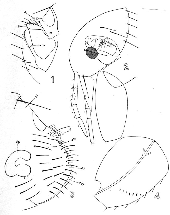
Plate III. LŒMOPSYLLA CHEOPIS, ROTHSCHILD.
139Thorax.—The pronotum is without a ctenidial comb, and has one row of about 14 bristles. The mesonotum, the broadest of the three thoracic nota, also has a single row of about 12 bristles. The metanotum has a single row of about the same number. The mesosternite contains about 5 bristles. The pleura of the metathorax is normally divided. The sternum contains 2 bristles, 1 anterior and 1 posterior. The episternum contains 1 bristle, and the epimerum contains 2 rows of bristles, an anterior row of 7 and an apical row of the same number.
Abdomen.—The first abdominal tergite contains 2 rows of bristles, an anterior and a posterior of about 6 bristles each, while the next 6 contains but a single row of about 14 bristles each, the lowest placed just below the stigma. From the seventh tergite springs a single antipygidial bristle. The sternites contain a single row of 8 or 10 bristles.
Legs.—The fore coxa is normally clothed. The fore femur has on its outer surface about 8 fine bristles. The mid femur has a single row of about 6 bristles, while the hind femur has a row of the same number. The hind coxa has on its inner surface a regular row of about 6 teeth. The hind tibia has on its posterior border 5 groups of spines in pairs, while on its outer surface there are about 8 small bristles in a row. The apical bristle on the second tarsal joint of the hind leg reaches to about the middle of the fifth tarsal article. The fifth tarsal article on all of the legs has 4 lateral spines and a subapical pair of hairs.
Modified segments.—(♂) The manubrium of the claspers is short and narrow. There are two free processes, the upper one, the finger, being broadest and wider at the tip than at the base, its upper border being more convex than the lower border and containing a number of bristles. The ninth sternite is club-shaped, is nearly straight on its dorsal margin, and the ventral margin contains a row of fine bristles from base to apex.
(♀) No bristles in front of the sensory plate. Along its apical margin externally there is a row of about 12 long bristles, and internally 140a row of less numerous, shorter bristles. Laterally there is a more or less regular row of about 8 bristles, and between this row and the apical row 3 or 4 more.
Description of Plate IV.
Fig. 1. Clasping organs, male. 10 T Tenth Tergite.
10 St Tenth Sternite.
P Process.
F Finger.
M Manubrium.
IX St Ninth Sternite.
Fig. 2. Head of female.
Fig. 3. Last tarsal joint of hind leg.
Fig. 4. Terminal abdominal segments, female. 8 T Eighth Tergite.
8 St Eighth Sternite.
10 T Tenth Tergite.
10 St Tenth Sternite.
Sp Spermatheca.
Fig. 5. Hind tibia.
CTENOPSYLLUS MUSCULI Dugés.
[Plate IV.]
Head.—The frons is prominent anteriorly, giving the head somewhat the shape of a fez. There are 4 spines on the posterior border of the gena. The antennal groove reaches to the top of the head. The maxillary palpi are shorter than the labial palpi, which reach to about two-thirds of the fore coxa and are 5-jointed. Maxilla triangular. Eyes absent. At the most prominent part of the frons anteriorly there are two short thick spines, while below these, running along the anterior margin, there are 5 bristles. Above there is an oblique row of 4 bristles, with 1 more placed near the top of the antennal groove. Between this oblique row and lower bristles there are numerous fine hairs. On the occiput there is a subapical row of about 7 bristles on each side, while in front of this are 3 oblique rows of bristles, the first containing 3, the second 4, and the third 5. On the posterior margin of the antennal groove there are several small hairs. On the first joint of the antenna there are about 3 hairs, while on the second joint there are 4 or 5, the longest somewhat longer than the third joint.
Thorax.—The pronotum has an anterior row of about 10 bristles, and a ctenidium of about 24 spines. The mesonotum contains about 4 rows of bristles, more or less regularly disposed, each row consisting of about 8 or 9 bristles. The metanotum has 2 rows of bristles, a posterior row of about 10 bristles, and an anterior of the same number, while there are several smaller bristles in front of this. The mesothorax contains about 10 bristles. The episternum of the metathorax has 2 bristles, and on the sternum there is 1 large one. The epimerum has 2 rows of 4 bristles each, with 1 large one at the apical margin.
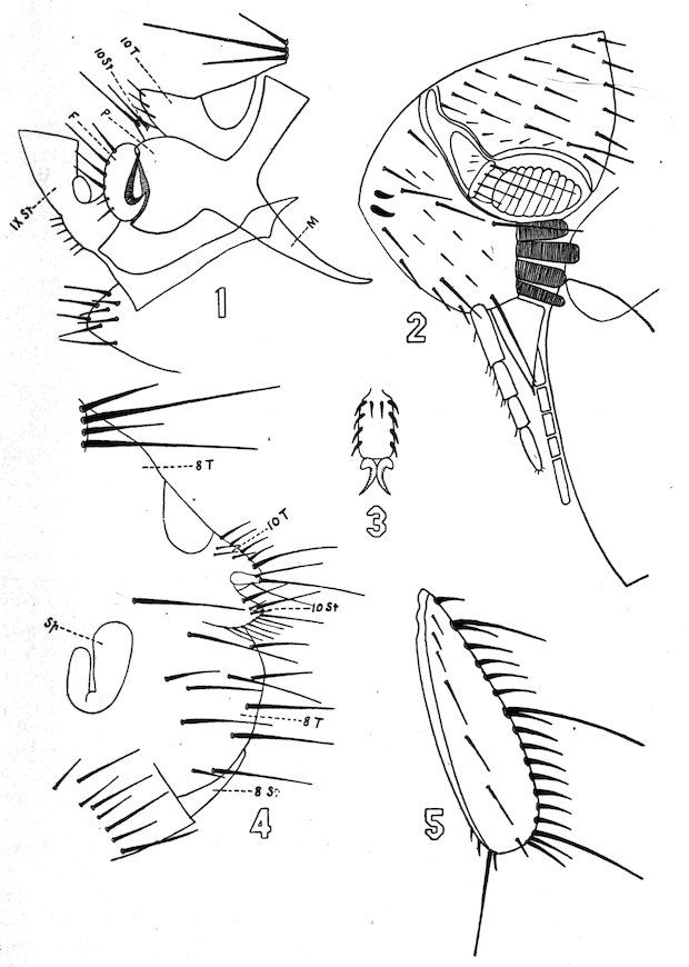
Plate IV. CTENOPSYLLUS MUSCULI, DUGES.
141Abdomen.—The first abdominal tergite has 2 rows of 10 bristles each, the posterior being comprised of the larger bristles. The next 6 tergites have 2 rows of bristles each, a posterior of large bristles, about 12 in number, and an anterior of smaller bristles, also 12 in number. On the apical edge of the metanotum there are 2 small teeth on each side. The first abdominal tergite contains 3 such teeth while the second and third have 1 each on each side. At the apex of the seventh tergite in the female there are 4 antipygidial bristles, sometimes 5. The male has but 3 antipygidial bristles. The abdominal sternites from the third to the sixth have a single row of 6 bristles. The seventh has a row of about 16 bristles.
Legs.—The fore coxa has about 32 large bristles more or less regularly disposed in 6 oblique rows. The hind coxa is without teeth on the inner surface. The mid femur is without bristles on its lateral surfaces. The hind femur is also without a row of bristles on its lateral surfaces. The spines on the posterior border of the tibia are single and in a close set row. The apical spines of the second tarsal joint of the hind legs are shorter than the third joint. The last tarsal joint on all the legs contains 4 lateral spines and a subbasal pair situated between the first lateral pair.
Modified segments.—(♀) Just beneath the pygidium is 1 long bristle. On the eighth tergite there is a patch of hairs, 6 of which are on the apical margin and about 5 or 7 anterior to these. The stylet is short, almost as wide at the base as at the tip, where there is a long hair. Posteriorly to this bristle there springs another one from the under surface.
(♂) Manubrium of the claspers is narrow, curved at the tip. The finger reaches to the level of the process, has a stout pedicle, is flat on its anterior border, and is decidedly convex on its posterior border, where there are 4 bristles. The shape of the ninth sternite is shown in the figure.
Description of Plate V.
Fig. 1. Clasping organs of male P Process.
F Finger.
M Manubrium.
IX St Ninth Sternite.
Fig. 2. Head of female.
Fig. 3. Terminal abdominal segments, females 8 T Eighth Tergite.
8 St Eighth Sternite.
10 T Tenth Tergite.
10 St Tenth Sternite.
Sty Stylet.
Sp Spermatheca.
Fig. 4. Hind coxa, inner surface.
142
PULEX IRRITANS Linnæus.
[Plate V.]
Head.—Evenly and abruptly rounded in both sexes. Frontal notch absent. Eye large. Maxillary palpi longer than the labial palpi. Labial palpi reach to about half the length of the anterior coxa and are 4-jointed. The mandibles are broad and markedly serrate. Maxillæ triangular. Antennal groove short and wide, closed behind, thickened on edges, and reaches to top of head in both sexes by chitinous thickening. Second joint contains 8 or 9 fine hairs, shorter than the third joint. Division of the third joint only to be seen on dorsal surface. Two bristles on the gena, one placed low down just above the maxilla, the other below the eye. From the lower margin of the gena occasionally may be seen a small tooth. One bristle on the occiput near the posterior lower angle. A few fine hairs on the posterior edge of the antennal groove.
Thorax.—The thoracic nota each contain a single row of about 10 or 12 bristles. There is no ctenidium on the pronotum. The mesosternite is narrow and is not divided by an internal incrassation. The episternum of the metathorax is large and contains about 2 or 3 bristles and is not quite separated from the sternum anteriorly. The epimerum has an anterior row of about 7 or 8 bristles and an apical row of about 6.
Abdomen.—Each of the abdominal tergites, with the exception of the first, has a single row of 8 or 10 bristles. The first has 2 rows of about 4 each. The sternites from third to seventh have a single row of about 6 bristles. There is one short antipygidial bristle on each side.
Legs.—The hind coxa has on its inner surface posteriorly a number of fine hairs, while anteriorly there are 10 or 12 teeth in an irregular line. The hind femur has on its inner surface a row of about 8 or 9 bristles. The spines on the posterior tibia are in pairs, and there are about 7 bristles in a line on its outer lateral surface. The apical bristle of the second tarsal joint of the hind leg reaches to about the middle of the fifth joint. The last tarsal joints of all the legs contain 4 lateral spines and a subapical pair, and between the third and last lateral spine there is a hair.
Modified segments.—(♀) The eighth tergite has no bristles above the pygidium but has numerous short stout bristles laterally and on and close to the apical margin. The stylet is short and stout and has at its tip a long hair. The tenth sternite and tergite contain numerous fine hairs, those on the sternite confined to the apical edge.
(♂) The male claspers are quite characteristic. The manubrium is large and curved and points ventrally. The claspers have two processes, the lower of which, with the finger, form together a kind of claw which is covered by the other process forming a flap, quite hairy on its upper margin. The ninth sternite is described very well by Rothschild[208] as “boomerang” shaped. The eighth tergite has a small manubrium.
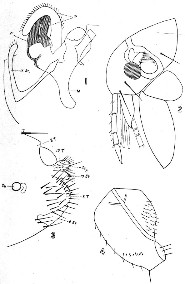
Plate V. PULEX IRRITANS, LINNÆUS.
143
Description of Plate VI.
Fig. 1. Clasping organs of male F Finger.
M Manubrium.
IX St Ninth Sternite.
Fig. 2. Head of male.
Fig. 3. Terminal abdominal segments, female 8 T Eighth Tergite.
8 St Eighth Sternite.
Fig. 4. Hind coxa and femur, inner surface.
CTENOCEPHALUS CANIS Curtis.
[Plate VI.]
Head.—Strongly and evenly rounded in both sexes. Eye large. Maxilla triangular. Maxillary palpi about as long as labial palpi. Labial palpi reach to two-thirds of anterior coxæ, 4-jointed. Seven spines along the lower margin of the gena. The posterior angle of the gena ends in a small tooth. Occasionally this may be absent. Antennal groove in the female reaches to within one-third of the top of head and is prolonged upwards by a chitinous thickening and in the male reaches almost to top of head. Two bristles on the gena, one placed well toward the anterior lower angle and the other in front of the eye. Usual number of bristles on posterior margin of the head, with 2 large ones back of the antennal groove. About 8 hairs on the second joint of the antenna nearly as long as the third joint.
Thorax.—A row of about 10 bristles on the pronotum, with a ctenidium of about 14 to 16 spines. Two rows of bristles on the mesonotum, a posterior of about 12, another of more numerous smaller bristles placed well anteriorly. The metanotum contains a single row of about 10 or 12 bristles. The episternum of the metathorax has 3 or 4 stout bristles, while the epimerum contains an anterior row of about 10 bristles and a posterior row of about 9.
Abdomen.—The first abdominal tergite contains 2 rows of about 4 bristles each, while the other tergites to the seventh contain a single row of from 12 to 16 bristles. The stigmata are large. There is a single antipygidial bristle on each side. The sternites from third to seventh have a single row of 4 bristles each.
Legs.—The hind coxa has on its inner side a patch of from 6 to 12 spines, while the hind femur has a row of 10 or 12 bristles on its inner surface. The spines on the posterior border of the hind tibia, with the exception of the apical, are in pairs, while in the apical group are about 3 stout bristles. The apical spine of the second joint of the hind leg reaches to nearly the middle of the fifth joint. On the fifth 144joint of all the legs there are 4 lateral spines and a subapical pair, and between the third and fourth lateral spines there is a hair.
Modified segments.—(♀) The eighth tergite has no hairs back of the stigma. The apical margin is rounded at the apex and contains 8 or 10 bristles. The stylet is short and wide and contains at its tip a long and a short bristle.
(♂) The manubrium is short and narrow. The movable finger of the clasper is short, thick, swollen at its middle, bluntly rounded at its extremity, and contains on its upper border numerous hairs and a few on its lower border.
Rothschild[209] has pointed out certain differences between the Ctenocephalus canis and Ctenocephalus felis. The differences are that in the female of the felis the head is longer and more pointed. This difference is not so pronounced in the male. Also certain differences in the shape of the claspers and the number of bristles in the episternum and epimerum of the metathorax and the hind femur, those in the C. canis being more numerous. Also that group of bristles on the posterior border of the hind tibia between the fifth pair and the apical bristles consists of two in the Ctenocephalus canis, while there is but a single bristle with a small hair in the Ctenocephalus felis.
REFERENCES.
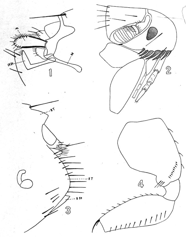
Plate VI. CTENOCEPHALUS CANIS, CURTIS.
145