THE ECTOPARASITES OF THE RAT.
Reading edition
By Nathan Banks,
Assistant Entomologist, Bureau of Entomology.
The ectoparasites of the rat fall naturally into three groups, the fleas, the lice, and the mites. These three groups are widely separated from each other, the mites belonging to the class Arachnida, having four pairs of legs, no segmentation to the body, no antennæ, and no compound eyes. The fleas and lice belong to the class Insecta. The lice are near the order Hemiptera, sucking insects without a complete metamorphosis, while the fleas are related to the Diptera and pass through a complete metamorphosis. All of these three groups, however, agree in one character—they are wingless. The mites and lice have flattened or depressed bodies, while the fleas have compressed bodies. All three groups have many other species which infest various other animals. Few, if any, of these parasites confine themselves to the rat, and all can walk or jump in the adult condition, so that they can easily transfer their attentions from one rat to another or to some other host. The majority of them are known to occur on mice, and several of the fleas and mites will readily attack man.
FLEAS—SIPHONAPTERA.
These wingless, compressed insects are known to all, but few have taken the trouble to look at them with much care. The adult female flea deposits her eggs among the hairs or fur of the host animal, but, unlike the eggs of many parasites, these are not fastened to the hairs and fall freely to the ground. These eggs are oval, whitish, and smooth, and about one-half millimeter long. The larvæ escape from the eggs in two to five days. They are enabled to break the eggshell by a slender process on the top of the head which disappears after the first molt. This larva is a slender, legless, cylindrical creature, whitish or yellowish in color, with a head and 13 segments. There are a few scattered hairs or bristles on the body and at the tip is a pair of corneous processes. On the upper part of the head is a pair of 70short, slender appendages, the antennæ or feelers. At the front of the head is a pair of biting jaws or mandibles. These larvæ feed on almost any kind of refuse; some have been reared on the sweepings from rooms. There is always some organic matter in this refuse, and this is doubtless their nourishment. The larvæ in houses usually crawl into cracks or under carpets and feed on the dust that occurs in such places. Those that infest wild animals probably feed on the refuse in the nests or retreats of these animals. They remain in the larval stage from a week to ten days, sometimes two weeks, molting the skin three times in this interval. Then they spin flat, white, silken cocoons, in which they transform to the pupal stage. Sometimes the cocoon is covered with particles of dust. In from five to eight days the adult flea emerges from the cocoon. The period of their transformation is affected by the temperature and moisture. In warm, damp weather a generation may develop in ten days or two weeks, but usually about eighteen days to three weeks elapse from the egg to adult. Although some moisture seems necessary to their development, an excess is apt to destroy the larvæ.
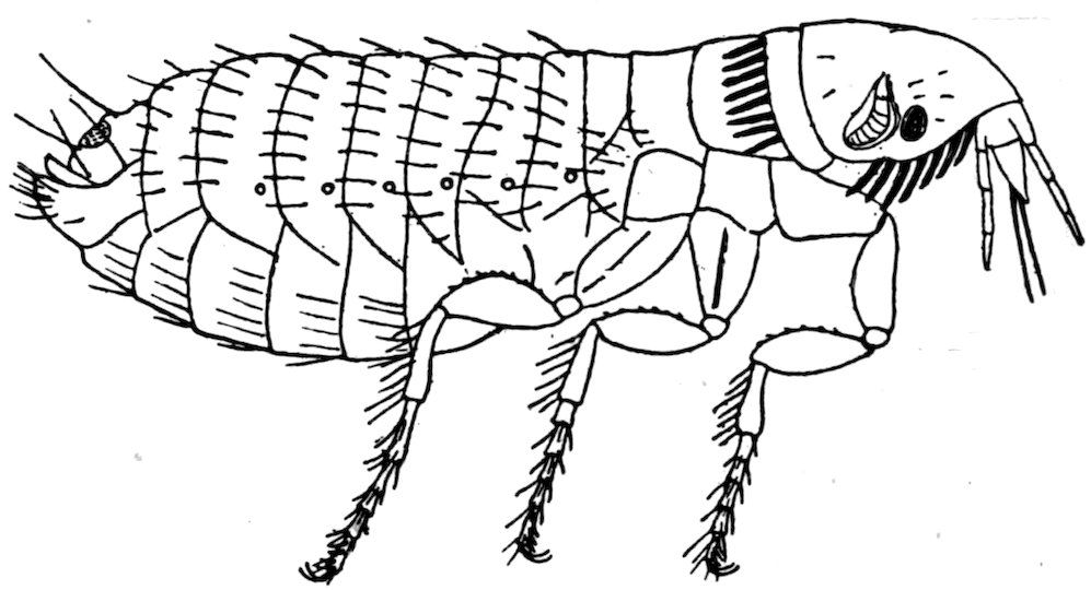
Fig. 6.—Flea, showing the various parts.
The leaping ability of adult fleas is familiar to all. No part of the leg is particularly enlarged, so that the jump is made by the entire leg as in the leaf-hopper insects, and not by the hind part of the leg as in grasshoppers and flea-beetles. The size of fleas is not as variable as in many insects. Most are about 2 to 3 millimeters long, while the range is about 1.5 to 6 millimeters. The adult flea has a hard, strongly chitinized body. The head is small, and on each side bears a short jointed antenna, which may repose in a groove or depression. Most species have a small, simple eye, but several forms are normally without eyes. The sides of the head below the antennæ are called the genæ. At the lower front end of the head is the mouth and mouth parts. The latter consist of a pair of triangular maxillæ with jointed maxillary palpi and a beak or proboscis made up of one median and four lateral pieces. The outer pair of lateral pieces is the labrum with the imperfectly jointed labial palpi. 71They serve as a sheath for the other organs, which are more slender. The inner pair of pieces are considered to be the mandibles and the median piece a labrum or hypopharynx. Others call this piece the unpaired piercing organ, the lingua, or the syringostome. There are other interpretations of the homologies of the mouth parts, but the above is the most generally adopted one. The labrum and the mandibles are roughened and constitute the piercing organs which the flea inserts into the host to tap a blood vessel. On the lower part of the head there is frequently a series or comb of stout spines. Similar spines sometimes occur on the posterior border of the pronotum. These series of spines are called “ctenidia,” and they are of great value in classification. Behind the head are three segments, or zoonites, each bearing a pair of legs. These together form the thorax. The upper surface is called the notum (pronotum, mesonotum, etc.). The sides are the pleura—sometimes “epimera” is used; and the ventral part is the sternum. Each of the thoracic segments has a spiracle, or a breathing pore, on each side. The first segment of the thorax, called the “prothorax,” is shorter than the others, and, as above stated, frequently has a row of ctenidia, or spines, on its posterior border. The next segment is the mesothorax, and the third the metathorax. The metathorax usually has some stout bristles in rows on its pleura, which are enlarged and called “epiphyses,” formerly called “squama aliforme.” The basal one or two segments are sometimes partly covered by the epiphyses or the metathorax. These segments consist of a dorsal plate, or tergite, and a ventral plate, or sternite. Behind the thorax is the abdomen of 9 apparent segments. Seven of these segments have a spiracle or breathing pore on the sides. The last segment, or pygidium, bears the genital organs; in the male certain processes called “claspers” at each side of the genital opening. The anal aperture is at the end of the ninth segment between the dorsal and ventral plates. The claspers have a main curved part, and a slender backward projection called the “manubrium,” and at the apex an articulated clawlike process called “the movable finger.” At the tip of the abdomen of the female there is a short median piece called the “style.” The legs consist of five parts: The coxa, a large basal piece; the trochanter, a minute piece at the end of the coxa; the femur, which is usually slightly swollen in the middle; the tibia, which usually has stout bristles or spines on its posterior side; and the tarsus, which consists of five parts or joints. The basal joint is often the longest, and the comparative lengths of these joints is expressed by a formula, as 60–45–32–18–30. The last, or fifth, joint has been called the “metatarsus,” but this name is better applied to the basal joint. At the tip of the last tarsal joint is a pair of stout claws. The coxæ of legs II and III show a longitudinal suture.
72Fleas as a rule prefer certain hosts, but are not as particular in this regard as are many parasites. Those species which are best known are found to attack several hosts, including man. This catholicity of taste is what makes them dangerous parasites, the possible transmitters not only of plague, but also of consumption, leprosy, etc. The fleas are treated by various writers under other names, such as Aphaniptera, and Suctoria. About 300 species are described, and perhaps as many more will be gathered by collectors. Formerly all fleas were kept in the genus Pulex; now they are arranged in many genera, and these genera grouped into families. No less than eight such families are recognized by some authorities on this group. The species that occur on rats belong to three families, which may be separated as follows:
1. Thoracic segments much shortened and constricted; labial palpi apparently not jointed; third joint of antennæ without subjoints; no ctenidia; abdomen of female becomes more or less swollen Sarcopsyllidæ
Thoracic segments not shortened nor constricted; labial palpi with joints; third joint of antennæ with several more or less distinct subjoints; ctenidia often present; abdomen of female never distinctly swollen 2
2. Posterior tibial spines in pairs Pulicidæ
Posterior tibial spines mostly single and more numerous Ctenopsyllidæ
CTENOPSYLLIDÆ.
To this family belongs the Ctenopsylla musculi Dugès.
This was formerly placed in the genus Typhlopsylla. The head is rather acute in front and has four ctenidia each side; the eyes are very small; the pronotal comb has 22 spines; each dorsal segment of the body has two rows of hairs; the basal row of smaller hairs. The proportions of joints in the hind tarsus are: 45–25–17–8–14. Length 1.8 to 2.5 millimeters. This species is abundant on rats and mice in Europe and other countries; recently it has been taken in California and Florida on rats and mice.
PULICIDÆ.
This family includes the greater number of fleas. They have been arranged in many genera, six of which have been taken from rats. These are separable as follows:
1. Head without ctenidia; eyes distinct 2
Head and pronotum with ctenidia; last tarsal joint with four pairs of lateral spines 5
2. Pronotum with ctenidia; female with one antepygidial bristle on each side Hoplopsyllus.
Pronotum without ctenidia 3
3. Last tarsal joint with four pairs of lateral spines; female with one antepygidial bristle each side 4
Last tarsal joint with five pairs of lateral spines; female with two to five antepygidial bristles each side Ceratophyllus.
734. Mesosternite very narrow, without internal rod-like incrassation from the insertion of coxa upward Pulex.
Mesosternite with a rod-like internal incrassation from the insertion of coxa upward Xenopsylla.
5. Eyes rudimentary; female with two to five antepygidial bristles each side Neopsylla.
Eyes distinct; female with but one antepygidial bristle each side Ctenocephalus.
Hoplopsyllus, one species, described as a Pulex.
Hoplopsyllus anomalus Baker.
The mandibles scarcely reach halfway down on the anterior coxæ; upon each are two large spines; the pronotal comb has about nine spines each side; and each abdominal segment has but a single row of bristles. The hind femora have six to eight bristles on the side; the proportions of the joints in the hind tarsus are: 26–16–8–5–13. Color, dark reddish brown. Female, 2.5 millimeters; male, 1.5 millimeters.
Described from a spermophile from Colorado and recorded by Doctor Fox and Professor Doane from Mus norvegicus from California.
Pulex.—Of this, the typical genus of the family, but one species has been recorded from rats.
Pulex irritans Linn.
The mandibles reach about halfway down on the anterior coxæ; the head is regularly rounded in front; there are no transverse rows of bristles on the vertex, and but one row of bristles on each abdominal tergite. The proportions of the joints in the hind tarsus are, 50–30–18–12–32. Color, usually yellow brown. Male, 1.6 to 2 millimeters; female, 2 to 3.5 millimeters.
This, the human flea, is quite cosmopolitan, but more abundant in warm countries than elsewhere. It occurs on many domestic animals and has frequently been taken from rats in California and elsewhere; it also occurs on skunks.
Xenopsylla.—This genus includes the following species, formerly placed in the genus Lœmopsylla.
Xenopsylla cheopis Rothschild.
The mandibles reach nearly to the end of the anterior coxæ; there are no ctenidia on the head or pronotum; the eyes are distinct; each abdominal tergite has but one row of bristles; the hind femur has a row of about eight bristles; the proportions of the joints in the hind tarsus are as follows: 46–30–16–10–20. Color, light brown. Male, 2.5 to 3.5 millimeters; female, 4 to 5.5 millimeters.
This is a true rat flea, but will readily bite man, and is the species chiefly concerned in transmitting the bubonic plague. It is widely distributed, especially in seaport towns.
Ceratophyllus.—Fleas of this genus are abundant on many kinds of small mammals, especially rodents. There are a great many species 74and some are so closely related that it is not easy to identify them. Of the eight species recorded from rats, four have been taken in this country. It is not practicable to tabulate these eight species, but the four that occur in our country may be arranged as follows:
1. Hind tarsal joint II with an apical spine much longer than joint III acutus.
Hind tarsal joint II with spines not longer than joint III 2
2. Pronotal comb of about 26 spines niger.
Pronotal comb of about 18 or 20 spines fasciatus and londiniensis.
Ceratophyllus niger Fox.
This species has the pronotal ctenidia of about 26 spines; there are a few hairs on the inner surface of hind femur; apical spines of second joint of hind tarsus not longer than third joint; three hairs in front of the eye and three in front of these; movable finger of claspers with five slender bristles on the outer edge. Color, very dark brown. Length 3.5 millimeters.
Taken in California from Mus decumans and from man.
Ceratophyllus acutus Baker.
This species is readily known by having a spine at tip of the second joint of hind tarsus longer than the third joint and reaching over onto the fourth joint; the abdominal tergites have each two rows of bristles; the male claspers are very large and long, sickle shaped. Color, pale brown. Length, 3 to 3.5 millimeters.
It was described from a spermophile, but Doctor Fox has taken it once from a rat in California.
Ceratophyllus fasciatus Bosc.
There are 18 or 20 spines in the pronotal comb; there are three bristles in front of eye and in female two, and in male four in front of these; there are three or four hairs on the inner surface of the hind femur; the proportions of joints in the hind tarsus are 50–33–20–11–21. The manubrium of the male claspers is very long and slender, and some of the bristles on the movable finger are as long as the joint. Length, male, 1.8 millimeters; female, 2.5 millimeters.
It has been recorded from California on rats, mice, skunks, and man. It is also common in Europe and elsewhere on rats, mice, and other small animals.
Ceratophyllus londiniensis Rothschild.
This is allied closely to C. fasciatus, and is best separated from that species by the shape and armature of the genital parts; the manubrium is not as long as in that species, and the bristles on the movable finger are shorter; the third joint of the maxillary palpi is proportionally longer than in C. fasciatus. There are three bristles in front of the eyes and four or five in front of these; there are a few hairs on the inner surface of the hind femur; the proportions of the joints in the hind tarsus are 46–30–18–11–18.
75It has been recorded by Doctor Fox from Mus rattus in California, and is known from rats and mice from several parts of Europe; the C. italicus Tiraboschi is the same species.
The four other species of this genus found on rats and not yet found in our country are closely related to C. fasciatus, and distinguished chiefly by the shape of the male genitalia.
Ceratophyllus mustelæ Wagner.
This species has no series of hairs on the inner surface of the hind femur; there are three bristles in front of the eye and six in front of these; the pronotal comb has 18 or 20 spines; the proportions of the joints in the hind tarsus are 47–37–20–13–20; the movable finger of the male clasper has a long process below not seen in other forms. Occurs (according to Rothschild) on rats in Europe.
Ceratophyllus pencilliger Grube.
This species also has no hairs on the inner surface of the hind femora. The pronotal comb has 18 spines; there are three bristles in front of the eye and four in front of these; the proportions of the joints in the hind tarsus are 52–36–23–14–24; the outer corner of the movable finger of the male clasper has two little rounded processes. It was described from Siberia, but according to Rothschild occurs on rats in Europe.
Ceratophyllus consimilis Wagner.
This species is very close to C. fasciatus, and has some fine hairs on the inner surface of the hind femur; there are but two bristles in front of the eye and in front of these a few finer hairs; the proportions of the joints in the hind tarsus are 42–30–20–11–19; pronotal spines 18. Occurs on rats in Russia.
Ceratophyllus lagomys Wagner.
This species also has a few fine hairs on the inner surface of the hind femur; 18 spines in pronotal comb; there are three bristles in front of eye and one in front of these; the proportions of the joints in the hind tarsus are 53–32–20–11–22; the outer corner of the movable finger of the male clasper has two little processes, similar to those on C. pencilliger. Occurs on rats in Europe.
Ctenocephalus.—The common fleas on cats and dogs, as well as on man, belong to two species long kept under one name (C. canis or C. serraticeps), but lately shown by Rothschild to be distinct. Both have a comb of 8 spines on the head and 16 spines in pronotal comb; the proportions of joints in the hind tarsus are 40–24–15–10–24. Both are occasionally taken on rats in this country. They may be separated as follows:
1. In the female the head is fully twice as long as high (seen from side); the first spine of genal comb is two-thirds the length of the second; in male the manubrium of claspers is barely enlarged at tip; and with two rows of hairs on disc of movable finger C. felis Bouché.
76 In the female the head is less than twice as long as high (seen from side); the first genal spine in the head comb is only about one-half the length of the second; in the male the manubrium of clasper is very distinctly enlarged at tip; but one row of hairs on the disc of the movable finger C. canis Curtis.
Neopsylla.—One species of this genus has been described from the brown rat in Europe.
Neopsylla bidentatiformis Wagner.
The eyes are very small; there are 4 pairs of lateral spines beneath the last joint of the hind tarsus; the comb on head consists of but 2 stout spines, below the middle of the antennæ; the pronotal comb has 18 spines; the proportions of the joints in the hind tarsus are 43–33–21–12–21.
Length: Male, 2 to 2.3 millimeters; female, 2.3 to 2.5 millimeters.
Not yet found in the United States; described from Russia.
SARCOPSYLLIDÆ.
The fleas of this family are commonly called “chigoes,” “jiggers,” or sand fleas. The head is usually larger proportionally than in the other fleas; there are no ctenidia on head or pronotum; the thoracic segments are extremely short, and in the female the abdomen enlarges with the development of the eggs. They do not hop about as other fleas, but remain on the spot to which they have attached until they die. Frequently the adjacent skin grows over them, forming a swelling of considerable size.
Three species belonging to two genera have been recorded from rats.
1. Angle of head acutely produced; fifth tarsal joint of hind legs without heavy spines; few spines on the legs Sarcopsylla.
Angle of head not produced, obtuse; fifth tarsal joint with heavy lateral spines, and other spines on other parts of the legs Echidnophaga.
Echidnophaga.—Two species of this genus are known from rats; one, however (E. gallinacea), can hardly be called a normal parasite, but rather of accidental occurrence. The genus has also been called Argiopsylla and Xestopsylla.
1. Bristles at end of second joint of hind tarsus about as long as next three joints; palpi about one-half the length of mandibles E. rhynchopsylla.
Bristles at end of second joint of hind tarsus about as long as next two joints; palpi about two-thirds the length of the mandibles E. gallinacea.
Echidnophaga rhynchopsylla Tiraboschi.
The body is about twice as long as broad and shining brown; there is but one hair in front of eye, and four on each metathoracic pleuron; mandibles larger than in E. gallinacea, and the spiracles are much higher up on the sides than in that species. Length, 1.4 to 1.8 millimeters.
Taken from Mus rattus in Italy.
77Echidnophaga gallinacea Westwood.
This species has the body almost as broad as long, and of a red-brown color; 1 bristle in front of eye and 6 on each metathoracic pleuron; each abdominal tergite has on each side near the median line a single hair; the spiracles are situated well down on the sides. Length: Male, 0.8 to 1.2 millimeters; female, 1 to 1.8 millimeters.
This species is a fairly common pest of poultry and dogs in warm countries, and is called the “chicken flea.” It has been taken from rats in Italy.
Sarcopsylla.—This genus includes the S. penetrans, which attacks the feet of various animals, including man, in the Tropics. This species has not yet been recorded from rats, but an allied species is described from Brazilian rats.
Sarcopsylla cæcata Enderlein.
Color, clear yellowish. Eyes rudimentary; lower anterior corner of coxæ prolonged in a tooth; tarsal joints very short; claws long, but little curved, and almost hair like. The body of a swollen female is about 5 millimeters long.
Taken from Mus rattus in Brazil.
LICE—ANOPLURA.
The insects known as Pediculi, or lice, are parasitic during their entire life on various mammals, including man. They are flat, rather elongate, wingless insects, with a small head and stout legs, which end in a strong claw, opposable to a projection at the tip of the penultimate joint. The simple antennæ, three to five jointed, are inserted in a concavity on the side of the head. The mouth parts are of a very peculiar nature, and not yet homologized with the cibaria of other insects. There is a short beak or proboscis in front, with recurved spines or hooks on its dorsal and lateral surfaces. Through this beak extends a slender stylet, that is formed of three parts; a ventral channeled piece, perhaps a labium; a dorsal piece, consisting of two pieces fused together, perhaps the maxillæ; and a median tube, possibly the hypopharynx. The stylet is used to pierce the skin of the host, and the blood is sucked up through it. There are no palpi. On each side of the head there is a small, simple eye. The thorax shows only incompletely the division into the three parts; there is a large spiracle above on each side. The abdomen shows eight segments, six of them have a spiracle, or breathing pore, on each side, the basal and apical segments being without them. All of the segments bear a few simple hairs or bristles; the longest are on the posterior segments. The legs are stout and prominent; they consist of a broad coxa, a small trochanter, a longer femur, a tibia with an apical process, and a tarsus of one joint and a very large terminal claw. At the apex of the tibia, just within the projection, is a sucking disc. This, the projection, and the claw form the apparatus to hold fast to the hair of the host.
78Lice usually walk sideways, but do not travel much, and they keep close to one host. The eggs are slightly elongate and fastened to the hair of the host. They hatch in about ten to fifteen days, the young coming out of the top of the egg. These young do not differ much in structure from the adults, but are paler in color. They molt their skin a few times, probably four, before they reach the mature condition. The males are less numerous than the females, and ordinarily smaller. There are several generations each year, dependent doubtless on the temperature; but the life history is not thoroughly known for any species. After sucking the blood the abdomen of the louse becomes somewhat distended, very noticeably so in some species.
The sucking habits of the lice render them dangerous parasites and capable of transmitting a disease from one host to another. Fortunately they do not readily change hosts so that they can not be considered quite as dangerous as some more active parasites. However, several species have already been shown to carry diseases in laboratory experiments. Therefore it is probable that some of them will be connected with the origin and diffusion of certain diseases of animals.
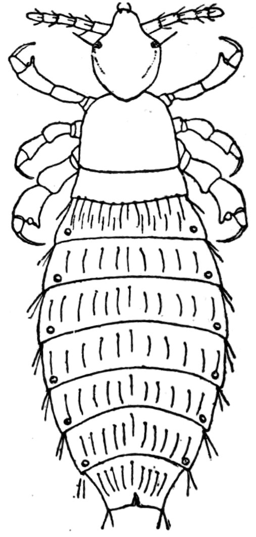
Fig. 7.—Louse (Polyplax spinulosa).
The Anoplura, or lice, have often been treated in connection with the Mallophaga, or biting lice. This is doubtless because they frequently occur on the same animal, and have a general resemblance to them. However, they have no real affinity to these insects, and the general opinion is that they are more or less related to the Hemiptera. Sometimes they are treated as a group or section of the Hemiptera, but also as a separate order, under various names as Siphunculata, Lipognatha, Pseudorhynchota, and Ellipoptera.
There are about 50 or 60 known species which are arranged in 15 genera and 4 families. Four species belonging to three different 79genera have been recorded from rats; a number of others are known from mice and other rodents, and some of these will probably yet be taken upon rats.
These four forms are separated, as follows:
1. Eyes large and distinct; beak very short; thorax plainly longer than broad Pediculus capitis.
Eyes small, beak longer; thorax about as broad as long 2
2. In male the pleura of abdominal segments 3 to 6 above and below have a prominent tooth-like projection; a tooth on the hind femur of female Hoplopleura acanthopus.
In male the pleura of abdominal segments without such projections; no tooth on hind femur of female 3
3. Last joint of antennæ much more slender than those before; an acute tooth at sides of segments 4 to 7; head much narrower in front; antennæ two-thirds as long as head Polyplax miacantha.
Last joint of antennæ not much more slender than others; head quite broad in front; antennæ as long as head Polyplax spinulosus.
Pediculus capitis De Geer.
It is pale grayish in color, with faint dark markings at the sides of the thorax and abdomen; the last segment of the abdomen in female is bilobed. The head is longer than broad and tapers in front. Length, 2 millimeters. This is the head louse of man, and is said to have been taken from rats, and is claimed to be able to transfer plague from rats to man. Its occurrence on rats, however, appears to be very uncommon.
Hopopleura acanthopus Burmeister.
In the male the pleura of the abdominal segments 3 to 6, which reach up on the dorsum and over on the venter, have at their inner ends a prominent projection, toothed in all except the third on dorsum and the sixth on venter, which are spine-like. The head is but little longer than broad, broad in front; and in the female there is a recurved tooth on each hind femur. The last segment of the female abdomen is bilobed behind. Length, 1.3 millimeters. It has been taken from rats in Europe, but is more common on species of Microtus.
Polyplax spinulosus Burmeister.
The sides of the abdominal segments are acute, but the males do not have the large tooth-like projection of Hoplopleura. The last segment of the female is truncate; the head is about as broad in front as behind, and the legs are very short and stout; the antennæ are as long as head, and the last joint is but little smaller than the others. Color, pale yellowish. Length, 1.4 millimeters.
This is the common rat louse, and is probably as widely distributed as its host. Specimens have been taken in both the eastern and western parts of the United States.
80Polyplax miacantha Speiser.
This differs from P. spinulosus in having a longer and narrower anterior part of head, in that the last joint of the antennæ is more slender, and the antennæ are only two-thirds as long as the head. The abdominal segments 4 to 7 show an acute process at the sides. Length, 1.5 to 1.75 millimeters.
Taken from rats in Abyssinia.
MITES—ACARINA.
The mites (order Acarina, class Arachnida) are readily known from the insects (fleas and lice) by having four pairs of legs, no antennæ, and the abdomen does not show any segmentation, nor is there usually any distinction between head and thorax. In some groups there is a small head-like part, called the capitulum. The mouth parts consist of a pair of mandibles (often styliform or needle-like), a lip, and a pair of palpi. In some forms there is a central piece, called the hypopharynx, and in other groups is a plate above the mouth parts, known as the epistome. The body usually shows more or less distinctly a division into two parts—the anterior, called the cephalothorax, and the posterior the abdomen. However, in many mites it is not possible to separate these parts, except that it is considered that the legs are borne by the cephalothorax. In many forms there is a small, simple eye each side on the cephalothorax, but many other forms are blind. Some species have a tracheal system, which opens in a pair of spiracles near the hind legs or near the anterior end of the body; other species have no definite respiratory system. The genital aperture is on the venter, usually between the legs. The legs consist of the usual joints—coxa, trochanter, femur, tibia, sometimes a metatarsus, and a tarsus. The tarsus terminates in a pair of claws, sometimes three or only one, and often a sucker or caroncle. Most mites are not parasitic; those species that are parasitic are often free in one stage. The parasitic mites suck the blood of their host, feed on the hair or dermal scales, or burrow in the skin. Some predaceous species inhabit animals to hunt and eat the parasitic mites that infest that animal.
The mites that occur parasitically on rats belong to four families: Sarcoptidæ, Cheyletidæ, Ixodidæ, and Gamasidæ.
1. A distinct spiracle or breathing pore on each side of body near coxæ III or IV 2
No such spiracle or pore visible 3
2. A small, distinct head part in front of the body; palpi three jointed; a granulate area around the spiracle; no sternal plate Ixodidæ.
No such head part; palpi five jointed; no granulate space around spiracle, but a long, chitinized piece reaching forward from it; a more or less distinct sternal plate Gamasidæ.
3. All legs simple, unmodified, ending in a stalked sucker Sarcoptidæ.
Front legs short, enlarged, and modified for clasping; all legs end in one or two stout claws Cheyletidæ.
81
IXODIDÆ.
The Ixodidæ, or ticks, are rather large, flat, leathery-skinned mites, which suck the blood of various animals. In the male the dorsum of the body is nearly covered by a corneous shield, while in the female this shield occupies only the anterior part of the body. In the female the body swells to enormous proportions as she engorges herself on the blood of the host. At the posterior margin of the body there are in many forms a series of lobes or festoons. There is no species of tick that is commonly found on rats, but four species that normally infest other animals have been taken from them.
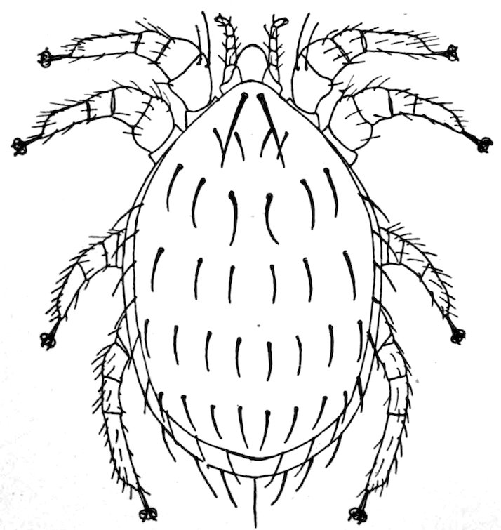
Fig. 8.—Mite (Lælaps echidninus).
1. On the venter is a groove in front of the anus and extending back each side; no festoons to posterior margin of body; palpi rather long Ixodes ricinus.
On the venter there is no groove in front of the anus, but usually one behind; festoons distinct in males and unengorged females 2
2. Palpi very short with transverse ridges; shield of female narrowed behind eyes; stigmal plate nearly round Margaropus annulatus.
Palpi short, without transverse ridges; shield of female not narrowed behind eyes; stigmal plate comma shaped Rhipicephalus sanguineus.
Palpi elongate, without ridges; shield of female broad; stigmal plate oval, Hyalomma aegypticum.
Ixodes ricinus Linné.
The shield of the female is elliptical, plainly longer than broad, sides not suddenly narrowed behind, and there is no eye-spot at each lateral corner. The coxa I has a long sharp spine.
This is a common European tick found on sheep, cattle, dogs, etc., and it has been taken a few times in this country. Neumann has recorded its capture from Mus decumans.
82Margaropus annulatus Say.
The shield is plainly longer than broad, with a distinct eye-spot at each lateral corner, and behind the eye the shield is suddenly narrowed; the coxæ of the female are without spines, but the male has 2 on coxæ I. This is the common cattle tick of the United States, and disseminates the Texas fever. Mr. Hunter has taken it once from a rat in a barn at Dallas, Tex.
Rhipicephalus sanguineus Latreille.
The shield of the female is oval, and longer than broad, with an eye-spot at each outer corner. Coxa I with 2 teeth; a smaller tooth on each of the other coxæ. Stigmal plate long, comma shaped. In the male there is a corneous plate each side of the anus, and on middle of posterior margin a projection, or short tail.
This species is common in tropical countries, and Nuttall has recorded specimens from the black rat in India.
Hyalomma aegypticum Linné.
The shield of the female is as broad as long, and the eye-spot is slightly above each outer corner. Coxa I has 2 large teeth, and a small tooth on each of the other coxæ. In the male there are 2 corneous plates each side of anus, and behind is a pair of small tubercles.
This is a common tick in the warmer parts of the Old World; and Nuttall has recorded young specimens on the black rat.
GAMASIDÆ.
The Gamasid mites, although much smaller than the ticks, are large enough to be seen by the naked eye. They are active, and most are not parasitic, at least for part of their time. The palpi are simple, of 5 joints; the mandibles are elongate, retractile, and usually chelate at tip. There are no eyes. The dorsum and often the venter shows one or more corneous shields or plates, frequently a number of them; one or two on the dorsum, and on the venter one between the coxæ, called the sternal plate; one behind this, the genital plate; one behind the latter, the ventral plate; and one surrounding the anus, the anal plate. Frequently some of these are absent or united to one of the others.
The legs are slender, usually of 6 joints, with a long tarsus that terminates in 2 claws, and often a sucker, or caroncle. The stigmata, or spiracles, are lateral above and between coxæ II and IV, and usually provided with a slender peritreme reaching forward toward the head. Nearly all the Gamasidæ deposit eggs, and the young often differ considerably from the adult in structure. There are two, and perhaps sometimes three, nymphal stages. In one of these nymphal stages the mite is apt to attach itself to an insect for the purpose of being carried to a similar locality, where it may feed and mature. The coprophagous and xylophagous insects are especially concerned in the diffusion of these mites.
83There are, however, quite a number of species that are genuine parasites of insects and other animals. Those occurring on rats belong to two genera, Myonyssus and Lælaps. They can be separated as follows:
1. Anal plate small, much smaller than the ventral plate Lælaps.
Anal plate large, larger than the ventral plate Myonyssus.
Myonyssus.—This genus is made by Tiraboschi for one species: Myonyssus decumani Tiraboschi.
Body oval; legs short and stout, all tarsi with a large caroncle with two short claws; coxæ II have a large tooth on the anterior border, none of coxæ with spines; sternal plate much broader than long, with three bristles each side; ventro-genital plate much longer than broad, broadest behind, bordered with bristles; anal plate very large, nearly one and a half times as broad as long; three large spines each side on venter. Length, 0.95 millimeter.
Found in Italy on Mus decumans.
Lælaps.—This genus embraces a large number of species, several of which occur on small animals, such as the muskrat, ground hog, and chipmunk. Three have been recorded from rats, one of these from California. The dorsal plate is covered with hairs or bristles, and there are usually stout bristles on the margins of the plates on the venter. There is also a bristle, or a spine, at the tip of the anal plate. The legs are short and stout, with a distinct caroncle, and two claws.
1. Dorsum with numerous fine hairs; no stout spines on coxæ L. stabularis.
Dorsum with fewer, but stouter spine-like bristles; each coxa has a stout spine 2
2. Body but little longer than broad; ventral plate longer than broad L. agilis.
Body much longer than broad; ventral plate about as broad as long L. echidninus.
Lælaps echidninus Berlese.
Dorsum of body almost wholly covered with a shield, with rows (six in front, eight behind) of stout, curved bristles, a longer pair near front margin, and some around lateral and posterior margins. Legs short and stout, tarsi about twice as long as preceding joint; each coxa bears a stout spine near middle. Palpi very short; sternum with three stout bristles or spines each side; ventral plate with four stout bristles each side; anal plate with a stout apical bristle, and a small one each side. Length, 1 millimeter.
Occurs commonly on rats in warm countries, and known from California. It may possibly aid in the transmission of disease.
Lælaps agilis Koch.
Similar in many respects to L. echidninus but differ in the shorter and proportionately broader body, barely longer than broad, and in the weaker and shorter spines on dorsum and on the ventral plates; there are also some small short spines on the general surface of the venter. Length, 0.7 millimeter.
84Recorded from rats from Europe and Africa.
Lælaps stabularis Koch.
The body is of the same general shape as in L. echidninus, but the dorsum is clothed with 12 to 18 rows of fine short hairs. The first pair of legs is more slender than in the other species, and the hind legs are also more elongate; the coxæ do not have the stout spines seen in the other species, and the bristles on the sternal and ventral plates are much less stout; the general surface of the venter has many hairs, the anal plate has a short apical bristle. Length, 1.2 millimeters.
Taken on the brown rat in Italy; also found in manure.
CHEYLETIDÆ.
This family consists of small, soft-bodied mites, that are parasitic or predaceous in habits. The palpi are small, three or four jointed; the mandibles are styliform and retractile; and the breathing spiracles open near the mouth parts. The species that occur on rats belong to the genus Myobia.
Myobia.—The body is elongate, fully twice as long as broad, tipped by a pair of long, stout bristles. The first pair of legs is enlarged and shortened, with a terminal hook to grasp hairs; the other legs are short, simple, and far apart. The palpi and mouth parts are very small, and the dorsum bears stout bristles. They are supposed to feed on the exudations of the skin, but it would seem more probable, from the nature of the mandibles, that they pierced the skin to secure food. All are very small, not one-half a millimeter long. Of the several species two have been recorded from rats.
1. Dorsum of female with spines all acute and sharp M. musculi.
Dorsum of female with some of the posterior spines flattened and rather scale like M. ensifera.
Myobia musculi Schrank.
This occurs on various mice and moles, and once recorded from the brown rat. It has been taken in this country on mice. It lives at the base of the hairs.
Myobia ensifera Poppe.
This was described from the brown rat in Europe. The female is separated from M. musculi by having about six of the posterior dorsal spines flattened and scale like; in the male the six dorsal spines are longer, and the small spines much smaller than in M. musculi.
SARCOPTIDÆ.
These are the itch and scab mites. The body is soft, rounded, and whitish in color. The legs are very short, of five joints, and end in one or two claws, and often a pediceled sucker. The palpi are small 85and short, of three joints, but the basal is usually united to the rostrum. There are no spiracles, and respiration is therefore through the general surface of the skin. The sexes are often quite different in structure. The females usually deposit eggs, the larvæ are hexopod, and there are two nymphal stages. They are all parasites, mostly on mammals and birds, and often burrow in the skin, causing mange, or scabies.
Only one species has been taken on rats; this belongs to the genus Notoedres.
Notoedres.—In this genus the third pair of legs of the male and the third and fourth of the female have no sucker at the tip. The anal opening is on the posterior part of the dorsum. The three known species are parasitic on mammals—one on the cat, one on the rabbit, and the third on rats.
Notoedres muris Mègnin.
This is a rounded mite, with finely striate skin, a small triangular rostrum; in front the four anterior legs project a little beyond the body, and each ends in a long pedicellate sucker; the third and fourth pairs of legs are not visible from above, and each ends in a long bristle. There are a few short hairs around the anal aperture and about ten others in front of these. The species measures about 0.3 to 0.4 millimeter long.
It usually occurs about the ears and the genital organs of the host, and has been taken from both the brown and black rat in Europe. The Sarcoptes alepis Railliet and Lucet is the same species.
DEMODECIDÆ.
Besides the mites above described, a form of Demodex has been recorded from rats, but the species is not given. These mites are very tiny, with elongate body, the posterior part annulate, the front part with eight very short legs. They inhabit the hair follicles of various mammals. That on the rat may have been only an accidental occurrence of some species normally on another animal.
87