Chapter IV · THE FLUIDS OF THE BODY (continued 2)
Reading edition
The Spleen.—The spleen is placed on the left side of the body, and rather towards the back. It rests between the stomach and the inner surface of the eighth, ninth, tenth, and eleventh ribs. It is quickly distinguished from other organs by its brown-purple colour, a sombre hue to which it owed its evil reputation with the humoralists. The liver’s yellow bile tinged man’s mental outlook, preventing him from seeing objects in their natural brightness; but the spleen made black bile, which, mounting to the brain, displayed its malign influence upon the action of that organ, as, or in, the worst of humours.
The spleen is invested with a capsule of no great toughness. Inside the capsule is “spleen-pulp.” When the fresh organ is cut across, it is seen that, although most of the pulp is of the colour of dark venous blood, it is mottled with light patches. In some animals—the cat, for example—these whitish patches are small round spots, regularly arranged at a certain distance from the capsule. The distinction into “red pulp” and “white pulp” marks a division into two kinds of tissue with entirely different functions. The white pulp is lymphoid tissue, lymph-follicles developed in the outer or connective-tissue coat of the branches of the splenic artery. Its function is to make lymphocytes, of which, for reasons which will shortly appear, the spleen needs an abundant supply. The constitution of the red pulp is entirely different, and peculiar to the spleen. The branches of the splenic artery divide in the usual way into smaller and still smaller twigs until the finest arterioles are reached; but these arterioles do not give rise to capillary vessels. At the point at which in any other organ their branches would attain the calibre of capillaries, the connective-tissue cells which make their walls scatter into a reticulum. They are no longer tiles with closely fitting, sinuous, dovetailed borders, but stellate cells with long delicate processes uniting to constitute a network. The blood which the arterioles bring to the pulp is not conducted by closed capillary vessels across the pulp to the commencing splenic veins. It falls into the general sponge-work. The venules commence exactly in the same way as the arterioles end. Stellate connective-tissue cells become flat tiles placed edge to edge. The endothelium of an arteriole might be likened to a column of men marching shoulder to shoulder, three or four abreast; the connective tissue of the pulp, to a crowd in an open place. The column breaks up into a crowd. On the other side the crowd falls into rank as the endothelium of veins. The capsule and the red pulp are largely composed of muscle-fibres. These relax and contract about once a minute. By their contraction the blood is squeezed out of the sponge.
If the spleen be enclosed in an air-tight box (an oncometer), from which a tube leads to a pressure-gauge—a drum covered with thin membrane on which the end of a lever rests, or a bent column of mercury on which it floats—the pressure-gauge shows the changes in volume of the spleen. The long end of the lever, which records the variations of pressure in the gauge, may be made to scratch a line on a soot-blackened surface of travelling paper. A record of the variations in volume of the organ, which can be studied at leisure, is thus obtained. It shows that the spleen is sensitive to every change of pressure in the splenic artery. Small notches on the tracing correspond to the beats of the heart. Larger curves record the changes of blood-pressure due to respiration. A long slow rise and fall marks the rhythmic dilation and contraction of the spleen itself.
One of the three large arteries into which the cœliac axis divides delivers blood to the spleen direct from the aorta. The splenic vein joins the portal vein shortly before it enters the liver. Thus the spleen is placed on a big vascular loop which directs blood, not long after it has left the heart, from the aorta, through the spleen, to the liver.
The peculiar construction of the splenic pulp which brings the blood more or less to rest within its sponge-work, and the transmission to the liver of the blood which leaves the spleen, indicate that it is an organ in which blood itself receives some kind of treatment. It is not passed through it, as it is through all other parts of the body, in closed pipes. The spleen is a reservoir, or a filter-bed, into which blood is received.
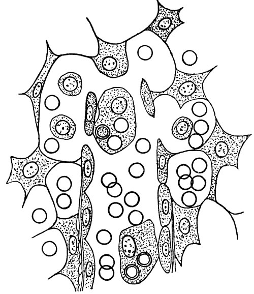
Fig. 5.—A Minute Portion of the Pulp of the Spleen, very highly magnified.
Stellate connective-tissue cells form spaces containing red blood-corpuscles and leucocytes. In the centre of the diagram is shown the mode of origin of a venule. It contains two phagocytes—the upper with a nucleus, two blood-corpuscles just ingested, and one partially digested in its body-substance; the lower with two blood-corpuscles.
The red blood-corpuscles of mammals are cells without nuclei, and with little, if any, body-protoplasm. They are merely vehicles for carrying hæmoglobin. We should deny to them the status of cell, if it were possible to prescribe the limit at which a structural unit ceases to be entitled to rank as a cell. They are helpless creatures, incapable of renewing their substance or of making good any of the damage to which the vicissitudes of their ceaseless circulation render them peculiarly liable. It is impossible to say with any approach to accuracy how long they last, but probably their average duration is comparatively short. The spleen is a labyrinth of tissue-spaces through which at frequent intervals all red corpuscles float. If they are clean, firm, resilient, they pass through without interference. If obsolete they are broken up. In the recesses of the spleen-pulp, leucocytes overtake the laggards of the blood-fleet, attach their pseudopodia to them, draw them into their body-substance, digest them. The albuminous constituent of hæmoglobin they use, presumably, for their own nutrition. The iron-containing colouring matter they decompose, and excrete in two parts; the iron (perhaps combined with protein); the colouring matter, without iron, as the pigment, or an antecedent of the pigment, which the liver will excrete in bile. Hæmoglobin is undoubtedly the source of bilirubin, and general considerations lead to the conclusion that it is split into protein, iron, and iron-free pigment in the spleen; but the details of this process have never been checked by chemical analysis. Neither bile-pigment nor an iron compound can be detected in the blood of the splenic vein. The only evidence of the setting free of iron in the spleen is to be found in the fact that the spleen yields on analysis an exceptionally large quantity of this metal (the liver also yields iron), and that the quantity is greatest when red corpuscles are being rapidly destroyed.
As a rule, it is very difficult to detect leucocytes in the act of eating red corpuscles; but under various circumstances their activity in this respect may be stimulated to such a degree as to show them, in a microscopic preparation, busily engaged in this operation. The writer had the good fortune to prepare a spleen which proved to be peculiarly suitable for this observation (Fig. 5). His method was an example of the way in which a physiological experiment ought not to be conducted. Having placed a cannula in the aorta of a rabbit, just killed with chloroform, he was proceeding to wash the blood out of its bloodvessels with a stream of warm normal saline solution, when the bottle from which the salt-solution was flowing overturned. Fearing lest an air-bubble should enter the cannula, he hastily poured warm water into the pressure-bottle, and threw in some salt, in the hope that it would make a solution of about 0·9 per cent. The salt-solution was allowed to run through the bloodvessel for rather more than an hour. When sections of the spleen were cut, after suitable hardening, every section was found to be packed with leucocytes gorged with red corpuscles. Some of the corpuscles had just been ingested; from others the hæmoglobin had already been removed. It may be that, for some unknown reason, the destruction of red corpuscles was occurring in this particular rabbit with unusual rapidity at the time when it was killed; but it seems more probable that the animal’s leucocytes were provoked to excessive activity by changes in the red corpuscles brought about by salt-solution which was either more or less than “toxic.” As a score of attempts to reproduce the experiment, with solutions of different strengths, have failed, it is impossible to be sure that this is a valid explanation.
There must be something in the condition of worn-out red corpuscles which either makes them peculiarly attractive to predatory leucocytes or renders them an exceptionally easy prey. It does not require much imagination to picture the drama which is enacted in the spleen. Slow-moving leucocytes are feeling for their food. The majority of red corpuscles pass by them; a few are held back. The leucocytes, like children in a cake-shop, cannot consume all the buns. A selection must be made, and preference is given to the sticky, sugary ones. Red corpuscles when out of order show a tendency to stick together. When blood is stagnating in a vein, or lying on a glass slide in a layer thin enough for microscopic examination, its red discs are seen after a time to adhere together in rouleaux. The parable of a child in a cake-shop is not so fanciful as it may appear.
The differentiation of function of organs is not as sharp as was formerly supposed. Evidence of their interdependence is rapidly accumulating. The activity of various organs is known to result in the formation of by-products termed “internal secretions,” which influence the activity of other organs, or even of the body as a whole. The spleen enlarges after meals. This may be merely connected with the engorgement of the abdominal viscera which occurs during active digestion, or it may indicate, as some physiologists hold, that an internal secretion of the spleen aids the pancreas in preparing its ferments. The spleen enlarges greatly in ague and in some other diseases of microbial origin. This has been regarded as evidence that it takes some part in protecting the body against microbes. But whatever may be the accessory functions which it exercises, they are not of material importance to the organism as a whole, seeing that removal of the spleen causes no permanent inconvenience either to men or animals. Its blood-destroying functions are taken on by accessory spleens, if there be any, and by lymphatic glands. The marrow of bone also becomes redder and more active. Under certain circumstances, red corpuscles, or fragments of red corpuscles, are to be seen within liver-cells; but it is uncertain whether blood-destruction is a standing function of the liver.