Chapter X · MUSCLE (continued 2)
Reading edition
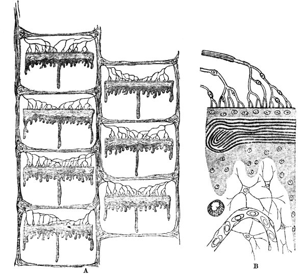
Fig. 18.—Electric Organ of a Skate in Longitudinal Section— A, Slightly, B, Highly Magnified.
A shows the compartments into which septa of fibrous tissue divide the organ. In the centre of each compartment is a disc formed from a modified muscle-fibre. Nerves ramify in abundance on its anterior surface. B, a minute portion of a disc. At the top are seen nerve-fibres in delicate nucleated sheaths; then follow the nucleated layer with which they come in contact, the contorted laminæ which represent the striations of the muscle-fibre, the granular nucleated substance of its posterior layer, some connective tissue, a capillary bloodvessel containing oval nucleated corpuscles. In a tissue space, a single coarsely granular leucocyte is to be seen.
The usefulness of a torpedo’s electric organs is unmistakable. They are powerful enough to paralyse every animal that touches its back, whether foe or little fish suitable for food. But of what service is its feeble battery to a skate? This and the allied question as to the advantages which can have accrued to the ancestors of the torpedo who first began to change innocent muscle into a weapon of offence are usually answered by pointing to the liability of flat fish lying on the bottom of the sea to become resting-places of parasites, corallines, and other fixed growths. Very mild shocks would suffice to disturb the peace of would-be settlers. In the same way, the electric organs of fresh-water fish may, when rudimentary, have protected the skin from invasion by moulds.
Luminous Glands.
If it be difficult, when considering the dispersal of energy as mechanical work, heat, or electricity, by living tissues, to bring the phenomena into line with those of which physics takes experimental cognizance, how are we to approach the problems involved in the generation of light? Yet the photogenic property of protoplasm is widely distributed. Protozoans and various other invertebrate animals cause the so-called phosphorescence of the sea. The abysmal depths of ocean are lighted by forests of luminous polyps, and traversed by fishes whose heads are furnished with lamps. By her own light the female glow-worm enables her winged mate to keep his tryst. Fireflies (Lampyrus) flash amongst the orange-trees of Italy, and blaze (Pyrophorus) beneath the mangoes of Ceylon.
Luminous organs vary too widely in structure to allow us to pick out, as in the case of electric organs, the features which are common to them all. In Pyrophorus the organ is a double mass of cylindrical cells near the tip of the abdomen. The cells are set vertically to the surface, and are supported by a tubular membrane. Their substance contains a kind of fat. Beneath them there is a layer of cells, not luminous, but evidently a part of the photogenic apparatus, containing chalky granules. The organ is well supplied with nerves and with respiratory tubes (tracheæ).
More interesting than its structure is the study of the peculiar character of the light which the organ emits. It gives a spectrum which extends from the red (beyond Fraunhofer’s line B of the solar spectrum) to the first blue rays (F). It shows no lines. Green rays appear only when the light is bright, and then they are the brightest of all the rays. The light is practically destitute of actinic or chemical rays. A photographic plate may be exposed for several minutes, almost without changing, to the light of a firefly bright enough to enable one to read with ease in a dark room; whereas light of equal brilliance from any other source would change it in the fraction of a second. Nor are heat-rays mixed with the light. Measurements show that the activity of the photogenic organs does not give rise to any greater rise of temperature than would occur in the case of any other gland.
The contrast between the emission of light by an animal and its production in any other manner is very striking when the physical evidence, or want of evidence, of what happens in the protoplasm which produces it is considered. The fact that no heat accompanies the light precludes us from attributing it to oxidation. If a firefly is enclosed in a vessel of oxygen, its lamp burns no brighter—clear evidence that its luminosity has nothing in common with the burning of a match or the glowing of a stick of phosphorus. Nor is the lamp put out when the insect is suddenly exposed to great cold (-100° C.). It continues to shine until the cold kills it. There is no relation between the luminosity of a firefly and the phenomenon termed “phosphorescence” by physicists. Sulphide of calcium—the substance used for rendering matchboxes visible in the dark—returns light which it has absorbed. A firefly’s power of emitting light is in no wise affected by keeping it for a long while in the dark.
Like all other events in vital chemistry, the generation of light by protoplasm is due to a process of fermentation. The luminous organs may be crushed, and the mixture of fermentable substance and ferment extracted with water. The extract is luminous. If an extract is prepared rapidly, and evaporated to dryness in vacuo, the residue glows when moistened with water. That two substances are present in the extract, one (luciferin) fermentable, the other (luciferase) a ferment, is proved by the following experiment: A certain quantity of extract is divided into two portions. One part (A) is allowed to glow until its capacity for emitting light is exhausted. The other portion (B), as soon as it is separated, is heated to 55° to kill the ferment. B still contains luciferin; A contains luciferase, although all its luciferin has been used up. Recombined, the extract is luminiferous.