Chapter XI · THE NERVOUS SYSTEM
Reading edition
Twenty-five years ago a new process was introduced for colouring the elements which by their combination make up the nervous system. With its aid anatomists discovered the inadequacy of their conceptions of nerve-cells. It was already known that a nerve-fibre—that is to say, its essential part, its core—is a part of a cell, the body and other parts of which are situate within the brain or spinal cord, or in one of their dependents, a ganglion. But the new method showed the nerve-cell as more elaborate in form than anything which had been imagined hitherto; and since the word “cell” was often loosely used when the cell-body alone was referred to, it seemed worth while to give the unit of structure a new name. The term “neurone” was introduced to emphasize its functional individuality. The nervous system is an association of neurones.
By the extremely simple expedient of placing a small block of nerve-tissue in bichromate of potassium, and then transferring it to nitrate of silver, jet-black pictures of nerve-cells are obtained showing with amazing completeness all the details of contour of their bodies and all the intricacies of branching of their limbs. The most surprising feature of the process is the absence of confusion in its results. Dyes were in use which stained one kind of cell better than another, or picked out a particular part—usually the nucleus—of every cell. If the chrome-silver process had acted in the same way, a dense black preparation in which no details could be distinguished would have been the result. But instead of treating all cells alike, the process blackens one cell here and another there, leaving hundreds or thousands untouched. It shows no preference for any particular kind of cell. In one section large cells are picked out, in another small ones; in a third no nerve-cells are blackened, but connective tissue is brought into view. When the block of tissue soaked with bichromate of potassium is immersed in a solution of nitrate of silver, the chromate escapes from it into the surrounding liquor much more quickly than the nitrate gets in; and when at last the nitrate of silver enters, it finds that some of the cells have fixed the chromate in their substance. This retained chromate combines with silver. The product is rapidly reduced to a black subchromate. No explanation of the fixing of the chromate by individual cells has yet been offered. It is a remarkable fact that another process which similarly makes choice amongst the elements has since been introduced, giving even more valuable results. Pieces of fresh tissue are placed in a very dilute solution of methylene blue. When staining is satisfactory, nerve-cells alone take up the dye. The selection of individual nerve-cells is not carried so far as it is by the chrome-silver method, but it is exhibited to a certain extent. It is probable that nerve-cells live (in a physiological sense) longer than other tissue-elements. Methylene-blue contains some easily removable oxygen of which the oxygen-starved nerve-cells take advantage. The reduced methylene-blue remains in their substance, so that when the preparation is reoxidized by exposure to air the pattern of the nerve-cells is rendered conspicuous. When a few cells are selected, it is, presumably, because they were the only ones alive at the time when the dye entered the tissue. Preparations made from the wall of the alimentary canal seem to justify this simple explanation. They show patches in which muscle-fibres are stained, patches in which there is no staining, and intermediate zones in which nerve-cells are coloured and muscle-fibres are not. But the hypothesis is inadequate to meet all cases. When first employed, the blue was injected into the animal in successive doses until it killed it. The staining was believed to occur intra vitam. Subsequently it was found that its application to fresh tissue, or, for certain results, to tissue which has been kept for some hours, is equally effective.
Without an understanding of the nature of the two new processes, and of the character of the results which they yield, it would be impossible for the reader to realize the extraordinary advance in our knowledge of the finer structure of the nervous system which has marked the period during which they have been employed.
The chrome-silver process is the more useful for the central nervous system. Methylene-blue gives better results with tissues containing minute nerve-cells and the branches of nerves. The latter method has revealed such a profusion of nerve-twigs as would never have been suspected but for its use. Consider, for example, the lining epithelium of the lungs (p. 168). Every one of its flattened cells has its own nerve twig or twigs. They lie between the cells. They give branchlets which enter them. A similar statement might be made regarding the richness of the nerve-supply of the muscle-fibres of the alimentary canal, or of the cells of glands, and possibly of other tissues. Each fresh success achieved in the application of the method makes a further revelation of the abundance in which nerves are distributed, increasing our sense of the dependence of all vital processes upon nervous control, and our appreciation of the unifying and integrating importance of the nervous system.
The term “neurone” is used by certain writers with a view to emphasizing their belief, not in the functional individuality alone of the unit of structure, but also in its anatomical isolation. The peculiarity of the methods of coloration which we have described lies, as already pointed out, in their selecting the cells which happen to be in a particular nutritive condition, and ignoring their neighbours. Hence pictures of separate and discrete units are obtained. This proves the nutritive autonomy of the cells, but it does not necessarily follow that A is not structurally connected with B, and B with C. Impulses are passed along the chain from A to C. Functionally, therefore, they are linked together; but until the question as to the way in which contact is established is settled, it is as well to think of the neurones as anatomically discrete.
It would be impossible in this book to describe all the varieties of neurone, for nothing is so characteristic of these elements as their enormous range both in size and form. It may be truly described as having no limits. Each of the two electric organs of Malapterurus is governed by a single neurone. Its cell-body is a fifth of a millimetre or more in diameter—large enough to be seen with the naked eye—and traversed by capillary bloodvessels. The axon of this nerve-cell—its single nerve-fibre—ramifies to supply a separate branch to each of the 2,000,000 chambers of the electric organ, and each branch breaks up into a bunch of twigs within the chamber. Contrast with such a giant cell as this one of the granules of the retina or cerebellum, the smallest cells to be found in the body, yet each a perfect neurone, exquisitely elaborate in form.
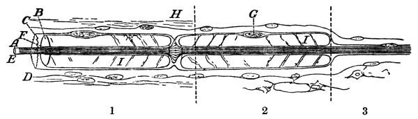
Fig. 19.—A Nerve-Fibre consisting of A, the Undivided, Fibrillated Axon of a Nerve-Cell, with its Various Wrappings.
In segment 1 the wrappings comprise B, a tube of phosphatic fat (myelin), interrupted at H, a node of Ranvier; C, a delicate membrane (sarcolemma); D, connective tissue; E, the rind of the axon; F, a tubular space containing lymph, between the axon and its sheath of myelin; G, nucleus of an enwrapping cell. At I the myelin is seen to be divided into overlapping conical rings. 2, The medullated nerve-fibre, running an isolated course, is merely enclosed in a tube of connective tissue containing lymph. 3, As a “grey” or “non-medullated” fibre, the axon has lost its myelin sheath.
As types for description we may take one of the motor cells of the spinal cord and a granule of the cerebellum. Every nerve-fibre which supplies a group of voluntary muscle-fibres is a thread drawn out from a large cell-body which lies in the grey matter of the spinal cord or of the axis of the brain. The fibres pass out in the anterior root of a spinal nerve or in a cranial nerve. The cell-body may have a diameter of as much as 100 µ (1 µ = 0·001 millimetre). In shape it is like a very irregular starfish, owing to its being continued into several, usually four or five, thick tapering branching limbs or processes, known as dendrites, in addition to its slender thread-like axon. From its origin in a cell-body to its destination in a muscle—it may be a few inches, or it may be a yard away—the axon is an unbroken thread. A short distance from the cell-body it enters a tubular sheath, which protects and insulates it, recalling the covering of gutta-percha in which the wires of a telegraph cable are enclosed. The sheath is of a phosphatic fat, invested and held in place by a delicate transparent membrane, neurilemma. Beneath this membrane nuclei occur at regular intervals, and midway between each two nuclei the sheath is cut across by a septum. Such interruptions or nodes show that the sheath is not a part of the nerve, if the term is used in the most restricted sense. Each internode is a cell which has been wrapped round the nerve for its protection. The axon with its sheath is spoken of as a nerve-fibre. A large number of nerve-fibres bound together by connective tissue constitute a nerve. In some cases the axon before it leaves the spinal cord, but after it has entered its myelin sheath, gives off one or two lateral branches (“collaterals”), which return to arborize in the grey matter of the cord. It does not appear that they are always present in the case of the motor neurones of the spinal or cranial nerves—probably they are usually omitted—but collaterals are important features of the large neurones of the cortex of the cerebrum and cerebellum (Figs. 23, 24). Usually two, three, or four such branches start off at right angles from the axon, and after a time turn back towards the surface, dividing into a few extremely slender branches. Their purpose is an enigma. Possibly they bind a group of cells together in functional unison. Such an explanation would seem reasonable in the case of an arrangement of collaterals on the plan we have just described; but in various situations in the brain cells are seen of which the axons, instead of becoming nerve-fibres, break up completely into collaterals, which branch repeatedly.
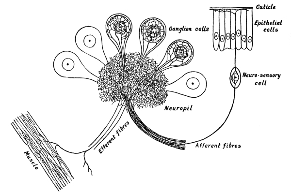
Fig. 20.—A Ganglion of a Leech.
Pear-shaped cells are set round a felt-work of nerve-fibrils (neuropil). A neuro-sensory cell is shown with one fibre directed peripherally, branching on the surface; and one directed centrally, ramifying in the neuropil. Several very slender fibrils from the neuropil pass up the stalk of each ganglion-cell. They join a network near its surface. This net is connected by radiating fibrils with a coarser net which surrounds the nucleus. From the central net a relatively stout fibril passes to muscle-fibres.
By various methods it may be shown that dendrites, cell-body, and axon contain fibrils (Fig. 22). These neuro-fibrillæ lie parallel to one another in the axon. Where it divides they are distributed amongst its branches. Possibly they also branch. In the neurones of Malapterurus, already referred to, this would appear to be inevitable. The discovery of neuro-fibrillæ seemed to carry us a step nearer to a comprehension of the physics of nervous conduction. They clearly indicate that particles of the substance of a nerve-fibre are oriented in the direction in which impulses pass. It is a structural differentiation similar to the fibrillation of muscle, and probably of the same order—a response to the same demand. But when we examine the arrangement of the fibrils in a cell-body and its dendrites, the appearances which we discover serve to perplex us. They complicate instead of simplifying our mental picture of the conduction of nervous impulses. The coarsest and most distinct neuro-fibrillæ are to be found in annelids, the ganglion-cells of a leech, for example, affording excellent preparations. These cells are pear-shaped, with a single stalk. As is usual in invertebrate animals, they do not exhibit separate dendrites and axon, but dendrites and axon pass out from the cell in the common stalk. The bodies of the cells are set round a felted mass of nerve-filaments, into which their stalks break up. Just beneath the surface of the stalk of one of these cells two or three very fine neuro-fibrillæ are to be seen. A single, much coarser fibril occupies its axis. The fine fibrils join a network at the periphery of the cell-body. The thick fibril is connected with a coarser network which surrounds the nucleus. Radiating threads unite the finer with the coarser net. It has been suggested that afferent impulses ascend the fine fibrils, pass from the finer to the coarser net, and take their exit by the thick fibril, which can be traced into a motor nerve. Such a transit could not, so far as one can imagine, have any effect upon the distribution of the impulses which pass through the neurone; besides, there are reasons for believing that the course taken by impulses which are delivered to the ganglion by sensory nerves is determined by the felt-work in its centre, the neuropil. It is probable that during their passage through the cell-body impulses acquire the energy requisite to discharge the muscles to which the motor-fibre carries them. In vertebrate animals, sensory nerves are branches of neurones of which the cell-bodies lie in cranial or spinal ganglia. They resemble the ganglion-cells of the leech in as much as they are unipolar; both branches, the one which collects impulses from sense-organs, and the one which distributes them to the spinal cord, come off from the cell in a common trunk which afterwards divides, although the unipolar condition of the cell of the spinal ganglion is not primitive, but acquired. In the earliest stages of its growth the cell is bipolar. Its two ends subsequently grow together for a certain distance, the common portion being the vertical limb of the T (cf. Fig. 21, which shows the growth of a granule of the cerebellum). The body of the cell contains a network not unlike the network of the leech. It is probably related to what may be termed the charge of the neurone, the development of a suitable degree of force in the impulses which pass through it.
The neuro-fibrillæ of a large nerve-cell, such as a motor cell of the spinal cord, are exceedingly slender (Fig. 22). They branch and reunite. A certain number gather towards the axon; but the majority pass through the cell from one dendrite to another, or from one branch of a dendrite to another branch. It is very tempting to suppose that neuro-fibrillæ are connected with conduction. When first discovered they were regarded as conducting strands; but it is evident that they are not comparable with telephone wires or other isolated or separate conductors. There are good reasons for regarding dendrites as collecting processes, taking up impulses from the end-twigs of the nerves which branch in the grey matter around them, passing them through the cell-body into the axon. The continuation of neuro-fibrillæ from dendrite to dendrite seems to be irreconcilable with the hypothesis that they are disposed in the lines of conduction.
In common with those of various other types of neurone, the dendrites of spinal motor cells are beset with “thorns.” These projections are not rugosities or serrations, but short, delicate threads which stand out at right angles from the dendrites (cf. Fig. 1). About a dozen years ago, the writer made a careful investigation of these structures; at a time when most anatomists regarded them as artifacts. He found that their claim to be regarded as parts of the neurone is as good as that of its axon or its dendrites; although never seen on certain types of cell, the thorns, of cells which carry them, are perfectly definite in arrangement and spacing. In some kinds of cell they are more numerous, in others less. Neuro-fibrillæ, as we now know them, had not been discovered at the date when this investigation was undertaken; but on various grounds the conclusion was arrived at that thorns are the cell-ends of fibrils which pass from the end-twigs of arborizing axons into dendrites. Upon this conclusion was based an hypothesis of conduction which is here submitted, not because there is not much to be said against it—or, at any rate, many a hiatus in knowledge to be filled—but because it happens to be the writer’s own. The chrome-silver and methylene-blue methods which reveal the existence of thorns do not stain neuro-fibrillæ. They colour the soft protoplasm in which fibrils are embedded. By modifying the chrome-silver method in every way which still allows a result to be obtained, it was found that thorns sometimes appear as comparatively long slender filaments, at others as shorter filaments ending in minute knobs, or as filaments bearing two or three dots; or finally no filaments are visible, but the dots are in the position which they would occupy if fibrils were present, but not stained. From this it was argued that the soft protoplasm which during life surrounds the filament as a continuous film, either falls back towards the cell after death or is made to shrink into the cell by reagents. This accounts for the appearance of rod and knob. What is supposed to happen may be illustrated by dipping a wire in treacle. At first, when the wire is withdrawn, it is surrounded with a film. Then the film gathers into droplets. It was suggested that the entrance of impulses into dendrites, their conduction across the space which separates the end-twigs of axons from the dendrites into which their impulses pass, is by means of the thorns, although the thorns are not in themselves conductors. Conduction occurs only when films of cytoplasm surround the thorns. The first effect of impulses is to call out the films, in the same kind of way that a current of electricity converts a row of falling drops into a continuous stream. A succession of impulses, by adding to the number of the filaments which are enveloped in cytoplasm, or by increasing the amount of cytoplasm investing certain groups of filaments, increases the openness of the path. Sleep is a condition in which all paths are open. Hence no impulses are effective. Wakefulness, alertness, depends upon the closing of all paths save those which are actually in use. We may go further. The power of concentrating attention is the power of limiting the spread of nerve-impulses in the brain. Alcohol opens extra paths; the concentrated effort which was making progress with a problem becomes more diffuse. The first effect appears in greater brilliance of thought, gained at some sacrifice of cogency. Unexpected analogies are discovered. Imagination takes a wider range. But as the dose is increased, a condition akin to sleep is set up. Nerve-impulses become ineffective because, many paths being open, they do not attain a sufficient intensity in any set of paths. These few illustrations are given for the sake of showing the need of a theory of the opening and closing of paths. It is not suggested that they favour the particular hypothesis here set forth as to the structural arrangement which provides the paths and regulates their accessibility.
Recent discoveries in the finer structure of the central nervous system have provided many problems which at present appear insoluble. One of the discoveries most difficult to make use of in constructing theory is the existence of extracellular or pericellular nets, which have the appearance of extraordinarily delicate cases of wire-netting immediately surrounding the nerve-cells. It is somewhat remarkable that the spacing of the nets is often very similar to, if not identical with, the spacing of thorns. While some anatomists look upon the nets as nervous, others regard them as pertaining to the connective tissue of the nervous system. At present it is not known how impulses get across from the finest visible twigs of arborizing axons to the dendrites of the neurones which they influence. The wealth of structural detail which recent research has revealed is an embarrassment to anyone who tries to devise a scheme. Not improbably, pericellular nets are intermediate factors in the exchange; or, if not the nets, the structures whose existence is indicated by the appearance of the nets. In the case of many of the finer markings which staining methods bring into view, it is impossible to say whether they indicate the presence during life of the structure as it appears to be, or whether the markings are due to coagulation of plasma or to strain caused by shrinkage in coagulating agents. In a sense this is not of much consequence. Coagulation in a uniform pattern would mean the existence of an architectural substructure which determines the pattern. We may be looking at the cake or at the tin the cake was baked in.
There is a danger of seeing too much in a nerve-cell when examining it under the highest powers of the microscope, and of endeavouring to picture in too much detail the arrangements which regulate the flow of impulses. Its markings are so complicated as to suggest to the mind of the observer that it is itself a microcosm—a nervous system in miniature. Neuro-fibrillæ appear to offer many alternative paths within the cell. It is unlikely that such a way of looking at the unit of structure is the right one. A certain motor cell of the spinal cord is connected by its axon with thirty or forty separate muscle-fibres; but there is no reason for thinking that the fibres ever contract save as a single group. The axon consists of parallel fibrillæ, but these do not appear to be needed as separate conductors; an impulse travels down the fascicle. It does not appear to be necessary in the case of a motor cell, and presumably the statement holds good for the large cells of the cerebellum and cerebrum to picture any arrangement for the simultaneous conduction in its axon of several impulses, or for the conduction of one impulse along one of its fibrillæ and a different one along another. What is necessary is that this particular efferent path Z should be accessible from every other part of the nervous system—from A to Y. If, merely for the sake of filling the space which would otherwise be blank in the mental picture, we imagine a pericellular net connected by thorns with the body and dendrites of the nerve-cell Z, then the net is the meeting-ground of all the routes through which Z is called into action. A nerve-wave from any of the neurones A to Y, breaking upon this net, passes along the thorns into the protoplasm of Z.
In size a granule of the cerebellum presents a marked contrast to a motor cell of the spinal cord; yet it is formed on essentially the same plan. From its minute round body (about 8 µ in diameter) four or five slender dendritic processes are drawn out. Each dendrite ends in a little bunch of twigs, resembling fingers curved over the palm. Its single slender axon runs towards the surface of the cortex. As the granules lie at a considerable depth, this course is, for those which distribute to the most superficial layers, a long one. They pass from the granular to the molecular layer between the big cells of Purkinje. When the axon has reached a certain level in the molecular layer, it divides into two threads which run for a great distance, right and left.
The granules of the cerebellum have a curious developmental history. Every neurone in the body has a lifelong existence. Except for the rare accident of its destruction by disease it occupies its station to the hour of death. But at the time of birth many neurones are still immature. Not all the granules of the cerebellum have yet assumed their permanent form or situation. Beneath the pia mater there is still a layer of minute undifferentiated cells. These, as they grow into granules, elongate, in the first instance, into long spindles. Subsequently they sink down through the molecular layer and between the cells of Purkinje, leaving the poles of the spindle as the right and left divisions of the axon (Fig. 21). It is interesting to learn that such a migration is possible. It is also of interest to find that a tiny granule of the cerebellum goes through the same stages in attaining its adult form as one of the large cells of a spinal ganglion.
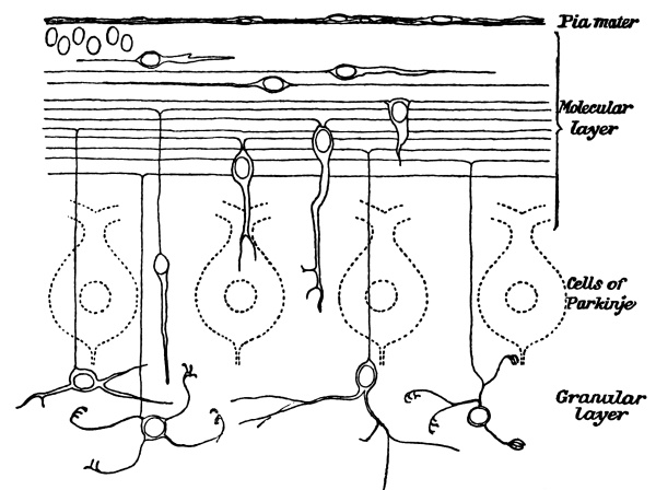
Fig. 21.—The Growth and Migration of Granules of the Cerebellum.
Half a dozen nuclei of as yet undeveloped granules are seen lying beneath the pia mater. From this level to the bottom of the drawing granules are shown in successive stages of growth. These developing granules, selected from various preparations of the cortex of the cerebellum, were drawn from nature.
There are many different types of neurone. Any attempt to describe them, or to give an account of the various details of structure which recent improvements in technique have enabled anatomists to observe, would fill a lengthy treatise; and would, moreover, be beside our aim, which is limited to obtaining such an idea of the unit of the nervous system as will enable us to form a conception, however crude, of the way in which it works. From the brief account that has been given, it will be evident that anatomists are approaching to an understanding of the mechanism. It will also be evident that they have already more information than they can apply. They are cognizant of many details of structure which they cannot interpret in terms of function; and at the same time are aware of wide gaps in their knowledge regarding facts which are essential to the construction of any scheme. This much is clear: A sense-cell on the surface or beneath it is touched (probably entered) by the ultimate twig of the outer limb of a neurone whose cell-body lies in a spinal ganglion, while its inner limb, as a fibre of a posterior root, enters the spinal cord. In the spinal cord the root-fibre splits into an ascending and a descending division which rain branches into the grey matter over a considerable area above its point of entrance, and a smaller area below it. The finest twigs of these branches are to be seen in the vicinity of the cell-bodies and dendrites of certain other neurones. The axons of these second links arborize in a similar way in the vicinity of large motor cells, whose axons in turn become fibres of anterior roots. (For simplicity’s sake no reference is made to hosts of other neurones which link the ganglion-cell and the motor cell to other cells higher in the cord or brain.) An impulse generated in the sense-cell on the surface of the body runs up the root neurone into the cord, where the ultimate twigs of the posterior root-fibre offer it a wide choice of distribution. Following the path of least resistance, it passes into neurone No. 2. Again, the arborization of No. 2 offers it alternative paths. It makes a choice which lands it in No. 3. No. 3 passes the impulse on to the muscle-fibres with which it is connected. Three points are especially worthy of attention: (1) The impulse has a wide (literally, an unlimited) choice of routes. The skin of the finger is touched. Any muscle may respond, although resistance is so graded as to cause the impulse to seek in the first instance the group of muscles which is most often required to act in consequence of stimulation of the finger. This means, we may suppose, that it follows the chain which, having the smallest number of links, offers least resistance. If it cannot get through to these muscles, owing to the fact that other impulses, acting simultaneously, either increase the resistance in this particular path, blocking its way, or reduce the resistance in an alternative path, it spreads farther afield. (2) Owing to the ramification of the root-fibre which conveys it to the cord, an impulse is not limited to a single line of distribution. It reaches many secondary links. It may therefore influence various effector neurones simultaneously. For example, a stimulus which calls extensor muscles into action, at the same time inhibits their flexor antagonists. (3) The path which it finally takes is accessible to all other impulses. Its root neurone was peculiar to itself. Link No. 2 was more or less a common path. Neurone No. 3 is open to every impulse which traverses the nervous system.
Anatomy justifies the construction of the scheme just outlined. But there are many points regarding structure upon which a physiologist desires information, many details that he wants to see filled in. How is the impulse passed from the arborization of axon No. 1 to the dendrites of neurone No. 2? By what structural arrangement is resistance introduced, and how is it regulated, if it varies? Supposing the resistance to be higher in one path than in another, or supposing that more force is needed to enable an impulse to invade a wider field, how is additional energy supplied? To the first question no answer can be given at present—the mechanism by which impulses are transferred from one neurone to another is unknown; yet it is convenient to find a name for the junction of axon-endings and dendrites. It is termed a “synapse,” on the understanding that the word involves no hypothesis as to its structural nature. It is generally held that resistance is introduced into nerve-circuits at synapses; although this again is a provisional statement. The phenomena for the explanation of which the idea of synaptic resistance was introduced, may be accounted for on a purely anatomical basis of distribution. The extent to which one neurone influences another may depend upon the size of the brush of fibrils with which its axon touches it. If a certain force is needed to discharge a neurone, a nerve-current must either have a sufficiently high potential when it reaches it, or it must act upon it for a sufficient length of time. There is little to choose between the arguments which place the resistance at the synapse and those which transfer it to the nerve-cell body.
As a mechanism the nervous system is unthinkable, unless we picture its units as independent, yet capable of forming associations; as functionally discrete, yet entering into functional continuity. When acting, they act as chains. Impulses run from link to link, from the end-twigs of an axon of one cell to the dendrites of the next. Neurones are so arranged as to make it impossible for impulses to escape backwards out of dendrites into axon-twigs. In this respect the system is valved. But there is no reason for thinking of the substance of the neurone as polarized in any way. The physical accompaniment of an impulse—the electric variation—travels with equal facility up and down its axon.
There is no evidence of any specificity of neurones; on the contrary, it is clear that impulses of every kind—that is to say, from every source, for we recognize no specificity of impulses—can travel equally well through neurones of all forms. At every junction, in passing through each synapse, they are delayed. It takes at least 0·01 second (less if the knee-jerk be a true reflex action) for a message delivered to the cord by a sensory root to reach a motor root. This hundredth of a second—the sum of the delays entailed in fording two or three synapses—is regarded as the minimum reflex time. To it must be added, in considering any particular reflex action, the time taken in travelling up sensory and down motor nerves. Delay indicates resistance. If a sensory stimulus be not sufficiently pronounced to provoke a reflex action, the reflex may be obtained on intensifying it. Prolonging or repeating the stimulus—really the same thing, since sensory impulses are rhythmic, not continuous—has a far more potent effect than increasing its force. The resistance of synapses gives way after a number of impulses have bombarded them. The desire of brushing a fly from the skin, if resisted, becomes intolerably urgent after a time. A persistent outflow of impulses produced by the irritation of a spot in the cortex of the brain overwhelms the nerve-muscle system in an epileptic fit. The following is an experiment illustrating the spread of impulses from their customary path to another less often used: A piece of blotting-paper, wet with vinegar, is placed on the inner side of the thigh of a brainless frog. There is no use in trying the experiment on a frog which retains its brain; the substitution of one action for another would be an exhibition of the adaptation of means to end—a demonstration of the animal’s right of choice. Besides, the frog might choose not to act, and so the experiment would fail. The brainless frog wipes off the blotting-paper with the foot of the same side. This foot is then fixed so that the action cannot be performed, and the blotting-paper replaced. After a longer interval the frog removes it with its other foot. Evidently it is more difficult for the impulses generated by the irritation which the vinegar causes to get across the cord than it is for them to reach motor neurones on the same side. Evidently, too, the continued irritation of the vinegar adds to the travelling power of the impulses. They are strengthened until they are capable of overcoming the resistance in the longer path. “Resistance in conductors” and “potential of current” are terms with which the study of electricity has rendered us familiar; but it must be evident from the experiment just described that these terms are not really applicable to nervous phenomena, convenient though they may be for use in an allegorical sense. Holding the foot does not, by any mechanism which we can recognize, switch off the shorter circuit, yet the impulses abandon it for the longer path. There is no evidence of a struggle to free the foot that has been fixed, coincident with the spread of impulses, as they gather sufficient strength to reach the nervous mechanism of the other leg. The right foot not being available, the impulses choose the route to the left foot. Any attempt to explain this in terms of resistance and potential involves the formulation of a number of subsidiary hypotheses; easy to devise, no doubt, but stultifying to the explanation exactly in proportion as they complicate it. Yet the hypothesis of lines of greater and of less resistance (keeping as far away from electrical analogies as possible) is essential to any explanation of nervous phenomena, and is, moreover, justified by the evidence available. There are two causes in chief upon which it depends: (1) The greater the number of neurones in a linear chain, the greater is the number of synapses to be traversed. If A, B, C are in the same circuit, the sum of their resistance has to be overcome. (2) The greater the number of neurones amongst which a nerve-current has to be subdivided, the smaller the charge available for each of them. Imagine
B
A
C
so placed as to divide B and C, the charge delivered by A between. This arrangement has, probably, an anatomical expression which accounts for the relative ease or difficulty of a path, even on the supposition that impulses do not open out as they advance—do not spread along all the branches into which an axon divides—but keep to a given line. The axon of neurone A divides, to branch about B, C, and D; but its representation in the several pericellular nets (the expression may pass for the sake of the simplicity which it introduces into the picture) is unequal. In the vinegar experiment the impulses delivered to the spinal cord by the root-ganglion neurone A pass to neurone B of the posterior horn. B’s axon arborizes more freely about the cell-body of neurone C in the anterior horn of the same side than it does about neurone D in the anterior horn of the opposite side. Hence the impulses generated by the vinegar stimulate C, sufficiently to discharge it, so long as that road is open, more quickly than they stimulate D. That C should be dischargeable only so long as the foot is free implies that the activity of the neurone is in some way conditioned by its relation with the muscles which it innervates. When the foot is held this relation is interfered with, giving to the impulses generated by the continued action of the vinegar time to overcome the resistance of D.
The simile of the opening up of paths is fairly applicable to the results which follow the use of artificial stimuli. Neurones seem to link up in series under the influence of the impulses which bombard them, popping like fireworks united by a common fuse.
Experimental evidence points to the following conclusions: (1) Resistance is offered at a synapse. This resistance must be overcome before an impulse can get through from neurone 1 to neurone 2. (2) The impulse does not, properly speaking, pass from 1, through 2. It infects 2, causing it to discharge a fresh impulse. (3) Time is of the essence of this process. Either the impulses head up at the synapse, or, passing through into the neurone, they produce a cumulative effect within it, which provokes it to discharge. (The latter hypothesis, which is the less likely of the two, transfers the resistance from the synapse to the neurone to be infected.) These conclusions are based upon experiments of the following kind: The minimal stimulus which will evoke a reflex action is determined. A stronger stimulus is then applied. The reflex occurs more promptly, and is more pronounced. But on further increasing the stimulus, it is found that the limit of effectiveness is soon reached. The proportional relation of response to stimulus is much less evident than it is when the experiment is tried with a nerve-muscle. Choosing a reflex action easily provoked, the afferent path is stimulated with an electric current interrupted fifty times a second. The impulses which flow down the efferent path to the muscle follow one another at the rate of about ten a second. A column of nerve-fibres within the spinal cord is stimulated fifty times a second. Again, the discharge into anterior roots has the natural rhythm of about ten. The cortex of the “motor area” of the great brain is stimulated with a rapidly interrupted current. The muscles which it governs contract with their natural rhythm. The cortex is sliced away, and the stimulus applied to the white matter beneath. A similar result is obtained. Evidence such as this points to an independence of action on the part of the neurones which one can express only in terms of resistance and explosion. But there is another line of thought which leads to the development of a picture of the working nervous system which seems at first sight incompatible with the one that we have sketched. The phenomenon of the knee-jerk (p. 274) reveals a nervous system so intimately linked together, so homogeneous, so mobile, that no event, however trivial, occurs in any part without sending a vibration throughout the rest. Instead of a multitude of batteries enveloped in a labyrinth of wires interrupted by myriads of switches which are crackling on and off, the image of a sheet of water better figures our conception—a material so frictionless that it is a-ripple from side to side and end to end, from the most distant rivulet which feeds it to the farthest trickle in which it drains away. It is a fluid in a state of infinite commotion, the movements of its particles varying in amplitude from tremulous quiverings which scarcely frost the silver of its surface to waves which, breaking on the muscular system, throw it up in heaps. The vinegar experiment seems to demand a scheme of batteries and wires. The knee-jerk points to a continuous conducting medium. Other phenomena suggest the superposition of the two pictures; the conception of a nervous system consisting of a uniform medium conducting, not indifferently in all directions, but with such freedom that from our point of view the paths are infinite in number; and within this conducting medium nerve-cell bodies and their processes which collect and distribute groups of vibrations sufficiently strong in combination to produce visible effects. In order that one of these neurones may be stimulated to discharging-point, the medium by which it is surrounded must be thrown into such a state of agitation as suffices to infect it. The considerations which point to the formulation of this double or superposed scheme are such as follow: The passage of tone-impulses does not appear compatible with the ideas we have formed on other evidence of synaptic resistance and neuronic discharge. They are too feeble for such a mechanism. The short “reflex time” of the knee-jerk points to the passage of the agitation up a sensory root to the spinal cord, and through a non-resistant medium to the environment of the motor cells which it discharges, missing the neurone or neurones which intervene in the case of ordinary reflex actions. This is an illustration of the way in which tone-impulses, which we imagine as conducted by the non-resistant medium, pass over into discharges which produce visible effects. Again, the phenomena of inhibition appear to require the supposition of extra-neuronic conduction. Whenever a reflex path is in use, all other paths in its neighbourhood are closed. The passage of impulses leading to a particular reflex action is favoured by the suppression of conduction in its vicinity. When A is talking to D through the nerve-telephone, B and C are compelled to hold their peace. Inhibition is a phenomenon of universal occurrence. In relation to various actions, it is sufficiently pronounced to be visible in the effects which it produces. A simple experiment will illustrate this. Holding water in the mouth has no effect upon respiration, but during the act of swallowing respiratory movements are suspended. Whilst the swallowing reflex is occurring the respiratory reflex is inhibited. This might be attributed to the volitional control of respiration, and certainly when attention is being directed to the process volition plays a large part. But if a finger is placed on the pulse, it is possible to detect that, during the act of swallowing, the pulse quickens, owing to the suppression of the slowing action of the vagus upon the heart. Here is a case in which inhibition is in no degree a voluntary action. Nor is it of any value as an adjunct to the particular reflex with which it is associated. It is an illustration of the universal rule that activity of any one spot in the nervous system is the cause of the quieting of the surrounding area. Impulses which reflexly check the heart cannot get through the medulla oblongata whilst the swallowing impulses are traversing it. Inhibition has been described as a drainage of nerve-force into the active area. On the structural side it seems to require the conception of an extra-neuronic substance which, agitated in the vicinity of the cells which are to be discharged, is brought to rest around neighbouring cells. The promulgation through the nervous system of the state which, when it reaches the centres of consciousness, produces pain also seems to call for an hypothesis of extra-neuronic conduction.
Any reference to pain in a work on physiology needs a few words of preface, since popularly the term “pain” is used in various senses. When I see pink geranium and nasturtiums growing in the same flower-bed, I may exclaim: “It is positively painful.” The want of harmony, and at the same time the insufficiency of contrast, of chalky pink and translucent orange, jars my æsthetic sense. Dislikes, however well founded, are ruled out in thinking of the physiology of pain. Further, in defining pain, we must be careful to isolate the real thing, and not to confuse it with sensations which seem to lead up to it. If, putting my finger in a pair of pincers, I touch it as lightly as possible, the first sensation is one of contact; a little harder, and it becomes a sense of pressure; harder still, and all sense of contact or pressure is lost in pain. It is usual to regard pain as sensation carried to excess. But neither is this physiological. An excessively bright light or an excessively loud sound is disagreeable. It causes a sudden movement for the purpose of avoiding it—just such a movement as one would make if one touched a red-hot poker—but it is not, strictly speaking, painful. Not uncommonly in cases of accident or disease of the spinal cord a sharp distinction is drawn between the sense of touch and the capacity for experiencing pain. Below the injury the patient retains his sense of touch undiminished in acuteness, but no blow, or cut, or burn, causes him any pain. The pain caused by squeezing the finger in a pair of pincers is not, therefore, an excess of touch sensation. Pain begins to be experienced in the skin just when the object applied to it is affecting it to an extent which might do harm. If the point of a needle touches it, it causes pain as soon as the pressure is a trifle less than that needed to pierce its surface. A hot object begins to hurt when the temperature reaches 48° C.—almost enough to coagulate the tissue fluids. Pain is not a discriminative sensation. If I hold my arm out at right angles, I am conscious for the first few minutes of its weight, and have, besides, some sense of the traction exerted by the muscle of the shoulder. At the end of ten minutes these sensations are merged in pain, and for some time after lowering the arm the shoulder-muscle aches, much as it does in rheumatism. Pain is an effect upon consciousness, which absorbs, engulfs, and therefore obliterates sensation. To use an ancient phrase, “It is less that I feel pain than that I am pain.” If we speak of the capacity for pain as a sense, we may call it for the purpose of our present argument the “sense of damage.” The nerves of the skin are acutely affected by any agent which is likely to do harm. It is their business to convey to the central nervous system an influence which so affects it as to set up in consciousness the condition of pain. Sensations of damage evoke reflex movements by means of which the part of the body likely to be injured, or the whole body, is removed to a safe distance. It being the duty of the skin to give this warning, a service of nerves sensitive to noxious agents has been developed which scouts in co-operation with the services devoted to the recognition of physical contact and heat and cold (cf. p. 425). If, imagining that the fire has not been lighted, I touch an almost red-hot stove, I acquire quite a considerable amount of information of which I am able to make use. I gain an accurate notion of the situation of the stove, and I put the right part of my finger in my mouth. The skin sends to the brain the ordinary sensations of touch and pressure before the condition of pain is established. In seeking for a definition of pain, we must eliminate the two attributes which have characterized all the forms of stimulation which we have considered up to the present time: (1) The tendency to provoke movement; (2) the supply of information. If I am suffering from a whitlow, the last thing that I am disposed to do is to jerk my finger about. Although it enhances the urgency of skin-reflexes, pain, in general, inhibits movement instead of provoking it. This is well illustrated in pleurisy. So long as a man is healthy he is quite unconscious of the fact that at each respiration the lower part of the lung slides on the lining of the chest-wall; but commencing inflammation on the surface of one of the lungs causes intense susceptibility to friction, and the pain produces an effect which the man is quite unable to produce by an effort of will; it stops the movements of the chest on the damaged side. Pain is inhibitory, not stimulant. It is not, properly speaking, a sensation. Frequently being mixed with sensational elements, it conveys topographical information; but pure pain approaches in quality the nebulous sense of distress of a patient who, when asked where he felt it, replied: “Nowhere; but there is a deal of it in the room.”
Sufferers describe pain in figurative language, as “burning,” “stabbing,” “throbbing,” “aching,” and so forth. Two persons afflicted with the same lesion, the same source of pain, use approximately the same terms. Hence we cannot say that pains do not differ in character. But this is not a sufficient reason for assigning any specific quality to pain. It varies in severity, in continuity or intermittence, in suddenness of onset, in the sensations which accompany it, in the emotional tone to which the disturbance of the organ from which it proceeds gives rise, in the tenseness of the part affected and its consequent sensitiveness to a throbbing pulse. All these things make a complex of pain plus sensation, which causes toothache to differ from headache, and both from the pain of burned skin. But they do not give specific qualities to different varieties of pain. This being the case, there is no need to presume the existence of special nerve-endings for the reception of pain, or of a special region of the cortex of the brain for its reception. On the contrary, the evidence is conclusive that the nerve-fibres which serve the more highly specialized senses, which have well-defined connections in the cortex of the brain, do not convey the influence which enters consciousness as pain. It is the innumerable nerves which have no specialized receptors that take up pain. The afferent nerves of the viscera—the vagus and sympathetic—convey no impulses which enter consciousness, so long as the tissues which they supply are healthy. They have no representation in the cortex. The organs with which they are connected (with trivial exceptions, easily accounted for) are absolutely insensitive to injury. Before the virtues of chloroform were known—in the days when, however severe the operation, the patient had to nerve himself to bear it without an anæsthetic—surgeons proved that the liver or the intestines, or practically any other viscus, may be cut or cauterized without the patient being aware that it is being touched. The same is equally true of the brain itself. But if damage in a viscus is set up gradually, its nerves convey to the central system an agitation which has the most pronounced results upon consciousness, and on the way profoundly affects the reflex actions which the spinal cord can carry out, and also its capacity as a conductor. Once in his life, perhaps, a man passes a gall-stone; for generations such a thing may not have happened in his family. Yet the man finds that he is provided with a nervous apparatus which conveys to consciousness intensest pain.
It is difficult to think of pain as travelling along nerves in the form of rhythmic impulses, similar to those which produce in consciousness the effects which we have distinguished as sensations. A few lines above we stated that no impulses which affect consciousness normally travel up the vagus or the sympathetic nerve, limiting the term “impulse,” perhaps unjustifiably. The vagus conveys an influence which enters our experience, as hunger. Probably other states of feeling for which we have no names, which resemble pain and hunger and their opposites, are set up through the agency of visceral nerves.
Fifty years ago attention was called to the difficulty of finding pain-paths amongst the white tracts (nerve-fibres) of the spinal cord. It is as difficult to point them out now as it was then; but the inference that pain travels up the grey matter has given way to the “neurone theory”; under a misapprehension as the writer holds. Pain travels slowly. If one happens to notice a person who unsuspiciously touches a hot surface, one observes that an interval elapses between contact of his finger with the iron and the exclamation with which he “relieves his feelings.” It amounts to more than a second—if the iron is not very hot, to several seconds—whereas the “reaction-time” for touch is only one-seventh of a second. The slowness of movement of pain through the nervous system can on the neurone theory be explained only on the hypothesis that it travels from link to link along a very long chain of very short neurones. That pain is a state of the grey matter rather than a succession of impulses, and that (within the cerebro-spinal axis) the state is transmitted through an extra-neuronic medium, seems a simpler explanation.
The state set up in the segment of the cord in which afferent fibres, conveying pain from viscera, embouch affects its conductivity. It subdues reflex action through the segment, and at the same time facilitates or reinforces the transmission of sensory impulses towards the seat of consciousness. This shows itself in the apparent increased sensitiveness of the skin of the area of the surface supplied by the posterior root which joins the segment of the spinal cord into which the pain influence is also being poured. For example, afferent sympathetic nerves from the cardiac end of the stomach join the sixth and seventh thoracic spinal nerves. Other afferent fibres run up the vagus to the medulla oblongata. When the cardiac end of the stomach is diseased, pain is referred to the skin area supplied by the sixth and seventh dorsal roots. The ordinary inevitable stimuli acting upon this area cause pain. Experimental stimuli which elsewhere would be felt as touch or warmth are painful. The impulses to which they give rise pass through pain-agitated segments of the spinal cord. The vagus nerve carries its pain influence to the medulla oblongata. Now, it happens that the sensory nerve of the face—the fifth—spreads for a considerable distance up and down the axis of the brain. The fifth nerve in consequence pours its sensory impulses into a region which is pain-agitated by those fibres of the vagus which come from the cardiac end of the stomach. Hence disease of that organ gives rise also to an “illusion” of pain—pains and illusions of pain are philosophically indistinguishable—on the surface of the head. The viscera, having no direct access to consciousness, appear by deputy. When the stomach is distressed, it makes its appeal to the whole body politic for considerate treatment through certain nerves which have the privilege of appearing at Court. The message is misread as coming from the front of the chest—“heart-burn”—or from the shoulder, or from the scalp, or from the other skin areas which these nerves serve. When the liver is in trouble, consciousness, having no knowledge of its whereabouts—is it the business of hand and eye to explore another man’s liver, or incumbent upon the mind to accept their findings?—infers that the cry comes from the shoulder. Nor have the tissues beneath the root of the nail, or the muscle of the shoulder, or the pulp of a tooth, any direct representation in consciousness; but since the pain-condition in the grey matter converts it into a microphone, messages from neighbouring structures which otherwise would fail to arouse attention, after traversing the pain-segments of the nervous system, ring out clearly, and hence the mind locates approximately the “pain” of the whitlow, the muscle-ache, the decayed tooth. Sufferers from toothache are familiar with the phenomenon of the spread of pain from a definite spot to the whole jaw or the whole side of the head, dependent upon the spread of the pain-agitation from the segment of the axis of the brain in which the dental nerve ends to neighbouring segments. Our ability or inability to localize a pain does not depend upon the presence or absence of pain-nerves, but upon the existence or non-existence of nerves coming from the same organ, or from its neighbourhood, and capable of conveying impulses to the seat of consciousness. In passing through the part of the spinal cord or of the axis of the brain which is disturbed by the influence exercised by a damaged organ, silent impulses acquire force sufficient to render them audible, and combine with the pain to produce a feeling which consciousness can analyse, to a certain extent. Informed as to its whereabouts by these accentuated sensations, consciousness recognizes a sense of pain limited in its topographical extension.
Sneezing when a bright light falls upon the eye is a curious illustration of the exaggeration of the effectiveness of sensory impulses when they happen to be poured into an agitated segment of grey matter. About one person in every three is affected in this way. A friend of the writer, who was particularly sensitive, rising in the night because he heard his child cry, three times lighted a candle and three times sneezed it out before he could watch the application of match to wick without suffering from a nerve-storm. Some nervous dogs—especially fox-terriers—are very liable to this neurosis. Many persons who do not sneeze feel, when the sunshine stimulates their retinæ, a tickling in the nose. Again the illusion is to be traced to the door of the fifth nerve—the sensory nerve of the whole of the face. The nose is the true tip of the body. Morphologically it is anterior to the eyes. Just as the fifth nerve extends its distribution to the nose, so also its root-fibres extend their connection within the axis of the brain forwards, until they traverse the mid-brain, the primary centre of the optic nerve. A bright light, by stimulating the optic nerve, sets up a commotion in the mid-brain. The ordinary every-moment impulses from the nose, carried by the fifth nerve to this region, ought not to appear in consciousness at all; but owing to the excited condition in which they find the grey matter they assume an importance which does not belong to them, and discharge the reflex action of sneezing, just as they would do had one taken snuff. Several lessons are to be learned from this phenomenon—as, for example, one which cannot be too often impressed, that the impulses which appear in consciousness (or, more accurately, the impulses to which attention is directed) are but a most insignificant fraction of those delivered by sense-organs to the central nervous system. The impulses which give rise to the sensation of tickling in the nose are not exceptional impulses which happened to be started when the light fell on the eye. They were reaching the brain in a steady flow before the agitation of the mid-brain gave to them exceptional force. No consideration regarding the working of the nervous system has a more important bearing than this. We cannot picture to ourselves the activity of the sensory nervous system. Our experience is limited to the scattered sensations which we perceive. Are the sensory nerve-endings incessantly responding to external forces, throwing an almost continuous procession of impulses up each of the millions of nerve-fibres which connect them with the central system? Such a conception is probably nearer to the truth than the conception which we should develop if we trusted to experience. Yet even experience tells us that an infinity of messages is delivered to the brain, of which consciousness takes no account. Changing trains at a roadside station in France, my attention was attracted by an electric bell on the platform, which was ringing continuously. “Why does the bell ring?” I asked the station-master. “To make known that everything goes well,” was the response. “If it stops, something is wrong.” “But do you not become so accustomed to it that you cease to hear it?” “Yes, truly; it rings day and night. One does not pay attention to it until it has stopped.” Sensory impulses generated by the contact of my skin with the chair that I am sitting on are incessantly ringing the bell of consciousness. I should notice them immediately if they stopped. As it is, they do not attract my attention until they ring a little louder than usual, or until some particular group, owing to unrelieved pressure, produces a cumulative effect. Another lesson; that the condition of the nervous system, and therefore its conductivity, is determined at any given moment by the sensory impulses which are reaching it. We cannot describe the effect of a bright light as pain, yet it agitates the grey matter, altering its state, in the same way as the nerve-inflow which we recognize as pain. A wet rag on the forehead does not assuage a headache by cooling the brain (cf. p. 106). The headache is “in the scalp.” The cool wet rag diminishes the dilation of the bloodvessels of the forehead, and quiets the impulses from the skin which are pouring into a tract of grey matter pain-agitated by the influences ascending a visceral nerve—usually the vagus.
It is necessary to warn the reader that a reversion to the old idea of “conduction through grey matter”—i.e., otherwise than by a chain of neurones—is unorthodox. It is set forth here because it seems to the writer that the various phenomena which have to be accounted for fit in best with the hypothesis of a double path. If evidence of the anatomical possibility of extra-neuronic conduction is asked for, it may be pointed out that the chrome-silver and methylene-blue methods, upon which our knowledge of neurones is based, do not, in the very nature of the case, show that grey matter consists only of neurones and their obvious branches. As they select particular elements of structure, we can never by their use alone know what they fail to show. Attention may also be called to the fact that the same staining process which reveals pericellular nets (p. 301) shows also a structure resembling a network in the substance which intervenes between them. Truly the method is a rough one. It may well be thought that the nitric acid used to fix the tissue may cause strange coagulations with solution of uncoagulated substance; but, as was remarked with regard to the pericellular nets, regular patterns indicate architectural differentiation. But whether these nets do or do not give hints as to the nature of the conducting medium, there is no difficulty in finding sufficient material, after all the substance entering into the formation of the conducting neurones, as we imagine them, has been accounted for. Ex hypothesi, the conducting material is provided by the fibrils of the sensory nerves in their extensions beyond the limits to which the deposit of subchromate of silver extends, when the chrome-silver method of displaying neurones has been used. Sensation-impulses enter neuronic chains. The condition which, when it affects the seat of consciousness, is known as pain, progresses up the vertebrate neuropil.
Energy is developed within the nervous system. The force of impulses is adjusted to the resistance which they have to overcome. Stimulation of the millions of twigs of the vagus nerve in the lungs brings about the gentle movements of ribs and diaphragm which constitute peaceful respiration. A crumb of bread touching the mucous membrane of the larynx stimulates a few of the endings of the same vagus nerve. Like an avalanche, the impulses gather head as they advance, causing, not the diaphragm and intercostal muscles alone to do their utmost, but calling into action half a dozen accessory muscles of respiration. It is difficult to account for this reverberation of the messages which clamour for the ejection of the crumb of bread without figuring them as spreading from neurone to neurone, urging each in turn to deliver its maximal discharge.
Neurones are provided with material which serves as a store of energy. In their cell-bodies, including their dendrites, are to be seen coarse granules of nucleo-protein, which, being fitted in between groups of neuro-fibrillæ, assume an angular form. They are known as Nissl’s corpuscles, or are termed “tigroids,” owing to the spotted appearance which they give to the substance of a cell. If the nerve-cells of birds be examined just after they have alighted from a migratory flight, the granules are found to be few and small. In a bee returning to the hive at evening with its last load of pollen, they are smaller than they were when it commenced its morning’s work. They disappear in certain pathological conditions, and under the influence of various drugs; and since their presence is revealed by staining, their disappearance is spoken of as “chromatolysis.”
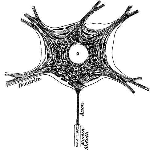
Fig. 22.—The Body of a Motor Neurone.
In its centre is a large clear spherical nucleus, with a nucleolus. The body-substance is prolonged into five dendrites and an axon. Neuro-fibrillæ are seen in dendrites and axon. They traverse the body of the cell in all directions, in little bundles which are separated by angular granules of stainable substance (tigroids).
The wasting of tigroids during functional activity proves clearly that nerve-cells do work, in the physical sense. Energy is expended in transmitting messages from receptor to effector, from sensory cell to muscles, from recipient nerve-ending to glands. Have nerve-cells any privileges or duties? Their functions, so far as we have considered them hitherto, are automatic, from a mechanician’s point of view. Their situation and connections determine the direction in which they conduct, and the degree in which they reinforce stimuli impressed upon the nervous system by the environment, including what may be termed the internal environment, food in the alimentary canal, secretions in ducts, and so forth. Have the cells any directive or executive functions? There is no evidence that they have; nor, it must be added, is there any line of reasoning which leads inevitably to the conclusion that they have not. Remembering that, until recently, it was the custom to solve all obscure problems and to shelve all difficulties by conferring human attributes upon nerve-cells and collections of nerve-cells, termed “centres,” a physiologist admits the negative with reluctance. The unconscious argument in the past used to run somewhat thus: “I decide to act or to abstain from action. The nerve-cell is the mechanism by means of which I decide. Therefore the nerve-cell decides.” (In the past a distinction was drawn between the cell-body and its processes, but that, we now see, was absurd.) It is very difficult to relinquish completely this attitude of mind. I feel, I remember, I will. There must be a something which feels, remembers, wills. But a physiologist finds in the nervous system no evidence of a capacity for any function other than that of conduction, with adjustment of the force of current. He can no more discover feeling, memory, or will in a chain of neurones than he can find music in a violin. He hears the strings singing in the breeze. He can twang them with an electric shock. But he has no vision of ghostly performers, no glimpse of the conductor’s baton. Yet he knows, as every sane man knows, that the neurones are the instruments played in the orchestra of mind. He knows that, while all are sounding, some are muted, in order that the others may produce a dominant effect. He knows, too, whenever he decides to continue writing or to close his notebook, that the conductor is raising the baton or allowing it to sink by his side.
A neurone or nerve-cell is a transmitting link. It is scarce a thing to wonder at that physiologists, having wrestled successfully with the superstition of the “pontifical nerve-cell,” are unwilling to reinstate it even as doorkeeper in a free church. It may be that it exercises some discretion in admitting impulses, but until its authority as a guardian of the path which stretches behind it has been established, it is better to regard it merely as a door which swings open whenever pressed with sufficient force.
Is it possible to classify neurones according to their function? They can be classified according to size, and, with some degree of completeness, according to form. But if, as we believe to be the case, size and form are governed by purely physical requirements, the divisions into which the cells fall have no physiological significance. The motor cells of the spinal cord and axis of the brain are large and irregular in shape. Their dimensions are clearly dependent upon the size, thickness rather than length, of the nerve-fibres which are drawn out from them. They discharge impulses to groups of voluntary muscle-fibres at a considerable distance. Small cells could not do the work. Precisely similar reasons can be given for the large size of the cells of Purkinje in the cerebellum, which transmit the elaborated product, as we may term it, of this organ to the great brain; and for the dimensions of the large pyramids of the great brain, which convey its decisions to the spinal cord. The small pyramids of the cortex of the great brain distribute the first crude impressions of sensations to neighbouring (association) areas of the cortex. A cell of Purkinje (Fig. 23) has a more complicated, and at the same time a more regular, form than any other nerve-cell. It resembles an exceedingly richly branched espalier pear-tree, set at right angles to the narrow convolutions of the cerebellum; a disposition easily accounted for, when the structure of the cortex of this organ is considered. Its outer layer in which the espalier processes ramify is traversed longitudinally by an infinity of nerve-threads, the bifurcated axons of granules. These granules are small neurones which take up impulses from afferent (“mossy”) fibres, and distribute them to the dendrites of the Purkinje cells—each collecting from a few fibrils only of the sensory channels. (The word “sensory” is used to indicate that sense-organs are their provenance, and not that their messages become sensations.) The numerous spreading branches of a Purkinje cell, disposed in a transverse plane, are obviously arranged to hold up and keep apart these myriads of longitudinal threads. A cerebral pyramid is shaped like a fir-tree. It is placed in a definitely stratified layer. By its branches it collects impulses from the superficial strata, which it transmits through its stem to the white matter beneath the cortex. The various parts of the central nervous system have work of different kinds to do, and we find interposed in the circuits which compose the several parts cells of various types. We speak of the large cells as “motor,” the granules as “sensory,” the small pyramids as “association” cells—such terms indicating the positions which they occupy in the arcs, but not defining their functions. Of specialization of function the physiologist cannot obtain a hint. He cannot classify nerve-cells in groups concerned in reflex action, in feeling, in remembering, in willing, in thought. On the contrary, he can assert with confidence that such distinctions are not to be drawn.
In various situations in the central nervous system a certain type of cell is found for which, in the present state of knowledge, it is impossible to account. We mention these cells lest it should be inferred, from what has been said above, that all neurones can be fitted into a simple scheme of conducting arcs. In the spinal ganglia there are neurones whose axons divide to form “baskets” around other ganglion-cells. In the cerebellum there are similar cells, the axons of which divide into branches, which break up to encase Purkinje-cells. Cells of the same kind are found in a few other situations. In some cases the end-branches which enter into the formation of the baskets are few in number, and thick and clumsy. They grasp the body of the cell which they surround, with gouty fingers, as it were. In other cases the basket is a tangle of fine threads. It is difficult to see what rôle cells of this kind can play in conduction. From the olfactory and optic centres nerve-fibres extend outwards to the olfactory bulb and retina. Here again is an arrangement which does not fit in with any scheme. We might multiply examples. But enough has been said, perhaps, to convey the impression which we wish to leave, that, although experiment abundantly proves that the nervous system consists of an association of sensori-motor conducting arcs, and although anatomical investigation demonstrates the existence of chains of neurones which take part in the formation of such arcs, it is impossible to reduce the system to schemata or to prepare diagrams in which all structural elements are, even hypothetically, fitted into place.
It may be convenient at this point to call attention to the differences which distinguish the sympathetic system—the ganglia and nerves of the viscera and bloodvessels—from the system devoted to bringing sense-organs into connection with the skeletal musculature which we have chiefly considered hitherto. The fibres of the posterior root of a spinal nerve which convey impulses from the skin and muscular sense-organs, and the fibres of its anterior root which convey impulses to skeletal muscles, have a similar diameter of about 15 µ. In addition to these, the roots contain fibres which carry impulses from and take them to the viscera. Those which bring impulses from the viscera vary greatly in thickness, some being as large as the other sensory nerves of the posterior root. The diameter of the fibres which go to the viscera is not more than one-fifth as great as that of the other fibres of an anterior root. Similar slender fibres are found in the vagus nerve. If all organs are removed from an animal’s chest and abdomen, a string of small pearl-like ganglia, united by a longitudinal cord, is seen lying on either side of the bodies of the vertebræ, one ganglion for each segment. This string of ganglia is termed the “sympathetic chain” (cf. p. 243). The small medullated fibres of the anterior spinal roots join these ganglia. Some of them arborize about their cells; some pass by them to arborize in ganglia which lie farther afield, on the course of the great bloodvessels and within the viscera. The axons of neurones whose cell-bodies are within a ganglion break up into bunches of non-medullated fibres. In this way the fibres of the sympathetic system are increased in number. Each of its neurones is a multiplying and distributing station. There is no evidence that it in any way serves as a “centre,” takes part in reflex action, or otherwise usurps the functions of the grey matter of the spinal cord. Nerve-cells are thickly strewn between the mucous membrane and the muscular coat, and again between the two layers of the muscular coat of the alimentary canal. It is not so certain that this system has no “central” functions. The remarkable degree in which the wall of the intestines retains its capacity for co-ordinated movement, after all nerves which reach it from the ganglia and through the vagus have been cut, suggests that the plexus of nerves within it does act to some extent as a reflex centre. If we leave the case of the intrinsic nervous system of the alimentary canal open, awaiting further proof, there is no reason for looking upon the sympathetic system as in any degree independent of the spinal cord and brain. It does its work on a large scale, and its work is of a low order. Nature does not need to connect up the viscera and bloodvessels with the central nervous system by means of fibres as thick as those used for skeletal muscles. It is more convenient to provide for the multiplication of the nerves—which must be extremely numerous, owing to the relatively minute size of the muscle-fibres for which they are destined—outside the central system than it would be to include the necessary distributive cells within it. Again, we find that a nerve-cell, when we see it at close quarters, shows no evidence of administrative capacity. Although of a different shape, a ganglion-cell of the sympathetic system is as large and as complex in form and structure as a pyramidal cell of the cortex of the brain; yet the work which it does is of a purely mechanical order. It receives, reinforces, transmits impulses which reach it from the central nervous system.
The often-repeated statement that a nerve-fibre is a drawn out process of a nerve-cell body has prepared the reader to anticipate that it dies when cut off from its central connection. When the axon is dead, the sheath which invests it rapidly loses its tubular character. If the situation of the cell-bodies of a nerve be known, it can be at once foretold on which side of the cut degeneration will occur. Suppose that the median nerve has been severed at the wrist. All nerve-fibres on the distal side of the wound must atrophy, whereas none of the fibres on the proximal side will be affected. The motor fibres have their cell-bodies in the spinal cord, the sensory in the spinal ganglia. Degenerations following lesions in the central nervous system have taught pathologists more about the course of the fibres in the white matter than any other class of observations. Degeneration above the lesion is spoken of as ascending, below as descending—not that it progresses upwards or downwards. It occurs throughout all the stretch of the fibre which has been isolated from its cell-body at the same time, or nearly so. The thought that impulses can no longer ascend or can no longer descend, as the case may be, has given sanction to the expressions “ascending” and “descending” degeneration.
Restoration to functional activity of tracts of fibres which have degenerated in the brain or spinal cord never occurs, but severed peripheral nerves regenerate. Not that fibres join cut end to cut end, however clean the wound. A wound in the wrist which has divided the median nerve may heal in a few days “by first intention,” so far as other tissues are concerned; but the patient does not for two or three months recover the power of using the muscles of the hand which the nerve supplied or the sense of touch in the area of skin to which it was distributed. The ends of the axons on the proximal side of the wound have to grow downwards to establish new connections in the muscles and in the skin. The interval which elapses between the healing of the wound in the wrist and the restoration of sensation and power of movement is occupied in their downgrowth.
The re-connection of regenerated nerves with their terminal apparatus presents to the mind a curious problem. There is no evidence that as function is re-established the brain has to re-learn the situation of the sensory spots on the skin, or to re-acquire skill in using the muscles which again come under its control. From the moment that the outgrowing nerves have recovered their terminal connections skin and muscles have their right representation in the brain, however much the two cut ends may have been twisted in their relation one to another. It seems inconceivable that each nerve-fibre can find its way to its original station; but if it does not, our conception of the mode of working of the nervous system still needs much refining from the telephone-exchange analogy by which we naturally help out our explanations. If a telephone cable has been severed, it can be made useful again only in one of two ways. Either the two segments of every wire that has been cut must be reunited, or the subscribers’ numbers must be redistributed.
The experiment of uniting the proximal segment of one nerve with the distal segment of another of a quite different function gives results which have an even more disconcerting effect upon our theory of the nervous system. The sympathetic cord of the neck and the vagus nerve lie very close together, alongside the carotid artery. The vagus is both afferent and efferent. The sympathetic is wholly efferent—i.e., it conducts impulses, which enter the sympathetic chain within the thorax, in the direction of the head. If both nerves are cut, and the end of the vagus turned round, so that it is in apposition with the upper end of the sympathetic, its regenerating fibres make their way along the sympathetic cord, headwards, to the superior cervical ganglion. They arborize about the bodies of its ganglion-cells, just as the sympathetic fibres used to do. The vagus is a nerve of many functions. Amongst others, it inhibits the contraction of the heart, constricts the bronchi of the lungs, dilates the bloodvessels of the intestines, and helps in regulating the movements of these viscera. After it has taken the place of the upper segment of the sympathetic it dilates the pupil, constricts the bloodvessels of the ear, erects the hairs of the head, as if to the manner born. To take another example, in a monkey the two nerves supplying respectively certain flexor and certain extensor muscles of the forearm were cut, and their ends crossed, so that flexor nerve-fibres grew down to extensor muscles, and extensor fibres to flexor muscles. There was no bungling of reflex actions or of voluntary actions when the new roads were first used. The monkey did not jerk its hand open when it tried to scratch or to grasp a nut.
When experimental data first began to accumulate, physiologists drew diagrams and made models of the nervous system in which they represented it as composed of conducting arcs. The arcs were superposed to indicate that they were of various grades—spinal for ordinary reflexes, bulbar for co-ordinated actions, through the grey matter in the centre of the great brain for “ideo-motor” actions, through the cortex of the great brain for voluntary acts. They spoke of authority and responsibility, comparing the nervous system to an army or a club. It is premature to attempt a theory of the nervous system compatible with recent discoveries regarding its structure and mode of working, but it is clear that the diagrams and metaphors to which we have just referred were misleading. In place of attempting to disarticulate the machine, we ought to emphasize its structural unity. The results obtained by uniting heterologous nerves cannot be explained by reference to a model made of wires and pieces of cork. They do not fit in with any organization of human units or with any postal system or telephonic apparatus for transmitting news. Probably the lines of thought which will prove most fruitful are somewhat as follows: (1) An efferent discharge occurs as the result of the opening of a circuit from a muscle back to the muscle. Afferent impulses—call them sensory, on the understanding that this does not imply that they appear in consciousness—are ceaselessly flowing from receptors to effectors in the muscle. A sensation—in the case of skeletal muscles usually a skin sensation—reinforces them to discharging-point. If the spinal cord has been severed from the brain, the up-and-down flow does not reach beyond its grey matter. It is short-circuited. If the brain is in normal connection with the spinal cord, sensory impulses travel upwards to its cortex (without, save in exceptional instances, arousing consciousness, or, as we should prefer to express it in this connection, without attracting attention) to a degree which varies with the several classes of receptor and with the animal. A monkey reduced to the condition of a “spinal animal”—i.e., with its spinal cord severed from its brain—is less competent than a dog, and a man is far less competent than a monkey. In other words, a man habitually uses his brain more than does a monkey, and a monkey more than a dog. The proportion which brain-weight bears to body-weight roughly indicates the part the brain plays in conducting the traffic of the body. (2) Communication within the nervous system is almost unrestricted. If, before the median nerve was divided at the wrist, receptor A usually initiated a current which passed through the circuit to effector X, and receptor B to effector Y, and if the new fibres which grew downwards lost their way so that the one which used to receive messages from A attached itself to B, and the one which used to transmit commands to X attached itself to Y, A is not thereby cut off from X, or B from Y. Such a mechanical association is restricted to our diagrams. It does not enter into Nature’s plan. The spinal cord is not scored with unchangeable paths. A messenger from A could always reach either X or Y. It was not the path, but the struggle with competing messengers, which directed him to X.
When we endeavour to picture the mechanism of the nervous system, we find ourselves faced by phenomena which appear irreconcilable. One set of observations leads to the conception of closed paths; another set points to an open conductor. The experimental crossing of nerves to which we have just alluded shows that the nervous system is adaptable, to a degree which seems extraordinary to anyone who attempts to compare it with any of Man’s devices for establishing communication. Paths appear to make themselves. On the other hand, the more important, and therefore dominant, reflex actions, such as swallowing, breathing, the maintenance of position, are due to the union of receptors and effectors by lines which are either reserved for their sole use, or, if shared by other currents, it is on the understanding that they have a first and altogether prepotent claim. No competing impulses can divert them or block their way. All reflexes which in the history of the race have established their right to dominance not only seize and hold a route through the nervous system, to the exclusion of all competitors, but, as we have already shown in the case of the swallowing impulse, the traffic in neighbouring routes is suspended for their benefit. At the other end of the scale we find reflexes which may be termed “occasional,” in that, although of frequent occurrence, they exhibit illimitable variability in form. Occasional reflexes require, as a preliminary to their transmission, that the afferent impulses which give rise to them should secure for a time the exclusive use of the motor neurones by which they are carried out. The receptors bring the motor neurones into tune with themselves, and while in tune they will respond to impulses from no others. But the tuning lasts for a short time only. Either receptor or neurone, or both, soon tire. There is no danger of a particular reflex being prolonged to the detriment of the organism as a whole. As an illustration of an occasional reflex, we may cite the scratching movement of a dog. Its skin is punctured by a flea. It scratches the place. A second flea bites it somewhere in the same neighbourhood. The dog does not shift its hind-foot so as to scratch midway between the two bites. It finishes out one scratch before paying attention to its second tormentor. The exact position to which the hind-foot is raised depends upon the position of the irritant; and since this may be shifted over a very considerable surface, the form of the reflex varies equally widely. Each of the very numerous receptors in the skin tunes a slightly different group of motor neurones; and since a second irritant may reinforce the first, instead of making an alteration in the group of neurones which the reflex is discharging, it is clear that there is no fixed path uniting receptor A with neurones X, Y, Z and receptor B with neurones W, X, Y. If, however, the second irritation occurs at a spot lying at a considerable distance from A, in place of reinforcing the scratching movement which A has set going, it weakens and shortens it. The receptor C, which is calling for the discharge of a markedly different set of motor neurones, tends to inhibit those which are already active. These results are tested with precision upon a “spinal dog” and with the aid of an electric needle, the other pole from the battery being a large flat plate placed in contact with the animal’s body. The conception of definite paths, to which the contemplation of permanent reflexes gives rise, is inappropriate to occasional reflexes. The latter show so wide a range of variability and adaptability as to prove that a given receptor may bring any of a great variety of groups of motor neurones into connection with itself; just as a given group of neurones may be played upon by impulses from a great number of different receptors. We have called it a tuning of the motor neurones. One metaphor is as good as another. The physical process which in the brainless frog underlies the preparation for discharging motor neurones in the spinal cord, on the same side as the leg on which vinegar is placed, so long as that leg is free, and on the opposite side, when that leg is fixed, is unknown. We seem to catch a glimpse of a doubleness of action, receptors in the muscles combining with receptors in the skin in determining the paths along which impulses shall be reflected—the efficient muscles sensitizing their own neurones to the tuning influence of impulses from sounding cutaneous nerve-endings. But it is impossible to formulate a working scheme in the present state of knowledge.
Sense-Organs and Nerve-Centres.—A vast amount of labour has been devoted to the study of the external form of the central nervous system and to unravelling its internal structure; to plotting out its various groups of nerve-cells, to disentangling its innumerable tracts of fibres. The surface of the brain and spinal cord has been mapped and measured. Every millimetre of its substance has been cut into sections on the micro-tome. Organs which, fifty years ago, appeared too complicated for investigation have been described in the minutest detail. An immense accumulation of data is available for purposes of reference; yet anyone who submits the theory of the nervous system as it is held at the present day to a general review must allow that the results of anatomical research enter but little into its construction. The reason for this is not far to seek. As knowledge has advanced, the apparent, or rather the expected, complication of the system has given place to ideas of unity and simplicity. Its external configuration and the varied arrangement in “nuclei” of its nerve-cells may, without impropriety, be described as accidental. The form of the body and the consequent location of the clients of the nervous system determine the disposition and degree of concentration of its various business centres. It shows, when followed throughout the whole animal kingdom, extreme variability of its constituent organs, with absolute uniformity of plan. Indeed, from the physiological point of view the term “organ” is scarce admissible. It implies diversity of function in too high a degree. The several parts into which the central nervous system is obviously divisible co-operate so intimately as to preclude us from thinking of them as separate organs.
If the citadel of the central nervous system is to be captured, all lines of approach must be tried. Its outward form must be studied, its minute structure examined with the microscope, its modifications in various animals compared, its development followed, its reactions to artificial stimuli tested, its pathological deficiencies and vagaries watched. Yet, of all the means which have been made use of in attempting to penetrate its secrets, the study of its history, by the methods of comparative anatomy and embryology, has probably contributed most to the development of sound ideas regarding the manner of its working. The first differentiation visible in the blastoderm—the globe of cells into which the ovum divides and out of which the embryo is built—has relation to the formation of the nervous system. If the earliest stages of its growth are followed, and the different phases through which it passes are compared with the forms which it assumes permanently in lower animals, the plan or type upon which it is constructed shows up distinctly. Looking down the line to the earliest vertebrata, we can discern clearly the form of nervous system possessed by their prototype. Not that this “ideal ancestor” ever existed. Experience teaches that it is unlikely that any animal that ever lived was absolutely regular and symmetrical in all its parts; nevertheless, the type can be presented in a perfectly regular scheme. The ideal ancestor of the vertebrata was segmented, like a caterpillar or a worm. Its mouth was not at the anterior extremity of the body, but two (or more) segments behind it. Every segment bore a sense-organ (at one period two sense-organs) on either side. Beneath each sense-organ there was a clump of “grey matter.” Each segment also contained (although not at the earliest epoch) two clumps of nerve-cells and neuropil in a more central situation. These “ganglia” were united by longitudinal and transverse commissures. They received the axons of the cells which lay in the clumps beneath the sense-organs. They gave axons to various muscles. Such is the type out of which the modern nervous system has developed: two separate sense-organs and a complete nervous system for each segment, the sense-organs connected with the ganglion of the same side, the ganglia of the two sides bound together across the middle line, and each row of sense-organs and each row of ganglia united by longitudinal commissures into a chain. From the nervous system as we see it now the majority of these segmental sense-organs have disappeared; but the mode of formation of the cerebro-spinal ganglia shows that they are the clumps of nerve-cells which lay beneath the vanished organs. In the nose and the eye the grey matter retains its original situation in the immediate vicinity of the receiving epithelial cells—as the olfactory bulb and the deeper (anterior) layers of the retina. The ganglia of the auditory nerve lie within the bones of the ear. Spinal ganglia are close to the spinal cord. Auditory and spinal ganglia contain only the cell-bodies of the first collecting neurones (sensory nerves) together with certain curious bracketing cells already referred to (p. 324), all the other constituents of the peripheral clumps of grey matter which are found in the olfactory bulb and retina having been withdrawn from the spinal ganglia into the axis of the brain and spinal cord.
The sense-organs in front of the mouth have had from the beginning an immense advantage over the others as observing-stations. Whereas the body-organs collected information regarding the things with which the animal came in contact, and consequently specialized in touch, pressure, temperature, and, in the case of fishes, sensitiveness to the chemical constitution of the medium in which the animal lived, the head-organs specialized in responsiveness to forces acting from a distance—particles suspended in air, vibrations of light, pulsations of sound. Sensitiveness to touch, if it is to be useful, must be widely distributed. The body-organs therefore broke into scattered groups of sense-cells. Touch-spots are scattered all over the surface, although they are set much closer together in the areas of skin which are usually the first to come into contact with external objects than they are elsewhere. The efficiency of the sense-organs of the head—nose, eye, and ear—depended upon their remaining compact. Progress in animal life, as we understand it—the rise from lower to higher forms—has depended upon increasing integration of the body and co-ordination of its functions. The nervous system is the agent which has accomplished this unification. Each step in advance has depended upon the provision of more nerve-tissue for the lacing together of the various parts. We have seen already (p. 329) how intimate is the union of receptors and effectors of every kind via the spinal cord and brain. The overwhelming predominance in the direction of action of the nose, the eye, and the ear has led to the accumulation in their vicinity of the ever-increasing grey matter. The cerebral hemispheres, or “great brain,” are pouched outgrowths from the first pair of ganglia directed towards the olfactory pits. The original eyes bore a similar relation to the second pair of ganglia—the epithet “original” implying that the eyes which we now use are not the organs with which our prevertebrate ancestors saw. First one of the original eyes disappeared, and then the other. The vestige of the second is still to be seen in the “pineal body” which is found on the dorsal side of the brain of every vertebrate animal—in a mammal deeply hidden in the cleft between the cerebrum and cerebellum. In place of the pineal eyes two other sense-organs have specialized as eyes. They are constructed on a different plan, being, to put it shortly, pineal eyes turned inside out; for whereas in the pineal eyes, as in most of the eyes of invertebrate animals, the rods and cones, which are the cells of the retina sensitive to light, are directed forwards towards the lens, the rods and cones of our permanent eyes are directed away from the source of light. This change has made it possible to provide more abundantly for their nutrition, and hence a greater power of discriminating separate points in space and of distinguishing colours is conferred upon them. The substitution of other sense-organs for the original eyes has complicated the pictures which are presented to us by a brain in its successive stages of growth; but it does not prevent us from recognizing the general plan. Probably the secondary eyes, like their predecessors, belonged to a pre-oral segment. The sense-organs of a segment behind the mouth developed into ears; and the ear was in its earliest phases, and still is, something more than an organ of hearing. Its semicircular canals give information of displacements in space. Knowledge of the position of its body is, to a fish, of far more importance than its ability to hear breakers on the rocks. Three looped tunnels, opening at either end into a common chamber, are hollowed in the bone which contains the ear (cf. Fig. 38). Placed at right angles one to the other, they occupy all three dimensions of space. Open a notebook until, one of its covers lying horizontally, the other is vertical, and place a sheet of paper vertically against the bottom of the pages. A curved line drawn on each of these three surfaces will represent the three semicircular canals. Arrange another notebook in the same way, and let the two rest on the table with the two vertical covers inclining one to the other, anteriorly, at an angle of 90 degrees. The six surfaces will be in the planes of the six semicircular canals. Within each bony canal is a membranous tube, to which nerves are distributed, filled with fluid. When the position of the head is changed, the fluid within the membranous tubes slides on their walls. It is left behind at the moment the movement commences. It overtakes its receptacle when the movement stops. The stimulus received by the nerve-endings is recognized as indicating an alteration in the orientation of the head. If the movement of the fluid is violent, as when one waltzes, the loss of the sense of position disconcerts the brain to such an extent that giddiness results. For a time the quiet assurance upon which so much depends, that one knows how the body stands in relation to its surroundings, gives way to a chaos of sensations. From the nature of the case, the information which the semicircular canals afford relates to change. They give no help in ascertaining the position of the head when it is at rest. This must be the reason, although the connection is not very clear, for the waning of the effect in consciousness when stimulation is prolonged, and also for the very marked after-sensation. At the commencement of a voyage attention may be unpleasantly attracted to the rolling of the ship. After a few days it ceases to be noticeable; yet when the voyager, the night after landing, wakes in the dark, he finds his bed-room as unsteady as his cabin. Rising hurriedly, the attempt to adjust his position to the heaving floor (we speak from personal experience) may result in a heavy fall. Although this phenomenon must be classed with other “after-sensations,” it is so prolonged as to suggest that consciousness, having become accustomed to a world which causes a backward and forward flow of endolymph, misinterprets the absence of sensation as indicative of change.