Part 1 — Comparison of the Appendages of Calymene and Ceraurus with those of Triarthrus.
Reading edition
As one may see by reading the above quotations from Doctor Walcott's descriptions, he found certain branchial organs in Ceraurus and Calymene which have not been found in other trilobites but otherwise the essential features of the appendages of all are in agreement.
Spiral Branchiæ.
It is now necessary to inquire if the thin sections can not be interpreted on the basis of trilobites with the same organs as Triarthrus. The interpretation of the structures seen in these translucent slices is exceedingly difficult, and Doctor Walcott deserves the utmost praise for the acumen with which he drew his deductions. Even with the present knowledge of Triarthrus, Isotelus, and Neolenus as a guide, I do not think it is safe to speak dogmatically about what one sees in them.
Walcott has summarized his results in his restoration of the appendages of Calymene (1918, pl. 33). The coxopodite supports a slender six-jointed endopodite as in Triarthrus, dorsal to which is a short setiferous epipodite which differs from the exopodite of Triarthrus, in being less long, unsegmented, and in having shorter setæ. Arising from the same part of the coxopodite with this epipodite is the bifurcate spiral branchia which has not been seen in this form in other trilobites. The evidence on which the existence of this organ is postulated consists of a series of sections across the thorax, the best of them figured by Walcott in his plates 2 and 3 (1881) and plate 27 (1918).
The specimens sliced were all partially or quite enrolled, and in that position one would expect to find the appendages so displaced that it would be only rarely that a section would be cut, either by chance or design, in such a direction as to show any considerable part of any one appendage. This expectation has proved true in regard to the endopodites, the sections rarely showing more than two or three consecutive segments. Sections like those shown in figures 1 and 2 in plate 2 (1881) seem to be unique. On the other hand, there are numerous slices showing the so-called spiral branchiæ. They show for the most part as a succession of rectangular to kidney-shaped spots of clear calcite.[1] Usually these clear spots are isolated, not confluent, but in a small number of specimens, perhaps three or four, the spots are connected in such a way as to show a zig-zag band which suggests a spiral. Such an explanation is of course entirely reasonable, but it would be surprising if so slender a spiral should be cut in such a way as to exhibit the large series of successive turns shown in many of these thin sections. Continuous sections of such organs should be no more common than continuous sections of endopodites.
One of the arguments against the interpretation of these series of spots as sections across spiral arms is that of probabilities. It is known from flattened specimens that Neolenus, Kootenia, Ptychoparia, Triarthrus, and Cryptolithus all have a single type of exopodite, consisting of a simple setiferous shaft. All these genera have been examined in a way that permits no doubt about the structure, and no trace of spiral arms has been detected. On the other hand, Walcott found spiral arms in three unrelated genera, Calymene, Ceraurus, and Acidaspis, all of the trilobites in which he found exopodites by the method of sectioning. What are the probabilities that genera of three different families, studied by means of sections, should agree in having a type of exopodite different from that of the five genera about whose interpretation there can be no doubt?
Another argument against the interpretation of the sections as spirals is that in any one line the individual spots are of roughly uniform size. This means of course that the spiral has been cut by a plane parallel to the tangent plane. This might happen once, just as once Doctor Walcott cut all six segments of a single endopodite, but that it should happen repeatedly is highly improbable. Moreover, there is a limit to the diameter of the section which may be made from these slender spirals. Most of the spots have one diameter about one half greater than the other, but others are from three to six times as long as wide. These last could obviously be cut only from a very large spiral, and they are therefore interpreted by Walcott as setæ of epipodites. Yet all gradations are found among the sections, from the long setæ to the short dots. (See pl. 27, 1918.) In referring to one slice, Walcott says (1918, p. 152):
In the latter figure and in figure 13, plate 27, the setæ of several epipodites appear to have been cut across so as to give the effect of long rows of setæ. The same condition occurs in specimens of Marrella when the setæ of several exopodites are matted against each other.
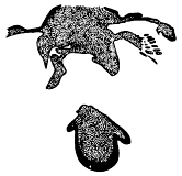
Fig. 12.—A slice of Ceraurus pleurexanthemus in which the exopodite happened to be cut in such a way as to show a part of the shaft and some of the setæ in longitudinal section. Specimen 80. × 4.
This is certainly an apt comparison, and equally true if Neolenus, Triarthrus, or Cryptolithus were substituted for Marrella.
Now consider the "epipodites." They are well shown in Calymene in the specimens illustrated on plate 27, figure 11 (1918), and plate 3, figure 3 (1881), and less clearly in one or two others. Slices 22 (pl. 27, fig. 12, 1918) and 80 (our fig. 12) show what is called the same organ in Ceraurus. It will be noted that all of these slices are cut in the same way, that is, more or less parallel to the under surface of the head, or, at any rate, on a plane parallel to a plane which would be tangent to the axial portion of the coiled shell. The sections which show the spirals best are those which are cut by a plane perpendicular to the long axis of the body. If one were to attempt to cut an enrolled Triarthrus in such a way as to get a section showing the length of the setæ, one would not cut a section perpendicular to the axis of the animal, nor, in fact, would he cut one parallel to the ventral plane, but it is obvious that in this latter type of section he would stand a better chance of finding a part of the plane of the exopodite coincident with the plane of his section than in the former. And that seems to be what has happened in these sections of Calymene and Ceraurus. If the exopodites were preserved, transverse sections were bound to cut across many sets of fringes, and the resultant slice would show transverse sections of the setæ as a series of overlapping spots. A few fortunately located sections in a more nearly horizontal plane might cut the setæ and occasionally the shaft of one or more exopodites in the longitudinal plane, and the resulting effect would produce the so-called "epipodites." A careful study has shown that no one of these epipodites is complete, and they do not have the palmate form shown in Walcott's figures.
And the last and most important argument against the spiral appendages is that certain slices, of both Calymene and Ceraurus, show definitely exopodites of exactly the type found in other trilobites. These are discussed later in the detailed description of the various slices.
If these series of spots are interpreted on the basis of the known structure of Triarthrus, they are of course a series of sections through the setæ of the exopodites. It will be shown in Part IV that these setæ are not circular in section, but flattened, in Cryptolithus even blade-like, and that they overlap one another. A section across them would give the same general appearance as, for instance, that shown in figures 4, 6, 9, and 10 of Walcott's plate 3 (1881).
When both endopodites and the "spiral branchiæ" are present in the same section (pl. 1, fig. 4; pl. 2, figs. 1, 2), the "spiral branchiæ" are dorsal to the endopodites, as the setæ of the exopodites would be expected to be. The specimens which show the clear spots connected, and which suggest a spiral (pl. 3, fig. 5), may seem at first sight to bear evidence against this interpretation, but one has only to think of the effect of cutting a section along the edge where the setæ are attached to the shaft of the exopodite of Triarthrus to see that such a zig-zag effect is entirely possible. One would expect to cut just this position only rarely, and, in fact, the zig-zags are seen in only three or four sections. The bifurcation of the basal segment of the "spiral branchiæ" (pl. 3, fig. 10, 1881) is probably more apparent than real, if indeed these basal segments have anything to do with the succeeding one.
A second peculiarity of Calymene, shown in Walcott's restoration, is the great enlargement of the coxopodites and of the distal segments of the endopodites of the fifth pair of appendages of the cephalon. This is based on the sections of plate 3, figures 6, 7, 8, 9, 10 (1881). After a study of the specimens I regret to find myself still unconvinced that the posterior cephalic appendages were any larger than those in front.
Ventral Membrane.
The most striking value of the thin sections of Ceraurus and Calymene, and therein they have a great superiority over all the other forms so far investigated, is that they show the extent of the body cavity and the position, though not the substance, of the ventral membrane. Transverse sections through Ceraurus (Walcott's pl. 1. figs. 1-5; pl. 2, figs. 1, 3, 1881) and Calymene (pl. 3, figs. 9, 10, 1881) show that the body cavity was almost entirely confined to the axial lobe. The longitudinal sections of Ceraurus (pl. 2, figs. 6, 8; pl. 4, fig. 8) and of Calymene (pl. 2, figs. 5, 7; pl. 5, figs. 1-4) show that the ventral membrane was exceedingly thin and was wrinkled transversely when the shell was enrolled.
The specimens of figures 1-3, plate 5 (1881) show the form of the ventral membrane more distinctly than any of the others. The section of figure 1 was cut just inside the dorsal furrow on the right side, and figure 2, which is on the opposite side of the same slice, is almost exactly on the median line. Figure 3 shows a section just inside the left dorsal furrow. Section 2 did not cut any of the appendages, and the ventral membrane is shown as a thickened, probably chitinous sheet thrown into low sharply crested folds equal in number to, and pointing in a direction just the reverse of, the crests of the segments of the thorax. Under the pygidium, where there would of course be less wrinkling, the folds are hardly noticeable. In the actual specimens one sees more plainly than in the figures the line of separation between the ventral membrane and the appendages, but the state of preservation of everything beneath the dorsal shell is so indefinite that one does not feel sure just what the connection between the appendages and the membrane was. In the original of figure 5, plate 2, which seems to have been cut so as to cross the appendages at their line of junction with the ventral membrane, there appear to be narrow chitinous (?) plates extending from the ventral membrane to the dorsal test.
Appendifers.
In Ceraurus there are regular calcareous processes which extend down from the dorsal test just inside the line of the dorsal furrow, and which undoubtedly serve as points of attachment of the appendages. These processes, which for convenience I have designated as "appendifers," are broken off in most specimens showing the lower surface of Ceraurus pleurexanthemus, but on certain ones cleaned with potash they are well preserved. Doctor Walcott showed them well in his figures of the lower surface of this species (1875, pl. 11; 1881, pl. 4, fig. 5), while the attempt of Raymond and Barton (1913, pl. 2, fig. 7) to show them by photography was not so successful.
There is one pair of appendifers on each of the thoracic segments and four pairs on the pygidium. On the cephalon there is one pair under the neck furrow, and a pair under the posterior glabellar furrows. These are not concealed by the hypostoma. Further forward, and completely covered by the hypostoma, are two much less strongly developed but similar ones, so that there are in all four pairs of appendifers on the cephalon, though it is extremely doubtful if the appendages were articulated directly to all of them. On a specimen of Ceraurus pleurexanthemus 30 mm. long on the median line, the dorsal furrows are 7.5 mm. apart at the anterior end of the thorax, and the tips of the appendifers of this segment are only 4 mm. apart. Each consists of a straight slender rod with a knoblike end projecting directly downward from the dorsal test, and supported by a thin calcareous plate which runs diagonally forward to the anterior edge of the segment directly under the dorsal furrow. On the pygidium three pairs of the appendifers have this form, while the fourth pair consist of low rounded tubercles which are concealed by the doublure. These appendifers are probably cut in many of Walcott's sections of Ceraurus, but owing to the state of preservation it is not always possible to determine what part is appendage, what part is body cavity, and what part is appendifer.
Nearly forty years ago Von Koenen (1880, p. 431, pl. 8, figs. 9, 10) described and figured the appendifers of Phacops latifrons. He found them to be calcareous projections on the hinder margin of each segment, converging inward, and about 1.5 mm. long. He correctly considered them as supports (Stützpunkte) for the feet.
Appendifers are well developed also in Pliomerops, and in well preserved specimens of Calymene senaria from Trenton Falls they are present, but instead of being rod-like processes, they are rather thick, prominent folds of the shell. They are also well shown in some of the thin sections. A specimen of Triarthrus (No. 229, our pl. 5, fig. 2) has broad processes extending downward from the lower side of the test below the dorsal furrows, much as in Calymene, and the individual of Cryptolithus shown in plate 8, figure 1, possesses slender appendifers. Two other specimens (Nos. 237 and 242) show them quite well. They were probably present in all trilobites, but seldom preserved. The appendifers have the same origin as the entopophyses of Limulus, and like them, may have relatively little effect on the dorsal surface.
Calymene senaria Conrad.
(Text figs. 13-16, 23.)
Illustrated: Walcott, Bull. Mus. Comp. Zool., Harvard Coll., vol. 8, 1881, pl. 1, figs. 6-10; pl. 2, figs. 5-7, 10; pl. 3, figs. 1, 3, 8-10; pl. 4, figs. 3, 7; pl. 5, figs. 1-6; pl. 6, figs. 1 (restoration), 2;—Proc. Biol. Soc. Washington, vol. 9, 1894, pl. 1. fig. 7 (restoration);—Geol. Mag., dec. 4, vol. 1. 1894, pl. 8, figs. 7, 8;—Smithson. Misc. Coll., vol. 67, 1918, pl. 26, figs. 1-7, 9-13; pl. 27, figs. 4, 5 (not 5a), 11 (not 12, Ceraurus), 13, 14, 15 (not Ceraurus); pl. 28, figs. 7, 8; pl. 33, fig. 1 (restoration); pl. 34, fig. 2; pl. 35, fig. 6.—Dames, N. Jahrb. f. Min., etc., vol. 1, 1880, pl. 8, figs. 1-5.—Milne-Edwards, Ann. Sci. Nat., Zoologie, ser. 6, vol. 12, 1881, pl. 11, figs. 19-32; pl. 12, figs. 33-41.—Packard, Amer. Nat., vol. 16, 1882, p. 796, fig. 12.—Bernard, The Apodidæ, 1892, text figs. 50, 52, 54;—Quart. Jour. Geol. Soc., London, vol. 50, 1894, text figs. 13, 15, 17.—Œhlert, Bull. Soc. Géol. France, ser. 3, vol. 24, 1896, fig. 12.—Beecher, Amer. Jour. Sci., vol. 13, 1902, pl. 5, fig. 7.
In both of Walcott's accounts (1881, 1918) of the appendages of Calymene and Ceraurus, he has described them together, so that those who have not taken time to study the illustrations and disentangle the descriptions are very apt to have a confused notion in regard to them. I have therefore selected from the original specimens those slices of Calymene which are most instructive, and bearing in mind the probable appearance of the appendages of an enrolled Triarthrus, have tried to interpret them. In such a method of study, I have of course started with a pre-formed theory of what to expect, but have tried to look for differences as well as likenesses.
Cephalic Appendages.
Antennules.—The evidence of antennules rests on a single slice (No. 78). The appendage in question is exceedingly slender and arises at the side of the hypostoma near its posterior end. It shows fine, slender segments, and curves first outward and then forward. If it is in its natural position, it is not an antennule, but the endopodite of the second or third pair of cephalic appendages. It is short, only about one-third the length of the hypostoma, but is doubtless incomplete. The two distal segments show a darker filling, indicating that they were hollow. Judging from analogy with other trilobites, the appendage is probably an endopodite and not an antennule. There can be no reasonable doubt, however, that Calymene possessed antennules.
Some idea of the form of the coxopodites of the cephalic appendages may be obtained from sections which cut in approximately the plane of the hypostoma. Such sections are shown in Walcott's photographs (pl. 26, figs. 4, 6, 11, 1918). Specimens 50 (fig. 4, our fig. 13), 51 (fig. 6), 6 (fig. 11), and 40 (our fig. 14) agree in showing two pairs of slender coxopodites which are attached at the sides of the hypostoma and run backward parallel and close to it, and two pairs of larger coxopodites which are behind the hypostoma, although the point of attachment of the third pair is in front of its tip. The anterior pair are apparently under-developed and no longer function as mouth parts, while the posterior two pairs are large and armed on their inner ends with spines. Specimen 78, which has already been mentioned in connection with the antennules, shows a second very slender appendage back of the so called antennule, which is equally slender, but is directed outward instead of forward. It seems not improbable, from their position and similarity, that these two are the endopodites of the first two appendages on one side of the hypostoma. Specimen 6 shows rather inadequately the endopodites of the second and third cephalic appendages. I have not found other slices showing endopodites of the cephalon. Walcott, in both his restorations, has shown enlarged, paddle-shaped dactylopodites on the distal ends of the fourth cephalic endopodites. The evidence for this rests principally on three slices, No. 38 (pl. 26, figs. 9, 10), 53 (pl. 26, fig. 12), and 43 (pl. 26, fig. 13). Of these, No. 43 may be dismissed at once as too poorly preserved to be interpreted. No. 53 does show a section of an appendage which seems to have an unusually wide dactylopodite, but this slice presents no evidence at all as to the appendage to which the dactylopodite appertains, nor can one even be sure that there has not been a secondary enlargement. Specimen 43 shows this feature much less definitely than is indicated by the published photograph and drawing. The segment in question is strongly curved, with a constriction possibly dividing it into two. If it is in its natural position in this section, it obviously belongs to one of the thoracic segments and not to the cephalon. With evidence of difference so unsatisfactory, I prefer to reconstruct the posterior cephalic endopodites on the same plan as those of the thorax.
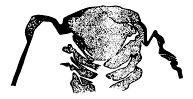
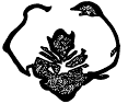
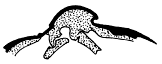
Fig. 13.—Slice through Calymene senaria in the plane of the hypostoma, showing the very slender coxopodites beside that organ, the spines on the inner end of one of the maxillulæ, and the anterior position of the attachment of all these appendages. From a photographic enlargement. Specimen 50. × 4. Fig. 14.—Slice through the hypostoma and thorax of Calymene senaria Conrad, showing the small size of the coxopodites nearest the hypostoma. Shell in black, appendages and filling of abdominal cavity dotted. From a photographic enlargement. Specimen 40. × 3.8. Fig. 15.—Transverse section of Calymene, showing method of articulation with the appendifer. The shell is in solid black, the filling of the appendage and appendifer stippled. Traced from a photographic enlargement of the slice. Specimen 63. × 7.
Exopodites.—Walcott admits that there is no direct evidence of spiral exopodites in the cephalon of Calymene. No one of the sections cutting through the plane of the hypostoma shows any trace of appendages which could be interpreted as exopodites.
Thoracic Appendages.
The large coxopodites of the anterior thoracic appendages are well shown in many specimens cut longitudinally, of which Nos. 23, 50, and 55 may be mentioned, since photographs of them have been published by Walcott (pl. 26, figs. 1-4, 1918). The endobases of all taper toward the proximal ends. Transverse slices show sections of the coxopodites which are no wider than those in longitudinal sections, indicating that they were not compressed but probably cylindrical. This is borne out by an individual (pl. 28, fig. 7, 1918) which is not a slice but an actual specimen, the body cavity of which was hollow, and, opened from above, shows the impressions of the last two coxopodites of the cephalon, and the first four of the thorax.
One transverse section (No. 63, see our fig. 15) is especially valuable, as it shows the method of articulation of the coxopodites with the dorsal skeleton. Another specimen (No. 73) shows that appendifers are present in Calymene, and while the appendifer does not retain its original form in slice No. 63, the section does show clearly that there was a notch in the inner (upper) side of the coxopodite into which the lower end of the appendifer fitted, thus giving a firm, articulated support for the appendage. This notch appears to be slightly nearer the outer than the inner end of the coxopodite, and since it must have made a kind of ball-and-socket joint, considerable freedom of movement was allowed. The appendage must have been held in place by muscles within the coxopodite and attached to the appendifer.
No slice which I have seen shows a continuous section through all the segments of an endopodite, but many, both longitudinal and transverse, show one, two, or as many as three segments.
Such sections as No. 120 show that the endopodites of the thorax were slender and composed of segments of rather uniform diameter. Other sections, notably No. 83, 154, and in, show that they tapered distally, and bore small spines at the outer end of each segment.
The exopodites of course furnish the chief difficulty in interpretation. Doctor Walcott finds two sets of structures attached to the coxopodite, a long, slender, spiral exopodite, and a short, broad epipodite with a fringe of long setæ. Since he has given the same interpretation for Calymene, Ceraurus, and Acidaspis, I have considered the question of all three together on a preceding page (p. 48), and given my reasons for regarding both structures as due to sections in different directions across setiferous exopodites.
Sections like those shown in figures 11, 13, and 14 of plate 27 (1918) happen to be cut in or near the plane of the setæ of an exopodite, and so show hairs of considerable length. Such sections are, as would be expected, very few in number, while sections like those shown on figures 4, 5, 7, and 9 of plate 27, which cut the setæ more nearly at right angles, are very common. Slices which give any definite idea of the form of the shaft of the exopodite are exceedingly rare. Perhaps the most satisfactory one is No. 23 (pl. 3, fig. 3, 1881), which shows the proximal part of a long, slender, unsegmented shaft, with the bases of a number of slender setæ. The organ is not complete, as would be inferred from the published figure, but the section cuts diagonally across it, and the total length is unknown. It is directed forward, like the exopodites of Neolenus, but whether or not this is a natural position is yet to be learned.
The proximal, non-setiferous portion of the exopodite is evidently at an angle with the setiferous part. Another similar exopodite is apparently shown by specimen 29 (pl. 3, fig. 9, 1881), which has a similar angulated shaft and just a trace of the bases of the setæ.
Pygidial Appendages.
That appendages were present under the pygidium is shown by longitudinal sections, but nothing is known of the detail of structure.
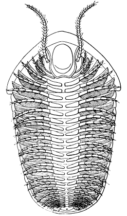
Fig. 16. Restoration of Calymene senaria Conrad, based upon data obtained from the study of the translucent sections made by Doctor Walcott. Prepared by Doctor Elvira Wood, under the supervision of the author. About twice natural size.
Relation of Hypostoma to Cephalon in Calymene.
In Calymene the shape of the hypostoma bears little relation to the shape of the glabella, and it is relatively smaller, both shorter and narrower, than in Ceraurus. In shape, neglecting the side lappets at the front, it is somewhat rectangular, but rounded at the back, where it is bifurcated by a shallow notch. The anterior edge has a narrow flange all across, which is turned at almost right angles to the plane of the appendage, and which fits against the doublure of the free cheeks at the sides and against the epistoma in the middle. The side lappets show on their inner (upper) surface shallow pits, one on each lappet, which fit over projections that on the dorsal surface show as deep pits in the bottom of the dorsal furrows in front of the anterior glabellar furrows. The appendifers on the head in Calymene take the form of curving projections of shell underneath the glabellar and neck furrows, and owing to the narrowness of the hypostoma, all these are visible from the ventral side, even with it in position. This shield extends back about 0.6 of the length of the cephalon, and to a point a little behind the second glabellar furrow from the back of the head.
In Doctor Walcott's restoration of Calymene he has represented all four pairs of biramous appendages as articulating back of the posterior end of the hypostoma. I think his sections indicate that the gnathobases of two pairs of these appendages rested alongside or beneath it, and in particular, the longitudinal sections (1881, pl. 5) would appear to show that the mouth was some distance in advance of its posterior end.
Restoration of Calymene.
(Text fig. 16.)
From what has been said above, it is evident that for a restoration of the appendages of Calymene considerable dependence must be placed upon analogy with other trilobites. Nothing is positively known of the antennules, the exopodites of the cephalon, or any appendages, other than coxopodites, of the pygidium, but all were probably present. It is inferred from the slices that the first two pairs of cephalic appendages were poorly developed, the endopodites short and very slender, the coxopodites lying parallel to the sides of the hypostoma and nearly or quite functionless. The gnathites of the last two pairs of cephalic appendages are large, closely approximated at their inner ends, and bear small tooth-like spines. The endopodites are probably somewhat better developed than the anterior ones and more like those on the thorax.
The coxopodites of the thorax appear to have had nearly cylindrical endobases which tapered inward. The endopodites were slender, tapering gradually outward, and probably did not extend beyond the dorsal test. Small spines were present on the distal end of each segment. Each exopodite had a long, slender, unsegmented shaft, to which were attached numerous long, overlapping, flattened setæ. The shaft may have been angulated near the proximal end, and may have been directed somewhat forward and outward as in Neolenus, but the evidence on this point is unsatisfactory. The number of pairs of appendages is that determined by Walcott from longitudinal sections, namely, four pairs on the cephalon beside the antennules, thirteen pairs in the thorax, and nine pairs on the pygidium.
Calymene sp. ind.
(pl. 6, figs. 4, 5.)
Illustrated: Walcott, Bull. Mus. Comp. Zool., Harvard Coll., vol. 8, 1881, pl. 6, figs. 5a, b;—Proc. Biol. Soc. Washington, vol. 9, 1894, pl. 1, fig. 10;—Geol. Mag., dec. 4, vol. 1, 1894, pl. 8, fig. 10;—Smithson. Misc. Coll., vol. 67, 1918, pl. 36, figs. 1, 2, 2a-d.—Milne-Edwards, Ann. Sci. Nat., Zoologie, ser. 6, vol. 12, 1881; pl. 12, figs. 44a, b.
In the United States National Museum there is a thin piece of limestone, about 3 inches square, which has on its surface eight jointed objects that have been called legs of trilobites. Two of these were figured by Walcott (1881, pl. 6, fig. 5). The slab contains specimens of Dalmanella and Cryptolithus, in addition to the appendages of trilobites, and is said by Doctor Ulrich to have come from the tipper part of the Point Pleasant formation (Trenton) on the bank of the Ohio River below Covington, Kentucky.
The specimens are all endopodites of long slender form, similar to those of Triarthrus, but since that genus does not occur in the Point Pleasant, it is necessary to look upon some other trilobite as the former possessor of these organs. Both Isotelus and Calymene occur at this horizon, and as the specimens obviously do not belong to Isotelus or Cryptolithus, it is probable that they were formerly part of a Calymene.
All the endopodites are of chitinous material, and the various specimens show, according to the perfection of their preservation, from four to six segments. The endopodite as a whole tapers but slightly outward, and the individual segments are of nearly equal length. They appear to be but little crushed, and are oval in section, with a crimped anterior and posterior margin. One or two show a median longitudinal ridge, such as is seen in some appendages of Triarthrus. Each segment is parallel-sided, with a slight expansion at the distal end, where the next segment fits into it.
Under the heading "Ordovician Crustacean Leg," Walcott (1918, p. 154, pl. 36, figs. 1,2) has recently redescribed these specimens, and thinks that they do not belong to Calymene, nor, indeed, to any trilobite. He concludes that they were more like what one would expect in an isopod. Passing over the fact that the oldest isopod now known is Devonian, the fossils in question seem to me quite trilobite-like. Walcott says:
The legs are associated with fragments of Calymene meeki but it is not probable that they belong to that species; if they did, they are unlike any trilobite leg known to me. The very short coxopodite and basopodite are unknown in the trilobites of which we have the legs, as they are fused into one joint forming the long protopodite in the trilobite. The distal joint is also unlike that of the trilobite legs known to us.
A great deal of Doctor Walcott's difficulty probably arises from his homology of the coxopodite of the trilobite with the protopodite of the higher Crustacea. The coxopodite of the trilobite is not fused with the basipodite, this latter segment always remaining free. Indeed, Walcott himself says of Neolenus (1918, p. 128):
Each thoracic leg (endopodite) is formed of a large elongate proximal joint (protopodite), four strong joints each about 1.5 times as long as wide (basopodite, ischiopodite, meropodite and carpopodite); two slender elongate joints (propodite and dactylopodite) and a claw-like, more or less tripartite termination.
Walcott's drawing (pl. 36, fig. 1) is a composite one, and while it shows eight segments, I was not able to count more than seven on any of the specimens themselves. In regard to the terminal segment, the dactylopodite of the limb shown in his plate 36, figure 2, is unusually long, and a comparison with other photographs published on the same plate shows that such long segments are unusual.
Proof that these are appendages of a Calymene is of course wanting, but there is no particular reason so far to say that they are not.
Measurements: Two of the more complete specimens, each showing six segments, are each 8 mm. long.
Somewhat similar to the specimens from Covington are the ones described by Eichwald (1825, p. 39, 1860, pl. 21), the specimens being from the Silurian of Gotland. The figure copied by Walcott (1881, pl. 6, fig. 4) has never been looked upon as entirely satisfactory evidence of the nature of the specimen, and so far as I know, the fossil has not been seen by any modern investigator.
Ceraurus pleurexanthemus Green.
(pl. 11; text figs. 12, 17-19, 21, 22, 24, 29, 30.)
Illustrated: Walcott, Ann. Lye. Nat. Hist. New York, vol. II, 1875, pl. 11;—31st Ann. Rept. New York State Mus. Nat. Hist, 1879, pl. 1, fig. 3;—Bull. Mus. Comp. Zool., Harvard Coll., vol. 8, 1881, pl. 1, figs. 1-5; pl. 2, figs. 1-4, 6-8; pl. 3, figs. 2, 4-7; pl. 4, figs. 1, 2, 4-6, 8; pl. 6, fig. 3; Smithson. Misc. Coll., vol. 67, 1918, pl. 26, figs. 8, 14, 15; pl. 27, figs. 1-3, 5a, 6-9, 12 (not Calymene), (not 15, Calymene); pl. 28, figs. 1-5; pl. 34, fig. 1; pl. 35, fig. 7.—Milne-Edwards, Ann. Sci. Nat., Zoologie, ser. 6, vol. 12, 1881, pl. 10, figs. 1-18.—Bernard, The Apodidæ, 1892, text figs. 46, 51.
Cephalic Appendages.
No trace of antennules has yet been found.
I find only three sections cut through the plane of the hypostoma of Ceraurus which show anything of the cephalic appendages, and no one of them is very satisfactory. The best is No. 22, the one figured by Walcott (pl. 3, fig. 2, 1881; pl. 27, fig. 12, 1918), but one should remember that this section is not actually cut in the plane of the hypostoma but is a slice diagonally through the head, cutting through one eye and the posterior end of the hypostoma. It shows what seem to be the coxopodites of the second, third, and fourth pairs of cephalic appendages, the exopodites of the third and fourth pairs, and the metastoma. If this interpretation is correct, the first pair of gnathites lay alongside the hypostoma or under its edge, and were feebly developed, the second pair were attached in front of the tip of the hypostoma, curved back close to it, and their inner ends reached the sides of the metastoma. The third and fourth pairs were back of the metastoma, the third pair was stronger than the second, and the fourth probably like the third.
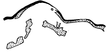
Fig. 17. Transverse section of Ceraurus pleurexanthemus, showing the relation of the coxopodite to the appendifer. Traced from a photographic enlargement of the slice. Specimen 128. × 4/5.
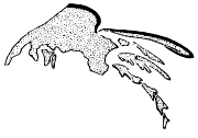
Fig. 18. Slice of Ceraurus pleurexanthemus, showing a nearly continuous section of an endopodite and an exopodite above it. The latter is so cut as to show only the edge of the shaft and the bases of a few setæ. Traced from a photographic enlargement. Specimen in. × 4.
Specimen 92 shows traces of the slender endopodites belonging to the cephalon, but no details. Specimen 22 shows on one side exopodites (epipodites of Walcott) belonging to the third and fourth cephalic appendages. That belonging to the third shows some long setæ and a trace of the shaft, while the one on the fourth appendage (third coxopodite) has a portion of a broad shaft and a number of long setæ. It should again be remembered that the slice does not cut through the plane of the exopodite, but across it at a low angle, so that a part but not all of the shaft is shown. On the other side of this slice there is a fairly good section of one of the thoracic exopodites. It is, however, turned around in the opposite direction from the others, as would be expected in an enrolled specimen.
Specimens 4 and 5 (pl. 1, figs. 4, 5, 1881) are slices cut diagonally through the head of Ceraurus, in front of the posterior tip of the hypostoma. They show fragments of endopodites and exopodites which may be interpreted as practically identical in form with those of the thorax. Due to the diagonal plane in which the section is cut, slice 5 shows the coxopodites of two pairs of appendages, one lying nearer the median cavity than the other. It is extremely difficult to visualize the interpretation of such sections.
Thoracic Appendages.
A transverse section through a thoracic segment (No. 128, our fig. 17) shows the relation of coxopodite to appendifer to be the same as in Calymene, the upper side of the coxopodite having a notch a little outward from the middle. After seeing that specimen, it is possible to understand slice No. 168, which shows longitudinal sections through a number of coxopodites of the thorax, with fragments of both exopodites and endopodites articulated at the distal ends. These and longitudinal vertical sections like No. 18 (pl. 2, fig. 8, 1881) show that the endobases taper inward, and the general uniformity in width in sections taken at various angles indicates that the coxopodites were not greatly flattened.
A unique slice (No. 111, pl. 2, fig. 2, 1881; pl. 27, fig. 1, 1918; our fig. 18) shows a nearly complete thoracic endopodite, and above it a part of the proximal end of the exopodite of the same segment. When one considers that out of over two thousand sections only this one shows the six successive segments of an endopodite, one realizes how futile it is to expect that dozens of the equally slender "spirals" should be cut so as to show practically all their turns.
This endopodite is slender, all the segments have nearly the same length and diameter, though there is a slight taper outward, each segment is expanded distally for the articulation of the next, and there are small spines on the distal ends of some of them. There is probably a terminal spine present, though it is neither so long nor so plainly visible as in Walcott's photograph.
The exopodite on this same specimen was evidently cut diagonally across near the setiferous edge, showing a section through the shaft and the bases of seven setæ (fig. 18). This section is so exactly what would be obtained by cutting similarly an exopodite of either Neolenus or Triarthrus that it should in itself dispose of the "spiral-exopodite" theory.
Several sections have already been illustrated showing sections across the setæ of the exopodites (pl. 3, figs. 4-6, 1881; pl. 27, figs. 3, 4, 9, 1918), and similar sections are not uncommon. Only a very few, however, show sections in the plane of the exopodite. If only No. 111, described above, were known, it would be inferred that the exopodite had a slender shaft as in Calymene, but another good slice, No. 80 (fig. 12, ante) shows that the blade was rather broad, though not so broad as in Neolenus. The other specimen is No. 22, which has already been discussed. The thoracic exopodite of this specimen has been very incorrectly figured by Walcott, as it shows no such palmate shaft as he has indicated, but a long blade-like one is outlined, though its entire width is not actually shown.
Pygidial Appendages.
Sections 14 and 18 (pl. 2, figs. 4, 8, 1881) prove the presence under the pygidium of three pairs of appendages, the coxopodites and fragments of endopodites of which are shown. Nothing is known of the exopodites.
Relation of Hypostoma to Cephalon.
In Ceraurus the body portion and posterior end of the hypostoma are roughly oval, about as wide as the glabella at its broadest part, and the posterior edge extends back to within 0.5 to 1 mm. of the neck furrow. The posterior pair of appendifers are behind the hypostoma, while the second pair are in front of its posterior end but escape being covered by it on account of its oval shape. At the anterior end the hypostoma is widened by the presence of two side lappets which extend beyond the boundaries of the glabella. In both Ceraurus and Cheirurus the anterior edge of the hypostoma fits against the doublure at the anterior margin of the head and the epistoma is either entirely absent or is so narrow as not to be seen in specimens in the ordinary state of preservation. A section across the cephalon of Ceraurus pleurexanthemus at the horizon of the eyes shows the sides of the hypostoma fitting closely against the sides of the glabella (Walcott's pl. 1, fig. 1). Further back on the head it is not in contact with the dorsal test, and the gnathobases extend beneath it.
Restoration of Ceraurus pleurexanthemus.
(pl. 11; text fig. 19.)
The restoration of the appendages of Ceraurus pleurexanthemus is a tentative one, based upon a careful study of the translucent sections prepared by Doctor Walcott. In no case among these sections is the actual test of any appendage preserved, and the real form of each part is generally obscured by the crystallization of the calcite which fills the spaces formerly occupied by animal matter.
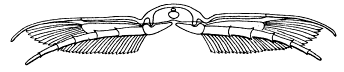
Fig. 19. Restoration of a transverse section of the thorax of Ceraurus pleurexanthemus Green, showing the relation of the appendages to the appendifers and the ventral membrane. The probable positions of the heart and alimentary canal are indicated.
No section shows anything which can be identified as any part of the antennules, so that these organs have been supplied from analogy with Triarthrus.
There are undoubtedly four pairs of biramous Cephalic appendages, but their points of attachment are not so obvious. There are two pairs of conspicuous appendifers on the posterior part of the cephalon and another pair almost concealed by the hypostoma. It is probable that the appendages of the cephalon were not attached directly beneath them, as the four pairs have to be placed within the space occupied by the three pairs of appendifers. As the mouth is in front of the posterior end of the hypostoma, the gnathites of the first pair of biramous appendages may have extended beneath that organ, or they may have lain beside it, and only become functional when the hypostoma was dropped down in the feeding position. The second pair of gnathites reached just to the tip of the hypostoma, and the other two pairs seemingly curved backward behind it.
The points of attachment on the thorax, as shown clearly in sections, were directly beneath the lower ends of the appendifers. The endopodites were long enough to reach to or a little beyond the outer extremities of the pleural spines, while the exopodites were apparently somewhat shorter. Each endopodite consisted of six short, fairly stout segments, each with at least two spines on the somewhat expanded distal ends. The exact form of the exopodites could not be made out. The shaft was apparently rather short, unsegmented, and fairly broad. The setæ appear from the sections to have been more or less blade-shaped and to have overlapped, as do those of the exopodites of Cryptolithus. Judging from their position in the sections, the setæ not only bordered the posterior side of the shaft, but radiated out from the end as well.
The pygidium shows three pairs of functional appendifers, hence three pairs of appendages have been supplied. There is a fourth pair of rudimentary appendifers, but as they are beneath the doublure they could not have borne ambulatory appendages.
The Appendages of Acidaspis trentonensis Walcott.
(pl. 6, fig. 6.)
A single individual of Acidaspis trentonensis, obtained from the same locality and horizon as the specimens of Triarthrus and Cryptolithus, when cleaned from the ventral side shows a number of poorly preserved endopodites which seem very similar in shape and position to those of Triarthrus. One endopodite on the right side of the head and the first five on the right side of the thorax are the best shown. All are slender, are directed first forward at an angle of about 45 with the axis, then, except in the case of the cephalic appendage, turn backward on a gentle curve and extend a little distance beyond the margin of the test, but not as far as the tips of the lateral spines of the thoracic segments.
The individual segments of the endopodites can not be seen clearly enough to make any measurements. On the fourth and fifth endopodites of the thorax, some of the segments seem to be broad and triangular as in Triarthrus. All that can be seen indicates that Acidaspis had appendages entirely similar to those of Triarthrus, but perhaps not quite so long, as they seem not to have projected beyond the limits of the lateral spines. There are no traces of antennules nor, unfortunately, of exopodites.
Measurements: Length 8 mm.
Walcott (1881, p. 206) stated that his sections had shown the presence in this species of legs "both cephalic and thoracic" and also the "spiral branchiæ." His specimens were from the Trenton at Trenton Falls, New York.