TUMORS OF THE BLADDER. DIAGNOSED BY MEANS OF THE ELECTRO-ENDOSCOPIC CYSTOSCOPE.
Reading edition
By Dr. Max Nitze.
In the following lines I wish to direct the attention of my English confreres to the value of the electro-endoscopic mode of examination of the male urinary bladder, invented by me. I believe I could not have chosen a more suitable theme for that purpose than a short report of the bladder tumors diagnosed by me cystoscopically; for the diagnosis of these new formations offers the greatest difficulty, and in most cases it has been impossible till now to prove their existence with accuracy without digital exploration of the bladder. By the new method of cystoscopical examination the conditions have entirely changed. One look into the bladder, illuminated as if by daylight, is generally sufficient to afford means for forming an opinion of all the questions coming into consideration—viz., size, form, and site of the tumor. The accompanying diagrams (Figs. 1, 2, 3, 4) may give an idea of the appearances which the different forms of bladder tumors present endoscopically. I regret that they cannot show the brightness of the light by which one sees the tumors during examination. The celebrated Vienna specialist, V. Dittel, is right in saying that "they offer sometimes truly charming pictures;" especially certain kinds of villous tumors, whose long slender villi floating in the liquid often present a splendid appearance. The following are the cases cystoscopically diagnosed by me.
Fig. 1.
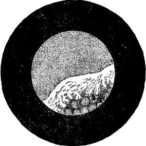
Case 1.—A man, aged fifty-five, under the care of Dr. Ch. Mayer, suffered from attacks of hæmaturia for thirty years. During the last six years he has had dysuria and inability to empty the bladder completely. The patient had been examined by the sound repeatedly by eminent surgeons and specialists, but none could give a certain diagnosis. On Nov. 11, 1886, I undertook the cystoscopic examination. I found on the anterior wall of the bladder a puffy swelling covered with white masses of mucus. (See Fig. 1.) The trigone was covered by a mass consisting of pointed papillæ. On account of the weakness of the patient extirpation was impossible. The patient became weaker
Fig. 2.
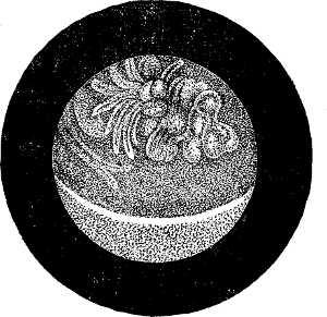
Case 2.—A man, aged fifty, was obliged to exert a strong pressure in order to empty the bladder. The flow of urine often stopped. He himself introduced a catheter, and on withdrawing it a piece of villous tissue was found. On Dec. 10, 1886, I saw, on cystoscopical examination, directly and immediately over the internal orifice of the urethra, a villous swelling hanging from the anterior wall of the bladder. (See Fig. 2.) On Jan. 15, 1887, extirpation of the tumor by means of the high section was performed by Professor v. Bergmann. The size of the tumor (which was as large as a pigeon's egg) and its position corresponded exactly to the endoscopic picture. The patient recovered.
Fig. 3.
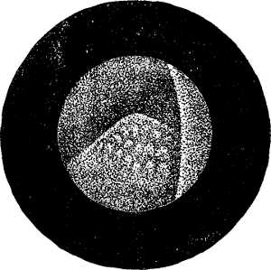
Case 3.—A patient under the care of Professor Madelung, aged fifty-five, suffered from attacks of hæmaturia. Examination by sound and rectal palpation had given me negative results. On Feb. 20, 1887, cystoscopical examination was made. On the left side of the trigone a tumor with a broad base was seen, which resembled somewhat a strawberry in size and form. (See Fig. 3.) On March 1, Professor Madelung undertook the extirpation of the tumor. The appearance corresponded exactly to the cystoscopic picture. The patient recovered.
Fig. 4.
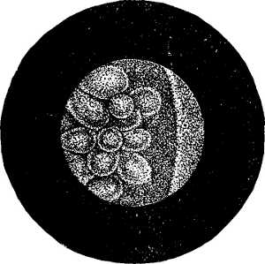
Case 4.—This was a patient on whom Dr. Israel had performed the high section a long time before, on account of a bladder tumor. The extent was so great that only its most prominent part could be removed. The microscopical examination proved the diagnosis of cancer. Quick healing took place. The patient became free from pain, and the urine became clear. In
[We omit the description of the additional cases.]
The above shortly described fifteen[9] cases of bladder tumors have been diagnosed by me cystoscopically during the last sixteen months. This is a proof, on the one hand, of the value of the cystoscopic examination; on the other hand, of the fact that the new formations in question are not of so rare occurrence as has been hitherto thought. I would like to emphasize that the important results were often obtained under the most difficult circumstances. In several cases the external orifice of the urethra was found abnormally small; in others (Cases 8 and 11) the examination was made during the occurrence of a continuous hemorrhage from the tumor; in one case (Case 1) I introduced the instrument through the center of the tumor, which bled on the slightest pressure. In spite of this the appearances were seen satisfactorily. In the first case a post mortem examination was made; in eight other cases (Cases 2, 3, 9, 10, 11, 13, 14, and 15) the tumor was extirpated, seven times by the high section—in one case, that of a woman, through the dilated urethra. In these nine cases the endoscopic appearances were in every important respect confirmed in the most perfect manner. In every case my opinion regarding the size, position, and form was found to be correct. It is only in those cases where the edges of the tumor overlap the short pedicle that the latter cannot be observed. Besides, the relative good results of the operations undertaken on account of the cystoscopic appearance may be emphasized. Of the eight patients from whom the tumors had been extirpated, none died from the result of the operation. Case 9 proved fatal on account of the progressive extension of the growth. In the eleventh case there was a recurrence, but the patient is still alive. Five patients (Cases 2, 3, 10, 13, 14) must be considered entirely cured. Case 15 is still under treatment, and, as the conditions of the patient are at present (ninth day after operation) in every way satisfactory, a complete recovery is anticipated.
Finally, on comparing the above cystoscopic appearances with the results obtained by other methods of examination, it must be observed that the examination of the urine, in most cases carefully made, had only in two cases shown the presence of villous tissue, which in one instance was brought out by the catheter. The rectal palpation, when made, had always given negative results. Further, the examination by means of the sound had been made in nine cases before the cystoscopic examination. In none of the cases had the sound revealed the presence of a tumor (which in two had attained the size of a small apple), although the examination was made by most experienced surgeons and eminent specialists. Those cases show how imperfect an instrument the sound is for the diagnosis of bladder tumors.
Only one method can compare with the cystoscope in giving valuable information regarding the size and nature of a bladder tumor—viz., the digital exploration of the internal surface of the bladder after a previous boutonniere, or the high section. The superiority of the cystoscopic method over the latter, on account of the smaller amount of inconvenience it causes the patient, need not be insisted on. The latter involves a cutting operation not free from danger, as well as deep narcosis, while the cystoscopic method is similar to a simple catheterization.
Fig. 5.
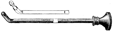
The accompanying diagram (Fig. 5) shows the instrument used by me for cystoscopic examination. It has been made by the Berlin instrument maker, Hartwig, according to my instructions. The source of the light (Mignon lamp) is cemented in a silver capsule, which is screwed into the distal end of the cystoscope. This instrument is superior to that made by Leiter, the Vienna instrument maker, because of its greater simplicity in construction, which allows the lamp to be easily replaced when necessary, and also on account of the greater length of the shaft.
I mention this because it differs from the explanation which Mr. Fenwick gave in his speech concerning my method of examination at the meeting of the Medical Society of London on Jan. 23, 1888. I must also strongly contradict Mr. Fenwick's statements concerning the share which he attributed to the Vienna instrument maker in the construction of the instrument. Leiter's connection with our instrument will be best explained when I say that he had to buy the patent[10] from me first in order to be allowed to make the instrument. Leiter has had no share in those peculiarities which characterize it as new. The introduction of the source of light into the organ had been practically brought about, the optical apparatus enlarging the view designed, the whole construction perfected, the instrument had proved itself useful in examining patients, and had been demonstrated by me in the Saechsisches Landes Medicinal Collegium before Leiter had any idea of the new invention! Also the eventual replacement of the first source of light (platinum wire) had been provided for.[11] Leiter has only made a few technical modifications on the finished instrument. I protest most emphatically against the incorrect explanations given by Mr. Fenwick, and against every connection of Leiter's name with my instruments. I hope to obtain in England the same generous recognition of my labors in this field that has been accorded to me in Germany.—Lancet.