PAPILLOMATOUS TUMOR OF THE BLADDER, DEMONSTRATED BY MEANS OF LISTER'S ELECTRO-CYSTOSCOPE.
Reading edition
By F. N. Otis, M.D., Clinical Professor, College of Physicians and Surgeons, New York.
A. G——, aged twenty-three, United States; single; barber.
The young man was referred to me by his former medical attendant, March 16, 1883. His urine was found to be slightly but distinctly tinged with blood, and contained some small clots as well as some pus and mucus. He complained of exquisite pain on urination, increased at the close, recurring every half hour. Through examination per rectum (a posteriori) unusual tenderness was found. Distinct increase in the density and thickness of the right inferior section of the bladder was recognized by the bimanual touch; a catheter was introduced, and three ounces of bloody urine removed. The bladder was then irrigated gently with a saturated solution of boric acid until the fluid returned clear. The catheter was then withdrawn, leaving about four ounces of the solution, of a temperature of 80°, in the bladder, as a preparation for its examination by the electro-cystoscope of Lister. The required current was furnished by the small six-cell battery of the Galvano-Faradic Co. The cystoscope was then introduced into the bladder, and the current turned on. The illumination was complete. Through the slightly rosy medium the small blood vessels in the bladder mucous membrane were distinctly seen. On the right side a deep red, granular-looking mass, with a wavy outline, was then distinctly observed, covering about one-fourth of the cystoscopic field. This appearance was verified by Drs. Abbe, Bangs, and W. K. Otis—the unanimous opinion being that it represented a papillomatous growth, to some extent covered by coagulated blood. Two days later a similar examination was made, under the influence of an anæsthetic, which corroborated the previous observations in every particular. (See illustration.)
DIAGRAM OF BLADDER, SHOWING LOCATION OF TUMOR AND POSITION OF CYSTOSCOPE.
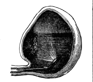
Some small filaments were subsequently removed with the lithotrite, but on microscopical examination nothing of diagnostic importance was discovered. From lack of the capacity of the bladder, the field was necessarily limited, nevertheless, a very excellent view of the tumor could be obtained. This is shown in the illustration, from a sketch made at the time of the first examination. It represents the position of the tumor and cystoscope when the best view of it was obtained.
On the following Monday the patient entered St. Luke's Hospital, and was operated upon by my associate, Dr. L. B. Bangs, Dr. Charles McBurney assisting. The high operation was performed, and the bladder being examined by means of an electric light, introduced through the suprapubic incision, the diagnosis made by the cystoscope was verified in every particular. The growth was then removed, as far as possible, with the scissors, and the surface cauterized with the Paquelin cautery. At the present writing the patient is going on toward a satisfactory recovery. The pathological examination made by Dr. Frank Ferguson, pathologist of St. Luke's Hospital, showed the neoplasm to be a simple papilloma.
This case is deserving of especial interest as being the first tumor of the bladder diagnosticated in this country by means of the cystoscope, and verified by subsequent
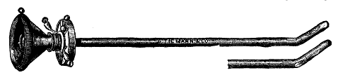
This consists of a metal tube, about seven inches long, of a caliber of 22 French, having at the proximal end a funnel shaped ocular opening; at the distal, a short beak, similar to that of the catheter coudé. A window of rock crystal is set in the end of this beak, behind which a small electric lamp, controlled by a switch at the ocular end, is placed. A rectangular prism, the hypothenuse plane of which is silvered, is placed in the end of the straight portion of the tube, its superior face being seen just anterior to the angle formed by the beak. The distended bladder is illuminated by the electric lamp, the rays reflected from its wall falling on the prism experience total reflection, an inverted image being formed within the tube. The size of the field thus obtained is greatly increased by means of a telescope introduced into the tube. The image seen through the cystoscope is an inverted image, but right and left are not transposed.
THE CYSTOSCOPE.
There can be no question as to the great prospective value of the electro-cystoscope in diagnosis of many difficulties to which the bladder is subject. A variety of foreign bodies have already been reported as made out by use of this instrument. The locality, size, and color of vesical calculi have been demonstrated in my own experience. In one instance two stones were seen where only one had been previously found, but this of course might with care have been effected by means of the lithotrite. But it is in the diagnosis of the tumors, and encysted or impacted calculi, that the most essential service may be anticipated from the use of the cystoscope. The orifices of the ureters are quite readily brought into the cystoscopic field, and it is more than probable that (perhaps through the introduction of some clear fluid with which blood does not readily mingle—glycerine, for instance) the true source of a previously doubtful hæmaturia will be demonstrated.—Medical Record.