Chapter XV · RHINOPLASTY (Surgery of the Nose)
Reading edition
Rhinoplastic operations serve to correct deformities of or restore the nose. Such operations may involve only a part of or the entire organ, hence may be termed partial or total. Furthermore, a fine distinction may be drawn between general rhinoplasty as applied to such deformities when caused by traumatism, the excision of neoplasms or destructive disease, whether such correction be partial or total, and cosmetic rhinoplasty when such corrections are made purely with the object of improving the nasal form when the deformity is either hereditary or the result of remote accident.
For some unaccountable reason the latter art has not met with the general favor the profession should grant it, yet the results obtained by such specialists as have undertaken this artistic branch of surgery have been all that could be desired, and have consequently added much to the comfort and happiness of the patient.
Without a comparatively thorough knowledge of the extent of cosmetic rhinoplasty it would be difficult to draw any conclusion as to the value of this art. If it has not met with the favor it deserves it is solely due to the fact that the art has been limited to the few, and the literature on the subject is so meager, indeed, that the surgeon has been compelled in many cases to trust to his own originality in undertaking an operation of this nature.
The limitation to rhinoplasty is due primarily to the artistic skill required to obtain results; secondly, to the risks involved by loss of tissue due to gangrene, imperfect healing or accidental interference, post-operatio; and thirdly, to scarring about the face as a result of the primary and secondary wounds; in fact, so much so that many surgeons prefer to allow a small defect to remain, to escape the risks involved in correcting them.
The author believes such fear misplaced, because with the methods of surgery of the present day and the proper knowledge of the art there need be little risk involved and the result expected should be as near perfect as human skill can make it.
True, a surgeon cannot be expected to build an entire nose from the skin or other tissue of the forehead or cheeks and make it a thing of aforethought beauty and shape, but if the result be no more than a curtain of skin to hide the hideous deformity he has done his share, and such result is the worst he might look forward to.
For the correction of nasal deformities the author will consider first such operations as involve the entire loss of the nasal organ or total rhinoplasty; thereafter partial loss of the nose, and lastly such cases involving no loss of tissue and dependent on malformation only under cosmetic rhinoplasty.
It is not here intended to lay down a law for the surgeon for the restoration of the entire or part of the nose for the reason that each case differs more or less; that in each case there is more or less tissue that may be utilized, and that there are many methods advanced for such procedure, but the author does desire to give to the operator a concise and comprehensive treatise on rhinoplasty and to illustrate the best of such operations as have been placed on record as a ready guide and for immediate reference—a matter of no small moment when this literature can be gained only by searching through innumerable medical journals and short references and in all languages of the civilized world.
In the chapter on history some idea of the time in which rhinoplasty has been practiced may be obtained. It is not deemed necessary to go into further historical facts here, except, perhaps, to divide the subject into the three most important schools or countries that have given individuality to the art.
THE CAUSES OF NASAL DESTRUCTION
The loss of the entire nose may be due to traumatism, actual amputation, the bites of man or beast, duels, the removal of neoplasms, gangrene after freezing or disease, rhinosclerosis, syphilis, the application of caustics, tubercular disease, lupus, cancer, and rarely congenital absence of the organ. The loss may be total or partial.
The extent of loss of substance in each case differs, and it is for this reason that surgeons have been compelled to originate many methods of operation, each having for its object to correct the deformity as neatly and as near to the normal as possible.
CLASSIFICATION OF DEFORMITIES
To give correctly a classification of nasal deformities would simply mean to mention each anatomical part or division of the nose referring to the deformity involving the same. For this reason such an arrangement would be uselessly extensive, but for the proper recording of such cases the author advises a systematic method of nomenclature in which the deformity is stated, as: left, unilateral deficiency of inferior lobule; or right, median third deficiency of nasal dorsum of the parts destroyed and mentioned as such.
Fig. 306.—Deficiency of Superior and Middle Third of Nose. (Saddle Nose.) Fig. 307.—Post-ulcerative Deformity of Superior Third of Nose.
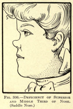
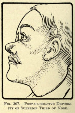
Fig. 308.—Loss of Right Ala, Lobule and Columna. Fig. 309.—Loss of Lobule, Inferior Septum and Columna.
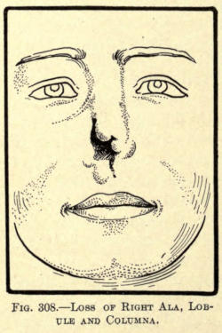
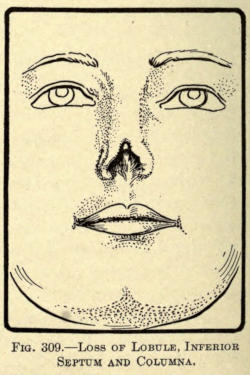
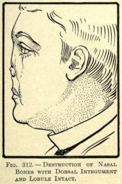
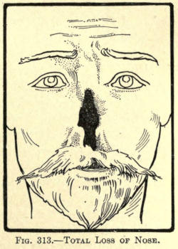
A fair idea of typical deformities may be obtained from the following illustrations in which deformities from the milder to the most extensive extent are shown. The types here shown are all pathological with the exception of Fig. 306, in which a saddle nose is illustrated which may or may not be the result of disease or traumatism.
Fig. 310.—Ulcerative Loss of Right Median Lateral Skin of Nose with Involvement of Ala. Fig. 311.—Loss of Nasal Bones and Partial Ulcerative Destruction of Dorsum, Lobule and Septum of Nose.
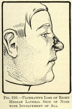
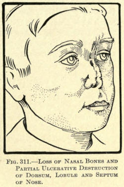
Fig. 312.—Destruction of Nasal Bones with Dorsal Integument and Lobule Intact. Fig. 313.—Total Loss of Nose.
Many other deformities of the nose exist, of course, such as lateral deviation, twists, etc., but as in most of such cases cosmetic rhinoplastic operations and subcutaneous injection are required for their correction, inasmuch as in these cases the skin is healthy and intact, they will be considered under that part of the chapter that has to do with purely cosmetic rhinoplasty or under the chapter on subcutaneous protheses.
SURGICAL TECHNIQUE
Before going into the individual methods involved in the correction of deformities of the nose, it is well here to go into the special details required for the performance of operations about the nose proper.
Anesthesia.—It may be well here to state that many of the smaller or cosmetic operations can and should be done under local anesthesia, and that the anterior nares should be plugged to prevent the blood from running into the pharynx, but in operations of greater extent the posterior nares should be plugged by Bellocq or other method, and that since the patient must be placed under a general anesthetic, some special plan must be followed to give the same.
The author has found no special apparatus on the market for this purpose. A most practical apparatus may be made as follows: A medium hard piece of rubber is cut into such shape as will fit into the patient’s mouth between the lips and the teeth. In its center a hole is made, into which a metal tube is fixed to which a rubber tube of three-fourth-inch diameter is securely fastened. This tube is connected by its distal end to the anesthetic container, which should be so constructed as to permit the required amount of air to be given with the anesthetic at the desired time.
Such an apparatus practically seals the oral orifice, and prevents blood from flowing into the mouth, gives the operator a free field to work in without the encumbrance of large external mouthpieces, and is one that in case of vomiting can be easily removed for the time being, and be replaced without interference to the surgeon.
Preparation and Cutting of Nasal Flaps.—Under a division of skin grafting some preliminary steps in the preparation and cutting of a nasal flap has been referred to, but the author thinks it timely to repeat here the necessity for a systematic method of procedure.
It is well for the surgeon to have fully decided upon the certain operative plan he is to follow several days prior to the operation. He must, especially in total rhinoplastic cases, prepare a paper or oiled silk model of the flap or flaps he has decided upon to take from the forehead or cheek, and to fold and bend this model into the place of the deformity to be overcome, to make sure of the result to be attained, allowing for the loss, if any, of mass by reason of the torsion of the flap at its pedicle.
If the hair of the frontal scalp lies within the flap outline, it should be shaven away well beyond the border to permit of unhindered work.
Thoroughly cleanse and keep clean with a suitable antiseptic the parts to be operated upon for at least twenty-four hours.
Place a rubber cap over the hair of the head, or a fixed gauze or waterproof arrangement to keep it in place.
If there be any hair adornment of the face remove it.
The surgeon should remember to get the flaps to be utilized on forming the lost parts of the nose, at least one third larger to overcome the consequent retraction.
Sterilized sutures, preferably silk of suitable size, should be ready and be cut of such length as will facilitate quick action.
Rubber tubes of proper diameter for insertion into the nares should be at hand if required.
When all is ready the operator is to proceed quickly and accurately, never changing his prearranged idea of the operation. His assistants should be ready to control by torsion or pressure the bleeding occasioned by cutting, since it covers the field of operation and hinders rapid work.
The surgeon in making flaps should use the greatest gentleness in handling them to prevent pressure gangrene. His finger tips are far better than fixation forceps. Sharp tenaculi may be employed with gentle traction only. Never permit the use of serrated forceps in autoplasty.
In cutting, employ the rules laid down under the principles of plastic surgery, and in dressing flap operations such methods as have been heretofore described.
Dressing.—Do not be too hasty in dressing such wounds, as early interference often results in partial if not total loss of the flap.
The author has found that in flap operations blood dressing under perforated rubber tissue is best. This helps to give nutriment to the parts and permits of free removal of the dressings. Never apply the blood treatment on gauze, since the latter is liable to become hard and attached to the suture lines, requiring undue force for its removal.
Care of the Nares.—Remove all packing from the nares before fixing the lobular section of the flap, and have all bleeding controlled before suturing the part of the flap intended for the columna. Blood clots tend to pressure and infection. If nare tubes are used rather let them remain in place for some time than to drag them forth forcibly.
The interior nose and nares can be kept clean by gentle irrigation through them.
Number of Operations.—Instruct the patient as to the probable outcome of the operation, and advise him that more than two or three operations may be necessary to correct the deformity.
Von Esmarch has said that twenty operations about the nose are none too many if the desired result can be obtained. Dieffenbach has said that it is more difficult to restore smaller nasal defects than those of greater extent.
The latter applies particularly to cosmetic operations in which the surgeon is compelled to work through small openings or incisions always with the view of leaving little if any scar, and to place such scar where it may be least observed.
The best cosmetic surgeon is he who can accomplish results with the least secondary disfigurement.
PROTHESES
When for any cause there is a loss of the entire nose, and the patient is unwilling to undergo surgical operation for its restoration, the surgeon may resort to the use of protheses or artificial noses.
Such noses are made of papier-maché, rubber, wood, or light metal, and painted to imitate the color of the skin of the individual. They should be made after a model previously prepared by molding the new organ upon the face of the patient or after such patterns as the surgeons may have to choose from, fitting the skin juncture accurately in such cases.
If the surgeon lacks such artistic ability, a sculptor should be employed to model the proper organ suitable for and on the face of the patient, from which a plaster cast or mold may be made from which the maker of protheses can work.
With the model in hand and no expert on protheses within reach, a skillful surgeon-dentist could easily make a vulcanized rubber nose, which may then be painted to suit.
Some method of attachment must be provided for, such as one or two soft rubber plugs or stems to fit into the nasal orifice or permanent fixture to the bridge of a pair of spectacles. Gums or pastes as advised with aural protheses may be of service.
Celluloid protheses should never be used because of their inflammable nature; furthermore, they are easily damaged or cracked. Wax noses are of little use, although resembling the normal very closely; they crack easily, and when soiled by dust or friction soon have to be replaced with new ones.
The following list of authorities shows the various materials employed by them for nasal protheses:
Martin—Porcelain.
Richter—Wood.
Debout—Rubber or silver covered with colored wax.
Mathieu—Aluminum.
Charrière—Silver.
NASAL REPLANTING
The plastic surgeon is often, especially in later years, called upon to attend to traumatic injuries of the nose. Sometimes there is a total severance of the nose; often a partial loss or injury, practically involving a loss of a part of the organ. Since the advent of the automobile such accidents are not unusual.
The author has found that a remarkable history lies back of the replanting of parts or all of the nose when found detached by accident or intent.
If the part cut from the nose or face has been not too severely bruised, it should be cleansed gently in a normal salt solution at about 100° F., and be sutured in place as quickly as possible. Partly separated sections should be treated in the same way. It is remarkable how Nature will take care of these traumatisms. So well did the executioners in India, where nasal amputation is a criminal sentence, know this that they destroyed the amputated organ by fire, so that the victim could not replant it upon himself.
Chelius successfully replanted a nose after it had been severed about an hour.
Hoffacker has replanted a number of noses cut off in the duels of Heidelburg students. In one case one and a half hours intervened between the accident and the operation.
In partial separations about the nose the flap, still hanging by a slight pedicle, should be brought in place by suture, and because of the peculiar hypertrophy that always follows the wounds one or two intraflap sutures should be employed to fix the part centrally to the deeper tissues, if any, to prevent the formation of clots that are liable to organize and encourage such enlargement.
Such sutures are only to be made when the flap is of sufficient size to necessitate them. If the hypertrophy or hyperplasia cannot be prevented by this means later cosmetic operations should be employed to make the parts heal into normal contour.
Blood dressings should be employed after the parts have been fixed by a number of fine silk sutures, the coaptation being made as neatly as possible to get the best results.
NASAL TRANSPLANTING
The making of a nose or part thereof from a nonpedicled flap of skin taken from the patient has met with more or less success in the remote past, but of later years such methods have fallen into disuse because of the many and better methods of modern times involving the use of flaps with nutrient pedicles.
Branca is said to have made a nose for a patient out of the skin of the arm of a slave.
Velpeau states that “In the land of the Pariahs the men in power had no scruples in having the nose of one of their subjects cut off to replace the lost organ of another.”
Van Helmont is said to have made a nose for a gentleman from the skin of the buttocks of a street porter.
Bünger, of Marburg, in 1822 made a total nose from the anterior thigh.
Several surgeons later than the above date have successfully restored parts of the nose by transplanting skin flaps from remote parts of the body, the method involved being practically what is now accomplished by the so-called skin-grafting methods of nonpedunculated flaps heretofore referred to.
While for small defects such procedure has proven quite successful, the employment of large flaps for nasal reconstruction has been exceedingly discouraging, although the author advises trying transplanting of such flaps when the patient hesitates giving up sufficient facial skin for rhinoplastic purposes for fear of disfiguring scars, or when there are untoward reasons.
In such event there is only the secondary wound to be considered apart from the death of the flap, and the minor operation about the remains of the nasal organ to permit of the fixation of the latter.
A thorough and practical knowledge of skin grafting is of the greatest necessity to the surgeon, because he must be ready to cope with any emergency in such cases, and thus be able to save a flap graft from death or partial gangrene, when he would otherwise fail.
TOTAL RHINOPLASTY
Pedunculated Flap Method
The most practical and safe methods of rebuilding the nasal organ have been those in which flaps having nutrient pedicles have been employed, whether these flaps be taken from the skin of the forehead, cheek, or both. These procedures are autoplasties, and may be grouped according to their peculiar differentiation into three classes, as follows:
The Indian or Hindu Method, in which the flap is made from the forehead.
The French Method, in which the flap is made from the tissue about the borders of the deformity.
The Italian Method, in which the flap is taken from some distant member or part of the body.
Furthermore, there are the combined methods of one or the other in which inverted skin flaps are used, or those lined with an osseous and cartilaginous support, and in some rare and rather unsuccessful cases by metallic supports.
The Indian or Hindu Method
The method of rebuilding the nose by taking one or two flaps from the forehead dates back to the Koomas, from whom the art of rhinoplasty has come down to the present time, all of the methods of to-day involving the utilization of the pedunculated flap being a result of their early surgical ingenuity.
Originally, their operation consisted of cutting an oval flap, having its pedicle as the root of the nose, and extending over the forehead, and upward vertically into the hair line. The flap thus made was dissected away from the bone and brought down by twisting it to the extent of a hundred and eighty degrees on its pedicle in front of the nasal deformity, the edges of which had been prepared to receive it. To hold the flap in position they resorted to some kind of clay, sutures being unknown to them.
The pedicle was cut after the flap had thoroughly united to the freshened borders of the deformed nose.
The steps of the operation as performed by them are shown in Figs. 314, 315, and 316.
Naturally, many improvements in the above method have been evolved, principally to overcome the extreme and injurious torsion of the pedicle, and from the desire on the part of the surgeon to bring about a better cosmetic result. Therefore, not only the position of the pedicle and its shape were altered, but also the size of the flap itself, as will be shown in the specific methods of the various authorities mentioned hereafter.
The author does not consider it necessary to go into chronological details of the evolvement of the art, and begs the surgeon to be content to learn of those operations and methods that have given the best result.
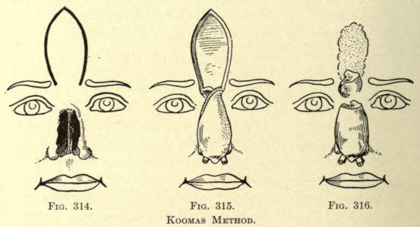
Fig. 314. Fig. 315. Fig. 316.
Koomas Method.
Where one surgeon has changed his incisions in the slightest direction and another has advised increasing the number of sutures is of little import to the operator of to-day; the gist of it all is the successful method for the successful outcome.
The first to be considered will be those methods wherein the vertical direction and the position of the pedicle have been similar to that of the Koomas. It will therein be noted that the principal change has been in the formation of the distal end of the flap with the object solely of forming a better base to the nose.
Graefe Method.—The flap was made in the shape of a heart with a rectangular addition at its upper or scalp border. The pedicle is made to lie between the inner limitations of the eyebrows (see Fig. 317).
The flap is twisted into position and sutured into the freshened remains of the nose, the pedicle being cut at a second operation after the flap has healed into place, which was about the tenth day.
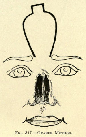
Fig. 317.—Graefe Method.
Delpech Method.—The shape of the frontal flap was cut in the form of a trident, as shown in Fig. 318.
The object of the arrangement was to give a rimlike lining to the two nostrils, the raw surfaces of the outer points being brought into contact with each other.
He also hollowed out a groove at the root of the nose, to better accommodate the pedicle when twisted. The steps are shown in Figs. 319 to 321. The pedicle was later severed when the conditions warranted it.
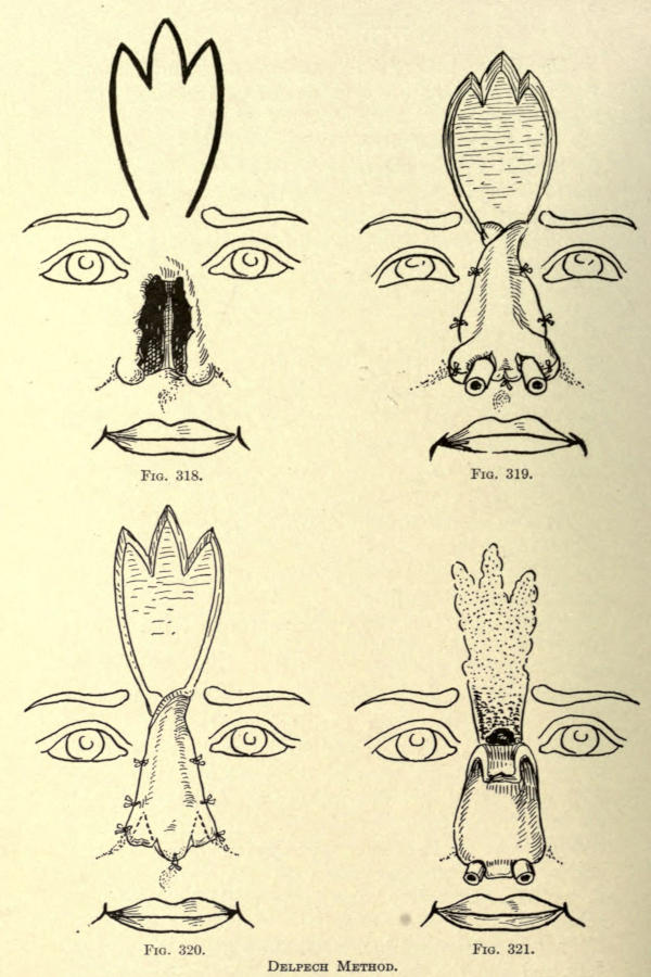
Fig. 318. Fig. 319. Fig. 320. Fig. 321.
Delpech Method.
Method of Lisfranc.—Lisfranc conceived the idea that if he carried down the one incision for the flap at the root of the nose somewhat lower than the other he would overcome some of the torsion at this point. This he consequently did, making the left incision half an inch lower than the right. The lateral incisions ascend at an angle of forty-five degrees (see Fig. 322), uniting in rectangular form at the scalp line, as shown, the rectangle of skin being utilized to make the subseptum.
Instead of sutures he dissected up the old nasal borders and slid the flap borders into this groovelike arrangement, holding it in place with the aid of sticking plasters.
With the above method the pedicle was allowed to remain intact. Fig. 323 shows the position of the flap, and the treatment of the subseptal section.
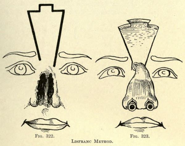
Fig. 322. Fig. 323.
Lisfranc Method.
Labat Method.—Labat uses a frontal flap shaped as in Fig. 324. The left bordering incision is carried down one half inch below the point of beginning on the right and carried downward in such manner that its lower point lies in a line with that of the right above it.
The object of this was to overcome torsion, and, where obtainable, the small triangle of healthy tissue at the root of the nose, as shown in the illustration, was dissected off from above downward, and turned downward with the cutaneous side facing the nasal chasm and its dissected side facing that of the flap. He avoids injury to the angular artery, as should be done in all cases. The pedicle was replaced at a second operation.
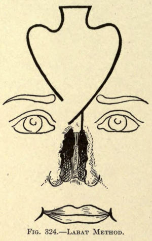
Fig. 324.—Labat Method.
Keegan Method.—Utilized a flap, shaped as in Fig. 325. The pedicle occupies the internal angle of the eye, care being taken to preserve the angular artery. The flap is mapped out obliquely, not perpendicularly. To get the best results he advises pasting a paper model upon the forehead to guide the operator in making the flap, which includes all the tissue down to the periosteum. Horsehair sutures are employed to approximate the parts accurately. The pedicle is divided in about twenty days, and a wedge-shaped piece of skin is excised at the root of the nose to prevent the tuberosity at this point of the new nose, so commonly observed with Indian-flap methods.
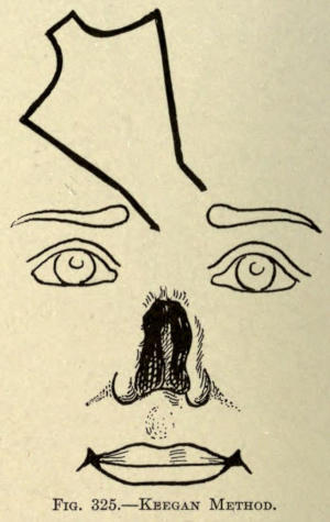
Fig. 325.—Keegan Method.
Duberwitsky Method.—The flap at its root resembles that of Labat, but at its superior border it formed an oval with an elongated point running into the hair line, which he divided, as shown in Fig. 326, to form the subseptum and nasal wings.
At the root the pedicle was about half an inch wide made in the oblique.
The middle section of the superior pointlike projection and intended for the subseptum was folded upon itself or doubled, as it were, to give support to the nasal point. The same was done with the alar or lateral sections, so as to line the nares with epitheliar surface to prevent contraction. The lower part of the nose was fixed into position by a harelip pin inserted transversely after all parts of the flap had been sutured into place.
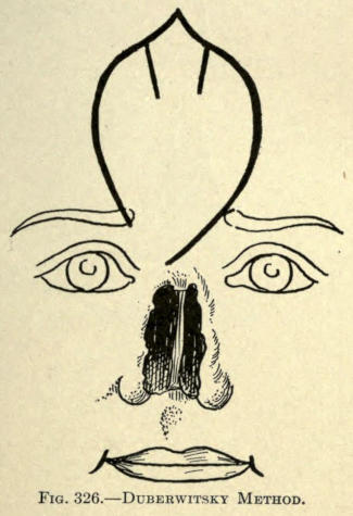
Fig. 326.—Duberwitsky Method.
Dieffenbach Method.—The flap is cut very much like that advised by Lisfranc, being wider only at its upper extremity, as shown in Fig. 327.
He advocates removing the remains of the old nose, almost circumscribing the nose, as shown in the illustration, except for the deep linear incision at the base of the nose on a level with the oval fissure, leaving a bridge of skin at either angle into which the square or septal part of the superior frontal flap is affixed.
The flap is made so that the right oblique line lies an inch above that of the left, the latter incision running into the angle formed at the root of the old nose caused by the ablation.
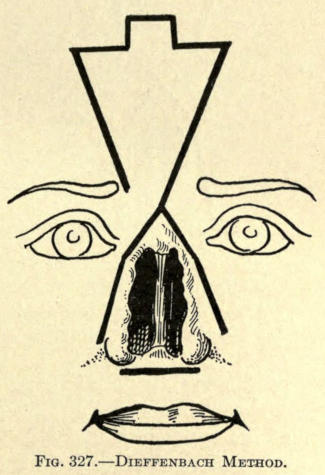
Fig. 327.—Dieffenbach Method.
Von Ammon Method.—The flap is cut at its superior border, similar to that of Keegan, but made in the perpendicular; the point of beginning, at the end of the right eyebrow, lies about an inch above the end of the incision of the opposite side, but in line with it (Fig. 328). The same method of removing the remains of the old nose advocated by Dieffenbach is followed as well as the lobial incision to receive the septal section.
The shape of this flap permits of bringing the secondary wound on the forehead more readily than where square exsections are resorted to.
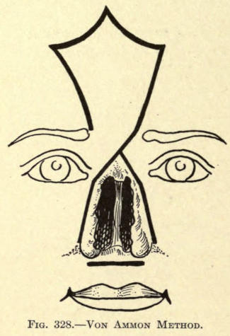
Fig. 328.—Von Ammon Method.
Auvert Method.—Like the method of Keegan, the frontal flap is made at an angle of forty-five degrees instead of the perpendicular, the flap being cut to the left of the median line. Its outline is shown in Fig. 329, and differs little at its superior extremity from that of Labat, except that it is made longer and narrower. The left lateral incision runs into the superior border of the old nose at the median line.
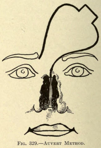
Fig. 329.—Auvert Method.
Von Langenbeck Method.—The flap is fashioned like that of Duberwitsky, but the left lateral incision enters the remains of the old nose, as Dieffenbach advised. The superior border was shaped, as shown in Fig. 330, to form the alæ and columna.
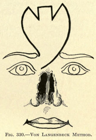
Fig. 330.—Von Langenbeck Method.
Petrali Method.—The shape of the flap is cut in ovate form with its rounded base near the hair line of the forehead. Petrali likens it to the form of the mulberry leaf. The left lateral incision dips down into the median line of the old deformity at its upper border.
The flap, after having been cut free, is folded upon itself along the median line, bringing the raw surfaces together along the dorsum of the new nose, thus giving body to the whole anterior nasal line. Presumably he introduces several sutures through the side of the flap to facilitate union along this line.
The method is illustrated in Figs. 331 and 332.
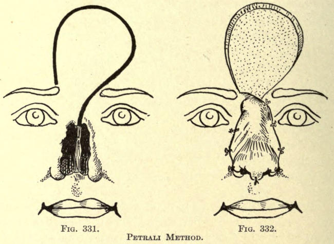
Fig. 331. Fig. 332.
Petrali Method.
Forque Method.—Herein the right lateral incision of the frontal flap is begun at a point above and corresponding to the middle of the eyebrow. The base is fashioned as shown in Fig. 333, and the left lateral incision is carried down to the median line of the old nasal defect, coming within the inner border of the eyebrow.
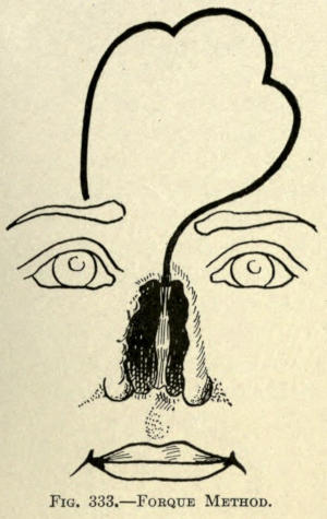
Fig. 333.—Forque Method.
D’Alguie Method.—This author conceived the idea of further relieving the torsion of the pedicle by making the frontal flap transverse along the forehead, instead of perpendicular.
The incision at the root of the nose is on a level and in line with the inner ends of the eyebrows. The left lateral incision is made to lie just above the eyebrow and the right sweeps upward and outward, as shown in Fig. 334.
The base is made with a rectangular projection to form the columna.
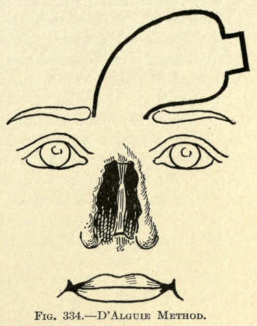
Fig. 334.—D’Alguie Method.
Landreau Method.—The direction of the frontal flap is transverse, but the root of pedicle, instead of having a downward direction, is so cut as to have its attachment upward, as shown in Fig. 335. This position of the pedicle thus overcomes to a great extent the torsion at this point. The flap must be cut somewhat longer in its transverse axis to allow for the higher position of the pedicle on the forehead.
The distal end of the flap is trident-shaped, as shown.
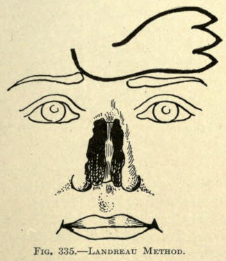
Fig. 335.—Landreau Method.
Langenbeck Method.—The flap is cut on an oblique line along its left border, running the incision down and across the root of the nose to the right while the right incision begins just under the eyebrow and extends less obliquely upward, as shown in Fig. 336. The base of the pedicle is fashioned as shown. The bordering remains of the old nose are removed.
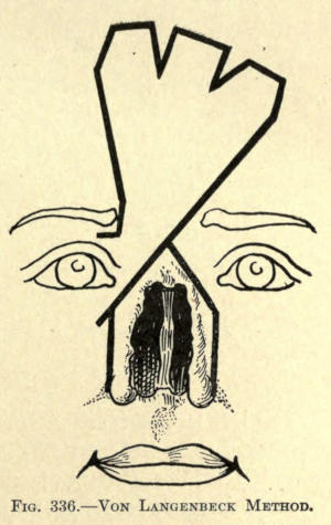
Fig. 336.—Von Langenbeck Method.
In another operation by the same operator the right incision was begun at a point above the eyebrow and carried transversely along to the rising point of the lateral. The left lateral incision was so made that it left an area of skin over the root of the nose, as shown in Fig. 337, which he dissected away, giving that part of the flap to cover it an opportunity to adhere, at the same time furnishing a nourishing area for its future life.
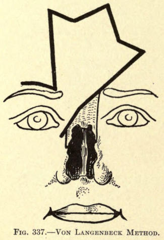
Fig. 337.—Von Langenbeck Method.
Szymanowski Method.—The flap is formed as shown in Fig. 338, the pedicle having its upper incision just below the end of the right eyebrow and the lower below the inner canthus on a line with the first, giving it an oblique position.
Just below the curvature of the basal incision two short incisions are made on either side into the forehead tissue with a view of rendering more flexible the skin to be utilized in correcting the secondary wound. The margin of the old nose is freshened.
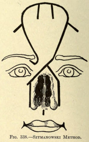
Fig. 338.—Szymanowski Method.
Labat, Blasius, Linhart Method.—These operators performed their operations in two sittings. In the first the incisions were so made at the base as to permit of that part of the flap intended for the rim of the nares to be tucked in, as it were, where these two triangular little folds were held in place by silk suture. When the parts had become thoroughly united, or at the second sitting, the entire flap was cut away and brought into place for the new nose. The object of this procedure was to give body to the wings of the nose and to overcome the consequent curling and contraction of the skin so commonly found with the single sitting operation.
This step marked the first advancement toward attaining much more successful results in total rhinoplasty by using skin-lined flaps, which not only added to the better nutriment to the part, but also gave support and firmness to the new organ.
The French Method
This method, per se, is not in itself sufficient to bring about a satisfactory result. The fundamental principle is that of the sliding flap of Celsus, and in which the two flaps intended to form the new nose are taken from the tissue of the cheek at either side of the remains of the old nose.
The total outcome is simply to bring before the opening a curtain of skin with a median scar running from the root to the lobule, which in itself is sufficient upon contraction to mar the result; furthermore, there are the two lateral wounds which have to be covered by skin grafts which, upon healing, have their tension of contraction, added to that of the median scar, with the result that the anterior nose becomes flattened and ugly, practically amounting only to an unevenly contracted curtain of marred skin.
The author would not advise resorting to such method, but, owing to the fact that a step in the advancement of the art was conceived under this particular method, space is given to the subject. This step, first introduced by Nélaton, consisted of allowing all of the cicatricial tissue of the old nose to remain with which the new nose could be built. As the possibility of this is rare in total rhinoplastic cases, the method is more useful in partial rhinoplastics, where it forms an important factor, as will be shown later under that subdivision.
Nélaton Method.—Two lateral flaps of triangular form, having their pedicles below the internal canthi, are cut from the cheeks, each flap containing all of the remains of the old nose. The entire inner borders of these flaps were freshened throughout their whole thickness.
In making the flaps, dissection is made down and through the periosteum, thus giving firmness and thickness to the new nose. The flaps are slid forward and sutured along the median line, leaving a triangular wound of the cheek on either side, as shown in Fig. 339.
To keep the raw surfaces in contact with the newly dissected area and to retain the nose in place as far as possible, a silver pin is inserted through the base of the new nose, going through the skin and remains of the old nose. It should be of sufficient length to permit holding a disk of cork at either end, beyond the skin and for the retention of the metal ring ends of a hook bent in inverted U-shape. The diameter of the latter bent wire is equal to that of the pin.
He claims for his method a perfect and fixed cicatrization of the newly placed parts.
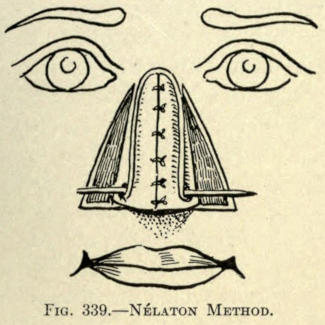
Fig. 339.—Nélaton Method.
Heuter Method.—The cheek flaps are cut from the cheeks, as shown in Fig. 340, leaving intact a triangular piece of skin with the object of giving support to the new nose. The inner and upper borders of the two flaps were stitched to the rim of this triangle, and then along the median line. The flaps are not made to include the periosteum, as in Nélaton’s method. The results thus obtained are not equal to the latter’s procedure.
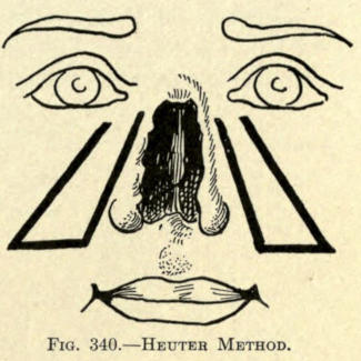
Fig. 340.—Heuter Method.
Bürow Method.—The cheek flaps are made as in Fig. 341. The projection intended for the subseptum is an elongated strip at the inferior border and inner angle of the left flap.
The shaded triangles at either extremity of the outer incisions show the removal of the skin at these points, to facilitate sliding of the flaps, adding, however, to the extent of cicatricial contraction upon final healing, with the resultant flattening of the new nose. The lobular prominence takes an upward position eventually, and altogether the extensive secondary wounds and the effect of their behavior does not warrant the use of this method.
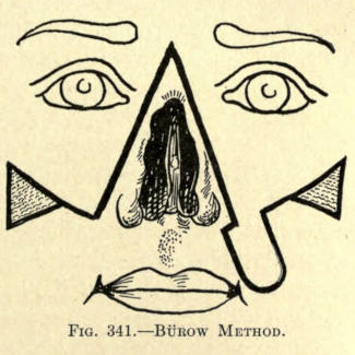
Fig. 341.—Bürow Method.
Szymanowski Method.—His method is an improvement on that of Bürow. The flaps, inclusive of considerable cellular tissue, are fashioned in Fig. 342, except under the two narrow extension flaps, which are to be utilized in building up the subseptum. Their raw surfaces are sutured together with silk. The flaps are united along the median line.
If the tissue from the cheeks do not permit of free sliding forward of the flaps, further incisions shown by the dotted lines over each malar prominence are made. The skin of the shaded irregular areas on either side is removed, as in the Bürow method.
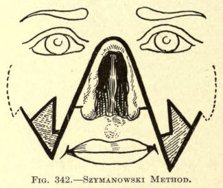
Fig. 342.—Szymanowski Method.
Serre Method.—The flaps are made to either side of the remains of the old nose, each leaving its pedicle about one fourth inch below the inner canthus of the eye. The flaps were cut rather obliquely, their bases extending somewhat below the nasal orifices. The remaining skin of the latter was dissected downward and folded down upon the median third of the lip. If cut in two sections their inner borders were sutured so that their raw surfaces faced each other. The object of the latter step was to form the subseptum, according to Lisfranc. The sections of skin lying with their bases on a level with the nasal orifices were dissected downward and united in the median line to assist in forming the end of the nose. All along the borders of the old nose were also dissected up where possible and folded inward, so that their raw surfaces would adhere to the new dorsum of the nose, and thus give it stability and form. These pieces of skin were united at the median line when possible.
The cheek flaps with indented bases were now brought forward and united, as shown in Fig. 343. The skin of the cheeks was dissected up to the extent of the dotted line in the former illustration, and when necessary two lower curved incisions were made to permit of free sliding. The skin of the cheeks was retained by three sutures at either side, as shown in Fig. 344. The subseptum may be made at the same sitting, or at a later operation.
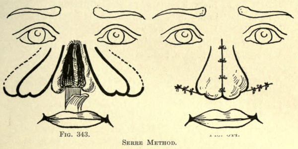
Fig. 343. Fig. 344.
Serre Method.
Syme Method.—The procedure is very like that of Heuter, except that the somewhat curved line making the inner borders of the flaps extended over the root of the old nose. The lower ends or bases of the two cheek flaps were stitched around and to the orifice to form the end of the nose, rubber tubes being used to form the nostrils, where they were retained until healing was complete.
Blasius Method.—He forms the cheek flaps in triangular form, including all of the tissue making up the buccal cavity. The outer or cheek incision is made through all of the tissue and extends to a point corresponding to a point a given distance beyond the angle of the mouth. The inner incision is made from a point just below the angle of the ala downward and through the thickness of the lip. A third incision unites the angle of the mouth with the outer incision. Both cheek flaps are made alike, each remaining attached along all of the remains of the old nose. They are now raised upward and inward, with their mucosa facing outward, and united along the median line. The raw cheek borders are now brought forward and held in place by suturing them at either side to the remaining rectangular flap of the upper lip. The formation of the subseptum is left for a second sitting. This method is not only too extensive, but too disfiguring to make its employment practicable. The mucous membrane would, of course, in time take on the function and appearance of skin, but the shape of the mouth never assumes a normal form, especially since there is quite a loss of the vermilion border at either side which is raised upward with the cheek flaps to assist in forming the base of the nose.
Maisonneuve Method.—Where there is more or less occlusion of the nares and yet an integumentary covering corresponding to the nose, as it might rarely be in congenital cases, Maisonneuve utilizes the sliding flap method to overcome the abnormality. In the case presented, the nasal orifices were hardly three sixty-fourths of an inch in diameter and about one inch apart. The correction was accomplished as follows, and shown in Fig. 345: An incision was made transversely outward from each nostril, then two converging incisions were made from both nares downward, meeting at the vermilion border of the lip in the form of a V, which were made to include the whole thickness of the lip. This flap was brought upward to form the subseptum. The skin to form the nasal lobule was now slid forward from either end of the incision and the subseptum sutured in place. Rubber tubes were employed to keep the nares distended and permit of the wings of the nose to form.
The defect in the upper lip was brought together as in a median harelip operation, the parts appearing after operation as illustrated in Fig. 346.
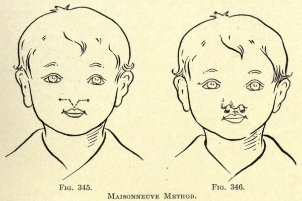
Fig. 345. Fig. 346.
Maisonneuve Method.
The Italian Method
In this classification of total rhinoplasty the skin flap is taken from another part of the body and not from the face. The integument of the arm is usually employed, the pedicle remaining intact until the flap has healed into place.
The method has been accredited to the Italian author-surgeon Tagliacozzi, but it was practiced long before his time; yet he was the first to fully describe the steps of the successful operation. It has been referred to quite fully under skin grafting.
The flap having an attached pedicle is cut from the entire thickness of the skin of the arm. The free end of the flap is sutured to the freshened borders of the old nose, and the arm is held in place until union has been established, when the pedicle is cut. There are no special advantages in this method, since the outcome is no better than that obtained with the Indian method; at best the result is merely the curtain of skin covering the defect, with the one thing in its favor—the avoidance of the frontal scar. Against this is the great discomfort the patient must suffer in having his arm retained in the necessary position to prevent movement and strain on the flap, to which may be added the danger of embolism occasioned by freeing the arm at the time the pedicle is cut. There is also difficulty of properly dressing the wounds, owing to the constrained position which consequently invite sepsis and imperfect healing. Hence, for total rhinoplasty, this method may be termed unsatisfactory; yet for certain partial rhinoplastic results it supersedes all other methods, as will be hereinafter shown.
To make the flap a pattern is laid upon the skin, from which it is to be made; it should be one third larger than the actual size of flap needed, to allow for contraction. The incisions should go through the entire thickness of the skin, leaving an attachment or pedicle, what in this case would be the part of the flap intended for the base of the nose, and directly opposite to those described heretofore.
The flap may be sutured in place immediately after the cutting, or it may be allowed to remain upon the arm until contraction has taken place in the flap, or the flap may first be modeled into nose shape and then sutured upon the freshened margins of the old nose.
The arm must in any of these methods be held in place during the days required to have the flap heal or unite with the facial tissue. The various operators have devised means to accomplish this. There is the linen network of bandages of Tagliacozzi, the harness of Berger, the starched linen and book-board affair of Sedillot, the one-piece suit of Lalenzowski, the leather sleeve and helmet of Graefe and Delpech and many others.
Having determined upon the method to be followed in securing the flap, the surgeon is advised to consider such apparatus as he may be able to procure to retain the parts, or to use his own ingenuity to construct one of plaster-of-Paris bandages to meet the requirements of the case at not only less expense, but with greater comfort to the patient. At best, any apparatus employed will do little to overcome the agony of the retained member, which must be held in position.
Various operators give this period between six and twenty days. The apparatus should be so constructed that dressings can be easily made without discomfort to the patient, and without doing damage to the parts, and also to expose the face of the patient as much as possible. The various operations employed to perform total rhinoplasty by the Italian method may now be considered.
Tagliacozzi Method.—This surgeon resorted to four steps to accomplish his operations, which were:
I. Massage of or stretching the skin of the part from which the flap is to be made.
II. Cutting the flap, and allowing the same to cicatrize.
III. Freshening the flap and suturing in place, and use of apparatus.
IV. Cutting the pedicle and making the subseptum.
The various details of these steps should be considered here, since the methods are practically the same for all other operations of this kind, except in certain particulars as to time and mode of procedure.
I. Massaging the tissue of the arm to render it supple. This is of some consequence, in some cases, where the skin is tense, but requires no especial description.
II. He then compressed a fold of the skin with a large forceps at the lower half of the biceps. Upon opening these forceps he forced a bistoury under the skin fold and cut down toward the elbow-joint a distance sufficient to form a flap. This gave him a piece of raised skin, attached at either end, double the size of that required to make the nose. Under this he introduced linen mesh dressings in the form of a seton, with the object of irritating the skin to encourage the circulation, and render it thicker by consequent suppurations and granulations. This was continued for fifteen days, when the skin was detached at its upper end, leaving it attached by the lower or wider pedicle intended for the base of the nose. The flap was now turned down and both flap and wound were allowed to cicatrize.
III. When the flap had become dry he fitted the linen bandage apparatus to retain the arm. Then the borders of the old nose were freshened. Thereafter he cut a paper pattern as a model for the new nose, upon which the margins and shape of the flap were cut. The flap was finally sutured in place, and the apparatus was tightened to prevent movement of the parts.
IV. After twenty days he cut the pedicle. The latter was then cut into, to divide it in three parts, which he formed into the subseptum and nasal wings, which were sutured in place, metal tubes being employed to keep the nares open.
Dieffenbach Method.—This surgeon followed seven steps to complete the operation, as follows:
I. The pattern of the new nose, cut one third larger, is fixed upon the skin of the arm, with the basic pedicle just above the fold of the elbow. Skin is now raised sufficiently to permit of its being incised, the incisions being made laterally, as shown in the dark lines in Fig. 347.
This gives a triangular flap, the apex lying upon the biceps and having two adherent pedicles at apex and base.
The base is now incised at one angle, transversely and again vertically, as shown. This incision liberates the part of the flap intended for one of the ala of the nose.
II. Diachylon plasters are placed under the flap to contract the arm wound immediately the bleeding has been arrested. The free angle of the base of the flap is now turned inward and under the attached part of the flap, as in Fig. 348, so that its margin protrudes from the other lateral incision, and its skin surface lying above the plaster. The edges of the flap are now stitched together, and the flap is allowed to lie cushionlike upon itself while the arm wound heals. This requires about six weeks.
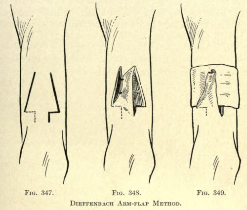
Fig. 347. Fig. 348. Fig. 349.
Dieffenbach Arm-flap Method.
III. The holding of the flap cushion in place by the use of splints of leather held in place by three needles. The latter are moved about, as the shape of the cushion becomes modeled, about every three weeks. The process ends when cicatrization of the flap or the newly formed nose has been accomplished, shown by firmness and contour.
IV. The margins of the old nose are freshened; the lateral incisions extend to the root of the nose, where they are united with an upward convex incision. The skin is well raised, gutterlike, from the deeper tissue, to assure of the best vascularity.
V. The upper or apex pedicle of the flap on the arm is cut (see Fig. 349), and the thickened roll of skin, or what may now be termed the new nose, is turned down toward the elbow. It is divided along the line where the two margins of skin had been sutured; in other words, it is laid open longitudinally.
VI. The nose thus prepared is brought into place before the freshened margins of the old nose and is sutured into place beginning at the root before the sides are coapted.
VII. At the end of fifteen days the pedicle attaching the nose to the arm is severed, the angle for the wing being cut slightly larger than that of the other side, which by this time has, of course, undergone full contraction. The subseptum is made out of the square projection folded upon itself, raw surfaces facing, and is brought into place by suturing it into an incision made in the lip at the required point.
Graefe Method.—This surgeon devotes six steps to his operation, as follows:
I. The borders of the old nose are freshened.
II. Sutures are passed through the raised skin of the borders of the old nose.
III. The flap is cut from the arm after a pattern made one fourth larger than the new nose required, leaving it attached by the small pedicle intended for the subseptum.
IV. The sutures where required are now passed through the flap, having already been placed through the old nasal borders and left untied. The forearm is drawn against the forehead and the arm is fixed in place with the retention apparatus. The sutures are now tied. They are allowed to remain in about four or five days, not long enough to irritate.
V. About the tenth day the head apparatus is removed and the pedicle of the arm flap is divided. The arm may now be carefully lowered to its normal position.
VI. The subseptum is not formed from the free end of the attached flap for several weeks. It is then divided by two parallel incisions directed outward. The septal section is folded upon itself, and inserted and sutured in place into an incision made into the upper lip.
Szymanowski Method.—This author advises making the base of the flap sufficiently wide, and of the form shown in Fig. 350, to permit of the three sections of skin of this part of the flap to be folded upon themselves before being sutured in place at the base of the nose, so as to form lined nares and a thickened and supportative subseptum.
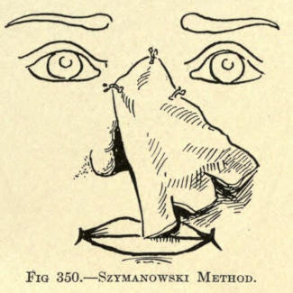
Fig. 350.—Szymanowski Method.
Fabrizi Method.—This author utilized the immediate method of flap fixation, but makes his flap of triangular form from the inner and upper skin of the forearm.
The transverse base is made to lie one half inch below the radio-ulnar space. The flap should be about three inches long and of about the same width. It is cut while the forearm is relaxed; bleeding is controlled by gentle pressure. In the meantime the cicatricial tissue of the old nose margins has been removed and the skin freshened to receive the flap.
To approximate the parts, the hand is laid palm down upon the shoulder; the resultant position of the arm and forearm are retained by bandages. The parts are now sutured. On the thirteenth day the line of division is traced out upon the arm with nitrate of silver, at the same time giving the flap somewhat the form required to give the nose its contour.
The next day the pedicle is cut and the arm is brought back into its normal position. With the division of the pedicle he advises including a portion of the aponeurosis and a few fibers of the supinator longus muscle.
The flap is allowed to remain free at its base until contraction and cicatrization have been established, when the subseptum and wings are made.
The position of the arm and the attached flap at the root of the nose is shown in Fig. 351.
He advises, when possible, to dissect up a flap of the cartilage of the old septum, letting it adhere at its lower border and turning it from below upward with the skin which covers it to form the subseptum. This will help to hold up the point of the nose firmly (an important matter because it is at this point that all noses constructed of skin flaps alone sink down for the want of suitable prop of tissue).
This cartilaginous flap he held in place with two pins thrust through the latter and the skin flap proper, and held them in place with a figure twist of silk. He removed the needles about the sixth day.
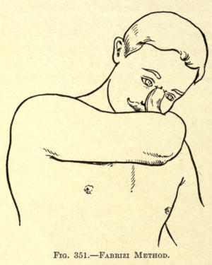
Fig. 351.—Fabrizi Method.
Steinthal Method.—This authority made the flap for the nose from the skin over the sternum, proceeding as follows:
“From the sternum I cut a flap of skin and periosteum in the form of a tongue whose lower base was five centimeters wide, and the summit forming the pedicle three centimeters wide; its length was twelve centimeters.
“I could have taken away with this flap some of the costal cartilage to utilize in making the wings of the new nose.
“I dissected up this flap and closed the wound over the sternum with sutures. The flap was then stitched to the forearm by its base into an incision of appropriate length made near the radius. (See Fig. 352.) The arm was properly fastened in a plaster apparatus and the flap enveloped in a dressing of borated vaselin. The forearm was held in front of the breast, an attitude easily retained. Twelve days later I cut the pedicle.
“I let a few days pass by, and then stitched the pedicle end of the flap to the root of the nose. A new plaster apparatus was put in a suitable position. The hand was placed on the forehead.
“Ten days after, I detached the flap from the arm and reformed the nose with the flap, which hung down like an apron. It is necessary to have a flap sufficiently long to fold in for the nostrils. I used bronze aluminum wires for all the sutures.”
The position of the hand while the flap was healing to the root of the old nose and the slight twist of the flap is shown in Fig. 353.
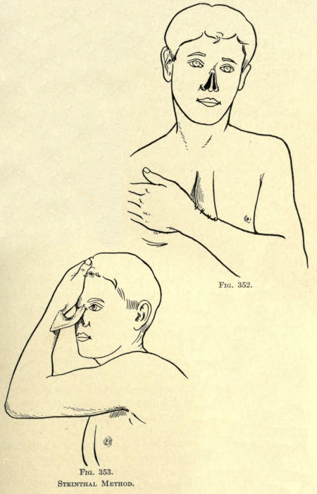
Fig. 352. Fig. 353.
Steinthal Method.
The Combined Flap Method
To overcome the consequent cicatricial contraction and falling in of the flap used to make the new nose by either of the three grand methods given, various surgeons have resorted to lining the flap with skin flaps, bringing their raw surfaces together so that the nose actually received in this way an integumentary lining.
While this had the tendency to thicken the new nose, it did not give the support necessary to it, especially at the lower third, and the lobule, at first quite satisfactory, resulted only in the appearance and form of a small tubercule of tissue, with a decided saddle effect above it. This combined method did overcome, however, the slow process of cicatrization, and its accompanying suppuration.
The raw surfaces of the two flaps, if properly brought together, healed upon themselves readily, as has been referred to in the lining or doubling in of the basal sections to form the nostrils and subseptum.
The method of lining the nasal flap in this manner is never sufficient to give a satisfactory result in total rhinoplastic cases, but may be of great service in restoring parts of the nose, as will be shown later.
The requirement is that of support, whether it be organic or inorganic, and these methods will be considered presently.
Volkmann Method.—This surgeon fashioned the frontal flap as shown in Fig. 354. This resulted in leaving a triangle of skin at the root of the nose, which he dissected up, down, to and inclusive of the periosteum, and turned downward so that its raw surface faced upward, as in Fig. 355. The flap was sutured into place to retain it.
The frontal flap was brought down, so that the two raw surfaces came together.
This method overcame the contraction of the flap over the nasal bridge or superior third of the new nose, and an excellent adhesion of that part of the flap to the denuded bone and flap resulted, but the same faults about the base were not mitigated.
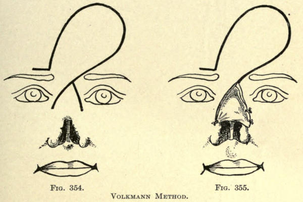
Fig. 354. Fig. 355.
Volkmann Method.
Keegan Method.—The frontal flap method of Keegan has been referred to. For the lining of the upper nose he cuts two flaps from the skin above the old nasal orifice, as shown in Fig. 356, which he turns down, raw surfaces out. This gave a lining to either side of the median line; the skin remaining intact between the two flaps gave additional prominence and support to the upper third of the new nose.
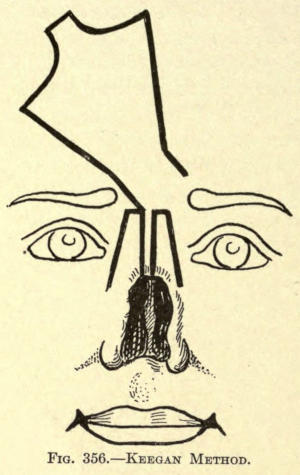
Fig. 356.—Keegan Method.
Verneuil Method.—Contrariwise to the methods just given, Verneuil, after cutting out the frontal flap, cuts the flap from the remaining sides of the old nose somewhat involving the skin of the cheeks, as in Fig. 357. This done, the frontal flap is simply turned down, raw surface out, and the cheek flaps are slid over it, bringing the raw surfaces together. The inner borders of the flaps were sutured in the median line, as shown in Fig. 358. The base of the nose is made from the frontal flap by any of the methods already given.
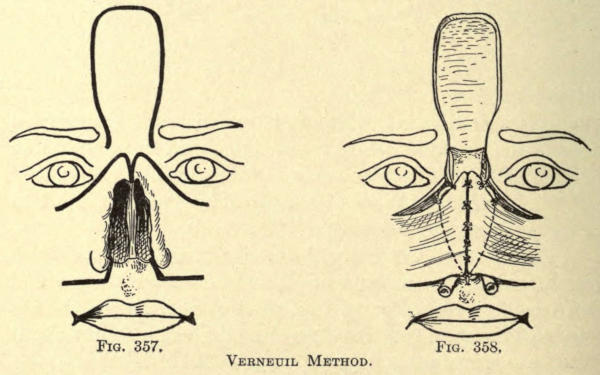
Fig. 357. Fig. 358.
Verneuil Method.
Thiersch Method.—The frontal flap is cut from the skin of the forehead in the shape shown in Fig. 359. Then two quadrilateral flaps are raised from the cheeks, as also illustrated. These are made wide enough that, when they were brought together, their inner borders could be made to face each other. In this position they were sutured along the median line, so as to give a double-gun-barrel form to the nose, with a septal wall between.
From the lower border the nostrils were formed, giving to the new nose a normal appearance, the continuous septum curving downward to form the subseptum, the whole being sutured to the remains of the old nose.
The frontal flap was now brought down over it, the raw surfaces facing each other, and sutured in place, as shown in Fig. 360. Later, Thiersch replanted the sides of the nose, to give it better contour, and attained a very satisfactory result. The frontal wound was covered with skin grafts, but the cheek wounds were allowed to heal by granulation. The cicatrization of the latter was not sufficient to effect the lower eyelids nor the angles of the mouth.
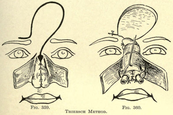
Fig. 359. Fig. 360.
Thiersch Method.
Helferich Method.—His is an ingenious application of the French method. Both flaps are cut from the cheeks; the lining flap was made from the left and the covering one from the right cheek. The shape of the flaps is shown in Fig. 361.
The lining flap is stitched along the freshened margin of the right side of the nose. The flap should be wide enough to give convexity to the nose, as shown in Fig. 362.
The covering or right flap, cut much larger, is now slid over this. It should be cut amply large to cover the flap just sutured in place. It is sutured on both sides of the nose to hold it in place, also at the inferior margin. The nose is lightly packed with iodoform gauze.
The pedicle of the right flap was cut after two and a half weeks and brought into place across the root of the nose, and sutured in place to give better contour to the part after freshening the skin about the left side of the nose at this point. He does not make a subseptum, but thinks the inferior base of the nose of sufficient size to hide the absence thereof.
The subseptum could, however, be readily made from the upper lip, as will be shown later.
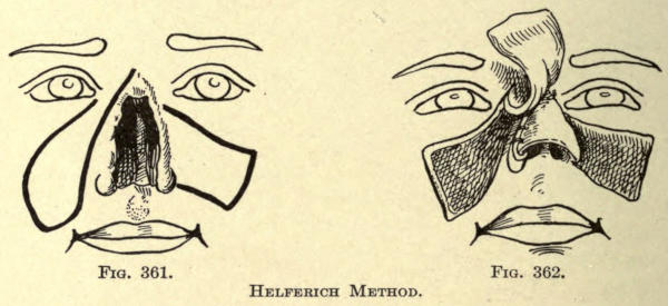
Fig. 361. Fig. 362.
Helferich Method.
Sedillot Method.—This operation is particularly efficacious in giving a splendid subseptum and support of the point of the nose, but does not overcome the falling-in of the whole anterior line, so common with all Indian-flap methods. A flap one centimeter wide and extending downward almost to the vermilion border is cut from the thickness of the upper lip, not including the mucous membrane, however. It is turned upward, as shown in Fig. 363.
The frontal flap is fashioned as shown, care being taken to cut a subseptal rectangle of greater length than usual, since it is intended to overlie the raw surface of the flap taken from the lip. It is rotated downward and sutured into place at both sides, and also to the lip flap, to assure of accurate union.
A lateral view of the nose as formed in this manner is shown in Fig. 364.
The free end of the septal flap is fixed into the superior lobial wound with a harelip pin. The lobial wound is sutured as in ordinary harelip operations. This method is particularly valuable in total rhinoplasties involving the columna and alæ in conjunction with flaps obtained by the Italian method.
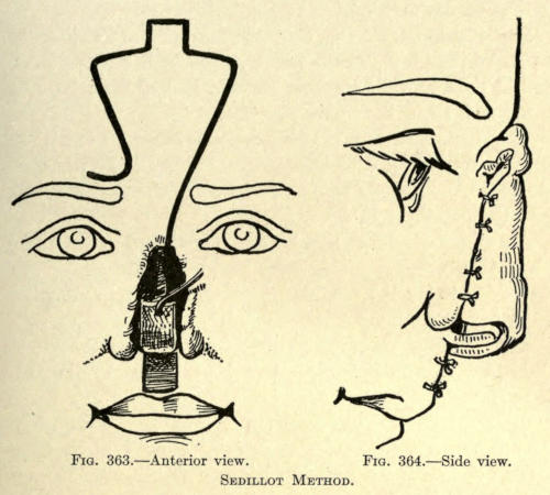
Fig. 363.—Anterior view. Fig. 364.—Side view.
Sedillot Method.
Küster-Israel Method.—A flap was taken from the arm by the Italian method, which was sutured to the remains of the old nose so that its raw surface looked upward, not downward, as in the ordinary case.
The flap was made sufficiently large to permit of building the wings and subseptum. After it had healed into place the pedicle was cut, and a frontal flap was cut from the forehead to cover it.
An unusually large flap was required to do this, since it had to overcome the greater curvature already given and added to by the arm flap, necessitating an extensive secondary wound.
The reverse order of procedure would be the more advisable for this reason, and is resorted to by the following:
Berger Method.—This surgeon makes the lining flap from the forehead. The secondary wound is at once closed. A flap is then made from the arm by the Italian method, and brought into place before the one just made. It should be of sufficient size to allow of building the base of the nose, which is done not later than three weeks after the pedicle of the arm flap is severed, which may be done at any time between the eighth and the twelfth day.
All the precautions are used as already given in the description of the Italian method. The arm is held in the position shown in Fig. 365.
Berger sutures the arm wound before bringing the flap into place upon the face to overcome the discomfort of suppuration to the patient.
The apparatus is fixed definitely after the patient has recovered from the anesthetic. Great care is exercised to prevent coryza from exposure. Dressings are made twice daily.
The pedicle is cut under local cocain anesthesia.
To make the subseptum and wings of the nose, the base of the flap is cut into three sections. The posterior surface is freshened and the parts are folded upon themselves and sutured into position.
Instead of employing rubber tubes, he resorts to a specially devised apparatus to retain two metal tubes in the nares, and at the same time make gentle pressure to the sides of the nose to mitigate the columna contraction. The latter is planted into a V-shaped incision made into the tissue of the upper lip at the proper place of attachment. The subseptum may be lined with a flap of mucosa dissected up from the floor of the inner nose.
For the wings of the nose, such tissue as may be of service to give them stability and structure is taken from the remains of the old nose.
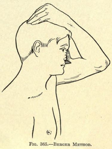
Fig. 365.—Berger Method.
The apparatus just mentioned and shown in Fig. 366 is used from the very first day until total cicatrization has taken place, and even for a longer period to aid in shaping the entire nose and the tendency to collapse has been overcome.
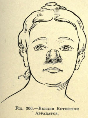
Fig. 366.—Berger Retention Apparatus.
Szymanowski Method.—A frontal flap, divided along the median line and shaped as outlined in Fig. 367, is made from the forehead.
Two triangular flaps are then raised from either side, and including the angle of the nose as shown. The divided frontal flap is now brought down in such manner that their raw surfaces meet, thus forming a vertical septum. The margins are united by suture, and the lower ends are fixed into a wound made for the purpose at the base of the nose, as shown in Fig. 368, to form the new subseptum.
The lateral triangular flaps are dissected up so that they can be readily slid forward toward the median line. Their inner freshened margins are sutured to the raw edge of the septum just made, and to themselves. The objection here is that there is a liability of considerable contraction of these lateral flaps, with a tendency to fall in and drag with them the new septum; and again, in total restorations, the upper third of the nose is only partially covered, and necessitates later upbuilding. The author finds difficulty in making the four margins thus brought together unite evenly throughout, and that a vertical contraction is caused by the cicatrization of the median marginal wound.
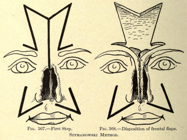
Fig. 367.—First Step.
Fig. 368.—Disposition of frontal flaps.
Szymanowski Method.
Goris Method.—The operation is performed as follows, having given very good results, according to the author:
I. The frontal flap is divided lengthwise so that its raw surfaces face each other. The resulting fold, representing the bridge of the nose, is held in place by catgut suture.
II. The skin to make the wings of the nose is folded in, as in the Langenbeck method.
III. A flap, half the thickness of the upper lip is brought up to form the new subseptum.
IV. Dissection and turning down the triangular flap of skin which surmounts the orifices of the old nose, and making it serve to line the lower part of the frontal flap.
V. Suturing the frontal flap thus modeled into two grooves made into the margins of the old nose along both sides to its base.
Organic Support of Nasal Flaps
It soon became evident to the rhinoplastic surgeon that without some support to the flap or flaps used for the construction of the new nose all of the preceding methods, as far as æsthetic results were concerned, were useless. Truly, the deformity lost its hideous appearance to a great extent, but the general results obtained hardly warranted a patient to undergo restorative operations of the nose. In fact, many surgeons advised against total rhinoplasty when practically all of the old nose was lost.
Langenbeck says “that total rhinoplasty, or even operation as to repair partial loss of the nose by the use of soft flaps, should not be undertaken. It is better to rely upon some prothesis.”
All that could be expected of utilizing the flap and making it heal into place had been accomplished up to about the year 1879. Thereafter many surgeons proceeded to evolve and use some kind of intranasal prothesis made of various inorganic materials. It may be stated, however, that Rousset in 1828 wrote: “Perhaps some day surgeons will give whatever shape they desire to the reconstructed nose. Then a frame of gold or silver, cleverly shaped and solidly fixed in the nose, will give the patient, at his own option, a Roman or Carthaginian nose, and to the ladies a choice of a roguish type, and to our Sultans a nose a la Roxelane.”
But it was after 1878 that such prothesis came into use, and these were at first made so that they might be removed at night and be replaced in the morning.
The intranasal supports were made of all kinds of material, such as gutta percha, gold plates, leaden devices, amber, silver, porcelain, celluloid, aluminum, platinum, etc.
With all due respect to the ingenuity of these inventions, especially that of Martin, which was made of platinum in the form of a St. Andrew’s cross, having at the four ends sharp pins which were driven and fixed into the skeleton of the nose, the use of these protheses resulted in nothing but failure.
The movable devices were a source of irritation and pressure, and could not overcome the consequent contraction of the flaps whether placed below a single flap or between two flaps, and the fixed protheses of whatever form or material caused so much pressure that gangrene resulted, and they had to be removed sooner or later.
Before the discovery of Gersuny, the author had many occasions to utilize such movable protheses in the correction of saddle noses. These were generally made of a silver shell, gutta percha, and later of decalcified bone, as advised by Senn. The former remained in place from six months to two and a half years, and then were thrown off or had to be removed because of irritation. The bone chips soon became absorbed, leaving the nose as before, or a thin median strip that became broken with the least violence, and then was absorbed.
In several cases where other surgeons had resorted to such protheses, the author was called upon at a later period to remove them.
While the immediate result is very gratifying, the ultimate result is worse than useless, since in the elimination of the foreign body the flap of the nose was married by cicatrices that added still further to the contraction and falling-in of the nose.
Periostitic Supports
Some other method had to be devised, and organic supports became known. These organic protheses were made of the tissue in the near vicinity of the flap, and at first formed a part thereof. The earlier method included only the periosteum; later bone and periosteum were added to the flap to give it shape and support, and lastly cartilage was employed for the purpose.
Of the methods employing only the periosteum, it may be said that what the surgeon expected of this membrane—namely, the springing up of bone cells—did not take place; at least, not to the extent desired. The very best to be attained was a thickening of flap in the membrane, but not sufficient to add necessary support to the nose.
Osteoperiostitic Supports
The inclusion of the periosteum-lined flap was soon abandoned, and recourse was had to such bone additions to the flaps as could be obtained from the vicinity of the nose.
The bone was removed with its periosteum, adherent or nonadherent to the flap, as will be shown by the methods described hereafter.
Both single and combined flap methods are employed as might be expected, following the procedures of the Indian, French, or Italian schools. The greatest credit for the methods herein involved belongs to the surgeons of Germany.
The earliest operation on these lines was that of König, who published his first successes in 1886.
König Method.—Extending upward from the root of the old nose, a flap is outlined in vertical ending at the hair line of the scalp, as shown in Fig. 369.
This flap was made about one centimeter wide, and is made to include the skin and periosteum. With the chisel a thin strip of bone is raised from the frontal bone to nearly the full length and width of the flap, making it an osteoperiostitic cutaneous section attached by its pedicle at the root of the nose.
This flap is brought down with bony surface outward, and the distal or skin end is fixed by suture into the upper lip at the point of the intersection of the subseptum.
Any of the soft parts of the old nose remaining are now dissected up toward the median line, and are folded upward and inward and sutured by their freshened margins to this median flap.
An Indian flap in oblique direction and of the form shown is cut from the skin of the forehead and rotated down into position before the bone-lined flap, and sutured into place.
He advises not to include the periosteum in the flap making up the subseptum, as it is likely to interfere with respiration. In fact, he deems it best to make the tegumentary flap sufficiently long to build the bone of the nose, doubling the raw edges upon themselves with a celluloid tube apparatus that may be removed for cleansing, and be kept in place long enough to give contour to the nares.
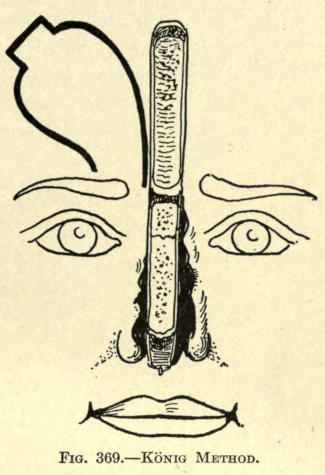
Fig. 369.—König Method.
Von Hacker Method.—The frontal flap was cut in the ordinary Indian method, and of the shape shown in Fig. 355. The skin at either side of the median line was dissected up to within four millimeters, leaving a strip eight millimeters wide from the root of the nose to the distal or scalp end. The two loose lips of the flap were brought together at the anterior median line by a few sutures to keep them in place.
This was done to give freedom to the surgeon while he detached a strip made of the periosteum and bone chiseled from the frontal bone. At the root of the nose or below the pedicle the bone was not included to the extent that it would interfere with torsion of the flap, and yet sufficient to allow the raw bone surface to fall upon what remained of the bony bridge of the old nose.
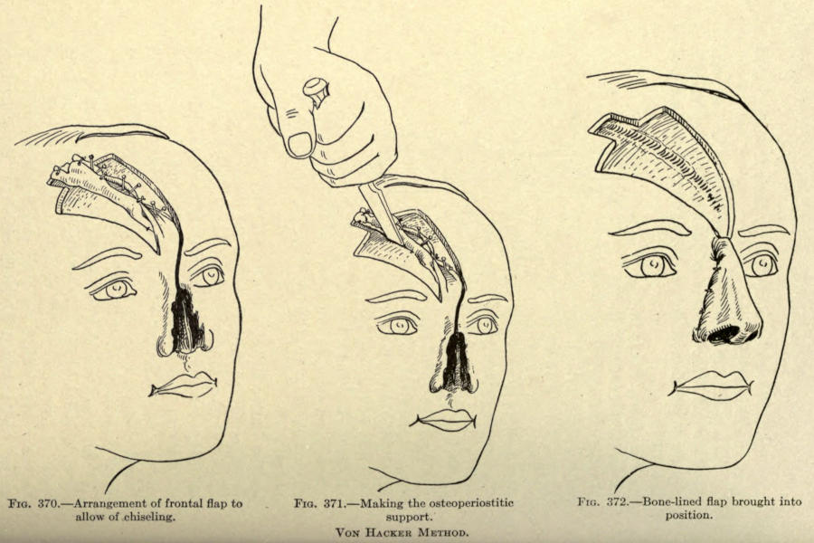
Fig. 370.—Arrangement of frontal flap to allow of chiseling.
Fig. 371.—Making the osteoperiostitic support.
Fig. 372.—Bone-lined flap brought into position.
Von Hacker Method.
He utilizes pins driven into the bone to outline this bony section, as shown in Fig. 370.
The latter is done in an oblique direction. See Fig. 371. The septal section is made to include the bone strip.
The bridge of bone holding the flap at its inferior end was now broken, leaving, however, the periosteum as part of the pedicle hinge.
The whole flap thus outlined was rotated downward into position and sutured, as shown in Fig. 372.
The margins at the base intended to form the subseptum were sutured behind the osseous structure, or, in other words, were doubled inward and fixed by suture. The bony strip was broken at the proper point to give prominence to the lobule.
The margins for the nostrils were turned inward and doubled on themselves, and sutured with silk.
Rubber tubes were left in the nares, for drainage and to keep them distended.
Rotter Method.—The frontal flap is made in the shape shown in Fig. 373, containing a section of the frontal bone and its periosteum. The width of the flap is about three and a half centimeters wide.
This flap is turned downward so that its raw surfaces look outward.
Owing to the loose adherence of the bony section to the skin flap, he allows the raw bone surface to granulate over for four weeks, to fix it more solidly to the soft parts.
The bone plate is then sawn into three sections made by two vertical incisions, made as shown in the illustration.
The median section forms the bridge and dorsal prominence of the nose.
The adherent skin of the lateral bony plates is dissected up sufficiently to permit of the proper formation of the sides and wings of the nose.
This gives a shape to the nose, as shown in Fig. 374.
The lateral margins of the integumentary flap are now sutured to the freshened margins of the old nose, and the remaining skin, if any, is made to cover the granulating surface; if this is lacking or insufficient, skin grafts are utilized to cover it completely.
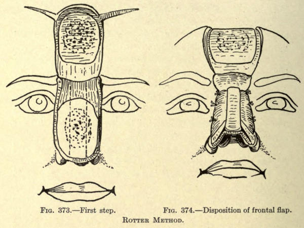
Fig. 373.—First step.
Fig. 374.—Disposition of frontal flap.
Rotter Method.
Schimmelbusch Method.—The principle herein is to give an osseous wall to the whole length of the restored nose, covering well the skin inside and outside, and, if possible, to fix the new nose solidly at the pyriform opening.
“I cut an osteo-cutaneous flap from the middle of the forehead, of a size proportional to the size and shape of the nose. Its pedicle between the eyebrows is two or three centimeters wide; it widens out superiorly to form seven to nine centimeters. It is triangular, and its base lies near the hair line. In cutting it out, preferably a little large, it goes at first to the bone, through skin and periosteum. With a large, sharp chisel, a thin bone plate throughout the whole extent of the cutaneous flap is detached. It is not always possible to make this a plate in one piece; it often breaks or gives off splinters. This is of no consequence, if care be taken not to lose them and to keep them adherent to the periosteum. They are attached as well as possible to the cutaneoperiostitic flap by passing threads crosswise from one edge of the flap to the other over bony surface, as in Fig. 375. The whole flap is then enveloped in iodoformed suture.
“The frontal wound I close at the same sitting by sliding large lateral flaps whose upper border follows the margin of the hair as far as the ears. These are freed completely, brought down and stitched, leaving eventually only a linear cicatrix on the forehead. The lateral loss of substance which results is healed by granulation, and the scars concealed by the hair.
“At first parts of the bone die; they ought to be expected to fall out; after four, six, or eight weeks the bone is completely covered with fleshy granulation, and adheres solidly to the flap. The prominent granulations are then scratched, or, better, trimmed away with the knife, and the whole surface is covered with Thiersch grafts.
“When the flap is thus furnished with skin within and without, it is put into place. I saw the bony plate with a fine-toothed saw from the grafted side; then I model the flap and place it on the loss of substance freshened by turning the grafted surface toward the interior of the nose by twisting its pedicle, as in Fig. 376. The osseous rim of the pyriform opening is uncovered at the moment of this freshening, and the bony edges of the flap are placed exactly on the bony edge of the aperture. The skin of the flap is then stitched at its lower margins to the skin of the cheeks. To preserve the height of the nasal profile and avoid displacing the bones of the nose, the nose is kept in place with a pin thrust through the nose, and furnished at each end with a rubber button. This aids to form the wings of the nose. If a subseptum is needed, it is made by taking from the skin that covers the circumference of the pyriform opening two small flaps, which are dissected from without toward the median line as far as the point where the septum is normally found.
“These are stitched at this point, first upon themselves, then to the end of the nose. Three weeks later the pedicle of the frontal flap is cut; it is turned, put in splints, and the stitching is finished.”
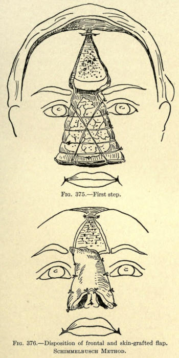
Fig. 375.—First step.
Fig. 376.—Disposition of frontal and skin-grafted flap.
Schimmelbusch Method.
Helferich Method.—A lining flap is made, according to the French method, from the one cheek, which is dissected up and turned over to bridge most of the loss of nasal tissue, and sutured to the opposite freshened margin, as showed in Fig. 377.
A frontal flap, as outlined in the same illustration, is now cut from the forehead, leaving a pedicle as shown, and containing a section of bone at its median line. This is rotated downward and into place, and sutured along the same margin to which the genian flap is fixed, as shown in Fig. 378.
When the frontal and genian flaps have become well united, the latter’s pedicle is cut when the freshened lateral margin of the frontal flap is sutured into place.
A subseptum is now made or deemed necessary by this surgeon.
At a later period the pedicle of the frontal flap is cut, and fixed by suture and some cutting, to reduce the resultant prominence thereof.
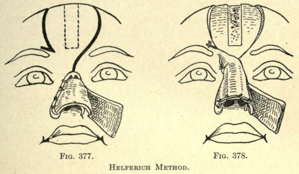
Fig. 377. Fig. 378.
Helferich Method.
Preidesberger Method.—This author cuts away the skin surrounding the arch of the old nose, and turns this flap downward to form the lining to the flap made from the forehead made in the same manner as Helferich.
The bone section is made in the median line, and is one centimeter wide and four long.
The frontal flap should be made long enough to permit of building a subseptum and the nostrils.
Krause Method.—This frontal cutaneo-osteo-periostitic flap is made according to the method of König.
After turning down the flap it was covered with a nonpedunculated skin flap taken from the upper part of the arm by transplanting after its subcutaneous fatty tissue had been removed. (See Fig. 379.)
This method necessitates a long-continued dressing of the forehead before the pedicle is cut, because of the needed nutrition to make the two flaps heal upon each other.
After union has been established the sides of the transplanted flaps are raised by dissection, as shown in Fig. 380, to expose the bone plate of the frontal flap. A median strip is left intact.
With a fine saw the bony plate is cut into three sections, making the narrowest the median.
The margins of the old nose are now freshened, and the combined flap is sutured along the sides, preserving what tissue the surgeon can use to add support to the nose, which is done by dissection and turning or folding, as heretofore described.
The lower or forehead flap is sutured to the soft parts of the old nose, and the transplanted lateral margins to the marginal skin of the cheeks, giving to the nose the appearance as shown in Fig. 381.
At a later period the pedicle is cut and the wound that cannot, at this time, be overcome by sliding of the adjacent skin, is covered by skin grafting.
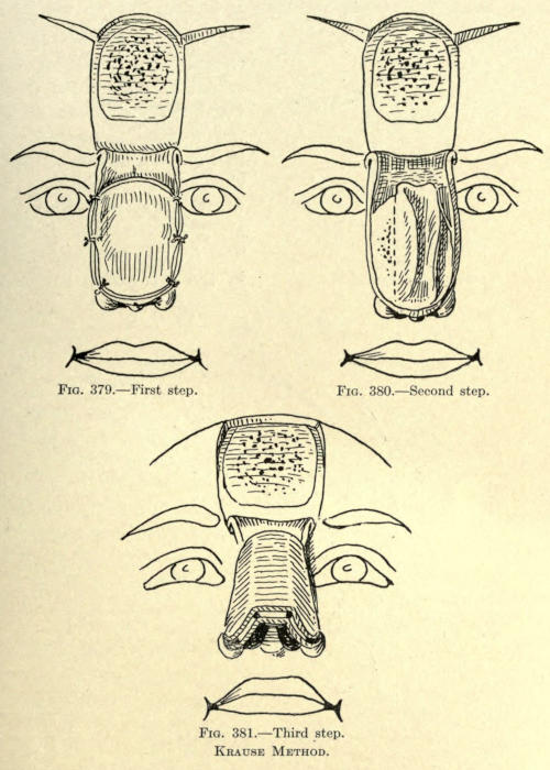
Fig. 379.—First step.
Fig. 380.—Second step.
Fig. 381.—Third step.
Krause Method.
Nélaton Method.—A lateral flap of skin is taken from the cheeks, beginning on a line with the root of the nose and as low as a point two thirds of its normal length. These flaps are made wide enough, so that when dissected up and folded inward they will meet on the median line, as shown in Fig. 382, having their raw surface facing outward. They are sutured along the median line. The frontal flap was cut in the form of a horse-shoe having its pedicle at the root of the nose just above the eyebrows, and being about three centimeters wide and six long.
The skin at the outer margins was dissected up from the bone, leaving sufficient attachment at its center to allow for a bony plate.
With a fine saw, and in the manner shown in Fig. 383, this plate was made from the frontal bone, being about two and a half centimeters wide and four long.
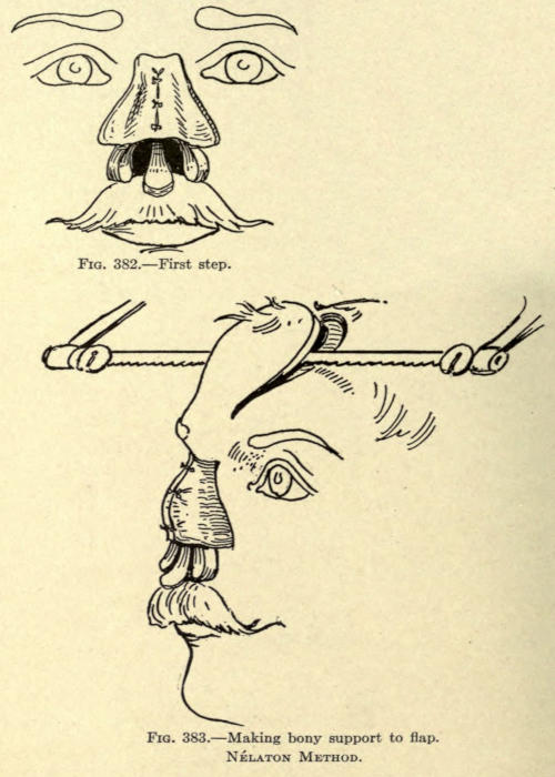
Fig. 382.—First step.
Fig. 383.—Making bony support to flap.
Nélaton Method.
There is some difficulty associated with the making of the flap, which ends at the superior border of the frontal, leaving the pedicle composed only of skin.
The flap is now turned down, exposing its raw surface. The bony plate is sawed through at the median line, as shown in Fig. 384, and the skin of the flap is also divided along this line, giving two partly bone-lined flaps.
The two flaps are now rotated downward before the lost nose, so that their raw surfaces face inward, and in this position they are sutured along the median line and the sides, as shown in Fig. 385.
The method gives an angular dorsum of satisfactory consistency to the new nose, but furnishes a serious drawback, in that the cicatrization along the median line is liable to affect the shape of the organ and leaves a prominent scar line. The use of two small pedicles is another objection in that the danger of gangrene is greater as the nourishment to each flap is less.
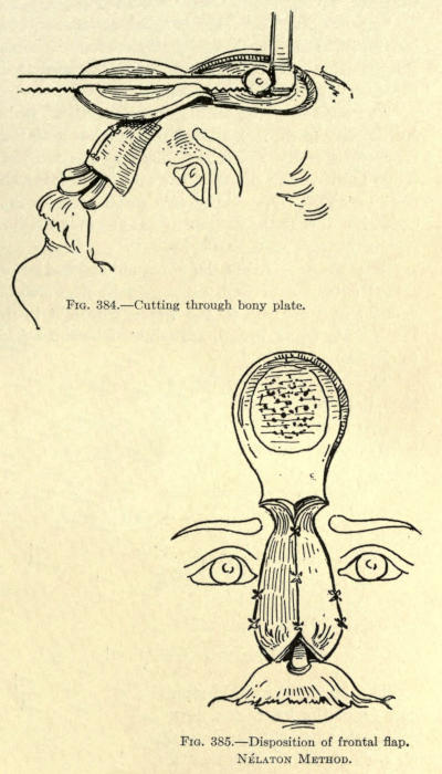
Fig. 384.—Cutting through bony plate.
Fig. 385.—Disposition of frontal flap.
Nélaton Method.
Israel Method.—From the ulnar side of the left forearm Israel cuts a skin flap, as shown in Fig. 386, with its smaller end nearest to the wrist, where it is detached, the pedicle being broad, assuring of better nourishment to the flap.
The narrow end of the flap is cut down to the bone, then the sides are dissected up until the borders of the ulna are reached on both sides, reserving an adherent strip about eight millimeters wide and six centimeters long.
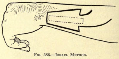
Fig. 386.—Israel Method.
The bone below this strip is now removed with the saw from the lower end upward, and ending about one centimeter beyond the base line of the flap, where the strip so made is left connected to the bone proper.
The flap is now raised gently and bent upward without breaking the bone. It is sawed half through, transversely, at a point corresponding to the lobule of the nose.
The flap is then enveloped in iodoform gauze, and the head, forearm, and arm are fixed in plaster of Paris, the forearm being bent at a right angle to the arm (see Fig. 387).
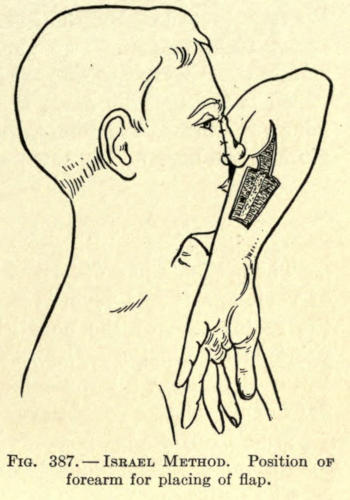
Fig. 387.—Israel Method. Position of forearm for placing of flap.
After nine days the osseous connection still remaining is severed, and the nose is modeled upon the forearm, as heretofore described in these operations, this surgeon using silver wire to retain the parts. The raw skin surfaces are allowed to heal upon each other and the flap is permitted to come in contact with the wound on the forearm temporarily, to which it might adhere, the gauze being now removed.
After twelve days the newly modeled nose is freed from such adhesions and kept from healing to the parts by using dressings between the flap and wound.
Five days after, the margins of the old nose are freshened in the form of an inverted V. If there be sufficient cicatricial tissue it is turned down, raw surface out, to line the new nose.
A prolongation of the pedicle is now cut, widening out toward the radial side of the arm, made obliquely, as shown, so that its pedicle now corresponds to a width of seven centimeters.
The whole flap except this newly formed pedicle is cut free of this forearm. The arm is put into the position shown in Fig. 387, and the freshened flap margins at the root, the whole length of the left side, and part of the upper right lateral. The plaster dressing to hold the arm in the proper position until complete union is established is used. This done, the pedicle is cut, and such minor operations are done to fix the remaining free margin and the base of the new nose.
Cartilaginous Support of Flap
The methods just described in which an osseous plate of various size and form is included with skin flaps for the restoration of the nose give undoubtedly the best rhinoplastic results. The new nose is given not only better shape, but a permanency of such form that skin flaps of themselves could never give.
The unfortunate factors in these osteo-cutaneous operations are the many difficulties experienced.
The cutting or making of the bony plate is no simple task.
The skin is an uncertain agent to employ, because of the peculiar contour of the bony surface from which the plate is to be removed. The chisel, no matter how dexterously used, is liable to cut through the entire bone thickness, which has occurred in several recorded cases.
There is also the possibility of necrosis of a part or all of the bony plate thus obtained, and where the latter is not lined interiorly there is the added danger of infection.
Furthermore, the secondary wound is more extensive; the bone exposed requires about a month’s time to granulate over before skin grafts can be successfully applied over it.
With the employment of a cheek-flap lining there is the added objection of cicatrization. The use of a flap from the arm is complicated and requires considerable time for the completion of the operation, and there is always the added danger of infection and consequent death of the osseous plate.
To overcome these many difficulties von Mangold advocates the use of a section of cartilage to support the anterior prominence of the nose.
It has been found, since the first attempt of and the successful result obtained in 1897 by this surgeon, that cartilage to be used for this purpose should be taken from the costal cartilage, where a strip of the required length and width can be obtained.
The results thus far recorded are excellent, and much is hoped for from this method, especially in the reconstruction of loss about the wing of the nose in partial rhinoplasties, where the convexed contour may be reproduced to a nicety.
The first attempt to support the flap for a total rhinoplasty by this method was made in 1902 by Charles Nélaton.
The use of cartilaginous supports may be combined with any of the methods given heretofore. The flap containing the cartilage may be lined or unlined. All tissue found about the old nose should, of course, be utilized to give added support and to reduce as far as possible extensive secondary cicatrization.
The combined Hindu and Italian methods give splendid results, the frontal flap and its support being brought down from the forehead, raw surface outward, and the arm or forearm flap being placed immediately in front of it.
The frontal flap with the support requires a preliminary operation to permit of the attachment of the cartilage. Fortunately, this step requires but little time and shows a very slight disfigurement during this period.
The secondary wound at the site of the cartilage excision requires little attention and heals readily, and the cicatrix involved is very small.
Steinthal proposes taking the flap and cartilage from the thoracic region, grafting it during the preparatory period to the forearm, from which it is transplanted to the face at a second sitting.
There is the objection to this method that it requires the arm to be retained in position for a very long time.
The author advocated the use of an arm flap made by the Italian method to line the one to be brought down from the forehead in cases of total rhinoplasty where little or no tissue can be obtained from the remains of the old nose. Such procedure reduces the time required by the Steinthal method to one half, and therefore greatly lessens the discomfort to the patient.
The fundamental principles as laid down by Nélaton are excellent, and may be applied to any modification of method the surgeon may decide upon where a section of costal cartilage is employed to support the flap, whether this be taken from the forehead, other parts of the face, or remote places.
The procedure of Nélaton is as follows:
Nélaton Method.—The method involved a preparatory and a final operation.
The preparatory operation has to do with obtaining and placing in position the section of cartilage under the skin flap wherever located.
The final operation may or may not consist of two sittings, the first being necessitated by the bringing upon the remains of the nose a flap of skin to line the one brought down in front of it and containing the support.
Preparatory Operation.—To begin properly, the frontal flap to be utilized is marked out on the forehead with nitrate of silver the day before the operation, so that its outline will be plainly discernible, and act as a guide for the placing of the cartilage. The shape of the flap is fashioned as shown in Fig. 388.
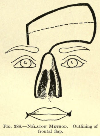
Fig. 388.—Nélaton Method. Outlining of frontal flap.
In the illustration is also shown the incisions later made to utilize the borders of the remaining nose to line the frontal flap. This is done by making an inverted V incision at a distance from the inner borders, corresponding to the lateral line of union of the frontal flap with the face. The resultant flap is turned down, raw surface outward, curtainlike, and is sutured to the frontal flap, where it falls into position.
The flap outline shows that its pedicle lies between the outer end of the inner third and above the right eyebrow and a little to the left of the median line at the root of the old nose. This will avoid considerable tension at this point, the rotation as made being ninety degrees.
Nearly horizontally, as shown in the figure, a line is drawn through the center of the flap, showing the position the strip of cartilage is to occupy.
This done, a pattern of the outline is cut from stiff paper or oiled silk to preserve as a guide for the making of the flap, it being understood that the outlining has been made to the measurement of the required nose, allowance being given for cicatricial contraction.
This done, the surgeon having prepared the skin about the costal prominences of the left thorax, he proceeds as follows:
A vertical line is drawn the width of two fingers to the right of the nipple, as shown in Fig. 389, the length of the line being obvious.
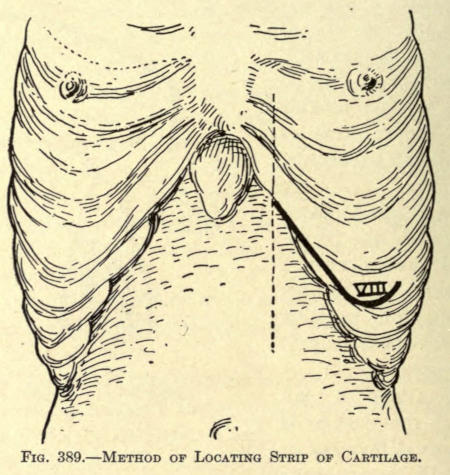
Fig. 389.—Method of Locating Strip of Cartilage.
Where the vertical crosses the eighth costal cartilage an incision is made downward over and not under the border of the cartilage.
The incision extends downward for a distance of eight centimeters, where it is turned upward at an angle, as shown, to a distance of three centimeters.
By separating the muscular aponeurosis made visible by this incision the lower edge of the eighth costal cartilage is exposed. The knife is moved along the lower edge of the cartilage, dividing the fibers of the insertion of the transverse muscle from without inward. The cartilage can now be grasped between the thumb and forefinger and be forced out of its normal position after a slight anterior dissection.
The union between cartilage and bone is exposed. The chisel is used to divide the cartilage about one centimeter from the rib, after the costal or inner extremity has been made.
The position of the hands and the exposed cartilage is shown in Fig. 390.
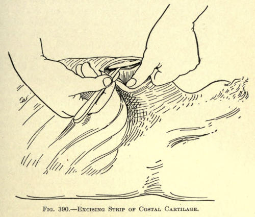
Fig. 390.—Excising Strip of Costal Cartilage.
This accomplished, the wound is temporarily dressed. The cartilage is then fashioned to suit the required size and shape.
It is thinned down on its lower surface to about three millimeters in diameter. This thickness is maintained to a length of two and a half centimeters, the part being intended for the subseptum.