F. SPECIALIZED HABITATS
Reading edition
(i) Fungi of dung and straw heaps
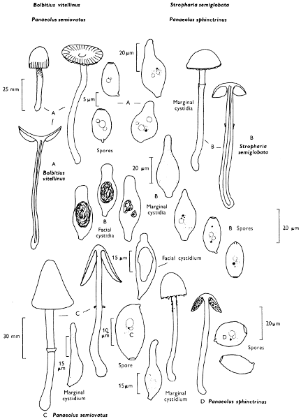
Bolbitius vitellinus (Fries) Fries Yellow cow-pat toadstool
Cap: width 20-40 mm. Stem: width 2-5 mm; length 30-60 mm.
Description:
Cap: chrome-yellow or lemon-yellow when young, paling with age at margin to become cinnamon-buff, bell-shaped but rapidly expanding to become plane or slightly umbonate, smooth, viscid but soon drying; margin striate then radially grooved, often split and the whole cap soon collapsing.
Stem: slender, whitish, cream colour to pale yellow, at apex covered in small, white floccose scales but downy at the base, fragile and soon collapsing.
Gills: adnexed or free, cinnamon-buff, thin and crowded.
Flesh: yellowish, very thin and lacking distinct smell.
Spore print: rust-brown.
Spores: long, yellow-brown under the microscope, ellipsoid with a very distinct germ-pore about 13 × 8 µm in size (11-15 × 6-9 µm).
Facial cystidia: rare, balloon-shaped.
Marginal cystidia: swollen, flask-shaped with a variable, elongate neck.
General Information: This fungus is common on horse droppings or other manures, but it may also be found amongst grass in pastures and in sand-dunes, and gardens on piles of rotting grass stems or straw. It is easily recognised by the colour and rapid expansion of the cap and the sudden collapse of the whole fruit-body. ‘Vitellinus’ means yolk of an egg and refers to the persistently bright yellow cap-centre, so obvious even when the fruit-body collapses. This collapsing is not one of autodigestion as described for members of the genus Coprinus. It is variable both in size and habitat, and I even have records of the fungus growing within herbaceous stems.
Illustrations: LH 153; WD 806.
Stropharia semiglobata (Fries) Quélet Dung-roundhead
Cap: width 10-35 mm. Stem: width 4-7 mm; length 25-50 mm.
Description:
Cap: hemispherical or slightly umbonate, sometimes flattened and hardly expanding even with age, very viscid, smooth, pale yellow-ochre or yellowish tan.
Stem: slender, straight, white then yellowish, smooth, viscid, but then dry and shiny below an imperfectly formed, thin ring.
Gills: adnate, almost triangular in shape, crowded, dark brown to purplish black, but with ochraceous areas at maturity.
Flesh: pale ochre.
Spore-print: purplish brown.
Spores: very long, dark brown under the microscope, smooth, ellipsoid with large germ-pore and about 18 × 10 µm in size (17-20 × 9-10 µm).
Facial cystidia: spindle-shaped, thin-walled and filled with amorphous contents which become yellow in solutions containing ammonia.
Marginal cystidia: spindle-shaped or flask-shaped, numerous, thin-walled and typically yellowing as above.
General Information: ‘Semiglobata’ means hemispherical and refers to the shape of the cap of S. semiglobata; it is a very variable fungus in both size of the cap and the prominence of the ring. The Dung-roundhead grows only on dung which is acidic in its soil status, whilst Panaeolus semiovatus (Fries) Lundell next described (p. 210) grows on slightly to distinctly base-rich dung. This may explain why in Britain the Dung-roundhead is the commoner of the two species. However, P. semiovatus was formerly placed in the genus Stropharia because of its blackish spores and distinct ring. The spores of Stropharia in the mass are violaceous black whilst those of P. semiovatus are brownish black. Under the microscope they are also differently coloured and have different chemical compositions as their reaction with dilute solutions of ammonia shows; the spores of the first species turn purplish olive in ammonia and those of the second species become very dark brown.
Illustrations: F 33b; Hvass 171; LH 153; NB 315; WD 753.
Plate 70. Dung-fungi
Larger illustration
Mottle-gills—on dung from Spring until Autumn.
Panaeolus semiovatus (Fries) Lundell
Cap: width 20-70 mm. Stem: width 5-10 mm; length 80-160 mm.
Description:
Cap: oval or bell-shaped, not expanding, dingy whitish or pale clay colour, smooth, slimy when moist, but soon drying and then becoming shiny, often wrinkled and cracked, and ornamented with fragments of veil at the margin.
Stem: dull, straight, rather rigid, tapering upwards, white, and striate at apex above a whitish erect and membranous, often collapsing, ring; yellowish below the ring and whitish and cottony at the slightly swollen base.
Gills: adnate, greyish then black, mottled and crowded.
Flesh: whitish or pale ochre.
Spore-print: black.
Spores: very long, very dark brown under the microscope with large obvious germ-pore and 18 × 10 µm (16-20 × 9-11 µm) in size.
Facial cystidia: flask-shaped and with amorphous contents.
Marginal cystidia: numerous, flask-shaped.
Panaeolus sphinctrinus (Fries) Quélet
Cap: width 15-35 mm. Stem: width 3-6 mm; length 60-95 mm.
Description:
Cap: bell-shaped, hardly expanding, expallent, dark grey to olivaceous black, much paler when dry and zoned when half dry; margin ornamented with a white fringe of veil fragments.
Stem: long, slender, straight, rather rigid but fragile, grey and completely powdered with white.
Gills: adnate, crowded and grey then blackened, mottled throughout except at the white fringed edge.
Flesh: reddish brown.
Spore-print: black.
Spores: long, very dark brown under the microscope, broadly lemon-shaped with large germ-pore, smooth and 14-15 × 9-10 µm in size (14-19 × 8-10 × 10-12 µm).
Facial cystidia: absent.
Marginal cystidia: numerous, cylindrical, flexuous and hyaline.
General Information: P. sphinctrinus is recognised by the overall grey colouration and very distinct white fringe to the cap-margin.
P. campanulatus (Fries) Quélet which is said to be common is in fact infrequent and many records really refer to P. sphinctrinus. The word semiovatus means half ovate and refers to the shape of the cap in P. semiovatus. Sphinctrinus means banded, referring to the zoned cap of the fungus when it is partially dry.
Illustrations: P. semiovatus—LH 145; WD 773. P. sphinctrinus—NB 415; WD 781.
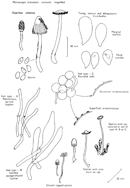
Coprinus cinereus (Fries) S. F. Gray Dung-heap ink-cap
Cap: width 20-40 mm. Stem: width 4-8 mm; length 50-100 mm.
Description:
Cap: oval then rapidly expanding, covered at first in a mass of dense, white or greyish woolly scales which break up into patches and finally leave the cap shiny, brownish grey at centre and striate and dark grey at the margin.
Stem: white, covered particularly towards the base with white, woolly scales, long, fragile, tapering upwards and at the base often elongated into a ‘tap root’ buried in the dung.
Gills: free, white but then rapidly dissolving into a black liquid.
Flesh: thin and whitish.
Spore-print: violaceous black.
Spores: medium sized, ellipsoid, smooth with a distinct germ-pore and 10-12 × 5-6 µm in size.
Facial cystidia: absent.
Marginal cystidia: inflated and large.
General Information: It is found on manure heaps, on straw dung and on silage heaps: very common throughout the year.
C. macrocephalus (Berkeley) Berkeley is very closely related to C. cinereus, but differs in having much larger spores over 12-15 × 7-9 µm, a long cap and a stem which lacks a rooting base.
Coprinus radiatus (Fries) S. F. Gray is smaller in stature and also differs in spore-size (11-14 × 6-7 µm). C. pseudoradiatus Kühner & Josserand is minute and has even smaller spores (7-9 × 4-5 µm). The dung-heap ink-cap has long been used by scientists in genetic studies, usually under the name of C. lagopus (Fries) Fries. However, this latter species, although similar, grows only on woodland detritus; it has narrower spores. The dung-heap ink-cap may be referred to in other books as C. fimetarius Fries or C. macrorhizus (Fries) Rea and whilst cinereus means grey referring to the colour, fimetarius means dung—from the habitat, and macrorhizus refers to the long rooting base found in some specimens.
Illustrations: LH 137; NB 4110; WD 814.
The genus Coprinus—or Ink-caps
The genus Coprinus is easily recognised from all other agarics by the structure and development of the fruit-body. In the field, most species of the genus can be recognised by the gradual conversion of the gills, and often the cap tissue into a black liquid resembling ink—hence the name inky-caps. The conversion of the gills to an inky mass is called autodigestion and the process is complete within a few hours; this mechanism enables spores to be dispersed immediately they have ripened. Unlike other agarics the spores are not shot off into the spaces between the gills, but directly into the air. The gills are parallel-sided in Coprinus and not wedge-shaped as in more normal agarics, and in order to achieve spore dispersal the gills must disintegrate; Coprini are very specialised.
Coprinus is a large genus with over seventy members in the British Isles, many of which are strictly dung-loving. It is impossible to give more than one example in full here, for although many of the large species can be recognised on sight the smaller ones require the aid of a microscope. The interested student must therefore refer to more advanced texts, but in order to demonstrate the diversity of the Coprini and how they are classified the following key to the sections of Coprinus will be found useful.
1. Cap naked of any veil fragments, either smooth or covered in minute hairs 2
Cap covered when young by powdery or hairy veil, particles of which either may persist on the cap until maturity or may disappear quickly 3
2. Cap completely naked—group Nudi, e.g. C. miser (Karsten) Karsten
Cap with hairs giving it a frosted appearance—group Setulosi, e.g. C. ephemerus (Fries) Fries, C. pellucidus Karsten and C. bisporus J. Lange
3. Veil on the cap composed under the microscope of rounded cells giving the cap a floccose powdery appearance—group Vestiti, e.g. C. patouillardii Quélet, C. niveus (Fries) Fries and C. ephemeroides (Fries) Fries
Veil on the cap composed under the microscope of elongate cells, either like thin-hairs or strings of sausages 4
4. Veil on the cap composed under the microscope of strings of sausage-shaped cells—group Lanatuli, e.g. C. cinereus, C. pseudoradiatus, C. radiatus (see p. 211)
Veil on the cap composed under the microscope of thick- or thin-walled, flexuous or straight, filamentous, hardly inflated cells—group Impexi, e.g. C. filamentifer Kühner, C. vermiculifer Dennis.
Plate 71. Dung-fungi—The genus Coprinus
Larger illustration
General notes on dung-loving fungi and their habitats
Dung fungi are highly satisfactory for demonstrating the diversity and morphology of a group of related organisms within a single ecological system, as representatives of most of the major groups of fungi usually grow on dung after a period of incubation. Dung will always produce characteristic fungi whatever time of year it is collected.
Dung is best incubated in a light place, for example on a window sill, in a warm room on layers of blotting paper or other absorbent material. For rabbit-pellets and samples of similar size petri-dishes are ideal, but for cow, horse and similar types of dung large covered dishes such as casseroles or sandwich containers are very good. Samples should not be kept in airtight containers for long periods of time as under such conditions animal life present rapidly breaks down the dung and induces anaerobic conditions. Instead larvae and earthworms should be excluded from the sample as they decompose the dung and inhibit fungal growth but their activity can be reduced, if causing a problem, by spraying the sample lightly with a proprietary fly-kill aerosol.
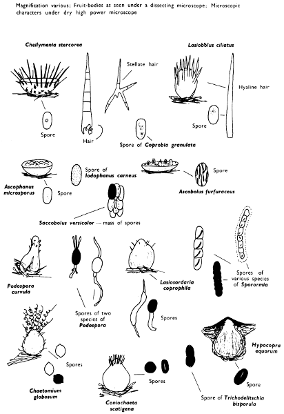
By keeping the dung under constant observation during incubation a whole succession of fungi can be seen. Thus the first fungi to appear are the moulds which although numerous need a microscope for their identification. The moulds are followed by a series of Ascomycetes (Sporormia & Sordaria with flask-shaped fruit-bodies and Iodophanus, Coprobia and Cheilymenia with disc-shaped fruit-bodies), which are best sought with the use of a powerful hand-lens or a stereoscopic binocular microscope when their full beauty will be revealed. However, because they need the aid of instruments even to see them they cannot be considered larger fungi. The fruit-bodies of the Basidiomycetes are readily seen with the naked eye, but a hand-lens is still very useful for observing features of the cap and stem, particularly the veil characters. The Basidiomycetes usually conclude the succession of fungi found on dung and soon after this state the dung is colonised by mosses and higher plants and later it is fully incorporated into the soil.
Plate 72. Dung-fungi: Cup fungi and allies
Larger illustration
Dung is a very useful substrate for studying succession. However, equally interesting results can be obtained from observing the fungi which appear on a stump, colonise a newly laid lawn, or indeed those growing on refuse such as a cast-out rug; microscopic and larger fungi are all to be found.
If the dung cannot be incubated immediately it should be dried quickly, for most dung-fungi will survive such treatment and grow when the sample is remoistened. The blotting-paper on which the dung is placed should be kept moist throughout the incubation period.
One large discomycete (up to 80 mm across) occurring on manure-heaps must, however, be mentioned, this is Peziza vesiculosa St Amans (see p. 200); the inner surface of this cup-fungus becomes detached from the flesh at maturity and forms blisters.
(ii) Fungi of bonfire-sites
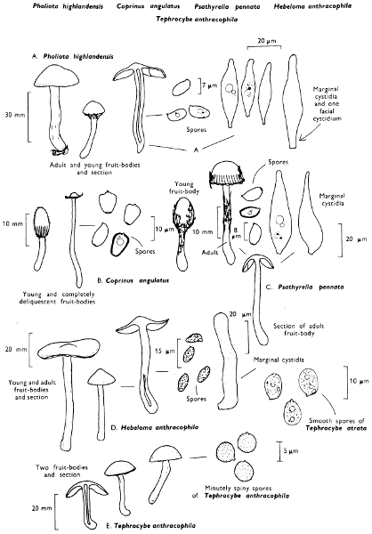
Pholiota highlandensis (Peck) A. H. Smith Charcoal pholiota
Cap: width 20-50 mm. Stem: width 4-8 mm; length 25-60 mm.
Description: Plate 73.
Cap: convex then flattened and slightly umbonate, smooth, very sticky at first, but becoming shiny when dry, orange-yellow to sand-colour; the margin is first incurved and ornamented with filaments from the veil, but these are soon lost.
Stem: dirty yellow, darker towards the base, cylindric or narrowed downwards and covered in small fibrillose scales.
Gills: clay-coloured then dull brown, adnate and crowded.
Flesh: yellowish.
Spore-print: dull rust-brown.
Spores: medium-sized, ellipsoid, smooth, dull brown under the microscope and 7-8 × 3-4 µm in size.
Facial cystidia: spindle-shaped with obtuse apex.
Marginal cystidia: similar to facial cystidia but usually smaller.
General Information: This fungus which occurs from spring to autumn is recognised by the habitat, colour of the fruit-body and the spore-size. It is known in many books as Flammula carbonaria (Fries) Kummer, but the genus Flammula is no longer used for it refers to a flowering plant in the buttercup family.
P. highlandensis is the same fungus as that referred to as Pholiota carbonaria by European Mycologists, but this name cannot be used for it refers to an entirely different N. American species. ‘Highlandensis’, in fact, refers to the locality where the present fungus was first found in the United States of America. The true P. carbonaria A. H. Smith has only been found once in Europe and this only recently in the south of England. It differs in the reddish orange scales on the stem; indeed it is a much brighter fungus than the common charcoal Pholiota.
Tephrocybe anthracophila (Lasch) P. D. Orton
Cap: width 1-4 mm. Stem: width 1 mm; length 2-5 mm.
Description: Plate 73.
Cap: blackish when wet, drying sooty brown, slightly depressed in the centre, smooth, and viscid.
Stem: sooty brown, tough and smooth.
Flesh: sooty brown.
Gills: whitish then grey, adnate and not very crowded.
Spores: medium sized, subglobose, 4-6 × 4-5 µm in diameter and minutely roughened.
Spore-print: white, not blueing in solutions of iodine.
General Information: T. atrata also grows on burnt soil and is very closely related, but differs in its spores being broadly ellipsoid and smooth. Mycena leucogala also grows on burnt soil (see p. 88).
Illustrations: T. anthracophila—LH 83. T. atrata—WD 4b.
Psathyrella pennata (Fries) Pearson & Dennis Bonfire brittle-cap
Cap: width 10-30 mm. Stem: width 1-3 mm; length 30-40 mm.
Description:
Cap: conical or bell-shaped then expanding and slightly umbonate, whitish because of a coating of dense fibrils, but soon becoming brownish as these are lost.
Stem: short, stout, white and densely floccose.
Gills: slightly adnate, pale brownish grey with pink tinge, then dark-brown.
Spore-print: purplish brown.
Spores: medium sized, oval, ellipsoid with an obvious germ-pore, purplish brown under the microscope and 8-9 × 4-5 µm in size.
Marginal & facial cystidia: flask-shaped, hyaline with either a short or long neck.
The brown-spored Hebeloma anthracophila Maire is similar.
Coprinus angulatus Peck Bonfire ink-cap
Cap: width 4-25 mm. Stem: 1-3 mm; length 15-30 mm.
Description:
Cap: dark red-brown at first, then orange-brown, especially at the margin and appearing as if frosted all over, conical at first but rapidly expanding at the margin and becoming grey-brown, strongly striate and deliquescent, leaving finally only a central red-brown umbo.
Stem: white and minutely hairy.
Gills: free, dirty whitish then black.
Spore-print: black-brown.
Spores: medium sized, dark brown under the microscope, lobed like the hat of a bishop and 8-11 × 6-8 × 5-7 µm in size.
Marginal cystidia: bottle-shaped, very variable.
Facial cystidia: similar to marginal cystidia.
General Information: It must be noted that this fungus has spores which require three quite different measurements to describe the dimension. Another species of Coprinus found on burnt soil is C. lagopides Karsten which resembles C. cinereus (Fries) S. F. Gray (p. 211); it is typified, however, by the rounded spores.
Plate 73. Fungi of bonfire-sites
Larger illustration
General notes on fungi of burnt sites
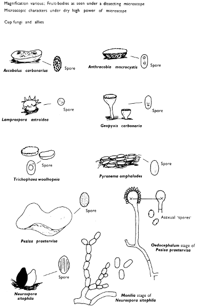
Several common fungi found at the sites of bonfires have their closest relatives amongst various groups of microscopic fungi more than amongst the large forms already discussed. Keeping a close watch at the site of a former bonfire day by day, week by week and month by month is very rewarding and shows a further example, like the dung habitat, of a tightly knit community of various groups of fungi.
Peziza repanda Persoon has been discussed in detail above (p. 200); its close relatives P. petersii Berkeley & Curtis (brown with grey tints and with spores finely warted and measuring 10-12 × 5-6 µm), P. praetervisa Bresadola (violet or mauve and with spores finely warted and measuring 11-13 × 6-8 µm), P. violacea Persoon (dark violet with smooth spores measuring 13-15 × 7-9 µm) and P. echinospora Karsten (dark chocolate brown with spores densely warted and 14-18 × 7-10 µm in size) all grow on the sites of old bonfires or around charred root stumps. Rhizina undulata also found by charred stumps has been described on p. 203. These are large to medium sized disc-fungi, but there are many much smaller species which cannot be dealt with here, such as species of Anthracobia and Trichophaea. Pyrenomycetes are also found on charred wood and soil. Probably the commonest species of fungus met with is a pale reddish orange to rose-pink disc-fungus seated on a white mycelial mat; this is Pyronema omphalodes (St Amans) Fuckel. Morchella esculenta St Amans and M. elata Fries (see p. 200) appear to grow on the sites of garden bonfires or where cinders have been spread on the soil surface. The stimulus for fruiting appears to be due to the release of mineral nutrients during the process of burning. Competition from other fungi appears to be reduced so rapid colonisation by the bonfire fungi (carbonicoles) after the period of sterilisation ensures their development. Many similar fungi were found about bomb- and shell-craters on the continent during the two World Wars.
One microscope fungus, however, must be mentioned when considering bonfires and that is Neurospora sitophila Shear & Dodge so much used in genetical studies. It can be found as the conidial state on burnt soil and is called ‘Baker’s mould’ because it is frequently found growing on refuse in the hot moist conditions of bakers’ kitchens.
Plate 74. Fungi of bonfire-sites
Larger illustration
(iii) Fungi of bogs and marshes
(a) Sphagnum bogs
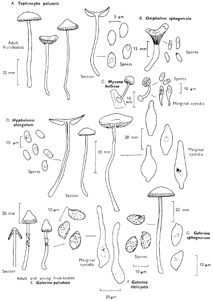
Hypholoma elongatum (Fries) Ricken
Cap: width 12-20 mm. Stem: width 3-5 mm; length 50-80 mm.
Description: Plate 75.
Cap: bell-shaped but rapidly expanding to become plane, honey-yellow with a greyish green tint, slightly striate at the margin and also with a few remnants of a fibrillose veil when very young, but these are soon lost.
Stem: slender, smooth, whitish at the apex and yellow-brown or honey-yellow below.
Gills: adnate and distant, pale ochraceous honey-yellow then lilaceous grey and finally sepia.
Flesh: yellowish in the cap, red-brown in the stem and lacking a distinct smell.
Spore-print: purplish brown.
Spores: long, ellipsoid, fairly thick-walled, olivaceous brown under the microscope and with a small germ-pore, smooth and 10-12 × 6-7 µm in size.
Marginal cystidia: flask-shaped and hyaline.
Facial cystidia: flask-shaped with contents which turn yellowish in solutions containing ammonia.
General Information: This fungus which appears from early summer to late autumn is recognised by the almost uniform ochraceous colour with hint of olive and its habit of growing in troops. The word elongatum means elongated and refers to the shape of the stem which pushes up through the Sphagnum and in order to disperse its spores it must elongate so that it just pushes up above the bog-surface. H. polytrichi is closely related to H. elongatum but has a paler cap and stem and it grows in moss, particularly Polytrichum in woodlands; the spores of H. polytrichi are paler, slightly narrower and slightly thinner, but they have a much more distinct germ-pore.
Both the above species have been formerly placed in Psilocybe, but they are more correctly classified in Hypholoma along with the sulphur-tuft fungus (see p. 64) because of the cortina-like veil and specialised facial cystidia.
Illustrations: WD 785.
Tephrocybe palustris (Peck) Donk
Cap: width 12-30 mm. Stem: width 3-5 mm; length 50-75 mm.
Description: Plate 75.
Cap: bell-shaped then plane-convex, but finally depressed at centre, watery buff to greyish with flush of ochre or smoky grey, striate to centre when moist, but drying out non-striate and uniformly ochraceous buff.
Stem: thin, rather long, smooth, similarly coloured to the cap or paler, fragile and whitish woolly at the base.
Gills: dirty whitish, adnate with a tooth and not very crowded.
Flesh: thin, watery buff, drying out ochraceous and with a strong smell of new meal.
Spore-print: white.
Spores: medium sized, hyaline under the microscope, oval, not turning blue-grey in solutions of iodine, and 6-7 × 4-5 µm in size.
Marginal and facial cystidia: absent.
General Information: This fungus which grows from late spring to autumn is usually associated with a greying and finally a killing of the Sphagnum, noticeable from a distance even in the absence of the fruiting-bodies as paler patches in the rich green bog. Another agaric found only in Sphagnum bogs is Omphalina sphagnicola (Berkeley) Moser with decurrent gills and long, elongate, hyaline spores.
At the margin of Sphagnum bogs, the fungus Mycena bulbosa can be found attached to the base of tufts of rushes.
Potting up a sward of Sphagnum and retaining it in a warm greenhouse during winter favours the bog agarics to fruit when other larger fungi are not available.
Mycena bulbosa (Cejp) Kühner
Cap: width 3-6 mm. Stem: width 1 mm; length 10-15 mm.
Description:
Cap: dirty white, greyish and very gelatinous.
Stem: very thin, hyaline with a very distinct hairy, basal disc.
Gills: crowded, adnexed, very short and whitish.
Spore-print: white, but because it is so small it is often difficult to see.
Spores: medium sized, hyaline under the microscope, ellipsoid, not blueing in solutions of iodine, and 8-10 × 4 µm in size.
Marginal cystidia: clavate or ventricose, hyaline and smooth.
Facial cystidia: absent.
Illustrations: T. palustris LH 83.
Galerina paludosa (Fries) Kühner
Cap: width 10-20 mm. Stem: width 3-5 mm; length 50-90 mm.
Description:
Cap: conico-convex expanding slightly but retaining the central umbo, striate to half-way, sand-colour to red-brown, hygrophanous, minutely floccose because of remnants of veil distributed over its surface, but soon becoming smooth.
Stem: long, buried amongst the Sphagnum, red-brown and flecked with white fibrils, except at the finely hairy apex, the fibrils typically form a distinct but easily lost ring.
Gills: almost horizontal, adnate to subdecurrent, pale at first and then rust-brown.
Spore-print: rust-brown.
Spores: medium-sized, ovate to slightly lemon-shaped, minutely warty, honey-brown under the microscope and about 10 × 6 µm in size, (9-11 × 6-7 µm).
Facial cystidia: absent.
Marginal cystidia: hyaline, almost cylindrical or bottle-shaped with an inflated base.
General Information: This species grows from spring to early autumn in Sphagnum bogs; several other species of Galerina are also found in the same localities:—
(i) G. sphagnorum (Fries) Kühner has a convex cap, fibrillose silky and ochraceous brown stem, but it lacks the ring-zone so typical of G. paludosa. The smell is like that of meal when crushed and the gills are emarginate.
(ii) G. tibiicystis (Atkinson) Kühner has a rapidly expanding cap which becomes plano-convex or depressed at maturity; it also lacks a ring-zone, but the stem in this species is finely hairy because of the presence of numerous pin-shaped cells which can be seen only with the aid of a lens. The gills are broadly adnate.
Illustrations: G. paludosa—LH 175.
Plate 75. Fungi of marshes
Larger illustration
(b) Alder-carrs
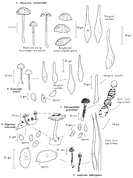
Naucoria escharoides (Fries) Kummer
Cap: width 12-30 mm. Stem: width 1-3 mm; length 25-45 mm.
Description: Plate 76.
Cap: pale yellowish ochre, but becoming darker ochraceous with age, scurfy, convex but then flattened, or with its edge upturned; the margin is slightly striate when moist.
Stem: slender, pale to dirty yellowish ochre but darker brown at base, slightly fibrillose, particularly at first because of filaments from a veil, but these are soon lost.
Gills: pale tan to brownish ochre with a paler, floccose margin, adnate and crowded.
Flesh: yellowish ochre but lacking a distinct smell.
Spore-print: clay-colour.
Spores: medium sized, almond-shaped, pale brown under the microscope, warted and 10-11 × 5-6 µm in size.
Marginal cystidia: swollen below, but drawn out into a hair-like apex.
Facial cystidia: absent.
General Information: Although this is a common species growing in damp places under alder it is difficult except with an expert eye to separate it from several closely related species which are also found in similar places. At present it is not known whether these fungi are favoured by the water-logged base-rich, reducing soils found nowhere else except under alder, or if they have a special relationship with the tree. There is ample evidence that soil conditions in alder woods are rather different from those found in other woodlands, but whatever the reason Naucoria escharoides is only found under alder—in fact this species has been placed in the genus Alnicola because of this character—cola meaning inhabitant and Alnus the tree of that name. Willow-carrs have not been as extensively studied as alder-carrs but there is evidence that a store of mycological information is still to be obtained from these places. Several species of Naucoria have been described from only willow-carrs, while others are to be found under both alder and willows; about eight species are known to grow under alder. The word escharoides means scab-like and refers to the cap which when freshly collected is minutely scaly and appears scabby.
Illustrations: LH 163; WD 671.
(iv) Fungi of beds of herbaceous plants
Beds of herbaceous plants provide protection for many small agarics and collecting can be conducted in these situations from spring to early winter. The buffered environments under the herbs is humid and relatively still, and this allows the development of the small often delicate fruit-bodies of certain species to continue unimpeded. Nettle-beds or mixtures of nettle and dog’s mercury have very rich floras under the shelter of their leaves and stems, either on the bare soil or plant debris.
On herbaceous stems
Coprinus urticicola (Berkeley & Broome) Buller
Cap: width 4-7 mm. Stem: width 1 mm; length 10-15 mm.
Description: Plate 76.
Cap: white then greyish, globose at first and then expanding to become plane with upturned margin covered, at first, with scales from a veil which at the centre are white-tipped with ochre.
Stem: white and slightly downy.
Spore-print: brownish black.
Spores: elliptic-ovoid, only slightly compressed with distinct germ-pore, dark brown under the microscope and 6-8 × 5 µm in size.
Marginal cystidia: ellipsoid to pyriform and hyaline.
Facial cystidia: elongate cylindric larger than marginal cystidia.
On bare soil
Leptonia babingtonii (Bloxam) P. D. Orton
Cap: 5-15 mm. Stem: width 1 mm; length 20-50 mm.
Description: Plate 76.
Cap: grey to sepia or greyish brown entirely scaly-hairy, at first, but then fibrillose.
Stem: silvery grey to grey-sepia and silky fibrillose.
Gills: greyish pink.
Spore-print: greyish pink.
Spores: very long, wavy angular in outline, very pale honey under the microscope and 14-20 × 7-9 µm.
Marginal cystidia: club-shaped or balloon-shaped and hyaline.
Facial cystidia: absent.
So very different to other species of Leptonia is it that it should be classified in Dr. Pilát’s genus Pouzaromyces.
Conocybe mairei Watling
Cap: width 5-10 mm. Stem: width 1 mm; length 10-40 mm.
Description:
Cap: pale to deep ochraceous or buff, minutely tomentose.
Stem: flexuous, whitish or very pale ochraceous.
Gills: pale buff then ochraceous.
Spore-print: ochraceous.
Spores: medium sized, ellipsoid or slightly almond-shaped with small germ-pore and 6-8 × 3-4 µm in size.
Flammulaster granulosa (J. Lange) Watling
Cap: 4-15 mm. Stem: width 1 mm; length 10-25 mm.
Description:
Cap: ochraceous to date-brown, darker at the centre and granular scaly throughout.
Stem: similarly coloured to the cap and similarly roughened, except for the slightly smoother paler apex.
Spores: ellipsoid to almond-shaped, very pale brown under the microscope and 8-10 × 4-5 µm in size.
Marginal cystidia: cylindric-wavy, hyaline.
Facial cystidia: absent.
Depending on the herbaceous constituents the fungus-flora will vary. Certain species are found on all sorts of herbaceous debris, but others are much more specific to their substrate preferences. Beds of Butterbur, Coltsfoot or Impatiens are also good hunting places, as are beds of sedges in fenland. In many of these localities agarics with reduced fruit-bodies looking like disc-fungi are frequently seen. We have already discussed the specific requirements of certain species of Marasmius (see p. 92).
Plate 76. Fungi of alder-carrs and from under herbaceous plants
Larger illustration
(v) Fungi of moss-cushions
Many small species grow amongst moss cushions on tree trunks, tucked in crevices in walls or on the tops of old buildings. However, there is one genus of agarics, i.e. Galerina which is probably more typical than any other of such situations. There are many members of this genus whose small caps are found in the autumn pushing up through the moss plants. Plate 78.
Galerina hypnorum (Fries) Kühner
Cap: width 4-6 mm. Stem: width 1 mm; length 20-40 mm.
Description:
Cap: hemispherical or bell-shaped, hygrophanous, orange-yellow, sand-colour, smooth and striate almost to the cap-centre.
Stem: smooth and similarly coloured to the cap.
Gills: yellow-tawny then rust-coloured, adnate emarginate, rather broad and somewhat distant.
Flesh: thin, yellow-tawny and with a smell of new meal.
Spore-print: rust-colour.
Spores: medium-sized, almond-shaped, golden yellow under the microscope, slightly roughened and 10-11 × 6-7 µm in size.
Marginal cystidia: flask-shaped or cylindrical with slight swelling at the apex.
Facial cystidia: absent.
Galerina mycenopsis (Fries) Kühner
Cap: width 6-15 mm. Stem: width 1 mm; length 30-60 mm.
Description:
Cap: similarly coloured to G. hypnorum, but with a few white silky fibrils.
Stem: coloured as the cap, but with white silky fibrils when young.
Gills and flesh: as in G. hypnorum, but it has no smell.
Spore-print: rust-colour.
Spores: medium-sized, ellipsoid, pale golden yellow under the microscope, smooth and 9-11 × 5-6 µm in size.
Marginal cystidia: club-shaped, cylindrical and with distinct rounded heads.
Facial cystidia: absent.
General Information: G. mniophila (Lasch) Kühner is similar to or slightly larger than C. mycenopsis, but differs in its dull honey-coloured cap and stem, and distinctly roughened spores. G. calyptrata P. D. Orton is small and has been long confused with G. hypnorum; it, however, is of a much brighter orange-colour, with distinct white fibrils on the cap and has spores which have a distinct envelope, sometimes separating as a loose covering. G. vittaeformis (Fries) Moser is a red-brown fungus with 2-spored basidia, facial cystidia, minutely hairy stem, and very rough spores; it grows in moss in pastures as well as on moss-cushions.
(vi) Heath and mountain fungi
(a) Moorland fungi
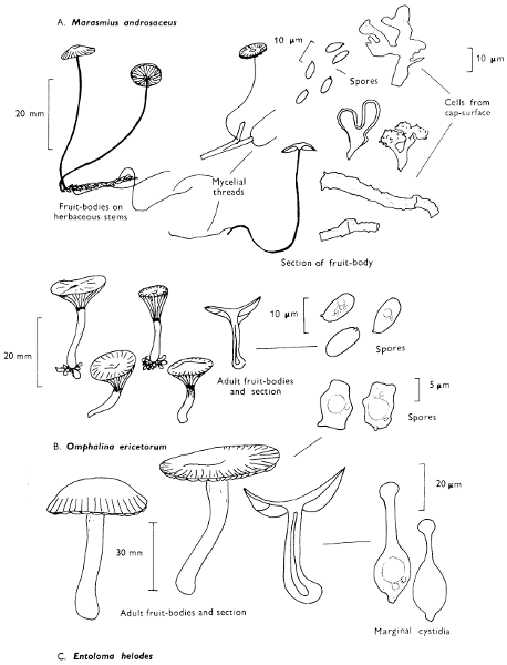
Marasmius androsaceus (Fries) Fries Horse-hair toadstool
Cap: width 5-15 mm. Stem: width 1 mm; length 30-60 mm.
Description: Plate 77.
Cap: whitish to pale smoke-brown with a distinct wine-coloured tinge, membranous, flattened, or umbilicate and radially wrinkled.
Stem: thread-like, black or very dark brown, horny and usually springing from a black horse-hair-like mycelium.
Gills: whitish or dirty flesh-colour, adnate and crowded.
Flesh: white in the pileus and black in the stem.
Spore-print: white.
Spores: medium-sized, pip-shaped, not blueing in solutions containing iodine and measuring 7-9 × 3-4 µm in size.
Marginal cystidia: oval or ellipsoid, covered on the upper half with small pimple-like projections.
Facial cystidia: absent.
General Information: This fungus is common in troops from late summer until winter on dead and dying heather. It is also found in woods on leaves and twigs, particularly in plantations on conifer needles. It is easily recognised by the dark horse-hair-like stem which becomes bent and twisted on drying and the small, pinkish flesh-coloured cap. The word androsaceus means, and refers to, the stem which resembles the tough and wiry fronds of some of the red algae, such as Ahnfeldtia which is found around our sea-shores.
Illustrations: LH 115; NB 471; WD 244.
Omphalina ericetorum (Fries) M. Lange
Cap: width 5-20 mm. Stem: width 2 mm; length 10-20 mm.
Description:
Cap: variable in colour, straw-colour, cream-colour, bistre or grey, convex then flat or slightly depressed, radially grooved to the centre when moist; the margin is scalloped.
Stem: slender, similarly coloured to the cap, except for a brownish wine-coloured zone at the very apex, thickened upwards and smooth with a white and woolly base.
Gills: adnate to decurrent, white then cream-colour or yellowish, triangular in shape, very distant and often connected by veins.
Flesh: pale cream-colour.
Spore-print: white.
Spores: medium sized, hyaline under the microscope, broadly ellipsoid, or pip-shaped, not becoming bluish grey in solutions of iodine, 8-10 × 5 µm in size.
Marginal and facial cystidia: absent.
General Information: This fungus is common and often in large troops on peaty ground in woods as well as in moorland and mountain regions. In mountains O. ericetorum must be carefully distinguished from some of the truly mountain species of Omphalina dealt with on p. 236. O. wynniae (Berkeley & Broome) P. D. Orton is similar but pale lemon-yellow and is found on stumps of conifers. The word ericetorum refers to the habit of growing on heaths—Erica is the Latin name for heath. In many books this same fungus is called O. umbellifera which reflects the shape of the cap—umbrella shaped.
Illustrations: Hvass 116; LH 99; NB 857; WD 299.
Entoloma helodes (Fries) Kummer
Cap: width 25-75 mm. Stem: width 2-6 mm; length 25-55 mm.
Description:
Cap: finely or minutely velvety at centre, fibrillose or white silky as if frosted towards the margin, sepia or bistre, or mouse-grey, dull-coloured but with a hint of violaceous brown.
Stem: equal or slightly thickened at the apex, sometimes club-shaped, thickened at the base, greyish brown and pale cream-colour at the base.
Flesh: dark sepia in the cap, whitish in the stem and smelling strongly of meal.
Plate 77. Moorland fungi
Larger illustration
Gills: white or whitish at first then dirty pinkish brown, adnate and emarginate.
Spore-print: dull salmon-pink.
Spores: medium to long, angular, ellipsoid-oblong, slightly cinnamon-colour under the microscope and 9-12 × 7-8 µm in size.
Marginal cystidia: conspicuous, spindle or bottle-shaped and with subcapitate apex.
Facial cystidia: absent.
Hypholoma ericaeum (Fries) Kühner
Cap: width 15-30 mm. Stem: width 4-7 mm; length 50-100 mm.
Description:
Cap: fleshy, convex, later becoming flattened but remaining slightly umbonate at the centre, viscid at first, smooth and shining when dry, bright reddish to sand-colour or brown.
Stem: slender, yellow above, brown below, smooth and tough.
Gills: adnate or adnexed, purplish black with a whitish margin and fairly crowded.
Flesh: yellowish or red-brown in the stem.
Spore-print: purple-brown.
Spores: long, dark purple-brown, broadly ellipsoid and 12-15 × 7-9 µm in size.
Marginal cystidia: cylindrical or flask-shaped.
Facial cystidia: flask-shaped and filled with contents which become yellowish in solutions containing ammonia.
Clavaria argillacea (Persoon) Fries
Fruit-body: height 20-60 mm.
Description:
Fruit-body: club-shaped, blunt or rounded at the apex, cylindrical or compressed and often grooved, yellow ochraceous or buff.
Stem: distinct but short and yellowish.
Flesh: yellowish.
Spore-print: white.
Spores: medium-sized, hyaline under the microscope, smooth and 10-11 × 5-6 µm in size.
All these three species are typical of bare peaty soil, or moss covered peat amongst or around Heather or Ling (Calluna vulgaris) bushes.
Plate 78. Moorland, moss-cushion and mountain fungi
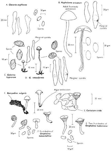
Larger illustration
(b) Mountain fungi and the so-called Basidiolichens
‘Basidiolichens.’
Plate 78.
Omphalina ericetorum (Fries) M. Lange has already been described (p. 232): it grows on acidic soils and ascends into mountain areas where it frequently grows on algal scum which accumulates around Sphagnum plants.
Under these conditions the algal cells enter the base of the fungus and grow in the cavity of the stem and amongst those hyphae which constitute the base. This association, however, appears to be much closer in the two lichens Coriscium viride (Acharius) Vain and Botrydina vulgaris Meneghini which have long been classified as species of lichen of unknown affinity because no perfect state was known. Coriscium viride consists of blue-green overlapping plates or scales with narrow rounded often paler margins and which dry out greenish brownish grey. Botrydina vulgaris, in contrast, consists of dark green, gelatinous blobs drying out greenish brown.
Coriscium is now considered to be an association of an algae and a Basidiomycete, the latter being the agaric, Omphalina hudsoniana (Jennings) Bigelow, which resembles O. ericetorum but for the pinkish coloured stem. Botrydina may be a complex of several separate associations of an algae with different species of Omphalina. In the high mountains the association is with O. luteovitellina (Pilát & Nannfeldt) M. Lange a small uniformly bright yellow agaric, whilst in Sphagnum bogs it is with O. sphagnicola (Berkeley) Moser. Myxomphalia maura (Fries) Hora, a fungus typical of burnt ground, is also reported to take up this association in lowland woods and O. velutina (Quélet) Quélet appears to be capable of forming a loose relationship with algal cells also. This is a most interesting association and research work is still at an early stage. In the tropics and subtropical regions of the world, similar associations are found on rotten and decomposing trunks and stumps. In these examples the Basidiomycetes are frequently fairy-clubs, particularly species of Multiclavula (‘many small clubs’). A few species of this genus may be found also in North temperate woodlands. Botrydina also grows in Europe with Stereum fasciatum (Schw.) Fries and Athelia viride (Bres.) Parm. (see p. 176), and Odontia bicolor (Fries) Quélet is rarely collected without green algal cells buried in the thallus. Perhaps associations like this are much commoner than at first supposed. Probably the most remarkable of this group of poorly known organisms is Cora pavonia (Sw.) Fries which produces masses of interlocking fans; it is tropical and found in Brazil.
Mountain fungi: general remarks
There are several groups of mountain fungi, some mycorrhizal formers, some which prefer peaty soil and some which are associated with algae forming a loose relationship—the Basidiolichens. When the mountain top is covered with such dwarf willows as Salix herbacea or S. reticulata the leaves are cast each year, woody tissue develops above and below the ground; in fact all the processes taking place in our familiar woodlands are also taking place in these communities, the only difference being that the trees are dwarf. Indeed it looks quite odd to see normal sized agarics growing amongst the woody stalks of dwarf trees, the leaves of which are often one-tenth the size of the fruit-bodies, but this is what happens.
The mycorrhizal formers in these conditions include species of Russula (e.g. Russula alpina Möller & Schaeffer, R. xerampelina var. pascua Favre (see p. 45)), Lactarius (e.g. Lactarius lacunarum Hora see p. 50), Cortinarius (e.g. C. anomalus (Fries) Fries see p. 42) and Amanita (e.g. Amanita nivalis Greville see p. 56). Subterranean fungi are also found, e.g. Elaphomyces see p. 244, and, just as woodlands, valley bottoms have a saprophytic ground flora of toadstools so do the high mountain ‘woods’, and many familiar fungi of the lowerland areas are to be found there also, e.g. Mycena epipterygia (Fries) S. F. Gray, Mycena olivaceo-marginata (Massee) Massee (see p. 88.)
The barer tops of the mountains, where large areas of moss are only to be found, support species of Hygrocybe, e.g. H. lilacina (Laestadius) Moser and H. subviolacea (Peck) P. D. Orton & Watling (see p. 97).
In the moist atmosphere on the hills in western Scotland, woodland-like floras containing familiar flowering plants are found on the mountain sides often much higher than in central Scotland. It is in such communities that typical woodland fungi are also to be found, e.g. Nolanea cetrata (Fries) Kummer (see p. 101).
(vii) Sand-dune fungi
Inocybe dunensis P. D. Orton
Cap: width 27-75 mm. Stem: width 4-10 mm; length 35-80 mm.
Description:
Cap: convex then expanded, usually broadly umbonate, pale or dirty ochraceous paler at the margin, reddish brown at the centre, smooth, radially fibrillose towards the margin and sometimes showing the remains of a pale greyish buff veil.
Stem: equal with marginate or rounded bulb at the base, white or whitish, then becoming discoloured pinkish or brownish, powdered with white, at first, but finally silky.
Gills: free or narrowly adnate, subcrowded, whitish then clay-buff, finally snuff-brown with whitish edge.
Flesh: white or whitish, tinted ochraceous or dirty pinkish and with strong smell of rancid oil.
Spore-print: snuff-brown.
Spores: medium to long, ellipsoid-oblong, indistinctly nodulose or wavy-angular and 9-12 × 6-7 µm in size.
Facial cystidia: swollen, spindle-shaped with short, broad neck, thick-walled and crested with crystals.
Marginal cystidia: spindle-shaped and crested with crystals.
General Information: This fungus is often buried to half-way in the sand of slacks near dwarf willows (Salix spp.). Three other species of Inocybe grow in dune-slacks I. halophila Heim, I. serotina Peck and I. devoniensis P. D. Orton, but all differ in their spores being smooth and elongate-cylindric. Astrosporina, a name referring to the shape of the spore, has been considered a genus of agarics in its own right and to this group I. dunensis would belong. However, as the members show the same range of characters as those species with the smooth spores it seems unnecessary to split Inocybe into two. The cystidia in many species are unusual, being crested with a bundle of crystals which have been reported as being calcium oxalate, although even the simplest school-laboratory tests have been rarely applied to them (see p. 84).
Plate 79. Sand-dune fungi
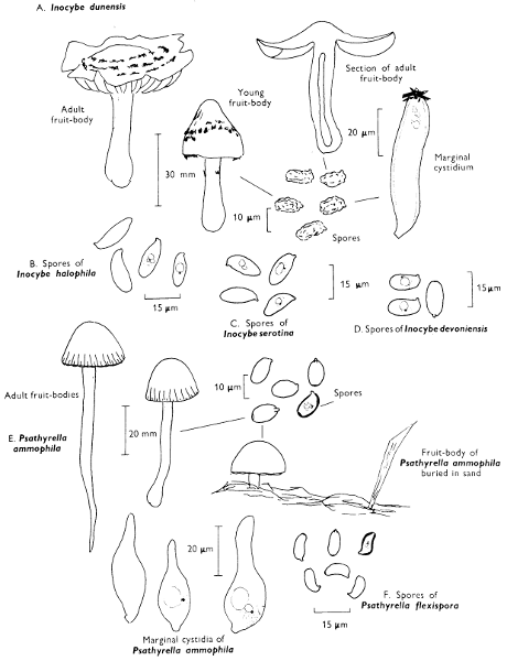
Larger illustration
Psathyrella ammophila (Durieu & Léville) P. D. Orton Sand-dune brittle-cap
Cap: width 20-40 mm. Stem: width 4-8 mm; length 40-80 mm.
Description: Plate 79.
Cap: semiglobate to convex, pale dingy clay-colour or dark tan to dirty brownish, non-striate, rather fleshy and usually sand covered.
Stem: deeply rooting in sand and club-shaped towards the base, similarly coloured to the cap except for the whitish apex.
Gills: adnate, subfuscous or dark dirt-brown.
Flesh: dirty buff and with no distinct smell.
Spore-print: pale snuff-brown with purplish flush.
Spores: long, ovoid, yellowish-grey brown under the microscope with a distinct germ-pore and 10-12 × 7 µm in size.
Marginal cystidia: balloon-shaped, obtuse or somewhat bottle-shaped and hyaline.
Facial cystidia: sparse, similar to the marginal cystidia, voluminous.
General Information: This is a very distinct fungus found amongst stems of Marram grass in sand-dune systems. At first sight it appears as if it is growing in the bare sand, but by careful excavation it usually is found attached to pieces of Marram grass, indeed the hyphae enter the roots of the grass, but apparently do not kill them.
This fungus was first described in the genus Psilocybe (see p. 114) because of its brownish purple spore-print, but the cap-surface is composed of rounded cells and so is related to all the other species of Psathyrella.
Psathyrella flexispora Wallace & P. D. Orton grows in similar habitats amongst Ammophila and other seashore grasses. It is easily recognised by the chocolate, umber or date-brown cap and the peculiar shaped spores, which look as if they have been slightly twisted during their development.
Stropharia coronilla (Fries) Quélet
, resembling a little mushroom (i.e. Agaricus) is also found in sand-dune systems and, just as species of Psathyrella, it possesses purplish black spores. However, the cap is ochraceous yellow with a whitish margin formed of veil fragments. The stem is white becoming yellow with age and possesses a narrow, white striate ring. The spores are ellipsoid and measure 8-9 × 4-5 µm and it has filamentous cells in the cap. Unlike P. ammophila it is not confined to sand-dune systems but it is also to be found in pastures and on heaths.
Plate 80. Sand-dune fungi
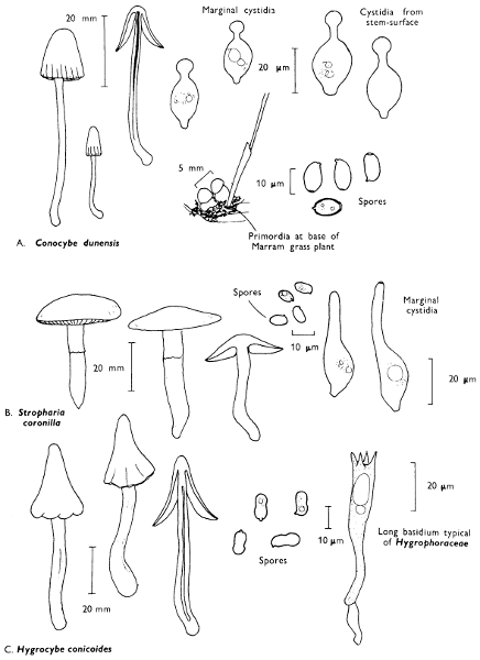
Larger illustration
Conocybe dunensis P. D. Orton Sand-dune brown cone cap.
Cap: width 10-30 mm. Stem: width 2-4 mm; length 40-100 mm.
Description: Plate 80.
Cap: conical then conico-expanded, date-brown, dull sand-colour or dark liver-colour, drying buff or ochraceous, expallent, not or indistinctly striate when moist.
Stem: whitish or pale ochraceous then darker ochraceous or dirty brownish from the base up, lower part whitish and buried in the sand.
Flesh: thin and pale ochraceous.
Gills: adnate, whitish but soon pale honey and finally rusty honey.
Spore-print: rust-brown.
Spores: long, ellipsoid or slightly amygdaliform, golden brown under the microscope with large germ-pore and 12-14 × 7-8 µm in size.
Facial cystidia: absent.
Marginal cystidia: capitate.
General Information: C. dunensis differs from C. tenera in its dull colours (see p. 116) and habitat preferences. Conocybe dunensis, Stropharia coronilla, the two species of Psathyrella are all dull-coloured. However, in the sand-dunes colourful agarics are also found. The most common is Hygrocybe conicoides (P. D. Orton) Orton & Watling; Laccaria maritima (Theodowicz) Moser is indeed an unusual but rewarding find. ‘Lac’ as in Laccaria is a red-brown resinous substrate produced by the lac-insect and resembles the cap colour of many species of the genus, including L. maritima, L. laccata and L. proxima (see p. 86). All these fungi were formerly placed in Clitocybe, but they differ in the warted or spiny spores which at maturity give the rather thick gills the appearance of being heavily talced. L. maritima can be distinguished from all other species of Laccaria by the elongated spores which are minutely spiny and not strongly warted as in L. laccata. Hygrocybe conicoides (P. D. Orton) Orton & Watling has a conical to conico-convex, acutely umbonate cap with wavy-lobed margin; it is scarlet or cherry-red, discolouring blackish with age or on bruising. The gills are at first chrome-yellow then become flushed red and the stem is yellow or greenish lemon becoming streaky blackish after handling. The spores are 10-13 × 4-5 µm in size and slightly French bean-shaped. It can be readily distinguished from close relatives, e.g. H. conica (Fries) Kummer by the gills soon turning reddish, the reddish cap and the narrow spores.
(viii) Subterranean fungi
General notes
The adaptive habit of growing completely submerged beneath the surface of the ground has developed in all the major groups of fungi. Thus the simplest form related to the common bread-mould have taken up the character just as certain relatives of the disc-fungi (discomycetes) and of the flask-fungi (pyrenomycetes). In the higher fungi in several foreign countries even agarics, polypores and stinkhorns have become hypogeous, but in this country we have a very depauparate flora composed of some twenty-eight species of false (Basidiomycete) truffle. The following key may assist in identifying the different groups of hypogeous fungi for some of these species are of commercial value and includes the French or Perigord truffle, Tuber melanospermum Vittadini which is used as a constituent of Pâté de Foie Gras, and many of the fungi used as poor quality substitutes. There is a long folk-history surrounding truffles and they have been utilised in the production of aphrodisiacs for centuries. Seeking them out was a difficulty and has been overcome in different countries in different ways. Thus in continental Europe, pigs have been used to sniff them out but on finding them the pigs cannot eat the truffles because of a ring placed through their nose. In Dorset a particular breed of dog was developed to do the same job—the Dorset hounds.
A simple key would read as follows:—
1. Spores produced on basidia 2
Spores produced in asci 4
2. Chambers throughout the inner tissue containing spores of approximately the same age 3
Chambers in the inner tissues containing spores found at different stages of development Hymenogaster
3. Basidiospores brown or greenish brown under the microscope, and black in mass Melanogaster
Basidiospores colourless or pale honey colour under the microscope and ochraceous in mass Rhizopogon
4. Asci globose, irregularly arranged within the fruit-body and quickly breaking down to shed the spores Elaphomyces
Asci globose or club-shaped and arranged in fertile areas which do not rapidly break down to shed the spores Tuber & relatives
Basidiomycetes
Rhizopogon roseolus (Corda) Fries Red truffle
Description:
Fruit-body: globular to tubiform and up to 60 mm broad, partly covered in mycelial cords, dirty white, later reddish-tawny gradually reddish and finally olive-brown, it soon becomes tawny on bruising when fresh and young.
Spores: medium sized, narrowly ellipsoid, smooth at first, hyaline then pale olive under the microscope and measuring 8-11 × 4 µm.
Habitat & Distribution: This fungus is not uncommon on the edges of paths, in pine woods just pushing up through the soil surface.
Ascomycetes
Elaphomyces granulatus Fries Harts’ truffle
Description:
Fruit-body: globose to ovoid, 20-40 mm broad, pale ochraceous, covered in small pyramidal warts, and when it is cut it shows three layers, an outer thin yellowish zone, an inner thicker compact white zone and within this a purplish black area full of spores separated into chambers by bands of sterile white tissue; the first two zones make up the ‘rind’.
Spores: spherical, blackish brown, warty, 24-32 µm in diameter; eight contained in globose asci.
Habitat & Distribution: This fungus is not uncommon in the surface layers of pine woods at the junction of needle debris and mineral soil. E. muricatus Fries is similar, but differs in the marbled flecked interior.
Tuber aestivum Vittadini English truffle
Description:
Fruit-body: subglobose except for basal flattening, up to 80 mm broad, covered in 5-6-sided pyramidal scales, dark brown to violaceous, white then greyish brown within, separated by a network of veins radiating from the basal cavity.
Spores: very large, ellipsoid, light or yellowish brown and ornamented with a prominent network, borne in two’s and sixes in subglobose asci and variable in size, 20-40 × 15-30 µm.
Plate 81. Subterranean fungi and fungus-parasites
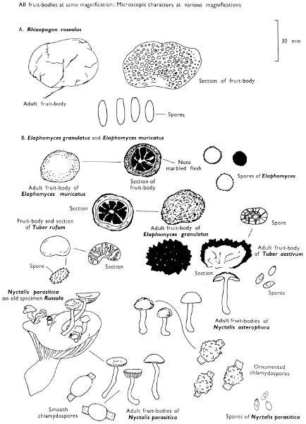
Larger illustration
Habitat & Distribution: This fungus is to be found buried in the surface layers of soil in beech woods. T. rufum is smaller and smoother and the spores are not crested but simply minutely spiny.
Illustrations: R. luteolus—Hvass 322; LH 215. El. granulatus—Hvass 325; LH 49. T. aestivum—LH 43. Melanogaster variegatus—LH 215 (see p. 243). Hymenogaster tener—LH 215 (see p. 243).
(ix) Fungal parasites
Nyctalis parasitica (Fries) Fries Pick-a-back-toadstool
Description: Plate 81.
Cap: bell-shaped then becoming expanded, silky dirty white, but gradually grey with a flush of lilac with age.
Stem: slender, white and smooth except for the base.
Gills: pallid but soon becoming brownish, adnate or adnate with tooth, thick and distant alternately long and short and contorted or united with age.
Flesh: dark brown.
Spore-print: buff.
Spores: small, hyaline under the microscope, ovoid, 5-6 × 3-4 µm but usually replaced completely or in part by ovoid, smooth, thick-walled and pale brownish asexually produced spores (chlamydospores) measuring about 15 × 10 µm in size.
Habitat & Distribution: This fungus grows in clusters on old decaying specimens of various species of Russula and Lactarius (Russulaceae)—see p. 45.
General Information: N. asterophora Fries is closely related and also grows on decaying specimens of various species of Russula, particularly R. nigricans (Fries) Fries. It differs, however, in the cap being fawn-coloured and very mealy when touched; it is recognised by the poorly formed often developmentally hindered gills on which chlamydospores are formed. Unlike the smooth asexual spores in N. parasitica this species has chlamydospores with conical, blunt humps—i.e. star-shaped; asterophora in fact means ‘I bear stars’. These fungi have been associated by some mycologists with the common chanterelle (Cantharellus cibarius Fries, see p. 162) in virtue of them possessing reduced fold-like gills. However, the fold-like gills are secondary in nature, correlated with the active production of chlamydospores and the suppression of the formation of basidiospores. The gills are not therefore of a primitive type. The genus Nyctalis is related to fungi such as Tephrocybe palustris (Peck) Donk (see p. 223).
There are several rather uncommon ‘agaric-parasites’ of agarics or other higher fungi, e.g. Volvariella surrecta (Knapp) Singer, but their formal description must be left to other more advanced texts. However, the intriguing bolete, Boletus parasiticus Fries, which grows on Scleroderma (earth-balls) in this country has been mentioned and figured previously (p. 35 & Plate 64). It is of interest to note that a close relative of B. parasiticus in Japan lives on another group of Gasteromycetes.
Illustrations: N. parasitica—F 11a; LH 81; WD 257. N. asterophora—LH 81; WD 258.
General notes on Fungicoles
Many beginners are confused on finding specimens which, although appearing agaric-like, are covered in long hairs or irregularly shaped bumps. Indeed many of these abnormalities are true agarics attacked by microscopic fungi, and I know of one textbook on mushrooms and toadstools which includes such an abnormality amongst the discussion on the normal fruit-bodies. Thus Sporadinia grandis Link, which is a primitive fungus, attacks many fungi reducing them to a grey velvety mass of fungal filaments. Specimens of several species of Mycena (p. 88) are common in autumn, covered in whiskers with small nobbles on the top. These whiskers are produced by the parasitic Spinellus megalocarpus (Corda) Karsten, another primitive fungus—a phycomycete.
In some wet seasons the orange and green coloured Lactarius deliciosus (Fries) S. F. Gray is to be found contorted and covered in small pinkish to lilac pimples of the ascomycete Byssonectria lactaria (Fries) Petch, and other species of Lactarius are attacked by Byssonectria viridis (Berkeley & Broome) Petch which converts the fruit-bodies into a hardened mass of green tissue. In North America, species of Lactarius are frequently attacked by Hypomyces lactifluorum (Schweintz) Tulasne and the whole fungus is reduced to a contorted acidic-smelling mass of fungal tissue with vivid orange pimples or warts on the outer surface. These parasitic fruit-bodies are eaten as a delicacy in their own right whereas the same consumer will be less enthusiastic about eating the same agaric before it is so deformed.
Boletes particularly B. subtomentosus Fries, B. chrysenteron St Amans and B. edulis Fries are frequently converted into yellow powdery masses due to the production of asexual spores of the fungus Sepedonium chrysospermum Fries; the sexual stage occurs on the remains after they have collapsed into the soil surface—this stage is called Apiocrea chrysosperma (Tulasne) Sydow. Several closely related fungi in the genus Hypomyces also attack agarics.
The yellow pustules found on the spore-bearing surface of the birch polypore Piptoporus (p. 142) is Hypocrea pulvinata Fuckel; it is only one of several lower fungi which grow on bracket fungi. The genus Cordyceps has been mentioned previously (p. 206) and in the discussion it was indicated that certain hypogeous fungi are attacked by members of this genus.
White gelatinous pustules found amongst the fruit-bodies of Stereum sanguinolentum (p. 176) have a hard white centre. On examination these ‘nuclei’ are aborted structures of the stereum covered in the jelly-fungus Tremella encephala Persoon. This fungus is apparently parasitic; it is closely related to Tremella foliacea and T. mesenterica described on page 184.