B. BRACKET-FUNGI AND THEIR RELATIVES
Reading edition
Key to major genera
A group of fungi which includes the bracket fungi, hedgehog fungi, fairy-clubs and their relatives; in the majority of species the margin continues to grow through the favourable part of the season and so often envelopes leaves, grass, etc.
1. Spore-bearing layer (hymenium) quite smooth, spread over veins or shallow pores; fruit-body top-shaped, fan-shaped or club-shaped, or spread over the substrate (resupinate) 2
Spore-bearing layer lining the inner surface of tubes or borne on warts or spines 17
2. Fruit-body club-shaped, coral-shaped or distinctly funnel-shaped, fan-like or resembling an agaric 3
Fruit-body resupinate or with poorly developed cap 11
3. Fruit-body coral-like or club-shaped with clubs grouped or branched 4
Fruit-body resembling an agaric or funnel-shaped to fan-shaped 9
4. Fruit-body large, branched with flattened and curled lobes and so resembling a cauliflower Sparassis
Fruit-body of single or grouped clubs or if branched then not resembling a cauliflower, the lobes being cylindrical or only slightly flattened and hardly bent 5
5. Fruit-body small arising from a seed-like structure or growing attached to dead herbaceous plant remains 6
Fruit-body medium to large, simple or branched and usually growing on the ground; one large species grows on wood 7
6. Fruit-body arising from a seed-like body embedded in the plant-tissue or found loose in the soil Typhula
Fruit-body on dead plant-remains but seed-like structure absent Pistillaria
7. Fruit-body much branched; spores ornamented (see also Thelephora below) Ramaria
Fruit-body simple or if with well-developed branches then spores smooth 8
8. Fruit-body branched irregularly with many to few branches, grey, white or drab-coloured; spores large, subglobose and smooth Clavulina
Fruit-body club-shaped or if branched then brightly coloured and spores not large and subglobose Clavaria, Clavulinopsis & Clavariadelphus
9. Fruit-body resembling an agaric with spores borne on fold-like, often forked and shallow ridges and veins, and often brightly coloured Cantharellus (compare carefully with Craterellus below)
Fruit-body funnel-shaped or fan-shaped 10
10. Fruit-body often drab colour or greyed with smooth or slightly veined outer surface Craterellus
Fruit-body wrinkled, irregular or smooth and powdery, lilaceous to chocolate-brown in colour Thelephora
11. Fruit-body sessile or resupinate and fleshy; spores borne on veins united to form shallow pores 12
Fruit-body resupinate or bracket-like, and spore-surface veined or rugulose but lacking distinct pores 13
12. Spores colourless Merulius
Spores brown Serpula
13. Spore-bearing layer containing long, brown spines Hymenochaete
Fruit-body lacking spines although often having encrusted sterile cells 14
14. Surface of fruit-body more or less radiately veined Phlebia
Surface of fruit-body not radiately veined 15
15. Spores brown Coniophora
Spores colourless 16
16. Flesh distinctly formed and fruit-body with or without a well formed cap Stereum & related genera
Flesh poorly differentiated and fruit-body lacking a cap members of the Corticiaceae (including Peniophora & Hyphodontia p. 176)
17. Spores borne on teeth or spines 18
Spore-bearing layer lining tubes or elongate pores 22
18. Fruit-body with central stem; agaric-like but not attached to cones 19
Fruit-body encrusting or bracket-like, or with lateral stem if resembling an agaric 20
19. Fruit-body fleshy Hydnum and related genera
Fruit-body rubbery or tough Hydnellum and related genera
20. Fruit-body growing attached to cones and cap with lateral stem Auriscalpium
Fruit-body not on cones and distinct stem lacking 21
21. Spores borne on a series of radially arranged knotches resembling gills Lentinellus
Spores borne on a resupinate layer of spines Mycoacia and related genera
22. Tubes free one from another Fistulina
Tubes united to form a distinct tissue 23
23. Fruit-body perennial and exhibiting more than one layer of tubes 24
Fruit-body annual although the fruit-body can persist in a dried depauperate form for several months 27
24. Spores brown 25
Spores colourless 26
25. Large, brown, sterile cells present in the tubes; spores simple Phellinus & Cryptoderma
Brown, sterile cells absent from tubes; spores complex Ganoderma
26. Large woody fruit-body with crust-like top Fomes
Medium sized to small, fleshy-tough fruit-body with downy or crust-like top Oxyporus, Fomitopsis & Heterobasidion
27. Spores borne in labyrinth-like or elongate pores, or cap either poorly developed or absent, and only resupinate pore-surface present 28
Spores borne in distinct pores on well-developed woody fruit-bodies 31
28. Spores borne in labyrinth-like pores Daedalea & Daedaleopsis
Spores borne in elongate pores like very thick gills, or fruit-body completely resupinate 29
29. Spore-layer lining elongate pores Lenzites (white) & Gloeophyllum (brown)
Spore-layer consisting of a resupinate pore-layer 30
30. Pore-layer totally resupinate; flesh very poorly developed Fibuloporia and related genera
Fruit-body resupinate or developing ill-formed caps at the margin; flesh well-developed and quite tough Datronia, Gloeoporus & Bjerkandera
31. Fruit-body with a distinct stem 32
Fruit-body sessile or with a poorly developed stem, or if merely with a basal swelling then pores bruising 33
32. Pores dark-coloured but spores pale-coloured in mass Coltricia (also see Phaeolus below)
Pores white or creamy, foot often darkened or black, and spores hyaline Polyporus
33. Pores brightly coloured, red, lilaceous or orange to apricot-colour 34
Pores never as brightly coloured, cream, white, grey or in some shade of brown 35
34. Pores red to orange-red Pycnoporus
Pores lilac to violaceous, or lilaceous orange to apricot colour Hapalopilus (orange-apricot) & Hirschioporus (lilaceous)
35. Pore-surface brown or dark grey and spores often colourless 36
Pore-surface white or creamy, or yellow; spores hyaline 38
36. Pore-surface firm and grey Bjerkandera
Pore-surface greenish yellow, bruising brown or yellow-brown and darkening with age 37
37. Fruit-body lacking a stem, rust-brown, breaking easily, cheesy and with silky sheen Inonotus
Fruit-body with a broad basal hump, fibrillose spongy with yellow margin to cap Phaeolus
38. Tubes forming a layer quite distinct from the flesh; fruit-body fleshy and tough 39
Tubes not forming a layer distinct from the flesh; fruit-body woody or corky 43
39. Pore-surface bright yellow; upper surface yellow or orange Laetiporus
Pore-surface white 40
40. Fruit-body medium to large, shell-shaped, whitish brown or silvery grey on top; on birch Piptoporus
Fruit-body often frond-like, infrequently shell-shaped and if on birch then small 41
41. Fruit-body fan- or frond-shaped, composed of innumerable more or less complete caps joined together at their base or to half-way Grifola & Meripilus
Fruit-body neither fan-shaped nor frond-shaped and compound 42
42. Fruit-body wholly pale-coloured white, cream, ivory, etc. Tyromyces
Fruit-body except pores usually some shade of brown Polyporus
43. Cap thick, corky or woody and pores medium or large Trametes & Pseudotrametes
Cap thin but leathery and pores small Coriolus
(i) Pored and toothed fungi
(a) Colonisers of tree trunks, stumps and branches
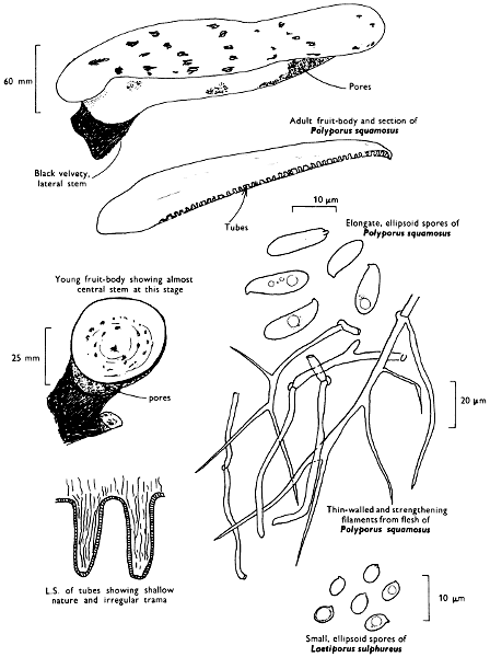
Polyporus squamosus Fries Scaly polypore
Cap: 100-300 mm. Stem: width 25-50 mm; length 25-75 mm.
Description:
Cap: fan-shaped or semicircular, spreading horizontally with age, ochre-yellow or straw-coloured with dark brown, flattened scales in concentric zones which are much more dense at the centre.
Stem: short, stout, white at apex and netted with pale creamy buff about middle, but dark brown or black towards the base and attached to the side of the cap.
Tubes: whitish to yellowish and decurrent with large, angular, irregularly fringed, whitish or cream-coloured pores.
Flesh: with strong, not very pleasant smell, cream-coloured or white.
Spore-print: white.
Spores: long, oblong or elongate ellipsoid, hyaline under the microscope (10-15 × 4-5 µm) and not blueing in solutions containing iodine.
Habitat & Distribution: An easily recognisable fungus growing on stumps and old living trees, especially of sycamore and elm where it often forms tiers of caps from late spring until autumn; however, they decompose rapidly and almost completely disappear by the next year when new fruit-bodies may appear in the same place, a phenomenon which may take place for several consecutive seasons.
General Information: The genus Polyporus is in most text-books, a big and unwieldy genus joining together all fleshy, annual fungi possessing tubes; even the boleti (see p. 32) have been included! Many of these species are now considered less closely related one to another than previously thought. Boleti differ from polypores, however, in their less tough and distinctly putrescent fruit-body, and in the fact that the margin of the cap extends but does not continue to grow during the life-cycle; the margin of the polypore fruit-body is active and may burst into growth again when favourable weather conditions occur. The ‘Scaly polypore’ has a flesh which consists of two types of hyphae: (i) hyphae of unlimited growth with abundant protoplasmic contents which stain easily and which collapse on drying; and (ii) thick-walled, strengthening hyphae which bind the thin walled hyphae together. Laetiporus sulphureus (Fries) Murrill ‘Sulphur polypore’ has a single type of hyphae in the tubes, i.e. thin walled generative, and only a few binding hyphae in the flesh. It has an orange cap with a rather thick, sulphur or chrome-yellow margin, sulphur-yellow tubes and pores and yellow, then pale buff, flesh. The spore-print is white and the spores hyaline, pip-shaped and medium sized, (5-7 × 4-5 µm).
Plate 44. Woody fungi: Spores white and borne within tubes—fruit-body annual
Larger illustration
Illustrations: P. squamosus—F 43b; Hvass 267; LH 75; NB 1291; WD 941. L. sulphureus—Hvass 268; LH 73; NB 1293; WD 942.
Some common annual polypores
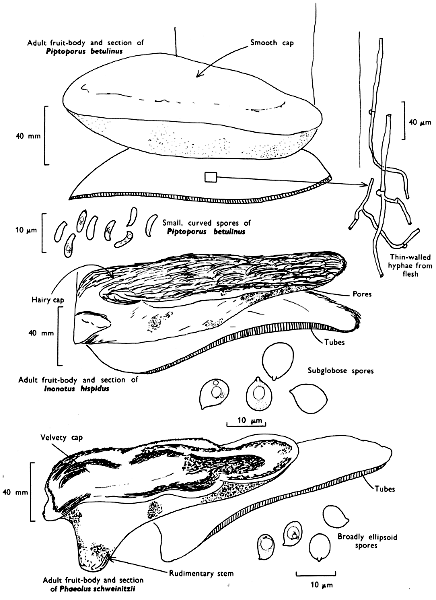
Piptoporus betulinus (Fries) Karsten Birch polypore
Cap: 75-200 mm, kidney-shaped or hoof-shaped, smooth, covered by a thin, separable and greyish silvery or pale brownish skin; cap-margin thick, incurved and projects beyond the tubes.
Stem: rudimentary, simply a small hump below which the fungus develops.
Tubes, pores and spore-print: white.
Spores: sausage-shaped, and thin-walled hyaline under the microscope and very narrow, (5-6 × 1-2 µm). It grows on birch throughout the country where it causes a sap wood-rot which finally converts the inner timber to a red-brown friable mass. The flesh, which contains thickened binding hyphae, is used for mounting insects and for sharpening knives, hence the common name ‘Razor-strop fungus’.
Illustrations: Hvass 269; LH 67; NB 1174; WD 933.
Inonotus hispidus (Fries) Karsten Shaggy polypore
Cap: 100-250 mm, kidney-shaped, yellow-brown to rust-brown, but finally almost black, at first covered with shaggy hairs, but these tend to mat together with age.
Stem: absent.
Tubes and flesh: rust-colour; pores at first yellow, but finally red-brown.
Spore-print: yellow-brown.
Spores: medium sized (8-9 × 7-8 µm) and globose under the microscope. It grows on various broad leaved trees, especially ash where it causes a spongy, white heart-wood rot. The flesh contains hyphae with thick, brown walls.
Illustrations: LH 63; WD 961.
Plate 45. Woody fungi—annual polypores
Larger illustration
Phaeolus schweinitzii (Fries) Patouillard
Cap: 100-300 mm, bracket-shaped or tub-shaped, dark brown with a knobbly, velvety, roughened and grooved surface; margin at first golden yellow.
Stem: absent or short, thick and brown.
Tubes and pores: greenish yellow.
Flesh: deep rust-brown.
Spore-print: greenish yellow.
Spores: medium sized, greenish under the microscope, ellipsoid and about 8 × 4 µm in size, (7-8 × 3-4 µm). This fungus is found on conifers or near conifer stumps where it is attached to the roots; it causes a brown cubical heart-wood rot; the flesh of the fruit-body is composed of only one type of hyphae.
Illustrations: LH 67; NB 1113; WD 951.
Meripilus giganteus (Fries) Karsten Giant polypore
Cap: 75-100 mm, or even up to 200 mm wide, grouped and forming a tuft of caps up to 750 mm across. The individual caps are fan-shaped, pliable, rather thin and yellow-brown to snuff-brown with their margins wavy and cream colour or yellowish.
Stem: replaced by a united mass of caps.
Tubes, pores and flesh: white and very soft, but becoming black on bruising.
Spores: small, pip-shaped, hyaline under the microscope and 5-6 × 4-5 µm. This fungus is a common sight forming masses at the base of broad-leaved trees; it is common on beech. It is a soft, fibrous polypore as a result of the lack in the flesh of thick-walled specialised hyphae.
Illustrations: Hvass 277; LH 73; NB 1294; WD 931.
The spores of all the annual polypores described above do not blue when placed in solutions containing iodine.
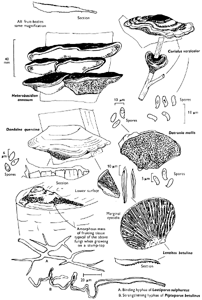
Coriolus versicolor (Fries) Quélet Many zoned polypore
Cap: 25-60 mm. Stem: absent.
Description: Plate 46.
Cap: semi-circular, flattened, thin, tough and flexible when fresh with the surface velvety and marked with smoother, paler concentric zones giving a pattern of yellow-brown, grey or darker greenish grey zones; the margin is thin and is the palest of the zones and may be wavy or lobed.
Tubes: white with small, round and rough-edged to angular white or cream-coloured pores which become yellowish with age.
Flesh: white, tough and continuous with the tube tissue and so not allowing one to detect any difference between the tissues.
Spore-print: white.
Spores: medium sized, oblong and hyaline under the microscope, and 6-8 × 2-3 µm; not blueing in solutions containing iodine.
Habitat & Distribution: Very common on stumps, trunks and fallen branches of various trees, especially beech; it is to be found throughout the year.
General Information: It is often associated with nodulose masses of fungal tissue which are covered in small poroid areas and are very confusing when found by the beginner; they are simply growth-forms of Coriolus versicolor; such forms are frequently found on old house-timbers exposed to the weather, particularly window frames where it forms a distinct rot. Its flesh consists of thin-walled hyphae and binding hyphae as in Polyporus squamosus as well as an additional thick-walled type called skeletal hyphae. It would appear that several polypores are capable of producing the amorphous growths mentioned above, some of which contain hyphal fragments called conidia.
The bands of colour on the cap of the ‘many zoned polypore’ are retained after drying and from a group of fruit-bodies the most attractively zoned can be selected, mounted on small pieces of wood or cardboard and fitted at the back with a pin. Such preparations make very attractive brooches and have been used even by modern designers to contrast with their fashion creations.
There are many pale tubed polypores growing on wood. Daedalea quercina Fries ‘Mazegill’, grows on oak and has irregular maze-like pores; Lenzites betulina (Fries) Fries, grows on birch, has tough plates which resemble the gills of an agaric. Datronia mollis (Fries) Donk forms thick spreading resupinate patches on beech, sometimes with irregular dark brown caps formed by the upturned margin. Several species of Tyromyces occur in Britain and are characterised by their white pores and tubes and the white or pale-coloured caps. Bjerkandera adusta (Fries) Karsten has a grey pore-surface and is also frequently found on beech.
Illustrations: F 44a; LH 69; NB 1173; WD 512.
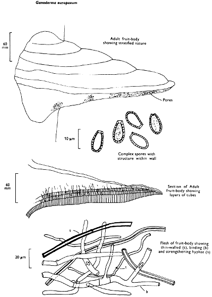
Ganoderma europaeum Steyaert Common ganoderma
Cap: 100-350 mm. Stem: absent.
Description: Plate 47.
Cap: bracket-shaped, rather flat at margin but humpy and irregular about the middle, frequently concentrically zoned, smooth and only slightly shiny; its margin is whitish or pale greyish.
Tubes: red-brown or cinnamon-brown, obscurely layered and with small, white pores flushed with pale cinnamon-brown, but deep red-brown when rubbed or with age.
Flesh: with a fragrant smell, deep red brown and felty-fibrous.
Spore-print: dark cinnamon-brown.
Spores: long, oval with truncate apex, smooth, but reticulate on the inner surface of the inner wall giving the spores a patterned appearance when seen under the microscope; 10-11 × 6-7 µm in size.
Habitat & Distribution: This fungus is common on various trees, especially beech and can be found throughout the year.
General Information: This common Ganoderma is perennial and distinguished from other polypore groups by the complex spores. G. applanatum (Fries) Karsten is closely related, but differs in the thinner fruit-body with a thin margin, and the pale cinnamon-brown flesh; the flesh of both species contains thick-walled binding and strengthening hyphae as well as the generative hyphae.
So sensitive are the pores to bruising that if a drawing or writing is executed on the lower surface with a pin, needle or similar sharp instrument and the fungus dried, the red-brown lines produced are retained and the pattern preserved. Several fungus paintings prepared in this way were made in the early part of the century, many beautiful ones having originated in the eastern part of North America.
Plate 46. Woody fungi: Spores white and borne within tubes or on thickened plates
Larger illustration
Fomes fomentarius whose important characters are described below has frequently been confused with Ganoderma europaeum. It is common growing on birch in Scotland, but is less frequent south of Perth, and then grows probably more frequently on beech which is similar to the pattern found on the continent of Europe. However, it has grown in former periods in England on birch, for it was found commonly amongst birch timbers in an excavation of an early Mesolithic lake side village near Scarborough, Yorkshire.
Illustrations: NB 1253; WD 1602.
Some perennial polypores. Plate 48.
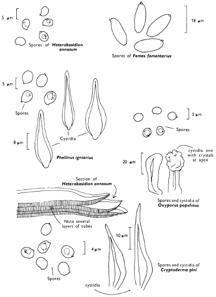
Fomes fomentarius (Fries) Kickx Tinder fungus
Cap: 90-300 mm, hoof-shaped, thick, broadly attached to the substrate, zoned with yellow-brown and shades of grey; its margin is blunt and fawn or pale brownish.
Tubes: layered, cinnamon-brown with pale cinnamon pores with a whitish bloom.
Flesh: cinnamon-brown or buff and woolly.
Spore-print: white.
Spores: elongate, ellipsoid, very long, hyaline under the microscope, 15-18 × 5-6 µm, and not ornamented. The flesh contains both thick- and thin-walled hyphae. It grows on birch and less frequently on beech. The flesh has been used in dentistry, in manufacturing fancy articles, such as mats, and was the basis of the tinder used in flint-boxes.
Illustrations: LH 65; NB 1171; WD 1001.
Phellinus igniarius (Fries) Quélet ‘Willow Fomes’
, grows on willows and causes their heart-rot. It is a rust-brown, woody fungus with a hard crust and brown tubes and flesh. The spore-print is white and composed of small, spherical, hyaline spores, 5-6 µm in diameter. The flesh contains thin- and thick-walled hyphae.
Illustrations: LH 63; WD 993.
Plate 47. Woody Fungi: Spores brown and borne within tubes—fruit-body perennial
Larger illustration
Oxyporus populinus (Fries) Donk
, grows on various sorts of broad-leaved trees, particularly poplars and often becomes covered in mosses and algae. It has a pale buff or cream-coloured cap, white flesh, pores, tubes and spores.
Illustrations: LH 67.
Cryptoderma pini (Fries) Imaz
, grows on conifers often several feet above the ground. It has a woody, deeply cracked upper surface, dark red-brown flesh, tubes and pores. Its spores are small, broadly ellipsoid and brown.
Heterobasidion annosum (Fries) Brefeld Root fomes
Variable, sometimes possessing a cap, sometimes resupinate except for the upturned margin, flattened or shell-shaped, red-brown to blackish at the centre but pale at the margin, which when seen from below is always white. The tubes are in layers and like the pores, flesh and spore-print are white. The spores are broadly ellipsoid, small, smooth, hyaline and 4-5 × 4 µm. The flesh is fairly tough as it contains both generative hyphae and skeletal hyphae. It is frequent on the roots and lower parts of stems of many trees and shrubs causing a rapid heart-rot of conifers and extensive damage to young trees.
Illustrations: LH 67; NB 1111; WD 981.
The spores of all the perennial polypores described above do not blue when placed in solutions containing iodine.
Plate 48. Woody fungi: Spores borne within tubes—perennial polypores
Larger illustration
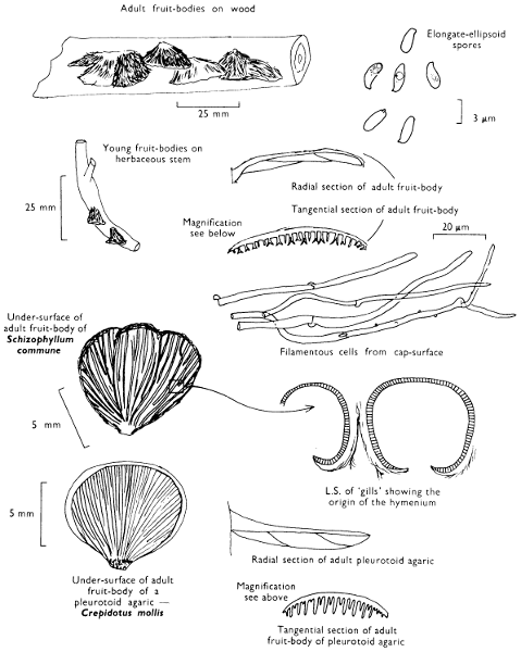
Schizophyllum commune Fries Split-gill fungus
Cap: 10-25 mm. Stem: width 2-4 mm; length 2-4 mm.
Description:
Cap: greyish fawn becoming whitish when dry, fan or kidney-shaped, often lobed and covered in close-set hairs and with incurved margin.
Stem: absent or the cap simply narrows into a stem-like bump.
Gills: replaced by a series of grey-brown plates which when dry appear as if to split longitudinally and their edges roll back.
Flesh: brownish but drying whitish.
Spore-print: white.
Spores: medium sized, oblong, hyaline under the microscope, not blueing in solutions containing iodine and 6-7 × 2-5 µm in size.
Facial and marginal cystidia: absent.
Habitat & Distribution: Grows on fallen branches, trunks, dead wood, etc.
General Information: Easily recognised by the ‘gills’ radiating from a point and becoming ‘split’ when dry. Specimens of Schizophyllum sealed by A. H. R. Buller in a tube in 1911 have been shown on remoistening to unroll their gills and shed variable spores, after 521⁄2 years—probably a world record! The split-gill is a rather unique British fungus which appears to be much more closely related to the polypores than to the agarics—although it has for a long time been associated with the Oyster mushroom (p. 74). In fact, the splitting gills are two adjacent shallow dishes with spores produced on their inner surfaces. The cups separate on drying and therefore only superficially resemble gills splitting down the centre.
Another fungus which can also be associated with the idea of cups is Fistulina hepatica Fries ‘the Beef-steak fungus’. This fungus is a polypore in the widest sense. It may grow up to 250 mm wide and is reddish-brown or liver-coloured with reddish tubes and pale flesh-coloured pores; the tubes although free are aggregated together and can be easily separated individually with the fingers. This fungus is edible although very strong in taste, it produces a serious decay of oaks.
Illustrations: S. commune—LH 105; NB 1256; WD 693. F. hepatica—F 43a (lower figure); Hvass 278; LH 75; NB 1292; WD 1014.
Plate 49. Woody fungi: Spores white and borne on split-‘gills’
Larger illustration
(b) Destroyers of timber in buildings
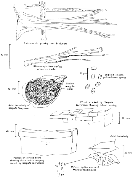
Serpula lacrymans (Fries) Karsten Dry-rot fungus
Description:
Fruit-body: usually widely spreading, but sometimes forming a distinct bracket with the upper surface silvery or smokey grey, flushed with lilac or rose or yellowish.
Stem: absent and replaced by a series of dirty white or greyish mycelial threads or strands which can be traced up to 100 mm over the substrate.
Flesh: thin, dirty yellowish and composed of only one type of hypha.
Spores: borne in shallow pores which are part of a complicated network of rust-brown folds and ridges.
Spore-print: rust-brown.
Spores: medium sized, golden yellow, thick-walled and broadly ellipsoid, and 8-10 × 5-6 µm in size.
Cystidia: absent.
Habitat & Distribution: On worked wood in buildings and less commonly in timber-yards. It can be found throughout the year.
General Information: This fungus forms fan-like structures and strands of mycelium which pass along beams and joists and through plaster. Where there is a bad case of dry-rot, the room or building will have an unpleasant musty smell and when actually growing the fungus exudes droplets of water on the mycelium and fruit-body, i.e. weeping, hence the name ‘lacrymans’—weepy. It is a very important and destructive agent causing damage to floors and skirting boards, to joists and beams. It is a frequent pest of old houses and therefore of many of our cities. This fungus does not appear to have been found in the wild in Europe, but there is a record from the Himalayas. There are, however, very closely related species found on soil or wood-detritus. The Dry-rot fungus darkens the wood and produces a rot which makes the wood crack into small cubes or rectangular blocks.
This fungus was formerly placed in Merulius, but this genus should be retained for hyaline-spored fungi, e.g. M. tremellosus Fries, a species which grows even in winter on stumps of various trees in our woods.
Illustrations: LH 53; WD 1033.
Plate 50. Dry-rot fungi—leathery and tough spores borne in shallow irregular pores
Larger illustration
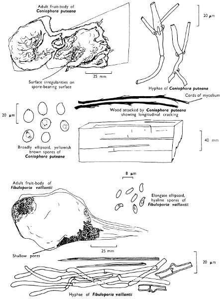
Coniophora puteana (Fries) Karsten Cellar or Wet-rot fungus
Description:
Fruit-body: variable in size, resupinate, composed of one type of hypha only and with a sterile whitish cream or yellow margin.
Spore-bearing tissue: an irregularly wrinkled or humpy, yellowish surface which then becomes olive-green or bronze-colour.
Spore-print: olivaceous brown.
Spores: olive-brown under the microscope, smooth, ellipsoid, thick-walled and 12-14 × 8-9 µm in size.
Cystidia: absent.
Habitat & Distribution: This fungus causes wet-rot in houses, but may also be found on stumps and fallen trunks in woodland.
General Information: The fungus causes a discolouration of worked timber and induces longitudinal cracking with only a few lateral hair-like cracks unlike timber attacked by the dry-rot fungus (see p. 154).
Illustrations: WD 1035.
Fibuloporia vaillantii (Fries) Bondarsev & Singer
Description:
Fruit-body: a resupinate layer of pores with cream-coloured or white sterile radiating margin.
Spore-bearing tissue: distributed within a series of small often shallow, white or ivory tubes.
Spore-print: white.
Spores: smooth, hyaline under the microscope, oblong 5-7 × 3-4 µm.
Cystidia: absent.
Habitat & Distribution: The dry-rot of houses, particularly in roof-systems.
General Information: Fibuloporia vaillantii is recognised by the white, resupinate pore-surface and fairly tough nature due to the presence of strengthening hyphae. Just as the genus Polyporus was found to be composed of several quite different elements (see pp. 140-44) and has since been split up into a number of different genera, the genus Poria has also been fragmented; one of the constituent genera is Fibuloporia. Amyloporia xantha (Fries) Bondarsev & Singer differs in having amyloid tissue and cystidia encrusted with crystals. The flesh contains both simple hyphae and thickened structural hyphae. It is yet another member of the large old unwieldy genus Poria and causes decay of worked wood, particularly the timbers of benching and staging in greenhouses. A. xantha has a sulphur-yellow pore-surface and is rather cheesy when handled.
Plate 51. Wet and Dry-rot fungi—leathery and tough and spores borne within shallow pores or on an uneven surface
Larger illustration
(c) Colonisers of cones
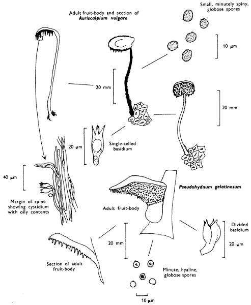
Auriscalpium vulgare S. F. Gray Ear-pick fungus
Cap: 8-12 mm. Stem: width 4-6 mm; length 40-75 mm.
Description:
Cap: kidney-shaped or semicircular, thin, date- or umber-brown, hairy, but paler towards the margin.
Stem: erect, slender, hairy, particularly at the base, and attached at the side of the cap (excentric).
Gills: replaced by flesh-coloured, then greyish brown spines.
Flesh: brown.
Spore-print: white.
Spores: small, hyaline, minutely spiny, spherical, 4-5 µm in diameter, and becoming blue-grey in solutions containing iodine.
Cystidia: flask-shaped with oily contents.
Habitat & Distribution: This fungus is always found on fallen pine-cones and occurs from early summer to autumn.
General Information: The ear-pick fungus is easily recognised by the slender, elegant habit, excentrically placed cap, substrate preference and dark colours. It cannot be confused with any other fungus. Recently it has been shown that the ‘agaric’ Lentinellus cochleatus (Fries) Karsten (p. 76) is more closely related to Auriscalpium than this fungus is to other spine-bearing forms and Lentinellus is to the other agarics. Both fungi possess thick-walled cells in the flesh and oil-containing hyphae; they are placed in the family Auriscalpiaceae.
Another laterally stemmed Hedgehog fungus differs in possessing distinctly gelatinised teeth and preference for conifer wood and not cones. Examination of the basidia of this fungus shows that it is more closely related to the jelly-fungi, Exidia and Tremella (p. 184) than to Hedgehog fungi such as Auriscalpium or Hyndum repandum Fries (p. 160). This false nature is reflected in the name of the genus to which it belongs, Pseudohydnum, and the very gelatinous texture in the specific name ‘gelatinosum’: the fungus is Pseudohydnum gelatinosum, or as it used to be called Tremellodon gelatinosum.
Illustrations: Auriscalpium vulgare—WD 1036. Pseudohydnum gelatinosum—WD 1059.
Plate 52. Tough or leathery fungi: Spores white and borne on spines—Ear pick fungus
Larger illustration
(d) Terrestrial forms
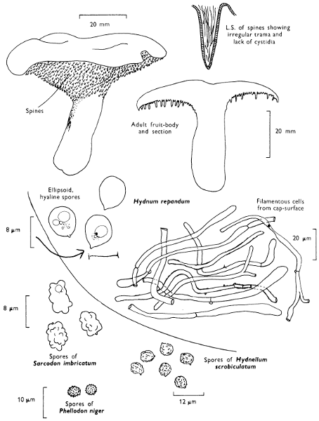
Hydnum repandum Fries Wood-hedgehog
Cap: 50-75 mm width. Stem: width 10-17 mm; length 45-65 mm.
Description:
Cap: rather thick, fleshy, pinkish buff or tan, paler at its incurved and often lobed margin.
Stem: short, stout and powdered with white roughenings and often attached to the cap to one side of the centre.
Gills: replaced by awl-shaped, pinkish buff spines which are unequal in length and run down the top of the stem.
Flesh: white, firm and with a pleasant smell.
Spore-print: whitish.
Spores: medium sized, hyaline under the microscope, smooth, broadly ellipsoid, 7 × 6-7 µm, and not becoming bluish grey in solutions containing iodine.
Cystidia: absent.
Habitat & Distribution: The ‘wood-hedgehog’ grows on the ground in mixed woods and is easily recognised by its colour and fleshy texture.
General Information: The closely related, smaller, red-brown species H. rufescens Persoon grows with conifers. Hydnum was formerly a genus which contained several entities, now not considered closely related. Thus the following genera have been delimited in addition to those related to Hydnum repandum and H. rufescens, and Auriscalpium described on p. 158.
Sarcodon: Fruit-body fleshy: spores brown and ornamented with irregular bumps, e.g. S. imbricatum (Fries) Karsten.
Phellodon: Fruit-body tough and fibrous: spores white and ornamented with small spines, e.g. P. niger (Fries) Karsten.
Hydnellum: Fruit-body tough and fibrous: spores brown and ornamented with irregular bumps and bosses, e.g. H. scrobiculatum (Secretan) Karsten.
Bankera: Fruit-body fleshy: spores white and ornamented with small spines, e.g. B. fuliginoalbum (Fries) Pouzar.
Illustrations: Hvass 280; LH 61; NB 1533; WD 534; Z 61.
Plate 53. Tough or leathery fungi: Spores whitish and borne on spines
Larger illustration
(ii) Chanterelles and relatives
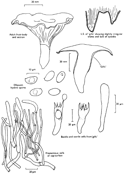
Cantharellus cibarius Fries Chanterelle
Cap: 30-100 mm. Stem: width 15-25 mm; length 30-70 mm.
Description:
Cap: convex then flattened, irregularly wavy, more or less top-shaped, depressed and smooth or slightly roughened at centre, egg-yellow or lemon-chrome with flush of orange and with the margin incurved at first.
Stem: short, stout, tapered downwards, fleshy and similarly coloured to the cap.
Gills: replaced by irregularly branched yellow folds which may form a network near the margin and at the apex of the stem over which the folds run down irregularly (decurrent).
Flesh: with pleasant, fruity smell, yellow but paler on drying.
Spore-print: pale cream-colour.
Spores: medium sized, ellipsoid, thin-walled, smooth, 8-10 × 5-6 µm in size and not becoming bluish grey in solutions containing iodine.
Marginal and facial cystidia: absent.
Basidia: 2-8 spored.
Habitat & Distribution: Very common in troops in deciduous woods especially those with beech and oak.
General Information: Easily recognised by its folds and absence of true gills beneath the cap and the overall yellow colour. This fungus is the edible chanterelle of the continental market, where it is considered of very high quality; it can be purchased in this country in tins. C. friesii Quélet is of a bright apricot colour with lilaceous or rose-coloured flesh. The ‘false chanterelle’ Hygrophoropsis aurantiaca (Fries) Maire already discussed (see p. 106) has true gills and is reddish orange in colour.
Illustrations: Hvass 182; LH 59; NB 1232; WD 831.
Plate 54. Fleshy but firm fungi: Spores pale-coloured and borne on irregular folds (False gills)
Larger illustration
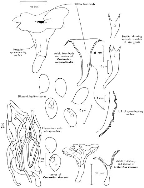
Craterellus cornucopioides (Fries) Persoon Horn of plenty
Cap: 22-80 mm. Stem: width 15-25 mm; length 25-80 mm.
Description:
Cap: funnel-shaped, membranous to leathery, but limp, dark brown or almost black in wet weather, but on drying becoming dull brown or sepia, slightly scaly and with irregularly wavy margin.
Stem: short, blackish and hollow.
Gills: absent, replaced by a smooth to irregularly wrinkled, ash-grey surface.
Flesh: sepia but drying out greyish ochre.
Spore-print: cream-colour.
Spores: medium sized, hyaline under the microscope, ellipsoid, smooth, 10-11 × 6-7 µm in size and not blueing in solutions containing iodine.
Marginal and facial cystidia: absent.
Basidia: usually 2-spored.
Habitat & Distribution: Often in very large troops in woods, especially under beech.
General Information: This fungus is recognised by the peculiar shape and dull colours which conceal it so well amongst the dead leaves and woodland debris; in the shade of the tree-canopy it is easily overlooked. Craterellus sinuosus (Fries) Fries is a much smaller species with dirty ochraceous fertile surface and brownish grey cap and stem.
‘Cornucopioides’ means like (oides) a horn of plenty, a familiar object in mediaeval paintings as part of heathen festivities full and overflowing either with fruit or wine, or both!
Illustrations: Hvass 186; LH 59; NB 1231; WD 834.
Plate 55. Fleshy but leathery fungi: Spores pale-coloured and borne on irregular wrinkles
Larger illustration
(iii) Fairy-club fungi
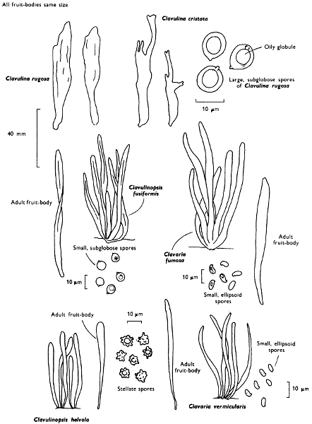
Clavulina rugosa (Fries) Schroeter Wrinkled club
Cap: absent. Fruit-body: length 50-100 mm; width 7-13 mm.
Description:
Fruit-body: club-shaped, simple with blunt apex or irregular blunt branches, white or dirty cream colour, often thickened upwards and marked with longitudinal wrinkles or grooves and the whole surface of the club bearing spores.
Stem: absent or extremely short.
Flesh: white.
Spore-print: white.
Spore: medium sized, broadly ellipsoid to subglobose, hyaline under the microscope and not turning bluish grey in iodine solutions, 9-10 × 7-8 µm in size.
Cystidia: absent.
Basidia: 2-spored.
Habitat & Distribution: Frequent on the ground in woods, especially in the shade of beech trees or in conifer plantations.
General Information: Two very closely related species are to be found in similar localities and are equally as common; they are C. cristata (Fries) Schroeter with strongly branched white fruit-body, each branch ending in pinkish or lavender-white, divided, sharply pointed branchlets and C. cinerea (Fries) Schroeter with irregular greyish or dark grey branches with a flush of violaceous.
These three species are very closely related; in fact so many intermediates between the extreme morphological forms are known that some authorities have considered them simply forms of a single species. All these species lack cystidia.
rugosa—wrinkled, referring to the spore-bearing surface.
cristata—crested, referring to the branchlets.
cinerea—ash-grey, referring to the colour.
All these species are often found blackened by the growth of the microscopic fungus, Helminthosphaeria clavariae (Tulasne) Fuckel.
Illustrations: C. rugosa—LH 55; WD 1045. C. cristata—LH 55; NB 1535; WD 1042. C. cinerea—WD 1041.
Plate 56. Fleshy but firm fungi: Spores pale-coloured and borne on club-shaped fruit-bodies
Larger illustration
Clavaria vermicularis Fries White spindles
Cap: absent. Fruit-body: width 6-10 mm; length 50-85 mm.
Description: Plate 56.
Simple or very rarely branched, but not forked below the soil-level, densely tufted, spindle-shaped, pure white with sharp, often slightly brownish, tips, when old it is wavy, often twisted, compressed and fragile.
Stem: absent.
Flesh: whitish.
Spore-print: white.
Spores: small, pip-shaped, smooth, hyaline under the microscope, 4-5 × 3 µm in size, and not becoming bluish grey in iodine solutions.
Cystidia: absent.
Habitat & Distribution: Common in autumn amongst grass in fields, less frequent in woods.
General Information: Clavulinopsis fusiformis (Fries) Corner, ‘Golden spindles’ is similar to C. vermicularis, but forms dense tufts of canary-yellow, very fragile clubs joined in 2’s or 3’s below the soil level; the spores are also slightly different, being almost globose, hyaline under the microscope and 5-7 µm in diameter.
Clavaria fumosa Fries is similar to C. vermicularis and forms tufts of very fragile mouse-grey clubs with brownish tips; it produces elongate ellipsoid spores measuring 6-8 × 3-4 µm which are hyaline under the microscope. C. vermicularis and C. fumosa differ from Clavulinopsis in hyphal construction, but the differences are rather difficult to demonstrate to the beginner. Clavulinopsis helvola favours similar habits to C. fusiformis and although yellow in colour differs in the more orange-yellow colouration, but more particularly in the spores being rounded, 5-6 µm in diameter with large angular spines.
The earth-tongues, i.e. members of the family Geoglossaceae which are also found in pastures belong to an unrelated group of fungi, the Ascomycetes. If the clubs are crushed and examined under the microscope rows of sacs (asci) containing long thread-like ascospores are found—no basidia are to be seen.
Illustrations: Clav. fusiformis—WD 1049. C. vermicularis—WD 10410. C. fumosa—Hvass 303; WD 10411. Clav. helvola—Hvass 300; WD 1051.
Plate 57. Club-shaped and coral fungi
Larger illustration
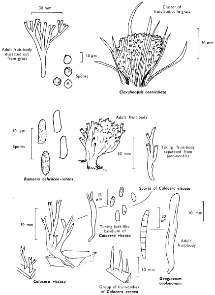
Clavulinopsis corniculata (Fries), Corner (p. 171).
Cap: absent. Fruit-body: complex; width 20-30 mm; length 20-40 mm.
Description: Plate 57.
Fruit-body: shape depending on the length of grass in which it grows but always branching strongly from its base, composed of a dense compact tuft of egg-yellow or orange-tawny branches which are either irregular or of equal length and so they form a flattened top to the fruit-body complex, the branchlets are slender, forked 2- or 3-times, with their apices narrowed or curved.
Stem: very downy at the base.
Flesh: pale yellow.
Spore-print: white.
Spores: medium sized, hyaline under the microscope, smooth, spherical and 5-7 µm in diameter, not becoming bluish grey in iodine solutions.
Cystidia: absent.
Habitat & Distribution: Common amongst grass in fields or on grassy path sides in woodland.
General Information: Clavulinopsis corniculata is recognised by the branched habit and the smooth spores; Ramaria ochraceo-virens is of similar form, but has an overall duller colour and turns green on bruising, grows in pinewoods and has finely roughened brownish spores. Calocera viscosa also has an erect, bright golden or orange-yellow fruit-body which becomes more orange on drying. It is repeatedly branched and usually has a long, tough-rooting base. However, the spore-print is dirty yellowish and the fruit-body, which grows on coniferous wood, is viscid and elastic, a character reflected in the name ‘viscosa’. Microscopically the basidium of Calocera is shaped like a tuning-fork and is not clavate as in Clavulinopsis corniculata. It appears to be more related to the jelly-fungi (see p. 180).
Illustrations: Clavulinopsis corniculata—LH 55; NB 6; WD 1043. Calocera viscosa—Hvass 304; LH 225; NB 1493; WD 1078.
Typhula erythropus Fries.
Cap: absent. Fruit-body up to 20 mm high.
Description:
Fruit-body: upper fertile portion club-shaped and not more than half the length, white, surmounting a reddish brown, thread-like, often wavy or twisted stem which is attached at its base to an ellipsoid bead-like structure, called a sclerotium.
Spore-print: white.
Spores: oblong, smooth, hyaline under the microscope, 6-7 × 2 µm in size and not becoming bluish grey in iodine solutions.
Cystidia: absent.
Habitat & Distribution: Not uncommon on dead leaves and twigs or dead herbaceous stems.
Pistillaria micans Fries.
Cap: absent. Fruit-body: up to 10 mm high.
Description:
Club-shaped or oblong, rose-pink hardly differentiated from the similarly coloured stem, and arising at most from a small pad of filaments.
Spore-print: white.
Spores: broadly ellipsoid to pip-shaped, smooth, hyaline under the microscope, about 10 × 6 µm (8-11 × 5-7 µm) in size and not becoming bluish grey in iodine solutions.
Cystidia: absent.
Habitat & Distribution: Not uncommon on dead herbaceous stems and leaves, especially those in damp places.
Illustrations: T. erythropus WD 10510. P. micans WD 1057.
General notes on the club-fungi
Early mycologists believed that the club-shaped nature of the fruit-body was important in the classification of these fungi. Thus the Earth Tongues (Geoglossum, see Plate 57), the Stag’s horn fungi and relatives (Xylosphaera see p. 204), both Ascomycete groups, the Dacrymycetales (a group of jelly-fungi, see p. 180) and the true fairy-clubs were all classified together. It was the ‘Father of Mycology’, the Swede, Elias Fries, who in 1821, as in many other groups of fungi, made an attempt to make some sense of the chaos. By very careful observations, and what is so amazing without using a microscope, he was able to separate the tough stemmed and gelatinous stemmed groups from the more slender or coral-like ones. Fries was a very keen observer and noticed features which many modern authorities miss in the field because they rely too heavily on the microscope. Fries’ system was used almost unchanged until the second half of this century; its beauty was its simplicity in that it joined together in one group all those fungi with simple basidia and the spore-bearing tissue distributed all around a simple club or around the branches of a complex fruit-body resembling a coral. However, by a careful examination of the microscopic structures, such as the spores and hyphae and the development of the fruit-body, it has been found necessary to separate these fungi still further. The ecology of the club-fungi has assisted in an understanding of these proposed divisions.
The larger many branched clavarias, more correctly placed in the genus Ramaria, are to be found on bare soil in woodlands and plantations; R. ochraceo-virens is common in conifer plantations and can be recognised by the long ornamented spores, which characterise this group of fungi, and the fact that the sandy-coloured fruit-body becomes dark olive-green on bruising (see p. 170).
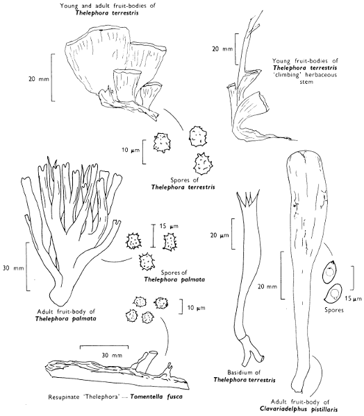
Clavariadelphus pistillaris is the largest of our simple club-fungi; it may grow up to 200 mm high and 50 mm wide. This fungus has a wrinkled outer surface and sometimes the apex of the club becomes flattened and lacks basidia; this suggests a possible relationship, perhaps evolutionary, to the primitive chanterelles (see p. 162)—also woodland fungi. Clavulina, a complex group of dull or whitish, branched fruit-bodies, has been described earlier and the genus is characterised by the large spores and 2-spored basidia; they are woodland fungi also.
The grassland species are often simple in structure belonging in the main to the genus Clavulinopsis (see p. 170) and the now much reduced genus Clavaria (see p. 168). Although really complex, some of these species of Clavulinopsis are branched only below the soil level and thus appear as single clubs amongst the grass. Perhaps the single club has evolved especially to grow amongst blades of grass. C. corniculata, however, is well branched and the head is tight and compact and often flattened close to the ground. The same fungus in woodland is more open and because of this it was thought to be a different species to the grassland form. It is the simple club which dominates the form of those species which grow on herbaceous debris and grass-stems; indeed several species of Typhula cause diseases of grass particularly those of lawns where they have suffered damage because of cold or long periods under the snow. Some of these small fungi produce a small hard mass of fungal tissue about the size of a lupin seed (called a sclerotium). This is a resting body from which the club-shaped almost filament-like fruit-body later develops.
Thelephora terrestris Fries Earth-fan
Cap: absent. Fruit-body: width 20-40 mm; height 30-50 mm.
Description:
Fruit-body: erect, fan-shaped or effused with upturned margin, tough but thin and fibrous, chocolate-brown or cocoa-coloured, scaly from radiating fibrils and with fringed, pale buff or wine-coloured margin.
Gills: absent and replaced by a wrinkled or irregularly granular, dark lilaceous grey or cocoa-coloured surface.
Flesh: brown and thin.
Spore-print: purplish brown.
Spores: medium sized, dark brown under the microscope, warted-angular and 8-9 × 6-7 µm in size.
Cystidia: absent but basidia often filled with brown contents.
Basidia: 2-4 spored.
Habitat & Distribution: Found on the ground in woods, especially pine woods; also on heathland growing up vegetation and incorporating it into the fruit-body’s shape.
General Information: There is some evidence to suggest that this fungus can form mycorrhiza with pine trees under certain conditions.
Although it may be easily passed over because it is perfectly camouflaged it is quite easy to recognise when collected. T. palmata (Bulliard) Patouillard, is a bigger, less frequently seen species more coral-like in shape; it also grows in pine woods. When the fruit-body of T. terrestris spreads over the soil or plant debris it resembles other members of the family to which it belongs, i.e. Thelephoraceae; species of Tomentella. They also have warty angular spores, purplish brown colours, and wrinkled or puckered spore-bearing surfaces. Tomentella spp., however, are resupinate or encrusting and so do not form caps, even at the margin of the fruit-body. Tomentella is one of the many genera which were classed collectively as resupinate fungi because they lack a cap and form crusts. This group ‘the resupinates’ consists of a whole series of quite unrelated fungi.
Illustrations: LH 53; NB 478.
Plate 58. Club and Fan-shaped fungi
Larger illustration
(iv) Resupinate fungi
When mycologists talk generally about ‘resupinates’ they are referring to a whole group of Basidiomycetes whose spore-bearing layer is exposed, the cap highly reduced or completely lacking, and the fungus adhering to the surface of the substrate which may be soil, wood, grasses, etc., at the point which would have been the cap of an agaric. Probably members of the group are the most commonly seen yet it is one of the most commonly ignored groups of fungi—by naturalists and mycologists alike; they form ‘white wash’ on old sticks, dark coloured discolourations on trunks, etc. It is an entirely artificial group of many quite unrelated elements united on the common factor of having either a reduced or primitive fruit-body consisting only of a sheet of tissue. However, these same fungi have a uniting factor in that they frequent the same ecological sites, e.g. on muddy soil in bogs, under overhangs of banks and stream sides, undersides of logs, trunks, branches and twigs, hidden in cracks of old stumps or spreading over carpets of conifer needles or dead leaves and sedges.
By studying the anatomy of the fruit-body and the characters of the spores certain relationships can be found which relate many of these fungi to several other groups of fungi we have dealt with in earlier chapters.
It is only possible to mention here the group as a whole for all the species really require very careful examination, often necessitating several hours of microscope work. They should be left by the beginner until more experience is obtained and advice of an expert easily available.
Although the group mainly contains saprophytes, a few are parasitic. ‘Silver-leaf disease’ of almonds and fruit trees is caused by Stereum (chondrostereum) purpureum (Persoon) Fries; it has a purple fruiting surface, and greyish upper surface when ever this is formed at the margin.
There are several species of Stereum in Britain, three species of which when handled in the fresh state stain red: S. sanguinolentum (A. & S.) Fries, a pale coloured species on conifer wood, S. rugosum (Pers.) Fries a similar coloured species on beech, birch and especially hazel, and S. gausapatum Fries an ochraceous yellow species on oak, often forming a pocket rot of the timber. However, the commonest member of the genus is an orange-tawny coloured species with a greyish buff, hairy cap, S. hirsutum (Willd) Fries. It grows on many trees of broad-leaved wood and can be found wherever twigs, branches, trunks or stumps have been lying out in the rain; it does not bleed.
Plate 59. Resupinate fungi
Larger illustration
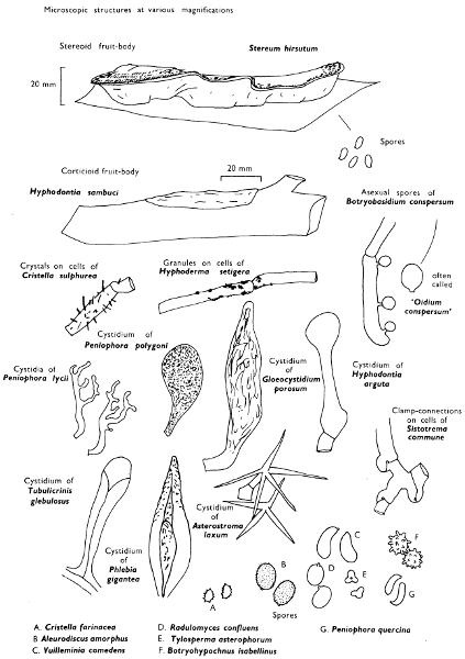
Those species of resupinate fungi which resemble members of this genus, i.e. those with a distinct tough, although poorly developed, cap, are called stereoid.
‘Red thread disease’ of grass which often causes unsightly red patches on lawns and school and corporation playing-fields is caused by Corticium fuciforme (Berkeley) Wakefield. Fungi belonging to this genus produce fruit-bodies which ‘scramble’ over the substrate; for example, if one searches old elder trees throughout the year one will certainly find a ‘white wash’ fungus of this type, Hyphodontia sambuci (Pers.) J. Eriksson. Fungi with this type of fruit-body are called corticoid.
The two major types are illustrated along with some of the bizarre microscopic structures one finds in the resupinates; such structures are useful in classification and identification, and their beauty and intricacy make up for the surprisingly simple fruit-body shape and texture.