VOLUME XI SLICE V Gassendi, Pierre to Geocentric
Reading edition
Articles in This Slice
GASSENDI, PIERRE GEFLE
GASTEIN GEGENBAUR, CARL
GASTRIC ULCER GEGENSCHEIN
GASTRITIS GEIBEL, EMANUEL
GASTROPODA GEIGE
GASTROTRICHA GEIGER, ABRAHAM
GATAKER, THOMAS GEIJER, ERIK GUSTAF
GATCHINA GEIKIE, SIR ARCHIBALD
GATE GEIKIE, JAMES
GATEHOUSE GEIKIE, WALTER
GATES, HORATIO GEILER VON KAISERSBERG, JOHANN
GATESHEAD GEINITZ, HANS BRUNO
GATH GEISHA
GATLING, RICHARD JORDAN GEISLINGEN
GATTY, MARGARET GEISSLER, HEINRICH
GAU, JOHN GELA
GAUDEN, JOHN GELADA
GAUDICHAUD-BEAUPRÉ, CHARLES GELASIUS
GAUDRY, JEAN ALBERT GELATI
GAUDY GELATIN
GAUERMANN, FRIEDRICH GELDERLAND (duchy)
GAUGE GELDERLAND (province of Holland)
GAUHATI GELDERN
GAUL, GILBERT WILLIAM GELL, SIR WILLIAM
GAUL GELLERT, CHRISTIAN FÜRCHTEGOTT
GAULT GELLERT
GAUNTLET GELLIUS, AULUS
GAUR (ruined city of India) GELLIVARA
GAUR (wild ox) GELNHAUSEN
GAUSS, KARL FRIEDRICH GELO
GAUSSEN, FRANÇOIS SAMUEL ROBERT LOUIS GELSEMIUM
GAUTIER, ÉMILE THÉODORE LÉON GELSENKIRCHEN
GAUTIER, THÉOPHILE GEM
GAUTIER D'ARRAS GEM, ARTIFICIAL
GAUZE GEMBLOUX
GAVARNI GEMINI
GAVAZZI, ALESSANDRO GEMINIANI, FRANCESCO
GAVELKIND GEMISTUS PLETHO, GEORGIUS
GAVESTON, PIERS GEMMI PASS
GAVOTTE GENDARMERIE
GAWAIN GENEALOGY
GAWLER GENELLI, GIOVANNI BUONAVENTURA
GAY, JOHN GENERAL
GAY, MARIE FRANÇOISE SOPHIE GENERATION
GAY, WALTER GENESIS
GAYA GENET
GAYAL GENEVA (New York, U.S.A.)
GAYANGOS Y ARCE, PASCUAL DE GENEVA (Switzerland)
GAYARRÉ, CHARLES ÉTIENNE ARTHUR GENEVA CONVENTION
GAY-LUSSAC, JOSEPH LOUIS GENEVA, LAKE OF
GAZA, THEODORUS GENEVIÈVE, ST
GAZA GENEVIÈVE, OF BRABANT
GAZALAND GENGA, GIROLAMO
GAZEBO GENISTA
GAZETTE GENIUS
GEAR GENUS, STÉPHANIE-FÉLICITÉ DU CREST DE SAINT-AUBIN
GEBER GENNA
GEBHARD TRUCHSESS VON WALDBURG GENNADIUS II.
GEBWEILER GENOA
GECKO GENOVESI, ANTONIO
GED, WILLIAM GENSONNÉ, ARMAND
GEDDES, ALEXANDER GENTIAN
GEDDES, ANDREW GENTIANACEAE
GEDDES, JAMES LORRAINE GENTILE
GEDDES, SIR WILLIAM DUGUID GENTILE DA FABRIANO
GEDYMIN GENTILESCHI, ARTEMISIA and ORAZIO DE’
GEE, THOMAS GENTILI, ALBERICO
GEEL, JACOB GENTLE
GEELONG GENTLEMAN
GEESTEMÜNDE GENTZ, FRIEDRICH VON
GEFFCKEN, FRIEDRICH HEINRICH GEOCENTRIC
GEFFROY, MATHIEU AUGUSTE
GASSENDI1 [Gassend], PIERRE (1592-1655), French philosopher, scientist and mathematician, was born of poor parents at Champtercier, near Digne, in Provence, on the 22nd of January 1592. At a very early age he gave indications of remarkable mental powers and was sent to the college at Digne. He showed particular aptitude for languages and mathematics, and it is said that at the age of sixteen he was invited to lecture on rhetoric at the college. Soon afterwards he entered the university of Aix, to study philosophy under P. Fesaye. In 1612 he was called to the college of Digne to lecture on theology. Four years later he received the degree of doctor of theology at Avignon, and in 1617 he took holy orders. In the same year he was called to the chair of philosophy at Aix, and seems gradually to have withdrawn from theology. He lectured principally on the Aristotelian philosophy, conforming as far as possible to the orthodox methods. At the same time, however, he followed with interest the discoveries of Galileo and Kepler, and became more and more dissatisfied with the Peripatetic system. It was the period of revolt against the Aristotelianism of the schools, and Gassendi shared to the full the empirical tendencies of the age. He, too, began to draw up objections to the Aristotelian philosophy, but did not at first venture to publish them. In 1624, however, after he had left Aix for a canonry at Grenoble, he printed the first part of his Exercitationes paradoxicae adversus Aristoteleos. A fragment of the second book was published later at La Haye (1659), but the remaining five were never composed, Gassendi apparently thinking that after the Discussiones Peripateticae of Francesco Patrizzi little field was left for his labours.
After 1628 Gassendi travelled in Flanders and Holland. During this time he wrote, at the instance of Mersenne, his examination of the mystical philosophy of Robert Fludd (Epistolica dissertatio in qua praecipua principia philosophiae Ro. Fluddi deteguntur, 1631), an essay on parhelia (Epistola de parheliis), and some valuable observations on the transit of Mercury which had been foretold by Kepler. He returned to France in 1631, and two years later became provost of the cathedral church at Digne. Some years were then spent in travelling through Provence with the duke of Angoulême, governor of the department. The only literary work of this period is the Life of Peiresc, which has been frequently reprinted, and was translated into English. In 1642 he was engaged by Mersenne in controversy with Descartes. His objections to the fundamental propositions of Descartes were published in 1642; they appear as the fifth in the series contained in the works of Descartes. In these objections Gassendi’s tendency towards the empirical school of speculation appears more pronounced than in any of his other writings. In 1645 he accepted the chair of mathematics in the Collège Royal at Paris, and lectured for many years with great success. In addition to controversial writings on physical questions, there appeared during this period the first of the works by which he is known in the history of philosophy. In 1647 he published the treatise De vita, moribus, et doctrina Epicuri libri octo. The work was well received, and two years later appeared his commentary on the tenth book of Diogenes Laërtius, De vita, moribus, et placitis Epicuri, seu Animadversiones in X. librum Diog. Laër. (Lyons, 1649; last edition, 1675). In the same year the more important Syntagma philosophiae Epicuri (Lyons, 1649; Amsterdam, 1684) was published.
In 1648 ill-health compelled him to give up his lectures at the Collège Royal. He travelled in the south of France, spending nearly two years at Toulon, the climate of which suited him. In 1653 he returned to Paris and resumed his literary work, publishing in that year lives of Copernicus and Tycho Brahe. The disease from which he suffered, lung complaint, had, however, established a firm hold on him. His strength gradually failed, and he died at Paris on the 24th of October 1655. A bronze statue of him was erected by subscription at Digne in 1852.
His collected works, of which the most important is the Syntagma philosophicum (Opera, i. and ii.), were published in 1658 by Montmort (6 vols., Lyons). Another edition, also in 6 folio volumes, was published by N. Averanius in 1727. The first two are occupied entirely with his Syntagma philosophicum; the third contains his critical writings on Epicurus, Aristotle, Descartes, Fludd and Lord Herbert, with some occasional pieces on certain problems of physics; the fourth, his Institutio astronomica, and his Commentarii de rebus celestibus; the fifth, his commentary on the tenth book of Diogenes Laërtius, the biographies of Epicurus, N.C.F. de Peiresc, Tycho Brahe, Copernicus, Georg von Peuerbach, and Regiomontanus, with some tracts on the value of ancient money, on the Roman calendar, and on the theory of music, to all which is appended a large and prolix piece entitled Notitia ecclesiae Diniensis; the sixth volume contains his correspondence. The Lives, especially those of Copernicus, Tycho and Peiresc, have been justly admired. That of Peiresc has been repeatedly printed; it has also been translated into English. Gassendi was one of the first after the revival of letters who treated the literature of philosophy in a lively way. His writings of this kind, though too laudatory and somewhat diffuse, have great merit; they abound in those anecdotal details, natural yet not obvious reflections, and vivacious turns of thought, which made Gibbon style him, with some extravagance certainly, though it was true enough up to Gassendi’s time—“le meilleur philosophe des littérateurs, et le meilleur littérateur des philosophes.”
Gassendi holds an honourable place in the history of physical science. He certainly added little to the stock of human knowledge, but the clearness of his exposition and the manner in which he, like Bacon, urged the importance of experimental research, were of inestimable service to the cause of science. To what extent any place can be assigned him in the history of philosophy is more doubtful. The Exercitationes on the whole seem to have excited more attention than they deserved. They contain little or nothing beyond what had been already advanced against Aristotle. The first book expounds clearly, and with much vigour, the evil effects of the blind acceptance of the Aristotelian dicta on physical and philosophical study; but, as is the case with so many of the anti-Aristotelian works of this period, the objections show the usual ignorance of Aristotle’s own writings. The second book, which contains the review of Aristotle’s dialectic or logic, is throughout Ramist in tone and method. The objections to Descartes—one of which at least, through Descartes’s statement of it in the appendix of objections in the Meditationes has become famous—have no speculative value, and in general are the outcome of the crudest empiricism. His labours on Epicurus have a certain historical value, but the want of consistency inherent in the philosophical system raised on Epicureanism is such as to deprive it of genuine worth. Along with strong expressions of empiricism we find him holding doctrines absolutely irreconcilable with empiricism in any form. For while he maintains constantly his favourite maxim “that there is nothing in the intellect which has not been in the senses” (nihil in intellectu quod non prius fuerit in sensu), while he contends that the imaginative faculty (phantasia) is the counterpart of sense—that, as it has to do with material images, it is itself, like sense, material, and essentially the same both in men and brutes; he at the same time admits that the intellect, which he affirms to be immaterial and immortal—the most characteristic distinction of humanity—attains notions and truths of which no effort of sensation or imagination can give us the slightest apprehension (Op. ii. 383). He instances the capacity of forming “general notions”; the very conception of universality itself (ib. 384), to which he says brutes, who partake as truly as men in the faculty called phantasia, never attain; the notion of God, whom he says we may imagine to be corporeal, but understand to be incorporeal; and lastly, the reflex action by which the mind makes its own phenomena and operations the objects of attention.
The Syntagma philosophicum, in fact, is one of those eclectic systems which unite, or rather place in juxtaposition, irreconcilable dogmas from various schools of thought. It is divided, according to the usual fashion of the Epicureans, into logic (which, with Gassendi as with Epicurus, is truly canonic), physics and ethics. The logic, which contains at least one praiseworthy portion, a sketch of the history of the science, is divided into theory of right apprehension (bene imaginari), theory of right judgment (bene proponere), theory of right inference (bene colligere), theory of right method (bene ordinare). The first part contains the specially empirical positions which Gassendi afterwards neglects or leaves out of account. The senses, the sole source of knowledge, are supposed to yield us immediately cognition of individual things; phantasy (which Gassendi takes to be material in nature) reproduces these ideas; understanding compares these ideas, which are particular, and frames general ideas. Nevertheless, he at the same time admits that the senses yield knowledge—not of things—but of qualities only, and holds that we arrive at the idea of thing or substance by induction. He holds that the true method of research is the analytic, rising from lower to higher notions; yet he sees clearly, and admits, that inductive reasoning, as conceived by Bacon, rests on a general proposition not itself proved by induction. He ought to hold, and in disputing with Descartes he did apparently hold, that the evidence of the senses is the only convincing evidence; yet he maintains, and from his special mathematical training it was natural he should maintain, that the evidence of reason is absolutely satisfactory. The whole doctrine of judgment, syllogism and method is a mixture of Aristotelian and Ramist notions.
In the second part of the Syntagma, the physics, there is more that deserves attention; but here, too, appears in the most glaring manner the inner contradiction between Gassendi’s fundamental principles. While approving of the Epicurean physics, he rejects altogether the Epicurean negation of God and particular providence. He states the various proofs for the existence of an immaterial, infinite, supreme Being, asserts that this Being is the author of the visible universe, and strongly defends the doctrine of the foreknowledge and particular providence of God. At the same time he holds, in opposition to Epicureanism, the doctrine of an immaterial rational soul, endowed with immortality and capable of free determination. It is altogether impossible to assent to the supposition of Lange (Gesch. des Materialismus, 3rd ed., i. 233), that all this portion of Gassendi’s system contains nothing of his own opinions, but is introduced solely from motives of self-defence. The positive exposition of atomism has much that is attractive, but the hypothesis of the calor vitalis (vital heat), a species of anima mundi (world-soul) which is introduced as physical explanation of physical phenomena, does not seem to throw much light on the special problems which it is invoked to solve. Nor is his theory of the weight essential to atoms as being due to an inner force impelling them to motion in any way reconcilable with his general doctrine of mechanical causes.
In the third part, the ethics, over and above the discussion on freedom, which on the whole is indefinite, there is little beyond a milder statement of the Epicurean moral code. The final end of life is happiness, and happiness is harmony of soul and body (tranquillitas animi et indolentia corporis). Probably, Gassendi thinks, perfect happiness is not attainable in this life, but it may be in the life to come.
The Syntagma is thus an essentially unsystematic work, and clearly exhibits the main characteristics of Gassendi’s genius. He was critical rather than constructive, widely read and trained thoroughly both in languages and in science, but deficient in speculative power and original force. Even in the department of natural science he shows the same inability steadfastly to retain principles and to work from them; he wavers between the systems of Brahe and Copernicus. That his revival of Epicureanism had an important influence on the general thinking of the 17th century may be admitted; that it has any real importance in the history of philosophy cannot be granted.
Authorities.—Gassendi’s life is given by Sorbière in the first collected edition of the works, by Bugerel, Vie de Gassendi (1737; 2nd ed., 1770), and by Damiron, Mémoire sur Gassendi (1839). An abridgment of his philosophy was given by his friend, the celebrated traveller, Bernier (Abrégé de la philosophie de Gassendi, 8 vols., 1678; 2nd ed., 7 vols., 1684). The most complete surveys of his work are those of G.S. Brett (Philosophy of Gassendi, London, 1908), Buhle (Geschichte der neuern Philosophie, iii. 1, 87-222), Damiron (Mémoires pour servir à l’histoire de philosophie au XVIIe siècle), and P.F. Thomas (La Philosophie de Gassendi, Paris, 1889). See also Ritter, Geschichte der Philosophie, x. 543-571; Feuerbach, Gesch. d. neu. Phil. von Bacon bis Spinoza, 127-150; F.X. Kiefl, P. Gassendis Erkenntnistheorie und seine Stellung zum Materialismus (1893) and “Gassendi’s Skepticismus” in Philos. Jahrb. vi. (1893); C. Güttler, “Gassend oder Gassendi?” in Archiv f. Gesch. d. Philos. x. (1897), pp. 238-242.
(R. Ad.; X.)
1 It was formerly thought that Gassendi was really the genitive of the Latin form Gassendus. C. Güttler, however, holds that it is a modernized form of the O. Fr. Gassendy (see paper quoted in bibliography).
GASTEIN, in the duchy of Salzburg, Austria, a side valley of the Pongau or Upper Salzach, about 25 m. long and 1¼ m. broad, renowned for its mineral springs. It has an elevation of between 3000 and 3500 ft. Behind it, to the S., tower the mountains Mallnitz or Nassfeld-Tauern (7907 ft.) and Ankogel (10,673 ft.), and from the right and left of these mountains two smaller ranges run northwards forming its two side walls. The river Ache traverses the valley, and near Wildbad-Gastein forms two magnificent waterfalls, the upper, the Kesselfall (196 ft.), and the lower, the Bärenfall (296 ft.). Near these falls is the Schleierfall (250 ft.), formed by the stream which drains the Bockhart-see. The valley is also traversed by the so-called Tauern railway (opened up to Wildbad-Gastein in September 1905), which goes to Mallnitz, piercing the Tauern range by a tunnel 9260 yds. in length. The principal villages of the valley are Hof-Gastein, Wildbad-Gastein and Böckstein.
Hof-Gastein, pop. (1900) 840, the capital of the valley, is also a watering-place, the thermal waters being conveyed here from Wildbad-Gastein by a conduit 5 m. long, constructed in 1828 by the emperor Francis I. of Austria. Hof-Gastein was, after Salzburg, the richest place in the duchy, owing to its gold and silver mines, which were already worked during the Roman period. During the 16th century these mines were yielding annually 1180 ℔ of gold and 9500 ℔ of silver, but since the 17th century they have been much neglected and many of them are now covered by glaciers.
Wildbad-Gastein, commonly called Bad-Gastein, one of the most celebrated watering-places in Europe, is picturesquely situated in the narrow valley of the Gasteiner Ache, at an altitude of 3480 ft. The thermal springs, which issue from the granite mountains, have a temperature of 77°-120° F., and yield about 880,000 gallons of water daily. The water contains only 0.35 to 1000 of mineral ingredients and is used for bathing purposes. The springs are resorted to in cases of nervous affections, senile and general debility, skin diseases, gout and rheumatism. Wildbad-Gastein is annually visited by over 8500 guests. The springs were known as early as the 7th century, but first came into fame by a successful visit paid to them by Duke Frederick of Austria in 1436. Gastein was a favourite resort of William I. of Prussia and of the Austrian imperial family, and it was here that, on the 14th of August 1865, was signed the agreement known as the Gastein Convention, which by dividing the administration of the conquered provinces of Schleswig and Holstein between Austria and Prussia postponed for a while the outbreak of war between the two powers. It was also here (August-September 1879) that Prince Bismarck negotiated with Count Julius Andrássy the Austro-German treaty, which resulted in the formation of the Triple Alliance.
See Pröll, Gastein, Its Springs and Climate (Vienna, 5th ed., 1893).
GASTRIC ULCER (ulcer of the stomach), a disease of much gravity, commonest in females, and especially in anaemic domestic servants. It is connected in many instances with impairment of the circulation in the stomach and the formation of a clot in a small blood-vessel (thrombosis). It may be due to an impoverished state of the blood (anaemia), but it may also arise from disease of the blood-vessels, the result of long-continued indigestion and gastric catarrh.
When clotting takes place in a blood-vessel the nutrition of that limited area of the stomach is cut off, and the patch undergoes digestion by the unresisted action of the gastric juices, an ulcer being formed. The ulcer is usually of the size of a silver threepence or sixpence, round or oval, and, eating deeply, is apt to make a hole right through the coats of the stomach. Its usual site is upon the posterior wall of the upper curvature, near to the pyloric orifice. It may undergo a healing process at any stage, in which case it may leave but little trace of its existence; while, on the other hand, it may in the course of cicatrizing produce such an amount of contraction as to lead to stricture of the pylorus, or to a peculiar hour-glass deformity of the stomach. Perforation is in most cases quickly fatal, unless previously the stomach has become adherent to some neighbouring organ, by which the dangerous effects of this occurrence may be averted, or unless the condition has been promptly recognized and an operation has been quickly done. Usually there is but one ulcer, but sometimes there are several ulcers.
The symptoms of ulcer of the stomach are often indefinite and obscure, and in some cases the diagnosis has been first made on the occurrence of a fatal perforation. First among the symptoms is pain, which is present at all times, but is markedly increased after food. The pain is situated either at the lower end of the breast-bone or about the middle of the back. Sometimes it is felt in the sides. It is often extremely severe, and is usually accompanied with localized tenderness and also with a sense of oppression, and by an inability to wear tight clothing. The pain is due to the movements of the stomach set up by the presence of the food, as well as to the irritation of the inflamed nerve filaments in the floor of the ulcer. Vomiting is a usual symptom. It occurs either soon after the food is swallowed or at a later period, and generally relieves the pain and discomfort. Vomiting of blood (haematemesis) is a frequent and important symptom. The blood may show itself in the form of a brown or coffee-like mixture, or as pure blood of dark colour and containing clots. It comes from some vessel or vessels which the ulcerative process has ruptured. Blood is also found mixed with the discharges from the bowels, rendering them dark or tarry-looking. The general condition of the patient with gastric ulcer is, as a rule, that of extreme ill-health, with pallor, emaciation and debility. The tongue is red, and there is usually constipation. In most of the cases the disease is chronic, lasting for months or years; and in those cases where the ulcers are large or multiple, incomplete healing may take place, relapses occurring from time to time. But the ulcers may give rise to no marked symptoms, and there have been instances where fatal perforation suddenly took place, and where post-mortem examination revealed the existence of long-standing ulcers which had given rise to no suggestive symptoms. While gastric ulcer is to be regarded as dangerous, its termination, in the great majority of cases, is in recovery. It frequently, however, leaves the stomach in a delicate condition, necessitating the utmost care as regards diet. Occasionally the disease proves fatal by sudden haemorrhage, but a fatal result is more frequently due to perforation and the escape of the contents of the stomach into the peritoneal cavity, in which case death usually occurs in from twelve to forty-eight hours, either from shock or from peritonitis. Should the stomach become adherent to another organ, and fatal perforation be thus prevented, chronic “indigestion” may persist, owing to interference with the natural movements of the stomach. Stricture of the pylorus and consequent dilatation of the stomach may be caused by the cicatrization of an ulcer.
The patient should at once be sent to bed and kept there, and allowed for a while nothing stronger than milk and water or milk and lime water. But if bleeding has recently taken place no food whatever should be allowed by the stomach, and the feeding should be by nutrient enemata. As the symptoms quiet down, eggs may be given beaten up with milk, and later, bread and milk and home-made broths and soups. Thus the diet advances to chicken and vegetables rubbed through a sieve, to custard pudding and bread and butter. As regards medicines, iron is the most useful, but no pills of any sort should be given. Under the influence of rest and diet most gastric ulcers get well. The presence of healthy-looking scars upon the surface of the stomach, which are constantly found in operating upon the interior of the abdomen, or as revealed in post-mortem examinations, are evidence of the truth of this statement. It is unlikely that under the treatment just described perforation of the stomach will take place, and if the surgeon is called in to assist he will probably advise that operation is inadvisable. Moreover, he knows that if he should open the abdomen to search for an ulcer of the stomach he might fail to find it; more than that, his search might also be in vain if he opened the stomach itself and examined the interior. Serious haemorrhages, however, may make it necessary that a prompt and thorough search should be made in order that the surgeon may endeavour to locate the ulcer, and, having found it, secure the damaged vessel and save the patient from death by bleeding.
Perforation of a gastric ulcer having taken place, the septic germs, which were harmless whilst in the stomach, escape with the rest of the contents of the stomach into the general peritoneal cavity. The immediate effects of this leakage are sudden and severe pain in the upper part of the abdomen and a great shock to the system (collapse). The muscles of the abdominal wall become hard and resisting, and as peritonitis appears and the intestines are distended with gas, the abdomen is distended and becomes greatly increased in size and ceases to move, the respiratory movements being short and quick. At first, most likely, the temperature drops below normal, and the pulse quickens. Later, the temperature rises. If nothing is done, death from the septic poisoning of peritonitis is almost certain.
The treatment of ruptured gastric ulcer demands immediate operation. An incision should be made in the upper part of the middle line of the abdomen, and the perforation should be looked for. There is not, as a rule, much difficulty in finding it, as there are generally deposits of lymph near the spot, and other signs of local inflammation; moreover, the contents of the stomach may be seen escaping from the opening. The ulcer is to be closed by running a “purse-string” suture in the healthy tissue around it, and the place is then buried in the stomach by picking up small folds of the stomach-wall above and below it and fixing them together by suturing. This being done, the surface of the stomach, and the neighbouring viscera which have been soiled by the leakage, are wiped clean and the abdominal wound is closed, provision being made for efficient drainage. A large proportion of cases of perforated gastric ulcer thus treated recover.
(E. O.*)
GASTRITIS (Gr. γαστήρ, stomach), an inflammatory affection of the stomach, of which the condition of catarrh, or irritation of its mucous membrane, is the most frequent and most readily recognized. This may exist in an acute or a chronic form, and depends upon some condition, either local or general, which produces a congested state of the circulation in the walls of the stomach (see Digestive Organs: Pathology).
Acute Gastritis may arise from various causes. The most intense forms of inflammation of the stomach are the toxic conditions which follow the swallowing of corrosive poisons, such as strong mineral acids of alkalis which may extensively destroy the mucous membrane. Other non-corrosive poisons cause acute degeneration of the stomach wall (see Poisons). Acute inflammatory conditions may be secondary to zymotic diseases such as diphtheria, pyaemia, typhus fever and others. Gastritis is also caused by the ingestion of food which has begun to decompose, or may result from eating unsuitable articles which themselves remain undigested and so excite acute catarrhal conditions. These give rise to the symptoms well known as characterizing an acute “bilious attack,” consisting in loss of appetite, sickness or nausea, and headache, frontal or occipital, often accompanied with giddiness. The tongue is furred, the breath foetid, and there is pain or discomfort in the region of the stomach, with sour eructations, and frequently vomiting, first of food and then of bilious matter. An attack of this kind tends to subside in a few days, especially if the exciting cause be removed. Sometimes, however, the symptoms recur with such frequency as to lead to the more serious chronic form of the disease.
The treatment bears reference, in the first place, to any known source of irritation, which, if it exist, may be expelled by an emetic or purgative (except in cases due to poisoning). This, however, is seldom necessary, since vomiting is usually present. For the relief of sickness and pain the sucking of ice and counter-irritation over the region of the stomach are of service. Further, remedies which exercise a soothing effect upon an irritable mucous membrane, such as bismuth or weak alkaline fluids, and along with these the use of a light milk diet, are usually sufficient to remove the symptoms.
Chronic Gastric Catarrh may result from the acute or may arise independently. It is not infrequently connected with antecedent disease in other organs, such as the lungs, heart, liver or kidneys, and it is especially common in persons addicted to alcoholic excess. In this form the texture of the stomach is more altered than in the acute form, except in the toxic and febrile forms above referred to. It is permanently in a state of congestion, and its mucous membrane and muscular coat undergo thickening and other changes, which markedly affect the function of digestion. The symptoms are those of dyspepsia in an aggravated form (see Dyspepsia), of which discomfort and pain after food, with distension and frequently vomiting, are the chief; and the treatment must be conducted in reference to the causes giving rise to it. The careful regulation of the diet, alike as to the amount, the quality, and the intervals between meals, demands special attention. Feeding on artificially soured milk may in many cases be useful. Lavage or washing out of the stomach with weak alkaline solutions has been used with marked success in the treatment of chronic gastritis. Of medicinal agents, bismuth, arsenic, nux vomica, and the mineral acids are all of acknowledged efficacy, as are also preparations of pepsin.
GASTROPODA, the second of the five classes of animals constituting the phylum Mollusca. For a discussion of the relationship of the Gastropoda to the remaining classes of the phylum, see Mollusca.
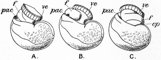
The Gastropoda are mainly characterized by a loss of symmetry, produced by torsion of the visceral sac. This torsion may be resolved into two successive movements. The first is a ventral flexure in the antero-posterior or sagittal plane; the result of this is to approximate the two ends of the alimentary canal. In development, the openings of the mantle-cavity and the anus are always originally posterior; later they are brought forward ventrally. During this first movement flexure is also produced by the coiling of the visceral sac and shell; primitively the latter was bowl-shaped; but the ventral flexure, which brings together the two extremities of the digestive tube, gives the visceral sac the outline of a more or less acute cone. The shell necessarily takes this form also, and then becomes coiled in a dorsal or anterior plane—that is to say, it becomes exogastric. This condition may be seen in embryonic Patellidae, Fissurellidae and Trochidae (fig. 1, A), and agrees with the method of coiling of a mollusc without lateral torsion, such as Nautilus. But ultimately the coil becomes ventral or endogastric, in consequence of the second torsion movement then apparent.
From Lankester’s Treatise on Zoology.
Fig. 1.—Three stages in the development of Trochus, during the process of torsion. (After Robert.)
A, Nearly symmetrical larva (veliger). B, A stage 1½ hours later than A. C, A stage 3½ hours later than B. f, Foot. op, Operculum. pac, Pallial cavity. ve, Velum.
The shell is represented as fixed, while the head and foot rotate from left to right. In reality the head and foot are fixed and the shell rotates from right to left.
The second movement is a lateral torsion of the visceral mass, the foot remaining a fixed point; this torsion occurs in a plane approximately at right angles to that of the first movement, and carries the pallial aperture and the anus from behind forwards. If, at this moment, the animal were placed with mouth and ventral surface turned towards the observer, this torsion carries the circumanal complex in a clockwise direction (along the right side in dextral forms) through 180° as compared with its primitive condition. The (primitively) right-hand organs of the complex thus become left-hand, and vice versa. The visceral commissure, while still surrounding the digestive tract, becomes looped; its right half, with its proper ganglion, passes to the left side over the dorsal face of the alimentary canal (whence the name supra-intestinal), while the left half passes below towards the right side, thus originating the name infra-intestinal given to this half and to its ganglion. Next, the shell, the coil of which was at first exogastric, being also included in this rotation through 180°, exhibits an endogastric coiling (fig. 1, B, C). This, however, is not generally retained in one plane, and the spire projects, little by little, on the side which was originally left, but finally becomes right (in dextral forms, with a clockwise direction, if viewed from the side of the spire; but counter-clockwise in sinistral forms). Finally, the original symmetry of the circumanal complex vanishes; the anus leaves the centre of the pallial cavity and passes towards the right side (left side in sinistral forms); the organs of this side become atrophied and disappear. The essential feature of the asymmetry of Gastropoda is the atrophy or disappearance of the primitively left half of the circumanal complex (the right half in sinistral forms), including the gill, the auricle, the osphradium, the hypobranchial gland and the kidney.
From Lankester’s Treatise on Zoology.
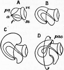
Fig. 2.—Four stages in the development of a Gastropod showing the process of body torsion. (After Robert.)
A, Embryo without flexure. B, Embryo with ventral flexure of the intestine. C, Embryo with ventral flexure and exogastric shell. D, Embryo with lateral torsion and an endogastric shell. a, Anus. f, Foot. m, Mouth. pa, Mantle. pac, Pallial cavity. ve, Velum.
In dextral Gastropods the only structure found on the topographically right side of the rectum is the genital duct. But this is not part of the primitive complex. It is absent in the most primitive and symmetrical forms, such as Haliotis and Pleurotomaria. Originally the gonads opened into the kidneys. In the most primitive existing Gastropods the gonad opens into the right kidney (Patellidae, Trochidae, Fissurellidae). The gonaduct, therefore, is derived from the topographically right kidney. The transformation has been actually shown to take place in the development of Paludina. In a dextral Gastropod the shell is coiled in a right-handed spiral from apex to mouth, and the spiral also projects to the right of the median plane of the animal.
When the shell is sinistral the asymmetry of the organs is usually reversed, and there is a complete situs inversus viscerum, the direction of the spiral of the shell corresponding to the position of the organs of the body. Triforis, Physa, Clausilia are examples of sinistral Gastropods, but reversal also occurs as an individual variation among forms normally dextral. But there are forms in which the involution is “hyperstrophic,” that is to say, the turns of the spire projecting but slightly, the spire, after flattening out gradually, finally becomes re-entrant and transformed into a false umbilicus; at the same time that part which corresponds to the umbilicus of forms with a normal coil projects and constitutes a false spire; the coil thus appears to be sinistral, although the asymmetry remains dextral, and the coil of the operculum (always the opposite to that of the shell) sinistral (e.g. Lanistes among Streptoneura, Limacinidae among Opisthobranchia). The same, mutatis mutandis, may occur in sinistral shells.
The problem of the causes of the torsion of the Gastropod body has been much discussed. E.R. Lankester in the ninth edition of this work attributed it to the pressure of the shell and visceral hump towards the right side. He referred also to the nautiloid shell of the larva falling to one side. But these are two distinct processes. In the larva a nautiloid shell is developed which is coiled exogastrically, that is, dorsally, and the pallial cavity is posterior or ventral (fig. 2, C): the larva therefore resembles Nautilus in the relations of body and shell. The shell then rotates towards the left side through 180°, so that it becomes ventral or endogastric (fig. 2, D). The pallial cavity, with its organs, is by this torsion moved up the right side of the larva to the dorsal surface, and thus the left organs become right and vice versa. In the subsequent growth of the shell the spire comes to project on the right side, which was originally the left. Neither the rotation of the shell as a whole nor its helicoid spiral coiling is the immediate cause of the torsion of the body in the individual, for the direction of the torsion is indicated in the segmentation of the ovum, in which there is a complete reversal of the cleavage planes in sinistral as compared with dextral forms. The facts, however, strongly suggest that the original cause of the torsion was the weight of the exogastric shell and visceral hump, which in an animal creeping on its ventral surface necessarily fell over to one side. It is not certain that the projection of the spire to the originally left side of the shell has anything to do with the falling over of the shell to that side. The facts do not support such a suggestion. In the larva there is no projection at the time the torsion takes place. In some forms the coiling disappears in the adult, leaving the shell simply conical as in Patellidae, Fissurellidae, &c., and in some cases the shell is coiled in one plane, e.g. Planorbis. In all these cases the torsion and asymmetry of the body are unaffected.
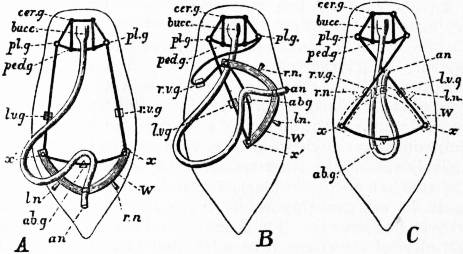
Fig 3.—Sketch of a model designed so as to show the effect of torsion or rotation of the visceral hump in Streptoneurous Gastropoda.
A, Unrotated ancestral condition. B, Quarter-rotation. C, Complete semi-rotation (the limit). an, Anus. ln, rn, Primarily left nephridium and primarily right nephridium. lvg, Primarily left (subsequently the sub-intestinal) visceral ganglion. rvg, Primarily right (subsequently the sub-intestinal) visceral ganglion. cerg, Cerebral ganglion. plg, Pleural ganglion. pedg, Pedal ganglion. abg, Abdominal ganglion. bucc, Buccal mass. W, Wooden arc representing the base-line of the wall of the visceral hump. x, x′, Pins fastening the elastic cord (representing the visceral nerve loop) to W.
The characteristic torsion attains its maximum effect among the majority of the Streptoneura. It is followed in some specialized Heteropoda and in the Euthyneura by a torsion in the opposite direction, or detorsion, which brings the anus farther back and untwists the visceral commissure (see Euthyneura, below). This conclusion has shown that the Euthyneura do not represent an archaic form of Gastropoda, but are themselves derived from streptoneurous forms. The difference between the two sub-classes has been shown to be slight; certain of the more archaic Tectibranchia (Actaeon) and Pulmonata (Chilina) still have the visceral commissure long and not untwisted. The fact that all the Euthyneura are hermaphrodite is not a fundamental difference; several Streptoneura are so, likewise Valvata, Oncidiopsis, Marsenina, Odostomia, Bathysciadium, Entoconcha.
Classification.—The class Gastropoda is subdivided as follows:
Sub-class I. Streptoneura.
Order 1. Aspidobranchia.
Sub-order 1. Docoglossa.
” 2. Rhipidoglossa.
Order 2. Pectinibranchia.
Sub-order 1. Taenioglossa.
Tribe 1. Platypoda.
” 2. Heteropoda.
Sub-order 2. Stenoglossa.
Tribe 1. Rachiglossa.
” 2. Toxiglossa.
Sub-class II. Euthyneura.
Order 1. Opisthobranchia.
Sub-order 1. Tectibranchia.
Tribe 1. Bullomorpha.
” 2. Aplysiomorpha.
” 3. Pleurobranchomorpha.
Sub-order 2. Nudibranchia.
Tribe 1. Tritoniomorpha.
” 2. Doridomorpha.
” 3. Eolidomorpha.
” 4. Elysiomorpha.
Order 2. Pulmonata.
Sub-order 1. Basommatophora.
” 2. Stylommatophora.
Tribe 1. Holognatha.
” 2. Agnatha.
” 3. Elasmognatha.
” 4. Ditremata.
Sub-Class I.—Streptoneura
In this division the torsion of the visceral mass and visceral commissure is at its maximum, the latter being twisted into a figure of eight. The right half of the commissure with its ganglion is supra-intestinal, the left half with its ganglion infra-intestinal. In some cases each pleural ganglion is connected with the opposite branch of the visceral commissure by anastomosis with the pallial nerve, a condition which is called dialyneury; or there may be a direct connective from the pleural ganglion to the visceral ganglion of the opposite side, which is called zygoneury. The head bears only one pair of tentacles. The radular teeth are of several different kinds in each transverse row. The heart is usually posterior to the branchia (proso-branchiate). The sexes are usually separate.
The old division into Zygobranchia and Azygobranchia must be abandoned, for the Azygobranchiate Rhipidoglossa have much greater affinity to the Zygobranchiate Haliotidae and Fissurellidae than to the Azygobranchia in general. This is shown by the labial commissure and pedal cords of the nervous system, by the opening of the gonad into the right kidney, and by other points. Further, the Pleurotomariidae have been discovered to possess two branchiae. The sub-class is now divided into two orders: the Aspidobranchia in which the branchia or ctenidium is bipectinate and attached only at its base, and the Pectinibranchia in which the ctenidium is monopectinate and attached to the mantle throughout its length.
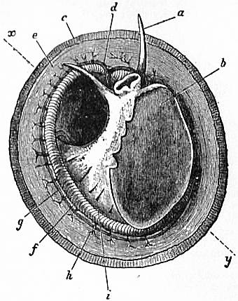
Fig. 4.—The Common Limpet (Patella vulgata) in its shell, seen from the pedal surface. (Lankester.)
x, y, The median antero-posterior axis. a, Cephalic tentacle. b, Plantar surface of the foot. c, Free edge of the shell. d, The branchial efferent vessel carrying aerated blood to the auricle, and here interrupting the circlet of gill lamellae. e, Margin of the mantle-skirt. f, Gill lamellae (not ctenidia, but special pallial growths, comparable with those of Pleurophyllidia). g, The branchial efferent vessel. h, Factor of the branchial advehent vessel. i, Interspaces between the muscular bundles of the root of the foot, causing the separate areae seen in fig. 5, c.
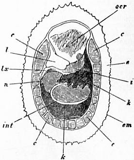
Fig. 5.—Dorsal surface of the Limpet removed from its shell and deprived of its black pigmented epithelium; the internal organs are seen through the transparent body-wall. (Lankester.)
c, Muscular bundles forming the root of the foot, and adherent to the shell. e, Free mantle-skirt. em, Tentaculiferous margin of the same. i, Smaller (left) nephridium. k, Larger (right) nephridium. l, Pericardium. lx, Fibrous septum, behind the pericardium. n, Liver. int, Intestine. ecr, Anterior area of the mantle-skirt over-hanging the head (cephalic hood).
Order I. Aspidobranchia.—These are the most primitive Gastropods, retaining to a great degree the original symmetry of the organs of the pallial complex, having two kidneys, in some cases two branchiae, and two auricles. The gonad has no accessory organs and except in Neritidae no duct, but discharges into the right kidney.
Forms adapted to terrestrial life and to aerial respiration occur in various divisions of Gastropods, and do not constitute a single homogeneous group. Thus the Helicinidae, which are terrestrial, are now placed among the Aspidobranchia. In these there are neither branchia nor osphradium, and the pallial chamber which retains its large opening serves as a lung. Degeneration of the shell occurs in some members of the order. It is largely covered by the mantle in some Fissurellidae, is entirely internal in Pupilia and absent in Titiscaniidae.
The common limpet is a specially interesting and abundant example of the more primitive Aspidobranchia. The foot of the limpet is a nearly circular disk of muscular tissue; in front, projecting from and raised above it, are the head and neck (figs. 4, 13). The visceral hump forms a low conical dome above the sub-circular foot, and standing out all round the base of this dome so as completely to overlap the head and foot, is the circular mantle-skirt. The depth of free mantle-skirt is greatest in front, where the head and neck are covered in by it. Upon the surface of the visceral dome, and extending to the edge of the free mantle-skirt, is the conical shell. When the shell is taken away (best effected by immersion in hot water) the surface of the visceral dome is found to be covered by a black-coloured epithelium, which may be removed, enabling the observer to note the position of some organs lying below the transparent integument (fig. 5). The muscular columns (c) attaching the foot to the shell form a ring incomplete in front, external to which is the free mantle-skirt. The limits of the large area formed by the flap over the head and neck (ecr) can be traced, and we note the anal papilla showing through and opening on the right shoulder, so to speak, of the animal into the large anterior region of the sub-pallial space. Close to this the small renal organ (i, mediad) and the larger renal organ (k, to the right and posteriorly) are seen, also the pericardium (l) and a coil of the intestine (int) embedded in the compact liver.
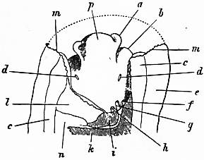
Fig. 6.—Anterior portion of the same Limpet, with the overhanging cephalic hood removed. (Lankester.)
a, Cephalic tentacle. b, Foot. c, Muscular substance forming the root of the foot. d, The capito-pedal organs of Lankester (= rudimentary ctenidia). e, Mantle-skirt. f, Papilla of the larger nephridium. g, Anus. h, Papilla of the smaller nephridium. i, Smaller nephridium. k, Larger nephridium. l, Pericardium. m, Cut edge of the mantle-skirt. n, Liver. p, Snout.
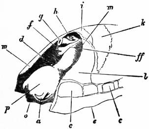
Fig. 7.—The same specimen viewed from the left front, so as to show the sub-anal tract (ff) of the larger nephridium, by which it communicates with the pericardium. o, Mouth; other letters as in fig. 6.
On cutting away the anterior part of the mantle-skirt so as to expose the sub-pallial chamber in the region of the neck, we find the right and left renal papillae (discovered by Lankester in 1867) on either side of the anal papilla (fig. 6), but no gills. If a similar examination be made of the allied genus Fissurella (fig. 17, d), we find right and left of the two renal apertures a right and left gill-plume or ctenidium, which here as in Haliotis and Pleurotomaria retain their original paired condition. In Patella no such plumes exist, but right and left of the neck are seen a pair of minute oblong yellow bodies (fig. 6, d), which were originally described by Lankester as orifices possibly connected with the evacuation of the generative products. On account of their position they were termed by him the “capito-pedal orifices,” being placed near the junction of head and foot. J.W. Spengel has, however, in a most ingenious way shown that these bodies are the representatives of the typical pair of ctenidia, here reduced to a mere rudiment. Near to each rudimentary ctenidium Spengel has discovered an olfactory patch or osphradium (consisting of modified epithelium) and an olfactory nerve-ganglion (fig. 8). It will be remembered that, according to Spengel, the osphradium of mollusca is definitely and intimately related to the gill-plume or ctenidium, being always placed near the base of that organ; further, Spengel has shown that the nerve-supply of this olfactory organ is always derived from the visceral loop. Accordingly, the nerve-supply affords a means of testing the conclusion that we have in Lankester’s capito-pedal bodies the rudimentary ctenidia. The accompanying diagrams (figs. 9, 10) of the nervous systems of Patella and of Haliotis, as determined by Spengel, show the identity in the origin of the nerves passing from the visceral loop to Spengel’s olfactory ganglion of the Limpet, and that of the nerves which pass from the visceral loop of Haliotis to the olfactory patch or osphradium, which lies in immediate relation on the right and on the left side to the right and left gill-plumes (ctenidia) respectively. The same diagrams serve to demonstrate the streptoneurous condition of the visceral loop in Aspidobranchia.
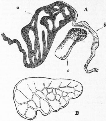
Fig. 8.—A, Section in a plane vertical to the surface of the neck of Patella through a, the rudimentary ctenidium (Lankester’s organ), and b, the olfactory epithelium (osphradium); c, the olfactory (osphradial) ganglion. (After Spengel.)
B, Surface view of a rudimentary ctenidium of Patella excised and viewed as a transparent object. (Lankester.)
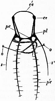
Fig. 9.—Nervous system of Patella; the visceral loop is lightly shaded; the buccal ganglia are omitted. (After Spengel.)
ce, Cerebral ganglia. c’e, Cerebral commissure. pl, Pleural ganglion. pe, Pedal ganglion. p′e, Pedal nerve. s, s′, Nerves (right and left) to the mantle. o, Olfactory ganglion, connected by nerve to the streptoneurous visceral loop.
Thus, then, we find that the limpet possesses a symmetrically disposed pair of ctenidia in a rudimentary condition, and justifies its position among Aspidobranchia. At the same time it possesses a totally distinct series of functional gills, which are not derived from the modification of the typical molluscan ctenidium. These gills are in the form of delicate lamellae (fig. 4, f), which form a series extending completely round the inner face of the depending mantle-skirt. This circlet of gill-lamellae led Cuvier to class the limpets as Cyclobranchiata, and, by erroneous identification of them with the series of metamerically repeated ctenidia of Chiton, to associate the latter mollusc with the former. The gill-lamellae of Patella are processes of the mantle comparable with the plait-like folds often observed on the roof of the branchial chamber in other Gastropoda (e.g. Buccinum and Haliotis). They are termed pallial gills. The only other molluscs in which they are exactly represented are the curious Opisthobranchs Phyllidia and Pleurophyllidia (fig. 55). In these, as in Patella, the typical ctenidia are aborted, and the branchial function is assumed by close-set lamelliform processes arranged in a series beneath the mantle-skirt on either side of the foot. In fig. 4, d, the large branchial vein of Patella bringing blood from the gill-series to the heart is seen; where it crosses the series of lamellae there is a short interval devoid of lamellae.
The heart in Patella consists of a single auricle (not two as in Haliotis and Fissurella) and a ventricle; the former receives the blood from the branchial vein, the latter distributes it through a large aorta which soon leads into irregular blood-lacunae.
The existence of two renal organs in Patella, and their relation to the pericardium (a portion of the coelom), is important. Each renal organ is a sac lined with glandular epithelium (ciliated cell, with concretions) communicating with the exterior by its papilla, and by a narrow passage with the pericardium. The connexion with the pericardium of the smaller of the two renal organs was demonstrated by Lankester in 1867, at a time when the fact that the renal organ of the Mollusca, as a rule, opens into the pericardium, and is therefore a typical nephridium, was not known. Subsequent investigations carried on under the direction of the same naturalist have shown that the larger as well as the smaller renal sac is in communication with the pericardium. The walls of the renal sacs are deeply plaited and thrown into ridges. Below the surface these walls are excavated with blood-vessels, so that the sac is practically a series of blood-vessels covered with renal epithelium, and forming a meshwork within a space communicating with the exterior. The larger renal sac (remarkably enough, that which is aborted in other Anisopleura) extends between the liver and the integument of the visceral dome very widely. It also bends round the liver as shown in fig. 12, and forms a large sac on half of the upper surface of the muscular mass of the foot. Here it lies close upon the genital body (ovary or testis), and in such intimate relationship with it that, when ripe, the gonad bursts into the renal sac, and its products are carried to the exterior by the papilla on the right side of the anus (Robin, Dall). This fact led Cuvier erroneously to the belief that a duct existed leading from the gonad to this papilla. The position of the gonad, best seen in the diagrammatic section (fig. 13), is, as in other Aspidobranchia, devoid of a special duct communicating with the exterior. This condition, probably an archaic one, distinguishes the Aspidobranchia from other Gastropoda.
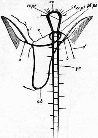
Fig. 10.—Nervous system of Haliotis; the visceral loop is lightly shaded; the buccal ganglia are omitted. (After Spengel.)
ce, Cerebral ganglion. pl.pe, The fused pleural and pedal ganglia. pe, The right pedal nerve. ce.pl, The cerebro-pleural connective. ce.pe, The cerebro-pedal connective. s, s′, Right and left mantle nerves. ab, Abdominal ganglion or site of same. o, o, Right and left olfactory ganglia and osphardia receiving nerve from visceral loop.
Fig. 11.—Nervous system of Fissurella. (From Gegenbaur, after Jhering.) pl, Pallial nerve. p, Pedal nerve. A, Abdominal ganglia in the streptoneurous visceral commissure, with supra- and sub-intestine ganglion on each side. B, Buccal ganglia. C, C, Cerebral ganglia. es, Cerebral commissure. o, Otocysts attached to the cerebro-pedal connectives. Fig. 12.—Diagram of the two renal organs (nephridia), to show their relation to the rectum and to the pericardium. (Lankester.) f, Papilla of the larger nephridium. g, Anal papilla with rectum leading from it. h, Papilla of the smaller nephridium, which is only represented by dotted outlines. l, Pericardium indicated by a dotted outline—at its right side are seen the two reno-pericardial pores. ff, The sub-anal tract of the large nephridium given off near its papilla and seen through the unshaded smaller nephridium. ks.a, Anterior superior lobe of the large nephridium. ks.l, Left lobe of same. ks.p, Posterior lobe of same. ks.i, Inferior sub-visceral lobe of same.
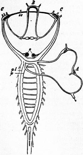
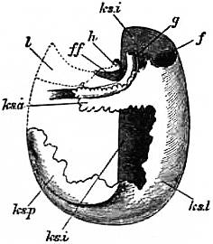
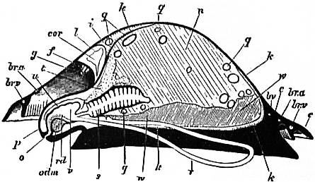
Fig. 13.—Diagram of a vertical antero-postero median section of a Limpet. Letters as in figs. 6, 7, with following additions. (Lankester.)
q, Intestine in transverse section. r, Lingual sac (radular sac). rd, Radula. s, Lamellated stomach. t, Salivary gland. u, Duct of same. v, Buccal cavity w, Gonad. br.a, Branchial advehent vessel (artery). br.v, Branchial efferent vessel (vein). bv, Blood-vessel. odm, Muscles and cartilage of the odontophore. cor, Heart within the pericardium.
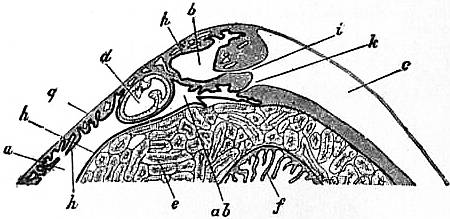
Fig. 14.—Vertical section in a plane running right and left through the anterior part of the visceral hump of Patella to show the two renal organs and their openings into the pericardium. (J.T. Cunningham.)
a, Large or external or right renal organ. ab, Narrow process of the same running below the intestine and leading by k into the pericardium. b, Small or median renal organ. c, Pericardium. d, Rectum. e, Liver. f, Manyplies. g, Epithelium of the dorsal surface. h, Renal epithelium lining the renal sacs. i, Aperture connecting the small sac with the pericardium. k, Aperture connecting the large sac with the pericardium.
The digestive tract of Patella offers some interesting features. The odontophore is powerfully developed; the radular sac is extraordinarily long, lying coiled in a space between the mass of the liver and the muscular foot. The radula has 160 rows of teeth with twelve teeth in each row. Two pairs of salivary ducts, each leading from a salivary gland, open into the buccal chamber. The oesophagus leads into a remarkable stomach, plaited like the manyplies of a sheep, and after this the intestine takes a very large number of turns embedded in the yellow liver, until at last it passes between the two renal sacs to the anal papilla. A curious ridge (spiral? valve) which secretes a slimy cord is found upon the inner wall of the intestine. The general structure of the Molluscan intestine has not been sufficiently investigated to render any comparison of this structure of Patella with that of other Mollusca possible. The eyes of the limpet deserve mention as examples of the most primitive kind of eye in the Molluscan series. They are found one on each cephalic tentacle, and are simply minute open pits or depressions of the epidermis, the epidermic cells lining them being pigmented and connected with nerves (compare fig. 14, art. Cephalopoda). The limpet breeds upon the southern English coast in the early part of April, but its development has not been followed. It has simply been traced as far as the formation of a diblastula which acquires a ciliated band, and becomes a nearly spherical trochosphere. It is probable that the limpet takes several years to attain full growth, and during that period it frequents the same spot, which becomes gradually sunk below the surrounding surface, especially if the rock be carbonate of lime. At low tide the limpet (being a strictly intertidal organism) is exposed to the air, and (according to trustworthy observers) quits its attachment and walks away in search of food (minute encrusting algae), and then once more returns to the identical spot, not an inch in diameter, which belongs, as it were, to it. Several million limpets—twelve million in Berwickshire alone—are annually used on the east coast of Britain as bait.
Sub-order 1. Docoglossa.—Nervous system without dialyneury. Eyes are open invaginations without crystalline lens. Two osphradia present but no hypobranchial glands nor operculum. Teeth of radula beam-like, and at most three marginal teeth on each side. Heart has only a single auricle, neither heart nor pericardium traversed by rectum. Shell conical without spire.
Fam. 1.—Acmaeidae. A single bipectinate ctenidium on left side. Acmaea, without pallial branchiae, British. Scurria, with pallial branchiae in a circle beneath the mantle.
Fam. 2.—Tryblidiidae. Muscle scar divided into numerous impressions. Tryblidium, Silurian.
Fam. 3.—Patellidae. No ctenidia but pallial branchiae in a circle between mantle and foot. Patella, pallial branchiae forming a complete circle, no epipodial tentacles, British. Ancistromesus, radula with median central tooth. Nacella, epipodial tentacles present. Helcion, circlet of branchiae interrupted anteriorly, British.
Fam. 4.—Lepetidae. Neither ctenidia nor pallial branchiae. Lepeta, without eyes. Pilidium. Propilidium.
Fam. 5.—Bathysciadidae. Hermaphrodite; head with appendage on right side; radula without central tooth. Bathysciadium, abyssal.
Sub-order 2. Rhipidoglossa.—Aspidobranchia with a palliovisceral anastomosis (dialyneurous); eye-vesicle closed, with crystalline lens; ctenidia, osphradia and hypobranchial glands paired or single. Radula with very numerous marginal teeth arranged like the rays of a fan. Heart with two auricles; ventricle traversed by the rectum, except in the Helicinidae. An epipodial ridge on each side of the foot and cephalic expansions between the tentacles often present.
Fam. 1.—Pleurotomariidae. Shell spiral; mantle and shell with an anterior fissure; two ctenidia; a horny operculum. Pleurotomaria, epipodium without tentacles. Genus includes several hundred extinct species ranging from the Silurian to the Tertiary. Five living species from the Antilles, Japan and the Moluccas. Moluccan species is 19 cm. in height.
Fam. 2.—Bellerophontidae. 300 species, all fossil, from Cambrian to Trias.
Fam. 3.—Euomphalidae. Also extinct, from Cambrian to Cretaceous.
Fam. 4.—Haliotidae. Spire of shell much reduced; two bipectinate ctenidia, the right being the smaller; no operculum. Haliotis.
Fam. 5.—Velainiellidae, an extinct family from the Eocene.
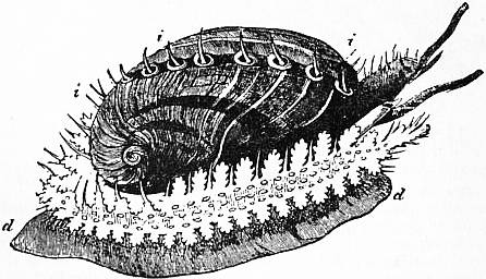
Fig. 15.—Halio tistuberculata. d, Foot; i, tentacular processes of the mantle. (From Owen, after Cuvier.)
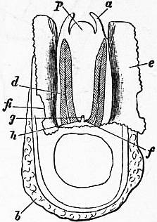
Fam. 6.—Fissurellidae. Shell conical; slit or hole in anterior part of mantle; two symmetrical ctenidia; no operculum. Emarginula, mantle and shell with a slit, British. Scutum, mantle split anteriorly and reflected over shell, which has no slit. Puncturella, mantle and shell with a foramen in front of the apex, British. Fissurella, mantle and shell perforated at apex, British.
Fam. 7.—Cocculinidae. Shell conical, symmetrical, without slit or perforation. Cocculina, abyssal.
Fam. 8.—Trochidae. Shell spirally coiled; a single ctenidium; eyes perforated; a horny operculum; lobes between the tentacles. Trochus, shell umbilicated, spire pointed and prominent, British. Monodonta, no jaws, spire not prominent, no umbilicus, columella toothed. Gibbula, with jaws, three pairs of epipodial cirri without pigment spots at their bases, British. Margarita, five to seven pairs of epipodial cirri with a pigment spot at base of each.
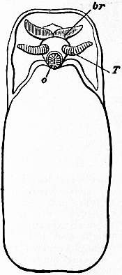
Fig. 16.—Scutum, seen from the pedal surface. (Lankester.) o, Mouth. T, Cephalic tentacle. br, One of the two symmetrical gills placed on the neck. Fig. 17.—Dorsal aspect of a specimen of Fissurella from which the shell has been removed, whilst the anterior area of the mantle-skirt has been longitudinally slit and its sides reflected. (Lankester.) a, Cephalic tentacle. b, Foot. d, Left (archaic right) gill-plume. e, Reflected mantle-flap. fi, The fissure or hole in the mantle-flap traversed by the longitudinal incision. f, Right (archaic left) nephridium’s aperture. g, Anus. h, Left (archaic right) aperture of nephridium. p, Snout.
Fam. 9.—Stomatellidae. Spire of shell much reduced; a single ctenidium. Stomatella, foot truncated posteriorly, an operculum present, no epipodial tentacles. Gena, foot elongated posteriorly, no operculum.
Fam. 10.—Delphinulidae. Shell spirally coiled; operculum horny; intertentacular lobes absent. Delphinula.
Fam. 11.—Liotiidae, shell globular, margin of aperture thickened. Liotia.
Fam. 12.—Cyclostrematidae. Shell flattened, umbilicated; foot anteriorly truncated with angles produced into lobes. Cyclostrema. Teinostoma.
Fam. 13.—Trochonematidae. All extinct, Cambrian to Cretaceous.
Fam. 14.—Turbinidae. Shell spirally coiled; epipodial tentacles present; operculum thick and calcareous. Turbo. Astralium. Molleria. Cyclonema.
Fam. 15.—Phasianellidae. Shell not nacreous, without umbilicus, with prominent spire and polished surface. Phasianella.
Fam. 16.—Umboniidae. Shell flattened, not umbilicated, generally smooth; operculum horny. Umbonium. Isanda.
Fam. 17.—Neritopsidae. Shell semi-globular, with short spire; operculum calcareous, not spiral. Neritopsis. Naticopsis, extinct.
Fam. 18.—Macluritidae. Extinct, Cambrian and Silurian.
Fam. 19.—Neritidae. Shell with very low spire, without umbilicus, internal partitions frequently absorbed; a single ctenidium; a cephalic penis present. Nerita, marine. Neritina, freshwater, British. Septaria, shell boat-shaped.
Fam. 20.—Titiscaniidae. Without shell and operculum, but with pallial cavity and ctenidium. Titiscania, Pacific.
Fam. 21.—Helicinidae. No ctenidium, but a pulmonary cavity; heart with a single auricle, not traversed by the rectum. Helicina. Eutrochatella. Stoastoma. Bourceria.
Fam. 22.—Hydrocenidae. No ctenidium, but a pulmonary cavity; operculum with an apophysis. Hydrocena, Dalmatia.
Fam. 23.—Proserpinidae. No operculum. Proserpina, Central America.
Order 2. Pectinibranchia.—In this order there is no longer any trace of bilateral symmetry in the circulatory, respiratory and excretory organs, the topographically right half of the pallial complex having completely disappeared, except the right kidney, which is represented by the genital duct. There is usually a penis in the male. The ctenidium is monopectinate and attached to the mantle along its whole length, except in Adeorbis and Valvata; in the latter alone it is bipectinate. There is a single well-developed, often pectinated osphradium. The eye is always a closed vesicle, and the internal cornea is extensive. In the radula there is a single central tooth or none.
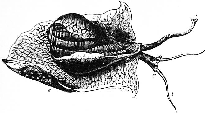
Fig. 18.—Animal and shell of Pyrula laevigata. (From Owen.)
a, Siphon. b, Head-tentacles. C, Head, the letter placed near the right eye. d, The foot, expanded as in crawling. h, The mantle-skirt reflected over the sides of the shell.
The former classification into Holochlamyda, Pneumochlamyda and Siphonochlamyda has been abandoned, as it was founded on adaptive characters not always indicative of true affinities. The order is now divided into two sub-orders: the Taenioglossa, in which there are three teeth on each side of the median tooth of the radula, and the Stenoglossa, in which there is only one tooth on each side of the median tooth. In the latter a pallial siphon, a well-developed proboscis and an unpaired oesophageal gland are always present, in the former they are usually absent. The siphon is an incompletely tubular outgrowth of the mantle margin on the left side, contained in a corresponding outgrowth of the edge of the shell-mouth, and serving to conduct water to the respiratory cavity.
The condition usually spoken of as a “proboscis” appears to be derived from the condition of a simple rostrum (having the mouth at its extremity) by the process of incomplete introversion of that simple rostrum. There is no reason in the actual significance of the word why the term “proboscis” should be applied to an alternately introversible and eversible tube connected with an animal’s body, and yet such is a very customary use of the term. The introversible tube may be completely closed, as in the “proboscis” of Nemertine worms, or it may have a passage in it leading into a non-eversible oesophagus, as in the present case, and in the case of the eversible pharynx of the predatory Chaetopod worms. The diagrams here introduced (fig. 19) are intended to show certain important distinctions which obtain amongst the various “introverts,” or intro- and e-versible tubes so frequently met with in animal bodies. Supposing the tube to be completely introverted and to commence its eversion, we then find that eversion may take place, either by a forward movement of the side of the tube near its attached base, as in the proboscis of the Nemertine worms, the pharynx of Chaetopods and the eye-tentacle of Gastropods, or by a forward movement of the inverted apex of the tube, as in the proboscis of the Rhabdocoel Planarians, and in that of Gastropods here under consideration. The former case we call “pleurecbolic” (fig. 19, A, B, C, H, I, K), the latter “acrecbolic” tubes or introverts (fig. 19, D, E, F, G). It is clear that, if we start from the condition of full eversion of the tube and watch the process of introversion, we shall find that the pleurecbolic variety is introverted by the apex of the tube sinking inwards; it may be called acrembolic, whilst conversely the acrecbolic tubes are pleurembolic. Further, it is obvious enough that the process either of introversion or of eversion of the tube may be arrested at any point, by the development of fibres connecting the wall of the introverted tube with the wall of the body, or with an axial structure such as the oesophagus; on the other hand, the range of movement of the tubular introvert may be unlimited or complete. The acrembolic proboscis or frontal introvert of the Nemertine worms has a complete range. So has the acrembolic pharynx of Chaetopods, if we consider the organ as terminating at that point where the jaws are placed and the oesophagus commences. So too the acrembolic eye-tentacle of the snail has a complete range of movement, and also the pleurembolic proboscis of the Rhabdocoel prostoma. The introverted rostrum of the Pectinibranch Gastropods presents in contrast to these a limited range of movement. The “introvert” in these Gastropods is not the pharynx as in the Chaetopod worms, but a prae-oral structure, its apical limit being formed by the true lips and jaws, whilst the apical limit of the Chaetopod’s introvert is formed by the jaws placed at the junction of pharynx and oesophagus, so that the Chaetopod’s introvert is part of the stomodaeum or fore-gut, whilst that of the Gastropod is external to the alimentary canal altogether, being in front of the mouth, not behind it, as is the Chaetopod’s. Further, the Gastropod’s introvert is pleurembolic (and therefore acrecbolic), and is limited both in eversion and in introversion; it cannot be completely everted owing to the muscular bands (fig. 19, G), nor can it be fully introverted owing to the bands (fig. 19, F) which tie the axial pharynx to the adjacent wall of the apical part of the introvert. As in all such intro- and e-versible organs, eversion of the Gastropod proboscis is effected by pressure communicated by the muscular body-wall to the liquid contents (blood) of the body-space, accompanied by the relaxation of the muscles which directly pull upon either the sides or the apex of the tubular organ. The inversion of the proboscis is effected directly by the contraction of these muscles. In various members of the Pectinibranchia the mouth-bearing cylinder is introversible (i.e. is a proboscis)—with rare exceptions these forms have a siphonate mantle-skirt. On the other hand, many which have a siphonate mantle-skirt are not provided with an introversible mouth-bearing cylinder, but have a simple non-introversible rostrum, as it has been termed, which is also the condition presented by the mouth-bearing region in nearly all other Gastropoda. One of the best examples of the introversible mouth-cylinder or proboscis which can be found is that of the common whelk (Buccinum undatum) and its immediate allies. In fig. 23 the proboscis is seen in an everted state; it is only so carried when feeding, being withdrawn when the animal is at rest. Probably its use is to enable the animal to introduce its rasping and licking apparatus into very narrow apertures for the purposes of feeding, e.g. into a small hole bored in the shell of another mollusc.
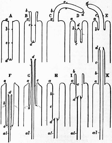
Fig. 19.—Diagrams explanatory of the nature of so-called proboscides or “introverts.” (Lankester.)
A, Simple introvert completely introverted.
B, The same, partially everted by eversion of the sides, as in the Nemertine proboscis and Gastropod eye-tentacle = pleurecbolic.
C, The same, fully everted.
D, E, A similar simple introvert in course of eversion by the forward movement, not of its sides, but of its apex, as in the proboscidean Rhabdocoels = acrecbolic.
F, Acrecbolic (= pleurembolic) introvert, formed by the snout of the proboscidiferous Gastropod. al, alimentary canal; d, the true mouth. The introvert is not a simple one with complete range both in eversion and introversion, but is arrested in introversion by the fibrous bands at c, and similarly in eversion by the fibrous bands at b.
G, The acrecbolic snout of a proboscidiferous Gastropod, arrested short of complete eversion by the fibrous band b.
H, The acrembolic (= pleurecbolic) pharynx of a Chaetopod fully introverted. al, alimentary canal; at d, the jaws; at a, the mouth; therefore a to d is stomodaeum, whereas in the Gastropod (F) a to d is inverted body-surface.
I, Partial eversion of H.
K, Complete eversion of H.
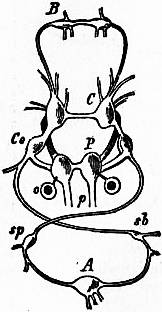
Fig. 20.—Male of Littorina littoralis, Lin., removed from its shell; the mantle-skirt cut along its right line of attachment and thrown over to the left side of the animal so as to expose the organs on its inner face. a, Anus. i, Intestine. r, Nephridium (kidney). r′, Aperture of the nephridium. c, Heart. br, Ctenidium (gill-plume). pbr, Parabranchia (= the osphradium or olfactory patch). x, Glandular lamellae of the inner face of the mantle-skirt. y, Adrectal (purpuriparous) gland. t, Testis. vd, Vas deferens. p, Penis. mc, Columella muscle (muscular process grasping the shell). v, Stomach. h, Liver. N.B.—Note the simple snout or rostrum not introverted as a “proboscis.” Fig. 21.—Nervous system of Paludina as a type of the streptoneurous condition. (From Gegenbaur, after Jhering.) B, Buccal (suboesophageal) ganglion. C, Cerebral ganglion. Co, Pleural ganglion. P, Pedal ganglion with otocyst attached. p, Pedal nerve. A, Abdominal ganglion at the extremity of the twisted visceral “loop.” sp, Supra-intestinal visceral ganglion on the course of the right visceral cord. sb, Sub-intestinal ganglion on the course of the left visceral cord.
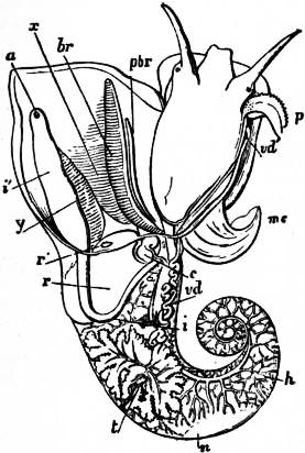
The very large assemblage of forms coming under this order comprises the most highly developed predaceous sea-snails, numerous vegetarian species, a considerable number of freshwater and some terrestrial forms. The partial dissection of a male specimen of the common periwinkle, Littorina littoralis, drawn in fig. 20, will serve to exhibit the disposition of viscera which prevails in the group. The branchial chamber formed by the mantle-skirt overhanging the head has been exposed by cutting along a line extending backward from the letters vd to the base of the columella muscle mc, and the whole roof of the chamber thus detached from the right side of the animal’s neck has been thrown over to the left, showing the organs which lie upon the roof. No opening into the body-cavity has been made; the organs which lie in the coiled visceral hump show through its transparent walls. The head is seen in front resting on the foot and carrying a median non-retractile snout or rostrum, and a pair of cephalic tentacles at the base of each of which is an eye. In many Gastropoda the eyes are not thus sessile but raised upon special eye-tentacles (figs. 25, 56). To the right of the head is seen the muscular penis p, close to the termination of the vas deferens (spermatic duct) vd. The testis t occupies a median position in the coiled visceral mass. Behind the penis on the same side is the hook-like columella muscle, a development of the retractor muscle of the foot, which clings to the spiral column or columella of the shell (see fig. 33). This columella muscle is the same thing as the muscles adhering to the shell in Patella, and the posterior adductor of Lamellibranchs.
The surface of the neck is covered by integument forming the floor of the branchial cavity. It has not been cut into. Of the organs lying on the reflected mantle-skirt, that which in the natural state lay nearest to the vas deferens on the right side of the median line of the roof of the branchial chamber is the rectum i′, ending in the anus a. It can be traced back to the intestine i near the surface of the visceral hump, and it is found that the apex of the coil formed by the hump is occupied by the liver h and the stomach v. Pharynx and oesophagus are concealed in the head. The enlarged glandular structure of the walls of the rectum is frequent in the Pectinibranchia, as is also though not universal the gland marked y, next to the rectum. It is the adrectal gland, and in the genera Murex and Purpura secretes a colourless liquid which turns purple upon exposure to the atmosphere, and was used by the ancients as a dye. Near this and less advanced into the branchial chamber is the single renal organ or nephridium r with its opening to the exterior r′. Internally this glandular sac presents a second slit or aperture which leads into the pericardium (as is now found to be the case in all Mollusca). The heart c lying in the pericardium is seen in close proximity to the renal organ, and consists of a single auricle receiving blood from the gill, and of a single ventricle which pumps it through the body by an anterior and posterior aorta. The surface x of the mantle between the rectum and the gill-plume is thrown into folds which in many sea-snails (whelks or Buccinidae, &c.) are very strongly developed. The whole of this surface appears to be active in the secretion of a mucous-like substance. The single gill-plume br lies to the left of the median line in natural position. It corresponds to the right of the two primitive ctenidia in the untwisted archaic condition of the molluscan body, and does not project freely into the branchial cavity, but its axis is attached (by concrescence) to the mantle-skirt (roof of the branchial chamber). It is rare for the gill-plume of a Pectinibranch Gastropod to stand out freely as a plume, but occasionally this more archaic condition is exhibited as in Valvata (fig. 30). Next beyond (to the left of) the gill-plume we find the so-called parabranchia, which is here simple, but sometimes lamellated as in Purpura (fig. 22). This organ has, without reason, been supposed to represent the second ctenidium of the typical mollusc, which it cannot do on account of its position. It should be to the right of the anus were this the case. Spengel showed that the parabranchia of Gastropods is the typical olfactory organ or osphradium in a highly developed condition. The minute structure of the epithelium which clothes it, as well as the origin of the nerve which is distributed to the parabranchia, proves it to be the same organ which is found universally in molluscs at the base of each gill-plume, and tests the indrawn current of water by the sense of smell. The nerve to this organ is given off from the superior (original right, see fig. 3) visceral ganglion.
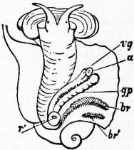
Fig. 22.—Female of Purpura lapillus removed from its shell; the mantle-skirt cut along its left line of attachment and thrown over to the right side of the animal so as to expose the organs on its inner face.
a, Anus. vg, Vagina. gp, Adrectal purpuriparous gland. r′, Aperture of the nephridium (kidney). br, Ctenidium (branchial plume). br′, Parabranchia (= the comb-like osphradium or olfactory organ).
The figures which are given here of various Pectinibranchia are in most cases sufficiently explained by the references attached to them. As an excellent general type of the nervous system, attention may be directed to that of Paludina drawn in fig. 21. On the whole the ganglia are strongly individualized in the Pectinibranchia, nerve-cell tissue being concentrated in the ganglia and absent from the cords. At the same time, the junction of the visceral loop above the intestine prevents in all Streptoneura the shortening of the visceral loop, and it is rare to find a fusion of the visceral ganglia with either pleural, pedal or cerebral—a fusion which can and does take place where the visceral loop is not above but below the intestine, e.g. in the Euthyneura (fig. 48), Cephalopoda and Lamellibranchia. As contrasted with the Aspidobranchia, we find that in the Pectinibranchia the pedal nerves are distinctly nerves given off from the pedal ganglia, rather than cord-like nerve-tracts containing both nerve-cells or ganglionic elements and nerve-fibres. Yet in some Pectinibranchia (Paludina) a ladder-like arrangement of the two pedal nerves and their lateral branches has been detected. The histology of the nervous system of Mollusca has yet to be seriously inquired into.
The alimentary canal of the Pectinibranchia presents little diversity of character, except in so far as the buccal region is concerned. Salivary glands are present, and in some carnivorous forms (Dolium) these secrete free sulphuric acid (as much as 2% is present in the secretion), which assists the animal in boring holes by means of its rasping tongue through the shells of other molluscs upon which it preys. A crop-like dilatation of the gut and a recurved intestine, embedded in the compact yellowish-brown liver, the ducts of which open into it, form the rest of the digestive tract and occupy a large bulk of the visceral hump. The buccal region presents a pair of shelly jaws placed laterally upon the lips, and a wide range of variation in the form of the denticles of the lingual ribbon or radula.
Well-developed glandular invaginations occur in different positions on the foot in Pectinibranchia. The most important of these opens by the ventral pedal pore, situated in the median line in the anterior half of the foot. This organ is probably homologous with the byssogenous gland of Lamellibranchs. The aperture, which was formerly supposed to be an aquiferous pore, leads into an extensive and often ramified cavity surrounded by glandular tubules. The gland has been found in both sub-orders of the Pectinibranchia, in Cyclostoma and Cypraea among the Taenioglossa, in Hemifusus, Cassis, Nassa, Murex, Fasciolariidae, Turbinellidae, Olividae, Marginellidae and Conidae among the Stenoglossa. It was discovered by J.T. Cunningham that in Buccinum the egg-capsules are formed by this pedal gland and not by any accessory organ of the generative system. Such horny egg-capsules doubtless have the same origin in all other species in which they occur, e.g. Fusus, Pyrula, Purpura, Murex, Nassa, Trophon, Voluta, &c. The float of the pelagic Janthina, to which the egg-capsules are attached, probably is also formed by the secretion of the pedal gland.
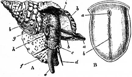
Fig. 23.—A, Triton variegatum, to show the proboscis or buccal introvert (e) in a state of eversion.
a, Siphonal notch of the shell occupied by the siphonal fold of the mantle-skirt (Siphonochlamyda). b, Edge of the mantle-skirt resting on the shell. c, Cephalic eye. d, Cephalic tentacle. e, Everted buccal introvert (proboscis). f, Foot. g, Operculum. h, Penis. i, Under surface of the mantle-skirt forming the roof of the sub-pallial chamber.
B, Sole of the foot of Pyrula tuba, to show a, the pore usually said to be “aquiferous” but probably the orifice of a gland; b, median line of foot.
Other glands opening on or near the foot are: (1) The suprapedal gland opening in the middle line between the snout and the anterior border of the foot. It is most commonly found in sessile forms and in terrestrial genera such as Cyclostoma; (2) the anterior pedal gland opening into the anterior groove of the foot, generally present in aquatic species; (3) dorsal posterior mucous glands in certain Cyclostomatidae.
The foot of the Pectinibranchia, unlike the simple muscular disk of the Isopleura and Aspidobranchia, is very often divided into lobes, a fore, middle and hind lobe (pro-, meso- and meta-podium, see figs. 24 and 25). Very usually, but not universally, the metapodium carries an operculum. The division of the foot into lobes is a simple case of that much greater elaboration or breaking up into processes and regions which it undergoes in the class Cephalopoda. Even among some Gastropoda (viz. the Opisthobranchia) we find the lobation of the foot still further carried out by the development of lateral lobes, the parapodia, whilst there are many Pectinibranchia, on the other hand, in which the foot has a simple oblong form without any trace of lobes.
The development of the Pectinibranchia has been followed in several examples, e.g. Paludina, Purpura, Nassa, Vermetus, Neritina. As in other Molluscan groups, we find a wide variation in the early process of the formation of the first embryonic cells, and their arrangement as a diblastula, dependent on the greater or less amount of food-yolk which is present in the egg-cell when it commences its embryonic changes. In fig. 26 the early stages of Paludina vivipara are represented. There is but very little food-material in the egg of this Pectinibranch, and consequently the diblastula forms by invagination; the blastopore or orifice of invagination coincides with the anus, and never closes entirely. A well-marked trochosphere is formed by the development of an equatorial ciliated band; and subsequently, by the disproportionate growth of the lower hemisphere, the trochosphere becomes a veliger. The primitive shell-sac or shell-gland is well marked at this stage, and the pharynx is seen as a new ingrowth (the stomodaeum), about to fuse with and open into the primitively invaginated arch-enteron (fig. 26, F).
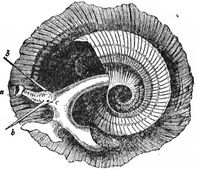
Fig. 24.—Animal and shell of Phorus exutus.
a, Snout (not introversible). b, Cephalic tentacles. c, Right eye. d, Pro- and meso-podium; to the right of this is seen the metapodium bearing the sculptured operculum.
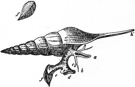
Fig. 25.—Animal and shell of Rostellaria rectirostris. (From Owen.)
a, Snout or rostrum. b, Cephalic tentacle. c, Eye. d, Propodium and mesopodium. e, Metapodium. f, Operculum. h′, Prolonged siphonal notch of the shell occupied by the siphon, or trough-like process of the mantle-skirt.
In other Pectinibranchia (and such variations are representative for all Mollusca, and not characteristic only of Pectinibranchia) we find that there is a very unequal division of the egg-cell at the commencement of embryonic development, as in Nassa. Consequently there is, strictly speaking, no invagination (emboly), but an overgrowth (epiboly) of the smaller cells to enclose the larger. The general features of this process and of the relation of the blastopore to mouth and anus have been explained in treating of the development of Mollusca generally. In such cases the blastopore may entirely close, and both mouth and anus develop as new ingrowths (stomodaeum and proctodaeum), whilst, according to the observations of N. Bobretzky, the closed blastopore may coincide in position with the mouth in some instances (Nassa, &c.), instead of with the anus. But in these epibolic forms, just as in the embolic Paludina, the embryo proceeds to develop its ciliated band and shell-gland, passing through the earlier condition of a trochosphere to that of the veliger. In the veliger stage many Pectinibranchia (Purpura, Nassa, &c.) exhibit, in the dorsal region behind the head, a contractile area of the body-wall. This acts as a larval heart, but ceases to pulsate after a time. Similar rhythmically contractile areas are found on the foot of the embryo Pulmonate Limax and on the yolk-sac (distended foot-surface) of the Cephalopod Loligo. The preconchylian invagination or shell-gland is formed in the embryo behind the velum, on the surface opposite the blastopore. It is surrounded by a ridge of cells which gradually extends over the visceral sac and secretes the shell. In forms which are naked in the adult state, the shell falls off soon after the reduction of the velum, but in Cenia, Runcina and Vaginula the shell-gland and shell are not developed, and the young animal when hatched has already the naked form of the adult.
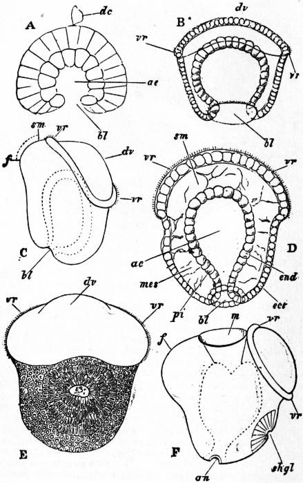
Fig. 26.—Development of the River-Snail, Paludina vivipara. (After Lankester, 17.)
dc, Directive corpuscle (outcast cell). ae, Arch-enteron or cavity lined by the enteric cell-layer or endoderm. bl, Blastopore. vr, Velum or circlet of ciliated cells. dv, Velar area or cephalic dome. sm, Site of the as yet unformed mouth. f, Foot. mes, Rudiments of the skeleto-trophic tissues. pi, The pedicle of invagination, the future rectum. shgl, The primitive shell-sac or shell-gland. m, Mouth. an, Anus.
A, Diblastula phase (optical section).
B, The diblastula has become a trochosphere by the development of the ciliated ring vr (optical section).
C, Side view of the trochosphere with commencing formation of the foot.
D, Further advanced trochosphere (optical section).
E, The trochosphere passing to the veliger stage, dorsal view showing the formation of the primitive shell-sac.
F, Side view of the same, showing foot, shell-sac (shgl), velum (vr), mouth and anus.
N.B.—In this development the blastopore is not elongated; it persists as the anus. The mouth and stomodaeum form independently of the blastopore.
One further feature of the development of the Pectinibranchia deserves special mention. Many Gastropoda deposit their eggs, after fertilization, enclosed in capsules; others, as Paludina, are viviparous; others, again, as the Zygobranchia, agree with the Lamellibranch Conchifera (the bivalves) in having simple exits for the ova without glandular walls, and therefore discharge their eggs unenclosed in capsules freely into the sea-water; such unencapsuled eggs are merely enclosed each in its own delicate chorion. When egg-capsules are formed they are often of large size, have tough walls, and in each capsule are several eggs floating in a viscid fluid. In some cases all the eggs in a capsule develop; in other cases one egg only in a capsule (Neritina), or a small proportion (Purpura, Buccinum), advance in development; the rest are arrested either after the first process of cell-division (cleavage) or before that process. The arrested embryos or eggs are then swallowed and digested by those in the same capsule which have advanced in development. This is clearly the same process in essence as that of the formation of a vitellogenous gland from part of the primitive ovary, or of the feeding of an ovarian egg by the absorption of neighbouring potential eggs; but here the period at which the sacrifice of one egg to another takes place is somewhat late. What it is that determines the arrest of some eggs and the progressive development of others in the same capsule is at present unknown.
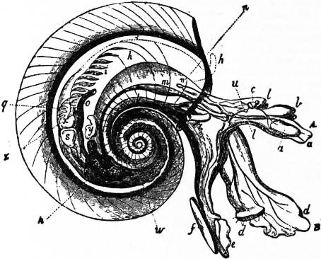
Fig. 27.—Oxygyrus Keraudrenii. (From Owen.)
a, Mouth and odontophore. b, Cephalic tentacles. c, Eye. d, Propodium (B) and mesopodium. e, Metapodium. f, Operculum. h, Mantle-chamber. i, Ctenidium (gill-plume). k, Retractor muscle of foot. l, Optic tentacle. m, Stomach. n, Dorsal surface overhung by the mantle-skirt; the letter is close to the salivary gland. o, Rectum and anus. p, Liver. q, Renal organ (nephridium). s, Ventricle. u, The otocyst attached to the cerebral ganglion. w, Testis. x, Auricle of the heart. y, Vesicle on genital duct. z, Penis.
In the tribe of Pectinibranchia called Heteropoda the foot takes the form of a swimming organ. The nervous system and sense organs are highly developed. The odontophore also is remarkably developed, its lateral teeth being mobile, and it serves as an efficient organ for attacking the other pelagic forms on which the Heteropoda prey. The sexes are distinct, as in all Streptoneura; and genital ducts and accessory glands and pouches are present, as in all Pectinibranchia. The Heteropoda exhibit a series of modifications in the form and proportions of the visceral mass and foot, leading from a condition readily comparable with that of a typical Pectinibranch such as Rostellaria, with the three regions of the foot strongly marked and a coiled visceral hump of the usual proportions, up to a condition in which the whole body is of a tapering cylindrical shape, the foot a plate-like vertical fin, and the visceral hump almost completely atrophied. Three steps of this modification may be distinguished as three families:—Atlantidae, Carinariidae and Pterotrachaeidae. They are true Pectinibranchia which have taken to a pelagic life, and the peculiarities of structure which they exhibit are strictly adaptations consequent upon their changed mode of life. Such adaptations are the transparency and colourlessness of the tissues, and the modifications of the foot, which still shows in Atlanta the form common in Pectinibranchia (compare fig. 27 and fig. 24). The cylindrical body of Pterotrachaea is paralleled by the slug-like forms of Euthyneura. J.W. Spengel has shown that the visceral loop of the Heteropoda is streptoneurous. Special to the Heteropoda is the high elaboration of the lingual ribbon, and, as an agreement with some of the opisthobranchiate Euthyneura, but as a difference from the Pectinibranchia, we find the otocysts closely attached to the cerebral ganglia. This is, however, less of a difference than it was at one time supposed to be, for it has been shown by H. Lacaze-Duthiers, and also by F. Leydig, that the otocysts of Pectinibranchia even when lying close upon the pedal ganglion (as in fig. 21) yet receive their special nerve (which can sometimes be readily isolated) from the cerebral ganglion (see fig. 11). Accordingly the difference is one of position of the otocyst and not of its nerve-supply. The Heteropoda are further remarkable for the high development of their cephalic eyes, and for the typical character of their osphradium (Spengel’s olfactory organ). This is a groove, the edges of which are raised and ciliated, lying near the branchial plume in the genera which possess that organ, whilst in Firoloida, which has no branchial plume, the osphradium occupies a corresponding position. Beneath the ciliated groove is placed an elongated ganglion (olfactory ganglion) connected by a nerve to the supra-intestinal (therefore the primitively dextral) ganglion of the long visceral nerve-loop, the strands of which cross one another—this being characteristic of Streptoneura (Spengel).
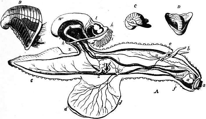
Fig. 28.—Carinaria mediterranea. (From Owen.) A, The animal. B, The shell removed. C, D, Two views of the shell of Cardiopoda.
a, Mouth and odontophore. b, Cephalic tentacles. c, Eye. d, The fin-like mesopodium. d’, Its sucker. e, Metapodium. f, Salivary glands. h, Border of the mantle-flap. i, Ctenidium (gill-plume). m, Stomach. n, Intestine. o, Anus. p, Liver. t, Aorta, springing from the ventricle. u, Cerebral ganglion. v, Pleural and pedal ganglion. w, Testis. x, Visceral ganglion. y, Vesicula seminalis. z, Penis.
The Heteropoda belong to the “pelagic fauna” occurring near the surface in the Mediterranean and great oceans in company with the Pteropoda, the Siphonophorous Hydrozoa, Salpae, Leptocephali, and other specially-modified transparent swimming representatives of various groups of the animal kingdom. In development they pass through the typical trochosphere and veliger stages provided with boat-like shell.
Sub-order 1.—Taenioglossa. Radula with a median tooth and three teeth on each side of it. Formula 3 : 1 : 3.
Tribe 1.—Platypoda. Normal Taenioglossa of creeping habit. The foot is flattened ventrally, at all events in its anterior part (Strombidae). Otocysts situated close to the pedal nerve-centres. Accessory organs are rarely found on the genital ducts, but occur in Paludina, Cyclostoma, Naticidae, Calyptraeidae, &c. Mandibles usually present. This is the largest group of Mollusca, including nearly sixty families, some of which are insufficiently known from the anatomical point of view.
Fam. 1.—Paludinidae. Pedal centres in the form of ganglionated cords; kidney provided with a ureter; viviparous; fluviatile. Paludina. Neothauma, from Lake Tanganyika. Tylopoma, extinct, Tertiary.
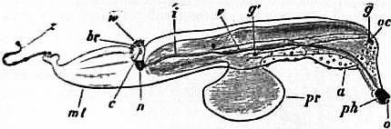
Fig. 29.—Pterotrachea mutica seen from the right side. (After Keferstein.)
a, Pouch for reception of the snout when retracted. c, Pericardium. ph, Pharynx. oc, Cephalic eye. g, Cerebral ganglion. g’, Pleuro-pedal ganglion. pr, Foot (mesopodium). v, Stomach. i, Intestine. n, So-called nucleus. br, Branchial plume (ctenidium). w, Osphradium. mt, Foot (metapodium). z, Caudal appendage.
Fam. 2.—Cyclophoridae. No ctenidium, pallial cavity transformed into a lung; aperture of shell circular; terrestrial. Pomatias, shell turriculated. Diplommatina. Hybocystis. Cyclophorus, shell umbilicated, with a short spire and horny operculum. Cyclosurus, shell uncoiled. Dermatocera, foot with a horn-shaped protuberance at its posterior end. Spiraculum.
Fam. 3.—Ampullariidae. To the left of the ctenidium a pulmonary sac, separated from it by an incomplete septum, amphibious. Ampullaria, shell dextral, coiled. Lanistes, shell sinistral, spire short or obsolete. Meladomus.
Fam. 4.—Littorinidae. Oesophageal pouches present; pedal nerve-centres concentrated; a pedal penis near the right tentacle. Littorina, shell not umbilicated, littoral habit. Lacuna, foot with two posterior appendages, marine, entirely aquatic. Cremnoconchus, entirely aerial, Indian. Risella. Tectarius.
Fam. 5.—Fossaridae. Head with two lobes in some Rhipidoglossa. Fossaria.
Fam. 6.—Purpurinidae, extinct.
Fam. 7.—Planaxidae. Shell with pointed spire; a short pallial siphon. Planaxis.
Fam. 8.—Cyclostomatidae. Pallial cavity transformed into a lung; pedal centres concentrated; a deep pedal groove. Cyclostoma, shell turbinated, operculum calcareous, British. Omphalotropis.
Fam. 9.—Aciculidae. Pallial cavity transformed into a lung; operculum horny; shell narrow and elongated. Acicula.
Fam. 10.—Valvatidae. Ctenidium bipectinate, free; hermaphrodite; fluviatile. Valvata, British.
Fam. 11.—Rissoidae. Epipodial filaments present; one or two pallial tentacles. Rissoa. Rissoina. Stiva.
Fam. 12.—Litiopidae. An epipodium bearing three pairs of tentacles and an operculigerous lobe with two appendages; inhabitants of the Sargasso weed. Litiopa.
Fam. 13.—Adeorbiidae. Mantle with two posterior appendages; ctenidium large and capable of protrusion from pallial cavity. Adeorbis, British.
Fam. 14.—Jeffreysiidae. Head with two long labial palps; shell ovoid; operculum horny, semicircular, carinated. Jeffreysia.
Fam. 15.—Homalogyridae. Shell flattened; no cephalic tentacles. Homalogyra, British. Ammoniceras.
Fam. 16.—Skeneidae. Shell depressed, with rounded aperture; cephalic tentacles long. Skenea, British.
Fam. 17.—Choristidae. Shell spiral; four cephalic tentacles; eyes absent; two pedal appendages. Choristes.
Fam. 18.—Assimineidae. Eyes at free extremities of tentacles. Assiminea, estuarine, British.
Fam. 19.—Truncatellidae. Snout very long, bilobed; foot short. Truncatella.
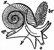
Fig. 30.—Valvata cristata, Müll.
o, Mouth. op, Operculum. br, Ctenidium (branchial plume). x, Filiform appendage (? rudimentary ctenidium).
The freely projecting ctenidium of typical form not having its axis fused to the roof of the branchial chamber is the notable character of this genus.
Fam. 20.—Hydrobiidae. Shell with prominent spire; penis distant from right tentacle, generally appendiculated; brackish water or fluviatile. Hydrobia, British. Baikalia, from Lake Baikal. Pomatiopsis. Bithynella. Lithoglyphus. Spekia, viviparous, from Lake Tanganyika. Tanganyicia. Limnotrochus, from Lake Tanganyika. Chytra. Littorinida. Bithynia, British, fluviatile. Stenothyra.
Fam. 21.—Melaniidae. Spire of shell somewhat elongated; mantle-border fringed; viviparous; fluviatile. Melania. Faunus. Paludomus. Melanopsis. Nassopsis. Bythoceras, from Lake Tanganyika.
Fam. 22.—Typhobiidae. Foot wide; shell turriculated, with carinated whorls, the carinae tuberculated or spiny. Typhobia. Bathanalia, from Lake Tanganyika.
Fam. 23.—Pleuroceridae. Like Melaniidae, but mantle-border not fringed and reproduction oviparous. Pleurocera. Anculotus.
Fam. 24.—Pseudomelaniidae. All extinct.
Fam. 25.—Subulitidae. All extinct.
Fam. 26.—Nerineidae. All extinct.
Fam. 27.—Cerithiidae. Shell with numerous tuberculated whorls; aperture canaliculated anteriorly; short pallial siphon. Cerithium. Bittium. Potamides. Triforis. Laeocochlis. Cerithiopsis.
Fam. 28.—Modulidae. Shell with short spire; no siphon. Modulus.
Fam. 29.—Vermetidae. Animal fixed by the shell, the last whorls of which are not in contact with each other; foot small; two anterior pedal tentacles. Vermetus. Siliquaria.
Fam. 30.—Caecidae. Shell almost completely uncoiled, in one plane, with internal septa. Caecum, British.
Fam. 31.—Turritellidae. Shell very long; head large; foot broad. Turritella, British. Mesalia. Mathilda.
Fam. 32.—Struthiolariidae. Shell conical; aperture slightly canaliculated; siphon slightly developed. Struthiolaria.
Fam. 33.—Chenopodidae. Shell elongated; aperture expanded; siphon very short. Chenopus, British. Alaria, Spinigera, Diartema, extinct.
Fam. 34.—Strombidae. Foot narrow, compressed, without sole. Strombus. Pteroceras. Rostellaria. Terebellum.
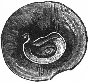
Fig. 31.—Shell of Crucibulum, seen from below so as to show the inner whorl b, concealed by the cap-like outer whorl a.
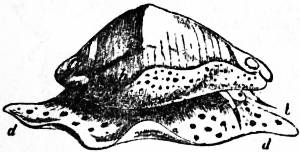
Fig. 32.—Animal and shell of Ovula.
b, Cephalic tentacles. d, Foot. h, Mantle-skirt, which is naturally carried in a reflected condition so as to cover the sides of the shell.
Fam. 35.—Xenophoridae. Foot transversely divided into two parts. Xenophorus. Eotrochus, Silurian.
Fam. 36.—Capulidae. Shell conical, not coiled, but slightly incurved posteriorly; a tongue-shaped projection between snout and foot. Capulus. Thyca, parasitic on asterids. Platyceras, extinct.
Fam. 37.—Hipponycidae. Shell conical; foot secreting a ventral calcareous plate; animal fixed. Hipponyx. Mitrularia.
Fam. 38.—Calyptraeidae. Shell with short spire; lateral cervical lobes present; accessory genital glands. Calyptraea, British. Crepidula. Crucibulum.
Fam. 39.—Naricidae. Foot divided into two, posterior half bearing the operculum; a wide epipodial velum; shell turbinated. Narica.
Fam. 40.—Naticidae. Foot large, with aquiferous system; propodium reflected over head; eyes degenerate; burrowing habit. Natica, British. Amaura. Sigaretus.
Fam. 41.—Lamellariidae. Shell thin, more or less covered by the mantle; no operculum. Lamellaria. Velutina. Marsenina, Oncidiopsis, hermaphrodite.
Fam. 42.—Trichotropidae. Shell with short spire, carinate and pointed. Trichotropis.
Fam. 43.—Seguenziidae. Shell trochiform, with canaliculated aperture and twisted columella. Seguenzia, abyssal.
Fam. 44.—Janthinidae. Shell thin; operculum absent; tentacles bifid; foot secretes a float; pelagic. Janthina. Recluzia.
Fam. 45.—Cypraeidae. Shell inrolled, solid, polished, aperture very narrow in adult; short siphon; anus posterior; osphradium with three lobes; mantle reflected over shell. Cypraea. Pustularia. Ovula. Pedicularia, attached to corals. Erato.
Fam. 46.—Tritonidae. Shell turriculated and siphonated, thick, each whorl with varices; foot broad and truncated anteriorly; pallial siphon well developed; proboscis present. Triton. Persona. Ranella.