VOLUME XI SLICE V Gassendi, Pierre to Geocentric (continued 2)
Reading edition
Fam. 47.—Columbellinidae. All extinct.
Fam. 48.—Cassididae. Shell ventricose, with elongated aperture, and short spire; proboscis and siphon long; operculum with marginal nucleus. Cassis. Cassidaria. Oniscia.
Fam. 49—Oocorythidae. Shell globular and ventricose; aperture oval and canaliculated; operculum spiral. Oocorys, abyssal.
Fam. 50.—Doliidae. Shell ventricose, with short spire, and wide aperture; no varices and no operculum; foot very broad, with projecting anterior angles; siphon long. Dolium. Pyrula.
Fam. 51.—Solariidae. Solarium. Torinia. Fluxina.
Fam. 52.—Scalariidae. Shell turriculated, with elongated spire; proboscis short; siphon rudimentary. Scalaria. Eglisia. Crossea. Aclis.
The three following families have neither radula nor jaws, and are therefore called Aglossa. They have a well-developed proboscis which is used as a suctorial organ; some are abyssal, but the majority are either commensals or parasites of Echinoderms.
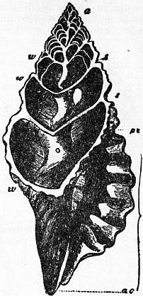
Fig. 33.—Section of the shell of Triton, Cuv. (From Owen.)
a, Apex. ac, Siphonal notch of the mouth of the shell. ac to pc, Mouth of the shell. w, w, Whorls of the shell. s, s. Sutures.
Occupying the axis, and exposed by the section, is seen the “columella” or spiral pillar. The upper whorls of the shell are seen to be divided into separate chambers by the formation of successively formed “septa.”
Fam. 53.—Pyramidellidae. Summit of spire heterostrophic; a projection, the mentum, between head and foot; operculum present. Pyramidella. Turbonilla. Odostomia, British. Myxa.
Fam. 54.—Eulimidae. Visceral mass still coiled spirally; shell thin and shining. Eulima, foot well developed, with an operculum, animal usually free, but some live in the digestive cavity of Holothurians. Mucronalia, foot reduced, but still operculate, eyes present, animal fixed by its very long proboscis which is deeply buried in the tissues of an Echinoderm, no pseudopallium. Stylifer, the operculum is lost, animal fixed by a large proboscis which forms a pseudopallium covering the whole shell except the extremity of the spire, parasitic on all groups of Echinoderms. Entosiphon, visceral mass still coiled; shell much reduced, proboscis very long forming a pseudopallium which covers the whole body and projects beyond in the form of a siphon, foot and nervous system present, eyes, branchia and anus absent, parasite in the Holothurian Deima blakei in the Indian Ocean.
Fam. 55.—Entoconchidae. No shell; visceral mass not coiled; no sensory organs, nervous system, branchia or anus; body reduced to a more or less tubular sac; hermaphrodite and viviparous; parasitic in Holothurians; larvae are veligers, with shell and operculum. Entocolax, mouth at free extremity, animal fixed by aboral orifice of pseudopallium, Pacific. Entoconcha, body elongated and tubular, animal fixed by the oral extremity, protandric hermaphrodite, parasitic in testes of Holothurians causing their abortion. Enteroxenos, no pseudopallium and no intestine, hermaphrodite, larvae with operculum.
Tribe 2.—Heteropoda. Pelagic Taenioglossa with foot large and laterally compressed to form a fin.
Fam. 1. Atlantidae. Visceral sac and shell coiled in one plane; foot divided transversely into two parts, posterior part bearing an operculum, anterior part forming a fin provided with a sucker. Atlanta. Oxygyrus.
Fam. 2.—Carinariidae. Visceral sac and shell small in proportion to the rest of the body, which cannot be withdrawn into the shell; foot elongated, fin-shaped, with sucker, but without operculum. Carinaria. Cardiopoda.
Fam. 3.—Pterotrachaeidae. Visceral sac very much reduced; without shell or mantle; anus posterior; foot provided with sucker in male only. Pterotrachaea. Firoloida. Pterosoma.
Sub-order 2.—Stenoglossa. Radula narrow with one lateral tooth on each side, and one median tooth or none.
Tribe 1.—Rachiglossa. Radula with a median tooth and a single tooth on each side of it. Formula 1 : 1 : 1. Rudimentary jaws present.
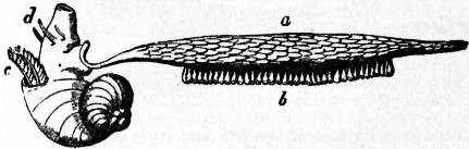
Fig. 34.—Female Janthina, with egg-float (a) attached to the foot; b, egg-capsules; c, ctenidium (gill-plume); d, cephalic tentacles.
Fam. 1.—Turbinellidae. Shell solid, piriform, with thick folded columella; lateral teeth of radula bicuspidate. Turbinella. Cynodonta. Fulgur. Hemifusus. Tudicla. Strepsidura.
Fam. 2.—Fasciolariidae. Shell elongated, with long siphon; lateral teeth of radula multicuspidate. Fasciolaria. Fusus. Clavella. Latirus.
Fam. 3.—Mitridae. Shell fusiform and solid, aperture elongated, columella folded; no operculum; eyes on sides of tentacles. Mitra. Turricula. Cylindromitra. Imbricaria.
Fam. 4.—Buccinidae. Foot large and broad; eyes at base of tentacles; operculum horny. Buccinum. Chrysodomus. Liomesus. Cominella. Tritonidea. Pisania. Euthria. Phos. Dipsacus.
Fam. 5.—Nassidae. Foot broad, with two slender posterior appendages; operculum unguiculate. Nassa, marine, British. Canidia, fluviatile. Bullia.
Fam. 6.—Muricidae. Shell with moderately long spire and canal, ornamented with ribs, often spiny; foot truncated anteriorly. Murex, British. Trophon, British. Typhis. Urosalpinx. Lachesis.
Fam. 7.—Purpuridae. Shell thick, with short spire, last whorl large and canal short; aperture wide; operculum horny. Purpura, British. Rapana. Monoceros. Sistrum. Concholepas.
Fam. 8.—Haliidae. Shell ventricose, thin and smooth, with wide aperture; foot large and thick, without operculum. Halia.
Fam. 9.—Cancellariidae. Shell ovoid, with short spire and folded columella; foot small, no operculum; siphon short. Cancellaria.
Fam. 10.—Columbellidae. Spire of shell prominent, aperture narrow, canal very short, columella crenelated; foot large. Columbella.
Fam. 11.—Coralliophilidae. Shell irregular; radula absent; foot and siphon short; sedentary animals, living in corals. Coralliophila. Rhizochilus. Leptoconchus. Magilus. Rapa.
Fam. 12.—Volutidae. Head much flattened and wide, with eyes on sides; foot broad; siphon with internal appendages. Valuta. Guivillea. Cymba.
Fam. 13.—Olividae. Foot with anterior transverse groove; a posterior pallial tentacle; generally burrowing. Olivia. Olivella. Ancillaria. Agaronia.
Fam. 14.—Marginellidae. Foot very large; mantle reflected over shell. Marginella. Pseudomarginella.
Fam. 15.—Harpidae. Foot very large; without operculum; shell with short spire and longitudinal ribs; siphon long. Harpa.
Tribe 2.—Toxiglossa. No jaws. No median tooth in radula. Formula: 1 : 0 : 1. Poison-gland present whose duct traverses the nerve-collar.
Fam. 1.—Pleurotomatidae. Shell fusiform, with elongated spire; margin of shell and mantle notched. Pleurotoma. Clavatula. Mangilia. Bela. Pusionella. Pontiothauma.
Fam. 2.—Terebridae. Shell turriculated, with numerous whorls; aperture and operculum oval; eyes at summits of tentacles; siphon long. Terebra.
Fam. 3.—Conidae. Shell conical, with very short spire, and narrow aperture with parallel borders; operculum unguiform Conus.
Sub-Class II.—Euthyneura
The most important general character of the Euthyneura is the absence of torsion in the visceral commissure, and the more posterior position of the anus and pallial organs. Comparative anatomy and embryology prove that this condition is due, not as formerly supposed to a difference in the relations of the visceral commissure which prevented it from being included in the torsion of the visceral hump, but to an actual detorsion which has taken place in evolution and is repeated to a great extent in individual development. In several of the more primitive forms the same torsion occurs as in Streptoneura, viz. in Actaeon and Limacina among Opisthobranchia, and Chilina among Pulmonata. Actaeon is proso-branchiate, the visceral commissure is twisted in Actaeon and Chilina, and even slightly still in Bulla and Scaphander; in Actaeon and Limacina the osphradium is to the left, innervated by the supra-intestinal ganglion. But in the other members of the sub-class the detorsion of the visceral mass has carried back the anus and circumanal complex from the anterior dorsal region to the right side, as in Bulla and Aplysia, or even to the posterior end of the body, as in Philine, Oncidium, Doris, &c. Different degrees of the same process of detorsion are, as we have seen, exhibited by the Heteropoda among the Streptoneura, and both in them and in the Euthyneura the detorsion is associated with degeneration of the shell. Where the modification is carried to its extreme degree, not only the shell but the pallial cavity, ctenidium and visceral hump disappear, and the body acquires a simple elongated form and a secondary external symmetry, as in Pterotrachaea and in Doris, Eolis, and other Nudibranchia. These facts afford strong support to the hypothesis that the weight of the shell is the original cause of the torsion of the dorsal visceral mass in Gastropods. But this hypothesis leaves the elevation of the visceral mass and the exogastric coiling of the shell in the ancestral form unexplained. In those Euthyneura in which the shell is entirely absent in the adult, it is, except in the three genera Cenia, Runcina and Vaginula, developed in the larva and then falls off. In other cases (Tectibranchs) the reduced shell is enclosed by upgrowths of the edge of the mantle and becomes internal, as in many Cephalopods. A few Euthyneura in which the shell is not much reduced retain an operculum in the adult state, e.g. Actaeon, Limacina, and the marine Pulmonate, Amphibola. The detorted visceral commissure shows a tendency to the concentration of all its elements round the oesophagus, so that except in the Bullomorpha and in Aplysia the whole nervous system is aggregated in the cephalic region, either dorsally or ventrally. The radula has a number of uniform teeth on each side of the median tooth in each transverse row. The head in most cases bears two pairs of tentacles. All the Euthyneura are hermaphrodite.
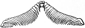
Fig. 35.—Acera bullata. A single row of teeth of the Radula. (Formula, x.l.x.)
In the most primitive condition the genital duct is single throughout its length and has a single external aperture; it is therefore said to be monaulic. The hermaphrodite aperture is on the right side near the opening of the pallial cavity, and a ciliated groove conducts the spermatozoa to the penis, which is situated more anteriorly. This is the condition in the Bullomorpha, the Aplysiomorpha, and in one Pulmonate, Pythia. In some cases while the original aperture remains undivided, the seminal groove is closed and so converted into a canal. This is the modification found in Cavolinia longirostris among the Bullomorpha, and in all the Auriculidae except Pythia. A further degree of modification occurs when the male duct takes its origin from the hermaphrodite duct above the external opening, so that there are two distinct apertures, one male and one female, the latter being the original opening. The genital duct is now said to be diaulic, as in Valvata, Oncidiopsis, Actaeon, and Lobiger among the Bullomorpha, in the Pleurobranchidae, in the Nudibranchia, except the Doridomorpha and most of the Elysiomorpha, and in the Pulmonata. Originally in this condition the female aperture is at some distance from the male, as in the Basommatophora and in other cases; but in some forms the female aperture itself has shifted and come to be contiguous with the male opening and penis as in the Stylommatophora. In all these cases the female duct bears a bursa copulatrix or receptaculum seminis. In some forms this receptacle acquires a separate external opening remaining connected with the oviduct internally. There are thus two female openings, one for copulation, the other for oviposition, as well as a male opening. The genital duct is now trifurcated or triaulic, a condition which is confined to certain Nudibranchs, viz. the Doridomorpha and most of the Elysiomorpha.
The Pteropoda, formerly regarded as a distinct class of the Mollusca, were interpreted by E.R. Lankester as a branch of the Cephalopoda, chiefly on account of the protrusible sucker-bearing processes at the anterior end of Pneumonoderma. These he considered to be homologous with the arms of Cephalopods. He fully recognized, however, the similarity of Pteropods to Gastropods in their general asymmetry and in the torsion of the visceral mass in Limacinidae. It is now understood that they are Euthyneurous Gastropods adapted to natatory locomotion and pelagic life. The sucker-bearing processes of Pneumonoderma are outgrowths of the proboscis. The fins of Pteropods are now interpreted as the expanded lateral margins of the foot, termed parapodia, not homologous with the siphon of Cephalopods which is formed from epipodia. The Thecosomatous Pteropoda are allied to Bulla, the Gymnosomatous forms to Aplysia. The Euthyneura comprises two orders, Opisthobranchia and Pulmonata.
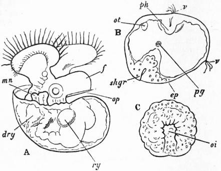
Fig. 36.
A, Veliger-larva of an Opisthobranch (Polycera). f, Foot; op, operculum; mn, anal papilla; ry, dry, two portions of unabsorbed nutritive yolk on either side of the intestine. The right otocyst is seen at the root of the foot.
B, Trochosphere of an Opisthobranch (Pleurobranchidium) showing—shgr, the shell-gland or primitive shell-sac; v, the cilia of the velum; ph, the commencing stomodaeum or oral invagination; ot, the left otocyst; pg, red-coloured pigment spot.
C, Diblastula of an Opisthobranch (Polycera) with elongated blastopore oi.
(All from Lankester.)
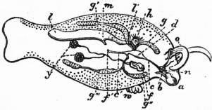
Fig. 37.—Phyllirhoë bucephala, twice the natural size, a transparent pisciform pelagic Opisthobranch. The internal organs are shown as seen by transmitted light. (After W. Keferstein.)
a, Mouth. b, Radular sac. c, Oesophagus. d, Stomach. c’, Intestine. f’, Anus. g, g′, g″, g″′, The four lobes of the liver. h, The heart (auricle and ventricle). l, The renal sac (nephridium). l′, The ciliated communication of the renal sac with the pericardium. m, The external opening of the renal sac. n, The cerebral ganglion. o, The cephalic tentacles. f, The genital pore. y, The ovo-testes. w, The parasitic hydromedusa Mnestra, usually found attached in this position by the aboral pole of its umbrella.
Order 1.—Opisthobranchia. Marine Euthyneura, the more archaic forms of which have a relatively large foot and a small visceral hump, from the base of which projects on the right side a short mantle-skirt. The anus is placed in such forms far back beyond the mantle-skirt. In front of the anus, and only partially covered by the mantle-skirt, is the ctenidium with its free end turned backwards. The heart lies in front of, instead of to the side of, the attachment of the ctenidium—hence Opisthobranchia as opposed to “Prosobranchia,” which correspond to the Streptoneura. A shell is possessed in the adult state by but few Opisthobranchia, but all pass through a veliger larval stage with a nautiloid shell (fig. 36). Many Opisthobranchia have by a process of atrophy lost the typical ctenidium and the mantle-skirt, and have developed other organs in their place. As in some Pectinibranchia, the free margin of the mantle-skirt is frequently reflected over the shell when a shell exists; and, as in some Pectinibranchia, broad lateral outgrowths of the foot (parapodia) are often developed which may be thrown over the shell or naked dorsal surface of the body.
The variety of special developments of structure accompanying the atrophy of typical organs in the Opisthobranchia and general degeneration of organization is very great. The members of the order present the same wide range of superficial appearance as do the Pectinibranchiate Streptoneura, forms carrying well-developed spiral shells and large mantle-skirts being included in the group, together with flattened or cylindrical slug-like forms. But in respect of the substitution of other parts for the mantle-skirt and for the gill which the more degenerate Opisthobranchia exhibit, this order stands alone. Some Opisthobranchia are striking examples of degeneration (some Nudibranchia), having none of those regions or processes of the body developed which distinguish the archaic Mollusca from such flat-worms as the Dendrocoel Planarians. Indeed, were it not for their retention of the characteristic odontophore we should have little or no indication that such forms as Phyllirhoë and Limapontia really belong to the Mollusca at all. The interesting little Rhodope veranyii, which has no odontophore, has been associated by systematists both with these simplified Opisthobranchs and with Rhabdocoel Planarians.
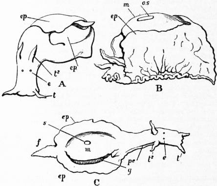
Fig. 38.—Three views of Aplysia sp., in various conditions of expansion and retraction. (After Cuvier.)
t, Anterior cephalic tentacles. t², Posterior cephalic tentacles. e, Eyes. f, Metapodium. ep, Epipodium. g, Gill-plume (ctenidium). m, Mantle-flap reflected over the thin oval shell. os, s, Orifice formed by the unclosed border of the reflected mantle-skirt, allowing the shell to show. pe, The spermatic groove.
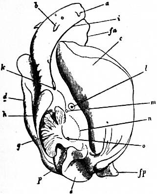
Fig. 39.—Aplysia leporina (camelus, Cuv.), with epipodia and mantle reflected away from the mid-line. (Lankester.)
a, Anterior cephalic tentacle. b, Posterior cephalic tentacle; between a and b, the eyes. c, Right epipodium. d, Left epipodium. e, Hinder part of visceral hump. fp, Posterior extremity of the foot. fa, Anterior part of the foot underlying the head. g, The ctenidium (branchial plume). h, The mantle-skirt tightly spread over the horny shell and pushed with it towards the left side. i, The spermatic groove. k, The common genital pore (male and female). l, Orifice of the grape-shaped (supposed poisonous) gland. m, The osphradium (olfactory organ of Spengel). n, Outline of part of the renal sac (nephridium) below the surface. o, External aperture of the nephridium. p, Anus.
In many respects the sea-hare (Aplysia), of which several species are known (some occurring on the English coast), serves as a convenient example of the fullest development of the organization characteristic of Opisthobranchia. The woodcut (fig. 38) gives a faithful representation of the great mobility of the various parts of the body. The head is well marked and joined to the body by a somewhat constricted neck. It carries two pairs of cephalic tentacles and a pair of sessile eyes. The visceral hump is low and not drawn out into a spire. The foot is long, carrying the oblong visceral mass upon it, and projecting (as metapodium) a little beyond it (f). Laterally the foot gives rise to a pair of mobile fleshy lobes, the parapodia (ep), which can be thrown up so as to cover in the dorsal surface of the animal. Such parapodia are common, though by no means universal, among Opisthobranchia. The torsion of the visceral hump is not carried out very fully, the consequence being that the anus has a posterior position a little to the right of the median line above the metapodium, whilst the branchial chamber formed by the overhanging mantle-skirt faces the right side of the body instead of lying well to the front as in Streptoneura and as in Pulmonate Euthyneura. The gill-plume, which in Aplysia is the typical Molluscan ctenidium, is seen in fig. 39 projecting from the branchial sub-pallial space. The relation of the delicate shell to the mantle is peculiar, since it occupies an oval area upon the visceral hump, the extent of which is indicated in fig. 38, C, but may be better understood by a glance at the figures of the allied genus Umbrella (fig. 40), in which the margin of the mantle-skirt coincides, just as it does in the limpet, with the margin of the shell. But in Aplysia the mantle is reflected over the edge of the shell, and grows over its upper surface so as to completely enclose it, excepting at the small central area s where the naked shell is exposed. This enclosure of the shell is a permanent development of the arrangement seen in many Streptoneura (e.g. Pyrula, Ovula, see figs. 18 and 32), where the border of the mantle can be, and usually is, drawn over the shell, though it is withdrawn (as it cannot be in Aplysia) when they are irritated. From the fact that Aplysia commences its life as a free-swimming veliger with a nautiloid shell not enclosed in any way by the border of the mantle, it is clear that the enclosure of the shell in the adult is a secondary process. Accordingly, the shell of Aplysia must not be confounded with a primitive shell in its shell-sac, such as we find realized in the shells of Chiton and in the plugs which form in the remarkable transitory “shell-sac” or “shell-gland” of Molluscan embryos (see figs. 26, 60). Aplysia, like other Mollusca, develops a primitive shell-sac in its trochosphere stage of development, which disappears and is succeeded by a nautiloid shell (fig. 36). This forms the nucleus of the adult shell, and, as the animal grows, becomes enclosed by a reflection of the mantle-skirt. When the shell of an Aplysia enclosed in its mantle is pushed well to the left, the sub-pallial space is fully exposed as in fig. 39, and the various apertures of the body are seen. Posteriorly we have the anus, in front of this the lobate gill-plume, between the two (hence corresponding in position to that of the Pectinibranchia) we have the aperture of the renal organ. In front, near the anterior attachment of the gill-plume, is the osphradium (olfactory organ) discovered by J.W. Spengel, yellowish in colour, in the typical position, and overlying an olfactory ganglion with typical nerve-connexion (see fig. 43). To the right of Spengel’s osphradium is the opening of a peculiar gland which has, when dissected out, the form of a bunch of grapes; its secretion is said to be poisonous. On the under side of the free edge of the mantle are situated the numerous small cutaneous glands which, in the large Aplysia camelus (not in other species), form the purple secretion which was known to the ancients. In front of the osphradium is the single genital pore, the aperture of the common or hermaphrodite duct. From this point there passes forward to the right side of the head a groove—the spermatic groove—down which the spermatic fluid passes. In other Euthyneura this groove may close up and form a canal. At its termination by the side of the head is the muscular introverted penis. In the hinder part of the foot (not shown in any of the diagrams) is the opening of a large mucus-forming gland very often found in the Molluscan foot.
With regard to internal organization we may commence with the disposition of the renal organ (nephridium), the external opening of which has already been noted. The position of this opening and other features of the renal organ were determined by J.T. Cunningham.
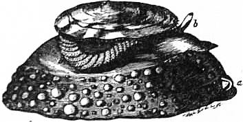
Fig. 40.—Umbrella mediterranea. a, mouth; b, cephalic tentacle; h, gill (ctenidium). The free edge of the mantle is seen just below the margin of the shell (compare with Aplysia, fig. 39). (From Owen.)
There is considerable uncertainty with respect to the names of the species of Aplysia. There are two forms which are very common in the Gulf of Naples. One is quite black in colour, and measures when outstretched 8 or 9 in. in length. The other is light brown and somewhat smaller, its length usually not exceeding 7 in. The first is flaccid and sluggish in its movements, and has not much power of contraction; its epipodial lobes are enormously developed and extend far forward along the body; it gives out when handled an abundance of purple liquid, which is derived from cutaneous glands situated on the under side of the free edge of the mantle. According to F. Blochmann it is identical with A. camelus of Cuvier. The other species is A. depilans; it is firm to the touch, and contracts forcibly when irritated; the secretion of the mantle-glands is not abundant, and is milky white in appearance. The kidney has similar relations in both species, and is identical with the organ spoken of by many authors as the triangular gland. Its superficial extent is seen when the folds covering the shell are cut away and the shell removed; the external surface forms a triangle with its base bordering the pericardium, and its apex directed posteriorly and reaching the the left-hand posterior corner of the shell-chamber. The dorsal surface of the kidney extends to the left beyond the shell-chamber beneath the skin in the space between the shell-chamber and the left parapodium.
When the animal is turned on its left-hand side and the mantle-chamber widely opened, the gill being turned over to the left, a part of the kidney is seen beneath the skin between the attachment of the gill and the right parapodium (fig. 39). On examination this is found to be the under surface of the posterior limb of the gland, the upper surface of which has just been described as lying beneath the shell. In the posterior third of this portion, close to that edge which is adjacent to the base of the gill, is the external opening (fig. 39, o).
When the pericardium is cut open from above in an animal otherwise entire, the anterior face of the kidney is seen forming the posterior wall of the pericardial chamber; on the deep edge of this face, a little to the left of the attachment of the auricle to the floor of the pericardium, is seen a depression; this depression contains the opening from the pericardium into the kidney.
To complete the account of the relations of the organ: the right anterior corner can be seen superficially in the wall of the mantle-chamber above the gill. Thus the base of the gill passes in a slanting direction across the right-hand side of the kidney, the posterior end being dorsal to the apex of the gland, and the anterior end ventral to the right-hand corner.
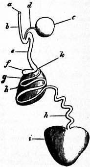
Fig. 41.—Gonad, and accessory glands and ducts of Aplysia. (Lankester.)
i, Ovo-testis. h, Hermaphrodite duct. g, Albuminiparous gland. f, Vesicula seminalis. k, Opening of the albuminiparous gland into the hermaphrodite duct. e, Hermaphrodite duct (uterine portion). b, Vaginal portion of the uterine duct. c, Spermatheca. d, Its duct. a, Genital pore.
As so great a part of the whole surface of the kidney lies adjacent to external surfaces of the body, the remaining part which faces the internal organs is small; it consists of the left part of the under surface; it is level with the floor of the pericardium, and lies over the globular mass formed by the liver and convoluted intestine.
Thus the renal organ of Aplysia is shown to conform to the Molluscan type. The heart lying within the adjacent pericardium has the usual form, a single auricle and ventricle. The vascular system is not extensive, the arteries soon ending in the well-marked spongy tissue which builds up the muscular foot, parapodia, and dorsal body-wall.
The alimentary canal commences with the usual buccal mass; the lips are cartilaginous, but not armed with horny jaws, though these are common in other Opisthobranchs; the lingual ribbon is multidenticulate, and a pair of salivary glands pour in their secretion. The oesophagus expands into a curious gizzard, which is armed internally with large horny processes, some broad and thick, others spinous, fitted to act as crushing instruments. From this we pass to a stomach and a coil of intestine embedded in the lobes of a voluminous liver; a caecum of large size is given off near the commencement of the intestine. The liver opens by two ducts into the digestive tract.
The generative organs lie close to the coil of intestine and liver, a little to the left side. When dissected out they appear as represented in fig. 41. The essential reproductive organ or gonad consists of both ovarian and testicular cells (see fig. 42). It is an ovo-testis. From it passes a common or hermaphrodite duct, which very soon becomes entwined in the spire of a gland—the albuminiparous gland. The latter opens into the common duct at the point k, and here also is a small diverticulum of the duct f. Passing on, we find not far from the genital pore a glandular spherical body (the spermatheca c) opening by means of a longish duct into the common duct, and then we reach the pore (fig. 39, k). Here the female apparatus terminates. But when the male secretion of the ovo-testis is active, the seminal fluid passes from the genital pore along the spermatic groove (fig. 39) to the penis, and is by the aid of that eversible muscular organ introduced into the genital pore of a second Aplysia, whence it passes into the spermatheca, there to await the activity of the female element of the ovo-testis of this second Aplysia. After an interval of some days—possibly weeks—the ova of the second Aplysia commence to descend the hermaphrodite duct; they become enclosed in a viscid secretion at the point where the albuminiparous gland opens into the duct intertwined with it; and on reaching the point where the spermathecal duct debouches they are impregnated by the spermatozoa which escape now from the spermatheca and meet the ova.
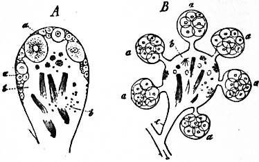
Fig. 42.—Follicles of the hermaphrodite gonads of Euthyneurous Gastropods. A, of Helix; B, of Eolis; a, ova; b, developing spermatozoa; c, common efferent duct.
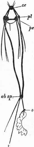
Fig. 43.—Nervous system of Aplysia, as a type of the long-looped Euthyneurous condition. The untwisted visceral loop is lightly shaded. (After Spengel.)
ce, Cerebral ganglion. pl, Pleural ganglion. pe, Pedal ganglion. ab. sp, Abdominal ganglion which represents also the supra-intestinal ganglion of Streptoneura and gives off the nerve to the osphradium (olfactory organ) o, and another to an unlettered so-called “genital” ganglion. The buccal nerves and ganglia are omitted.
The development of Aplysia from the egg presents many points of interest from the point of view of comparative embryology, but in relation to the morphology of the Opisthobranchia it is sufficient to point to the occurrence of a trochosphere and a veliger stage (fig. 36), and of a shell-gland or primitive shell-sac (fig. 36, shgr), which is succeeded by a nautiloid shell.
In the nervous system of Aplysia the great ganglion-pairs are well developed and distinct. The euthyneurous visceral loop is long, and presents only one ganglion (in Aplysia camelus, but two distinct ganglia joined to one another in Aplysia hybrida of the English coast), placed at its extreme limit, representing both the right and left visceral ganglia and the third or abdominal ganglion, which are so often separately present. The diagram (fig. 43) shows the nerve connecting this abdomino-visceral ganglion with the olfactory ganglion of Spengel. It is also seen to be connected with a more remote ganglion—the genital. Such special irregularities in the development of ganglia upon the visceral loop, and on one or more of the main nerves connected with it, are very frequent. Our figure of the nervous system of Aplysia does not give the small pair of buccal ganglia which are, as in all glossophorous Molluscs, present upon the nerves passing from the cerebral region to the odontophore.
For a comparison of various Opisthobranchs, Aplysia will be found to present a convenient starting-point. It is one of the more typical Opisthobranchs, that is to say, it belongs to the section Tectibranchia, but other members of the suborder, namely, Bulla and Actaeon (figs. 44 and 45), are less abnormal than Aplysia in regard to their shells and the form of the visceral hump. They have naked spirally twisted shells which may be concealed from view in the living animal by the expansion and reflection of the parapodia, but are not enclosed by the mantle, whilst Actaeon is remarkable for possessing an operculum like that of so many Streptoneura.
The great development of the parapodia seen in Aplysia is usual in Tectibranchiate Opisthobranchs. The whole surface of the body becomes greatly modified in those Nudibranchiate forms which have lost, not only the shell, but also the ctenidium. Many of these have peculiar processes developed on the dorsal surface (fig. 46, A, B), or retain purely negative characters (fig. 46, D). The chief modification of internal organization presented by these forms, as compared with Aplysia, is found in the condition of the alimentary canal. The liver is no longer a compact organ opening by a pair of ducts into the median digestive tract, but we find very numerous hepatic diverticula on a shortened axial tract (fig. 47). These diverticula extend usually one into each of the dorsal papillae or “cerata” when these are present. They are not merely digestive glands, but are sufficiently wide to act as receptacles of food, and in them the digestion of food proceeds just as in the axial portion of the canal. A precisely similar modification of the liver or great digestive gland is found in the scorpions, where the axial portion of the digestive canal is short and straight, and the lateral ducts sufficiently wide to admit food into the ramifications of the gland there to be digested; whilst in the spiders the gland is reduced to a series of simple caeca.
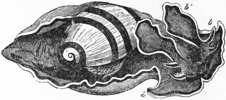
Fig. 44.—Bulla vexillum (Chemnitz), as seen crawling. á, oral hood (compare with Tethys, fig. 46, B), possibly a continuation of the epipodia; b, b′, cephalic tentacles. (From Owen.)
The typical character is retained by the heart, pericardium, and the communicating nephridium or renal organ in all Opisthobranchs. An interesting example of this is furnished by the fish-like transparent Phyllirhoë (fig. 37), in which it is possible most satisfactorily to study in the living animal, by means of the microscope, the course of the blood-stream, and also the reno-pericardial communication. In many of the Nudibranchiate Opisthobranchs the nervous system presents a concentration of the ganglia (fig. 48), contrasting greatly with what we have seen in Aplysia. Not only are the pleural ganglia fused to the cerebral, but also the visceral to these (see in further illustration the condition attained by the Pulmonate Limnaeus, fig. 59), and the visceral loop is astonishingly short and insignificant (fig. 48, e′). That the parts are rightly thus identified is probable from J.W. Spengel’s observation of the osphradium and its nerve-supply in these forms; the nerve to that organ, which is placed somewhat anteriorly—on the dorsal surface—being given off from the hinder part (visceral) of the right compound ganglion—the fellow to that marked A in fig. 48. The Eolid-like Nudibranchs, amongst other specialities of structure, possess (in some cases at any rate) apertures at the apices of the “cerata” or dorsal papillae, which lead from the exterior into the hepatic caeca. Some amongst them (Tergipes, Eolis) are also remarkable for possessing peculiarly modified cells placed in sacs (cnidosacs) at the apices of these same papillae, which resemble the “thread-cells” of the Coelentera. According to T.S. Wright and J.H. Grosvenor these nematocysts are derived from the hydroids on which the animals feed.
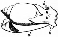
Fig. 45.—Actaeon. h, shell; b, oral hood; d, foot; f, operculum.
The development of many Opisthobranchia has been examined—e.g. Aplysia, Pleurobranchidium, Elysia, Polycera, Doris, Tergipes. All pass through trochosphere and veliger stages, and in all a nautiloid or boat-like shell is developed, preceded by a well-marked “shell-gland” (see fig. 36). The transition from the free-swimming veliger larva with its nautiloid shell (fig. 36) to the adult form has not been properly observed, and many interesting points as to the true nature of folds (whether parapodia or mantle or velum) have yet to be cleared up by a knowledge of such development in forms like Tethys, Doris, Phyllidia, &c. As in other Molluscan groups, we find even in closely-allied genera (for instance, in Aplysia and Pleurobranchidium, and other genera), the greatest differences as to the amount of food-material by which the egg-shell is encumbered. Some form their diblastula by emboly, others by epiboly; and in the later history of the further development of the enclosed cells (arch-enteron) very marked variations occur in closely-allied forms, due to the influence of a greater or less abundance of food-material mixed with the protoplasm of the egg.
Sub-order 1.—Tectibranchia. Opisthobranchs provided in the adult state with a shell and a mantle, except Runcina, Pleurobranchaea, Cymbuliidae, and some Aplysiomorpha. There is a ctenidium, except in some Thecosomata and Gymnosomata, and an osphradium.
Tribe 1.—Bullomorpha. The shell is usually well developed, except in Runcina and Cymbuliidae, and may be external or internal. No operculum, except in Actaeonidae and Limacinidae. The pallial cavity is always well developed, and contains the ctenidium, at least in part; ctenidium, except in Lophocercidae, of folded type. With the exception of the Aplustridae, Lophocercidae and Thecosomata, the head is devoid of tentacles, and its dorsal surface forms a digging disk or shield. The edges of the foot form parapodia, often transformed into fins. Posteriorly the mantle forms a large pallial lobe under the pallial aperture. Stomach generally provided with chitinous or calcified masticatory plates. Visceral commissure fairly long, except in Runcina, Lobiger and Thecosomata. Hermaphrodite genital aperture, connected with the penis by a ciliated groove, except in Actaeon, Lobiger and Cavolinia longirostris, in which the spermiduct is a closed tube. Animals either swim or burrow.
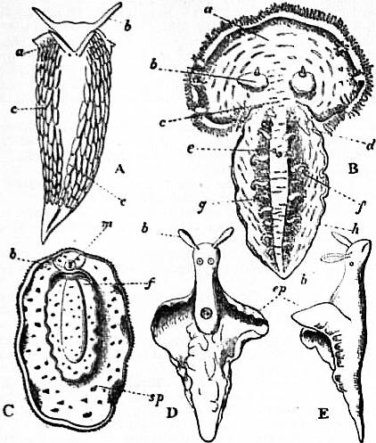
Fig. 46.
A, Eolis papillosa (Lin.), dorsal view.
a, b, Posterior and anterior cephalic tentacles. c, The dorsal “cerata.”
B, Tethys leporina, dorsal view.
a, The cephalic hood. b, Cephalic tentacles. c, Neck. d, Genital pore. e, Anus. f, Large cerata. g, Smaller cerata. h, Margin of the foot.
C, Doris (Actinocyclus) tuberculatus (Cuv.), seen from the pedal surface.
m, Mouth. b, Margin of the head. f, Sole of the foot. sp, The mantle-like epipodium.
D, E, Dorsal and lateral view of Elysia (Actaeon) viridis. ep, epipodial outgrowths. (After Keferstein.)
Fig. 47.—Enteric Canal of Eolis papillosa. (From Gegenbaur, after Alder and Hancock.) ph, Pharynx. m, Midgut, with its hepatic appendages h, all of which are not figured. e, Hind gut. an, Anus. Fig. 48.—Central Nervous System of Fiona (one of the Nudibranchia), showing a tendency to fusion of the great ganglia. (From Gegenbaur, after Bergh.) A, Cerebral, pleural and visceral ganglia united. B, Pedal ganglion. C, Buccal ganglion. D, Oesophageal ganglion connected with, the Buccal. a, Nerve to superior cephalic tentacle. b, Nerves to inferior cephalic tentacles. c, Nerve to generative organs. d, Pedal nerve. e, Pedal commissure. e′, Visceral loop or commissure (?).
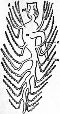
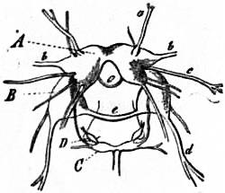
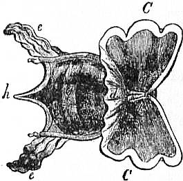
Fig. 49.—Cavolinia tridentata, Forsk. from the Mediterranean, magnified two diameters. (From Owen.)
a, Mouth. b, Pair of cephalic tentacles. C, C, Pteropodial lobes of the foot. d, Median web connecting these. e, e, Processes of the mantle-skirt reflected over the surface of the shell. g, The shell enclosing the visceral hump. h. The median spine of the shell.
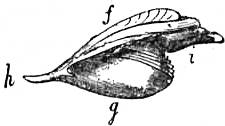
Fig. 50.—Shell of Cavolinia tridentata, seen from the side.
f, Postero-dorsal surface. g, Antero-ventral surface. h, Median dorsal spine. i, Mouth of the shell.
Fam. 1.—Actaeonidae. Cephalic shield bifid posteriorly; margins of foot slightly developed; genital duct diaulic; visceral commissure streptoneurous; shell thick, with prominent spire and elongated aperture; a horny operculum. Actaeon, British. Solidula. Tornatellaea, extinct. Adelactaeon. Bullina. Bullinula.
Fam. 2.—Ringiculidae. Cephalic disk enlarged anteriorly, forming an open tube posteriorly; shell external, thick, with prominent spire; no operculum. Ringicula. Pugnus.
Fam. 3.—Tornatinidae. Margins of foot not prominent; no radula; shell external, with inconspicuous spire. Tornatina, British. Retusa. Volvula.
Fam. 4.—Scaphandridae. Cephalic shield short, truncated posteriorly; eyes deeply embedded; three calcareous stomachal plates; shell external, with reduced spire. Scaphander, British. Atys. Smaragdinella. Cylichna, British. Amphisphyra, British.
Fam. 5.—Bullidae. Margins of foot well developed; eyes superficial; three chitinous stomachal plates; shell external, with reduced spire. Bulla, British. Haminea, British.
Fam. 6.—Aceratidae. Cephalic shield continuous with neck; twelve to fourteen stomachal plates; a posterior pallial filament passing through a notch in shell. Acera, British. Cylindrobulla. Volutella.
Fam. 7.—Aplustridae. Foot very broad; cephalic shield with four tentacles; shell external, thin, without prominent spire. Aplustrum. Hydatina. Micromelo.
Fam. 8.—Philinidae. Cephalic shield broad, thick and simple; shell wholly internal, thin, spire much reduced, aperture very large. Philine, British. Cryptophthalmus. Chelinodura. Phanerophthalmus. Colpodaspis, British. Colobocephalus.
Fam. 9.—Doridiidae. Cephalic shield ending posteriorly in a median point; shell internal, largely membranous; no radula or stomachal plates. Doridium. Navarchus.
Fam. 10.—Gastropteridae. Cephalic shield pointed behind; shell internal, chiefly membranous, with calcified nucleus, nautiloid; parapodia forming fins. Gastropteron.
Fam. 11.—Runcinidae. Cephalic shield continuous with dorsal integument; no shell; ctenidium projecting from mantle cavity. Runcina.
Fam. 12.—Lophocercidae. Shell external, globular or ovoid; foot elongated, parapodia separate from ventral surface; genital duct diaulic. Lobiger. Lophocercus.
The next three families form the group formerly known as Thecosomatous Pteropods. They are all pelagic, the foot being entirely transformed into a pair of anterior fins; eyes are absent, and the nerve centres are concentrated on the ventral side of the oesophagus.
Fam. 13.—Limacinidae. Dextral animals, with shell coiled pseudo-sinistrally; operculum with sinistral spiral; pallial cavity dorsal. Limacina, British. Peraclis, ctenidium present.
Fam. 14.—Cymbuliidae. Adult without shell; a sub-epithelial pseudoconch formed by connective tissue; pallial cavity ventral. Cymbulia. Cymbuliopsis. Gleba. Desmopterus.
Fam. 15.—Cavoliniidae. Shell not coiled, symmetrical; pallial cavity ventral. Cavolinia. Clio. Cuvierina.
Tribe 2.—Aplysiomorpha. Shell more or less internal, much reduced or absent. Head bears two pairs of tentacles. Parapodia separate from ventral surface, and generally transformed into swimming lobes. Visceral commissure much shortened, except in Aplysia. Genital duct monaulic; hermaphrodite duct connected with penis by a ciliated groove. Animals either swim or crawl.
Fam. 1.—Aplysiidae. Shell partly or wholly internal, or absent; foot long, with well-developed ventral surface. Aplysia. Dolabella. Dolabrifer. Aplysiella. Phyllaplysia. Notarchus.
The next six families include the animals formerly known as Gymnosomatous Pteropods, characterized by the absence of mantle and shell, the reduction of the ventral surface of the foot, and the parapodial fins at the anterior end of the body. They are all pelagic.
Fam. 2.—Pneumonodermatidae. Pharynx evaginable, with suckers. Pneumonoderma. Dexiobranchaea. Spongiobranchaea. Schizobrachium.
Fam. 3.—Clionopsidae. No buccal appendages or suckers; a very long evaginable proboscis; a quadriradiate terminal branchia. Clionopsis.
Fam. 4.—Notobranchaeidae. Posterior branchia triradiate. Notobranchaea.
Fam. 5.—Thliptodontidae. Head very large, not marked off from the body; neither branchia nor suckers; fins situated near the middle of the body. Thliptodon.
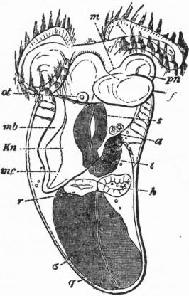
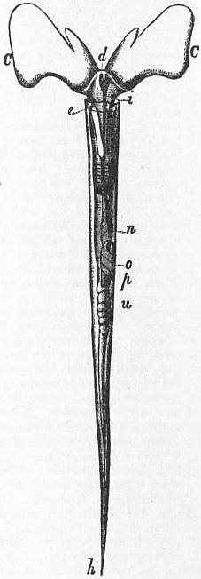
Fig. 51.—Embryo of Cavolinia tridentata. (From Balfour, after Fol.) a, Anus. f, Median portion of the foot. pn, Pteropodial lobe of the foot. h, Heart. i, Intestine. m. Mouth. ot, Otocyst. q, Shell. r, Nephridium. s, Oesophagus. σ, Sac containing nutritive yolk. mb, Mantle-skirt. mc, Sub-pallial chamber. Kn, Contractile sinus. Fig. 52.—Styliola acicula, Rang. sp. enlarged. (From Owen.) C, C, The wing-like lobes of the foot. d, Median fold of same. e, Copulatory organ. h, Pointed extremity of the shell. i, Anterior margin of the shell. n, Stomach. o, Liver. u. Hermaphrodite gonad.
Fam. 6.—Clionidae. No branchia of any kind; a short evaginable pharynx, bearing paired conical buccal appendages or “cephalocones.” Clione. Paraclione. Fowlerina.
Fam. 7.—Halopsychidae. No branchia; two long and branched buccal appendages. Halopsyche.
Tribe 3.—Pleurobranchomorpha. Two pairs of tentacles. Foot without parapodia; no pallial cavity, but always a single ctenidium situated on the right side between mantle and foot. Genital duct diaulic, without open seminal groove; male and female apertures contiguous. Visceral commissure short, tendency to concentration of all ganglia in dorsal side of oesophagus.
Fam. 1.—Tylodinidae. Shell external and conical; anterior tentacles form a frontal veil; ctenidium extending only over right side; a distinct osphradium. Tylodina.
Fam. 2.—Umbrellidae. Shell external, conical, much flattened; anterior tentacles very small, and situated with the mouth in a notch of the foot below the head; ctenidium very large. Umbrella.
Fam. 3.—Pleurobranchidae. Shell covered by mantle, or absent; anterior tentacles form a frontal veil; mantle contains spicules. Pleurobranchus. Berthella. Haliotinella. Oscanius, British. Oscaniella. Oscaniopsis. Pleurobranchaea.
Sub-order 2.—Nudibranchia. Shell absent in the adult; no ctenidium or osphradium. Body generally slug-like, and externally symmetrical. Visceral mass not marked off from the foot, except in Hedylidae. Dorsal respiratory appendages frequently present. Visceral commissure reduced; nervous system concentrated on dorsal side of oesophagus. Marine; generally carnivorous, and brightly coloured, affording many instances of protective resemblance.
Tribe 1.—Tritoniomorpha. Liver wholly or partially contained in the visceral mass. Anus lateral, on the right side. Usually two rows of ramified dorsal appendages. Genital duct diaulic; male and female apertures contiguous.
Fam. 1.—Tritoniidae. Anterior tentacles form a frontal veil; foot rather broad. Tritonia, British. Marionia.
Fam. 2.—Scyllaeidae. No anterior tentacles; dorsal appendages broad and foliaceous; foot very narrow; stomach with horny plates. Scyllaea, pelagic.
Fam. 3.—Phyllirhoidae. No anterior tentacles, and no dorsal appendages; body laterally compressed, transparent; pelagic. Phyllirhoë.
Fam. 4.—Tethyidae. Head broad, surrounded by a funnel-shaped velum or hood; no radula; dorsal appendages foliaceous. Tethys. Melibe.
Fam. 5.—Dendronotidae. Anterior tentacles forming a scalloped frontal veil; dorsal appendages and tentacles similarly ramified. Dendronotus. Campaspe.
Fam. 6.—Bornellidae. Dorsum furnished on either side with papillae, at the base of which are ramified appendages. Bornella.
Fam. 7.—Lomanotidae. Body flattened, the two dorsal borders prominent and foliaceous. Lomanotus, British.
Tribe 2.—Doridomorpha. Body externally symmetrical; anus median, posterior, and generally dorsal, surrounded by ramified pallial appendages, constituting a secondary branchia. Liver not ramified in the integuments. Genital duct triaulic. Spicules present in the mantle.
Fig. 53.—Halopsyche gaudichaudii, Soul. (From Owen.) Much enlarged; the body-wall removed. a, The mouth. c, The pteropodial lobes of the foot. f, The centrally-placed hind-foot. d, l, e, Three pairs of tentacle-like processes placed at the sides of the mouth, and developed (in all probability) from the fore-foot. o′, Anus. y, Genital pore. k, Retractor muscles. o and p, The liver. u, v, w, Genitalia. Fig. 54.—Ancula cristata, one of the pygobranchiate Opisthobranchs (dorsal view). (From Gegenbaur, after Alder and Hancock.) a, Anus. br, Secondary branchia surrounding the anus. t, Cephalic tentacles. External to the branchia are seen ten club-like processes of the dorsal wall, these are the “cerata” which are characteristically developed in another suborder of Opisthobranchs.
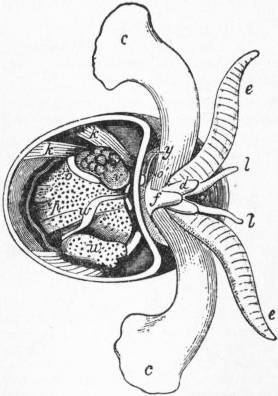
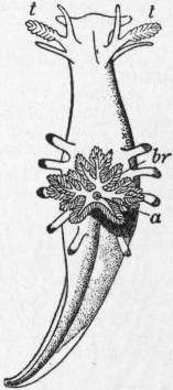
Fam. 1.—Polyceratidae. A more or less prominent frontal veil; branchiae non-retractile. Euplocamus. Polycera, British. Thecacera, British. Aegirus, British. Plocamopherus. Palio. Crimora. Triopa, British. Triopella.
Fam. 2.—Goniodorididae. Mantle-border projecting; frontal veil reduced, and often covered by the anterior border of the mantle. Goniodoris, British. Acanthodoris, British. Idalia, British. Ancula, British. Doridunculus. Lamellidoris. Ancylodoris, the only fresh-water Nudibranch, from Lake Baikal.
Fam. 3.—Heterodorididae. No branchia. Heterodoris.
Fam. 4.—Dorididae. Mantle oval, covering the head and the greater part of the body; anterior tentacles, ill-developed; branchiae generally retractile. Doris, British. Hexabranchus. Chromodoris.
Fam. 5.—Doridopsidae. Pharynx suctorial; no radula; branchial rosette on the dorsal surface, above the mantle-border. Doridopsis.
Fam. 6.—Corambidae. Anus and branchia posterior, below the mantle-border. Corambe.
Fam. 7.--Phyllidiidae. Pharynx suctorial; branchiae surrounding the body, between the mantle and foot. Phyllidia. Fryeria.
The last three families constitute the sub-tribe Porostomata, characterized by the reduction of the buccal mass, which is modified into a suctorial apparatus.
Tribe 3.—Eolidomorpha (Cladohepatica). The whole of the liver contained in the integuments and tegumentary papillae. Genital duct diaulic; male and female apertures contiguous. The anus is antero-lateral, except in the Proctonotidae, in which it is median. Tegumentary papillae not ramified, and containing cnidosacs with nematocysts.
Fam. 1.—Eolididae. Dorsal papillae spindle-shaped or club-shaped. Eolis, British. Facelina, British. Tergipes, British. Gonieolis. Cuthona. Embletonia. Galvina. Calma. Hero.
Fam. 2.—Glaucidae. Body furnished with three pairs of lateral lobes, bearing the tegumentary papillae; foot very narrow; pelagic. Glaucus.
Fam. 3.—Hedylidae. Body elongated; visceral mass marked off from foot posteriorly; dorsal appendages absent, or reduced to a single pair; spicules in the integument. Hedyle.
Fam. 4.—Pseudovermidae. Head without tentacles; body elongated; anus on right side. Pseudovermis.
Fam. 5.—Proctonotidae. Anus posterior, median; anterior tentacles, atrophied; foot broad. Janus, British. Proctonotus, British.
Fam. 6.—Dotonidae. Bases of the rhinophores surrounded by a sheath; dorsal papillae tuberculated and club-shaped, in a single row on either side of the dorsum; no cnidosacs. Doto, British. Gellina. Heromorpha.
Fam. 7.—Fionidae. Dorsal papillae with a membranous expansion; male and female apertures at some distance from each other; pelagic. Fiona.
Fam. 8.—Pleurophyllidae. Anterior tentacles in the form of a digging shield; mantle without appendages, but respiratory papillae beneath the mantle-border. Pleurophyllidia.
Fam. 9.—Dermatobranchidae. Like the last, but wholly without branchiae. Dermatobranchus.
Tribe 4.—Elysiomorpha. Liver ramifies in integuments and extends into dorsal papillae, but there are no cnidosacs. Genital duct always triaulic, and male and female apertures distant from each other. No mandibles, and radula uniserial. Never more than one pair of tentacles, and these are absent in Alderia and some species of Limapontia.
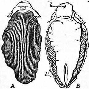
Fig. 55.—Dorsal and Ventral View of Pleurophyllidia lineata (Otto), one of the Eolidomorph Nudibranchs. (After Keferstein.)
b, The mouth. l, The lamelliform sub-pallial gills, which (as in Patella) replace the typical Molluscan ctenidium.
Fam. 1.—Hermaeidae. Foot narrow; dorsal papillae linear or fusiform, in several series. Hermaea, British. Stiliger. Alderia, British.
Fam. 2.—Phyllobranchidae. Foot broad; dorsal papillae flattened and foliaceous. Phyllobranchus. Cyerce.
Fam. 3.—Plakobranchidae. Body depressed, without dorsal papillae, but with two very large lateral expansions, with dorsal plications. Plakobranchus.
Fam. 4.—Elysiidae. Body elongated, with lateral expansions; tentacles large; foot narrow. Elysia, British. Tridachia.
Fam. 5.—Limapontiidae. No lateral expansions, and no dorsal papillae; body planariform; anus dorsal, median and posterior. Limapontia, British. Actaeonia, British. Cenia.
Order 2 (of the Euthyneura).—Pulmonata. Euthyneurous Gastropoda, probably derived from ancestral forms similar to the Tectibranchiate Opisthobranchia by adaptation to a terrestrial life. The ctenidium is atrophied, and the edge of the mantle-skirt is fused to the dorsal integument by concrescence, except at one point which forms the aperture of the mantle-chamber, thus converted into a nearly closed sac. Air is admitted to this sac for respiratory and hydrostatic purposes, and it thus becomes a lung. An operculum is present only in Amphibola; a contrast being thus afforded with the operculate pulmonate Streptoneura (Cyclostoma, &c.), which differ in other essential features of structure from the Pulmonata. The Pulmonata are, like the other Euthyneura, hermaphrodite, with elaborately developed copulatory organs and accessory glands. Like other Euthyneura, they have very numerous small denticles on the lingual ribbon. In aquatic Pulmonata the osphradium is retained.
In some Pulmonata (snails) the foot is extended at right angles to the visceral hump, which rises from it in the form of a coil as in Streptoneura; in others the visceral hump is not elevated, but is extended with the foot, and the shell is small or absent (slugs).
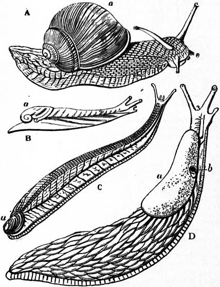
Fig. 56.—A Series of Stylommatophorous Pulmonata, showing transitional forms between snail and slug.
A, Helix pomatia. (From Keferstein.) B, Helicophanta brevipes. (From Keferstein, after Pfeiffer.) C, Testacella haliotidea. (From Keferstein.) D, Arion ater, the great black slug. (From Keferstein.) a, Shell in A, B, C, shell-sac (closed) in D; b, orifice leading into the sub-pallial chamber (lung).
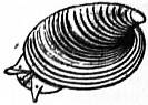
Fig. 57.—Ancylus fluviatilis, a patelliform aquatic Pulmonate.
Pulmonata are widely distinguished from a small number of Streptoneura at one time associated with them on account of their mantle-chamber being converted, as in Pulmonata, into a lung, and the ctenidium or branchial plume aborted. The terrestrial Streptoneura (represented in England by the common genus Cyclostoma) have a twisted visceral nerve-loop, an operculum on the foot, a complex rhipidoglossate or taenio-glossate radula, and are of distinct sexes. The Pulmonata have a straight visceral nerve-loop, usually no operculum even in the embryo, and a multidenticulate radula, the teeth being equi-formal; and they are hermaphrodite. Some Pulmonata (Limnaea, &c.) live in fresh waters although breathing air. The remarkable discovery has been made that in deep lakes such Limnaei do not breathe air, but admit water to the lung-sac and live at the bottom. The lung-sac serves undoubtedly as a hydrostatic apparatus in the aquatic Pulmonata, as well as assisting respiration.
The same general range of body-form is shown in Pulmonata as in the Heteropoda and in the Opisthobranchia; at one extreme we have snails with coiled visceral hump, at the other cylindrical or flattened slugs (see fig. 56). Limpet-like forms are also found (fig. 57, Ancylus). The foot is always simple, with its flat crawling surface extending from end to end, but in the embryo Limnaea it shows a bilobed character, which leads on to the condition characteristic of Pteropoda.
The adaptation of the Pulmonata to terrestrial life has entailed little modification of the internal organization. In one genus (Planorbis) the plasma of the blood is coloured red by haemoglobin, this being the only instance of the presence of this body in the blood of Glossophorous Mollusca, though it occurs in corpuscles in the blood of the bivalves Arca and Solen (Lankester).
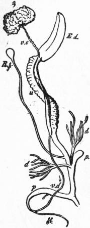
Fig. 58.—Hermaphrodite Reproductive Apparatus of the Garden Snail (Helix hortensis).
τ, Ovo-testis. ve, Hermaphrodite duct. Ed, Albuminiparous gland. u, Uterine dilatation of the hermaphrodite duct. d, Digitate accessory glands on the female duct. ps, Calciferous gland or dart-sac on the female duct. Rf, Spermatheca or receptacle of the sperm in copulation, opening into the female duct. vd, Male duct (vas deferens). p, Penis. fl, Flagellum.
The generative apparatus of the snail (Helix) may serve as an example of the hermaphrodite apparatus common to the Pulmonata and Opisthobranchia (fig. 58). From the ovo-testis, which lies near the apex of the visceral coil, a common hermaphrodite duct ve proceeds, which receives the duct of the compact white albuminiparous gland, Ed, and then becomes much enlarged, the additional width being due to the development of glandular folds, which are regarded as forming a uterus u. Where these folds cease the common duct splits into two portions, a male and a female. The male duct vd becomes fleshy and muscular near its termination at the genital pore, forming the penis p. Attached to it is a diverticulum fl, in which the spermatozoa which have descended from the ovo-testis are stored and modelled into sperm ropes or spermatophores. The female portion of the duct is more complex. Soon after quitting the uterus it is joined by a long duct leading from a glandular sac, the spermatheca (Rf). In this duct and sac the spermatophores received in copulation from another snail are lodged. In Helix hortensis the spermatheca is simple. In other species of Helix a second duct (as large in Helix aspersa as the chief one) is given off from the spermathecal duct, and in the natural state is closely adherent to the wall of the uterus. This second duct has normally no spermathecal gland at its termination, which is simple and blunt. But in rare cases in Helix aspersa a second spermatheca is found at the end of this second duct. Tracing the widening female duct onwards we now come to the openings of the digitate accessory glands d, d, which probably assist in the formation of the egg-capsule. Close to them is the remarkable dart-sac ps, a thick-walled sac, in the lumen of which a crystalline four-fluted rod or dart consisting of carbonate of lime is found. It is supposed to act in some way as a stimulant in copulation, but possibly has to do with the calcareous covering of the egg-capsule. Other Pulmonata exhibit variations of secondary importance in the details of this hermaphrodite apparatus.
The nervous system of Helix is not favourable as an example on account of the fusion of the ganglia to form an almost uniform ring of nervous matter around the oesophagus. The pond-snail (Limnaeus) furnishes, on the other hand, a very beautiful case of distinct ganglia and connecting cords (fig. 59). The demonstration which it affords of the extreme shortening of the Euthyneurous visceral nerve-loop is most instructive and valuable for comparison with and explanation of the condition of the nervous centres in Cephalopoda, as also of some Opisthobranchia. The figure (fig. 59) is sufficiently described in the letterpress attached to it; the pair of buccal ganglia joined by the connectives to the cerebrals are, as in most of our figures, omitted. Here we need only further draw attention to the osphradium, discovered by Lacaze-Duthiers, and shown by Spengel to agree in its innervation with that organ in all other Gastropoda. On account of the shortness of the visceral loop and the proximity of the right visceral ganglion to the oesophageal nerve-ring, the nerve to the osphradium and olfactory ganglion is very long. The position of the osphradium corresponds more or less closely with that of the vanished right ctenidium, with which it is normally associated. In Helix and Limax the osphradium has not been described, and possibly its discovery might clear up the doubts which have been raised as to the nature of the mantle-chamber of those genera. In Planorbis, which is sinistral (as are a few other genera or exceptional varieties of various Anisopleurous Gastropods), instead of being dextral, the osphradium is on the left side, and receives its nerve from the left visceral ganglion, the whole series of unilateral organs being reversed. This is, as might be expected, what is found to be the case in all “reversed” Gastropods.
The shell of the Pulmonata, though always light and delicate, is in many cases a well-developed spiral “house” into which the creature can withdraw itself; and, although the foot possesses no operculum, yet in Helix the aperture of the shell is closed in the winter by a complete lid, the “hybernaculum” more or less calcareous in nature, which is secreted by the foot. In Clausilia a peculiar modification of this lid exists permanently in the adult, attached by an elastic stalk to the mouth of the shell, and known as the “clausilium.” In Limnaeus the permanent shell is preceded in the embryo by a well-marked shell-gland or primitive shell-sac (fig. 60), at one time supposed to be the developing anus, but shown by Lankester to be identical with the “shell-gland” discovered by him in other Mollusca (Pisidium, Pleurobranchidium, Neritina, &c.). As in other Gastropoda Anisopleura, this shell-sac may abnormally develop a plug of chitinous matter, but normally it flattens out and disappears, whilst the cap-like rudiment of the permanent shell is shed out from the dome-like surface of the visceral hump, in the centre of which the shell-sac existed for a brief period.
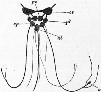
Fig. 59.—Nervous System of the Pond-Snail, Limnaeus stagnalis, as a type of the short-looped euthyneurous condition. The short visceral “loop” with its three ganglia is lightly-shaded.
ce, Cerebral ganglion. pe, Pedal ganglion. pl, Pleural ganglion. ab, Abdominal ganglion. sp, Visceral ganglion of the left side; opposite to it is the visceral ganglion of the right side, which gives off the long nerve to the olfactory ganglion and osphradium o.
In Planorbis and in Auricula (Pulmonata, allied to Limnaeus) the olfactory organ is on the left side and receives its nerve from the left visceral ganglion. (After Spengel.)
In Clausilia, according to the observations of C. Gegenbaur, the primitive shell-sac does not flatten out and disappear, but takes the form of a flattened closed sac. Within this closed sac a plate of calcareous matter is developed, and after a time the upper wall of the sac disappears, and the calcareous plate continues to grow as the nucleus of the permanent shell. In the slug Testacella (fig. 56, C) the shell-plate never attains a large size, though naked. In other slugs, namely, Limax and Arion, the shell-sac remains permanently closed over the shell-plate, which in the latter genus consists of a granular mass of carbonate of lime. The permanence of the primitive shell-sac in these slugs is a point of considerable interest. It is clear enough that the sac is of a different origin from that of Aplysia (described in the section treating of Opisthobranchia), being primitive instead of secondary. It seems probable that it is identical with one of the open sacs in which each shell-plate of a Chiton is formed, and the series of plate-like imbrications which are placed behind the single shell-sac on the dorsum of the curious slug, Plectrophorus, suggest the possibility of the formation of a series of shell-sacs on the back of that animal similar to those which we find in Chiton. Whether the closed primitive shell-sac of the slugs (and with it the transient embryonic shell-gland of all other Mollusca) is precisely the same thing as the closed sac in which the calcareous pen or shell of the Cephalopod Sepia and its allies is formed, is a further question which we shall consider when dealing with the Cephalopoda. It is important here to note that Clausilia furnishes us with an exceptional instance of the continuity of the shell or secreted product of the primitive shell-sac with the adult shell. In most other Mollusca (Anisopleurous Gastropods, Pteropods and Conchifera) there is a want of such continuity; the primitive shell-sac contributes no factor to the permanent shell, or only a very minute knob-like particle (Neritina and Paludina). It flattens out and disappears before the work of forming the permanent shell commences. And just as there is a break at this stage, so (as observed by A. Krohn in Marsenia = Echinospira) there may be a break at a later stage, the nautiloid shell formed on the larva being cast, and a new shell of a different form being formed afresh on the surface of the visceral hump. It is, then, in this sense that we may speak of primary, secondary and tertiary shells in Mollusca recognizing the fact that they may be merely phases fused by continuity of growth so as to form but one shell, or that in other cases they may be presented to us as separate individual things, in virtue of the non-development of the later phases, or in virtue of sudden changes in the activity of the mantle-surface causing the shedding or disappearance of one phase of shell-formation before a later one is entered upon.
The development of the aquatic Pulmonata from the egg offers considerable facilities for study, and that of Limnaeus has been elucidated by E.R. Lankester, whilst H. Rabl has with remarkable skill applied the method of sections to the study of the minute embryos of Planorbis. The chief features in the development of Limnaeus are exhibited in fig. 60. There is not a very large amount of food-material present in the egg of this snail, and accordingly the cells resulting from division are not so unequal as in many other cases. The four cells first formed are of equal size, and then four smaller cells are formed by division of these four so as to lie at one end of the first four (the pole corresponding to that at which the “directive corpuscles” are extruded and remain). The smaller cells now divide and spread over the four larger cells; at the same time a space—the cleavage cavity or blastocoel—forms in the centre of the mulberry-like mass. Then the large cells recommence the process of division and sink into the hollow of the sphere, leaving an elongated groove, the blastopore, on the surface. The invaginated cells (derived from the division of the four big cells) form the endoderm or arch-enteron; the outer cells are the ectoderm. The blastopore now closes along the middle part of its course, which coincides in position with the future “foot.” One end of the blastopore becomes nearly closed, and an ingrowth of ectoderm takes place around it to form the stomodaeum or fore-gut and mouth. The other extreme end closes, but the invaginated endoderm cells remain in continuity with this extremity of the blastopore, and form the “rectal peduncle” or “pedicle of invagination” of Lankester, although the endoderm cells retain no contact with the middle region of the now closed-up blastopore. The anal opening forms at a late period by a very short ingrowth or proctodaeum coinciding with the blind termination of the rectal peduncle (fig. 60, pi).
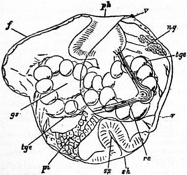
Fig. 60.—Embryo of Limnaeus stagnalis, at a stage when the Trochosphere is developing foot and shell-gland and becoming a Veliger, seen as a transparent object under slight pressure. (Lankester.)
ph, Pharynx (stomodaeal invagination). v, v, The ciliated band marking out the velum. ng, Cerebral nerve-ganglion. re, Stiebel’s canal (left side), probably an evanescent embryonic nephridium. sh, The primitive shell-sac or shell-gland. pi, The rectal peduncle or pedicle of invagination; its attachment to the ectoderm is coincident with the hindmost extremity of the elongated blastopore of fig. 3, C. tge, Mesoblastic (skeleto-trophic and muscular) cells investing gs, the bilobed arch-enteron or lateral vesicles of invaginated endoderm, which will develop into liver. f, The foot.
The body-cavity and the muscular, fibrous and vascular tissues are traced partly to two symmetrically disposed “mesoblasts,” which bud off from the invaginated arch-enteron, partly to cells derived from the ectoderm, which at a very early stage is connected by long processes with the invaginated endoderm. The external form of the embryo goes through the same changes as in other Gastropods, and is not, as was held previously to Lankester’s observations, exceptional. When the middle and hinder regions of the blastopore are closing in, an equatorial ridge of ciliated cells is formed, converting the embryo into a typical trochosphere.
The foot now protrudes below the mouth, and the post-oral hemisphere of the trochosphere grows more rapidly then the anterior or velar area. The young foot shows a bilobed form. Within the velar area the eyes and the cephalic tentacles commence to rise up, and on the surface of the post-oral region is formed a cap-like shell and an encircling ridge, which gradually increases in prominence and becomes the freely depending mantle-skirt. The outline of the velar area becomes strongly emarginated and can be traced through the more mature embryos to the cephalic lobes or labial processes of the adult Limnaeus (fig. 61).
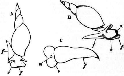
Fig. 61.—A, B, C. Three views of Limnaeus stagnalis, in order to show the persistence of the larval velar area v, as the circum-oral lobes of the adult. m, Mouth; f, foot; v, velar area, the margin v corresponding with the ciliated band which demarcates the velar area or velum of the embryo Gastropod (see fig. 4, D, E, F, H, I, v). (Original.)
The increase of the visceral dome, its spiral twisting, and the gradual closure of the space overhung by the mantle-skirt so as to convert it into a lung-sac with a small contractile aperture, belong to stages in the development later than any represented in our figures.
We may now revert briefly to the internal organization at a period when the trochosphere is beginning to show a prominent foot growing out from the area where the mid-region of the elongated blastopore was situated, and having therefore at one end of it the mouth and at the other the anus. Fig. 60 represents such an embryo under slight compression as seen by transmitted light. The ciliated band of the left side of the velar area is indicated by a line extending from v to v; the foot f is seen between the pharynx ph and the pedicle of invagination pi. The mass of the arch-enteron or invaginated endodermal sac has taken on a bilobed form, and its cells are swollen (gs and tge). This bilobed sac becomes entirely the liver in the adult; the intestine and stomach are formed from the pedicle of invagination, whilst the pharynx, oesophagus and crop form from the stomodaeal invagination ph. To the right (in the figure) of the rectal peduncle is seen the deeply invaginated shell-gland ss, with a secretion sh protruding from it. The shell-gland is destined in Limnaeus to become very rapidly stretched out, and to disappear. Farther up, within the velar area, the rudiments of the cerebral nerve-ganglion ng are seen separating from the ectoderm. A remarkable cord of cells having a position just below the integument occurs on each side of the head. In the figure the cord of the left side is seen, marked re. This paired organ consists of a string of cells which are perforated by a duct opening to the exterior and ending internally in a flame-cell. Such cannulated cells are characteristic of the nephridia of many worms, and the organs thus formed in the embryo Limnaeus are embryonic nephridia. The most important fact about them is that they disappear, and are in no way connected with the typical nephridium of the adult. In reference to their first observer they were formerly called “Stiebel’s canals.” Other Pulmonata possess, when embryos, Stiebel’s canals in a more fully developed state, for instance, the common slug Limax. Here too they disappear during embryonic life. Similar larval nephridia occur in other Gastropoda. In the marine Streptoneura they are ectodermic projections which ultimately fall off; in the Opisthobranchs they are closed pouches; in Paludina and Bithynia they are canals as in Pulmonata.
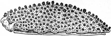
Fig. 62.—Oncidium tonganum, a littoral Pulmonate, found on the shores of the Indian and Pacific Oceans (Mauritius, Japan).
Marine Pulmonata.—Whilst the Pulmonata are essentially a terrestrial and fresh-water group, there is one genus of slug-like Pulmonates which frequent the sea-coast (Oncidium, fig. 62). Karl Semper has shown that these slugs have, in addition to the usual pair of cephalic eyes, a number of eyes developed upon the dorsal integument. These dorsal eyes are very perfect in elaboration, possessing lens, retinal nerve-end cells, retinal pigment and optic nerve. Curiously enough, however, they differ from the cephalic Molluscan eye in the fact that, as in the vertebrate eye, the filaments of the optic nerve penetrate the retina, and are connected with the surfaces of the nerve-end cells nearer the lens instead of with the opposite end. The significance of this arrangement is not known, but it is important to note, as shown by V. Henson, S.J. Hickson and others, that in the bivalves Pecten and Spondylus, which also have eyes upon the mantle quite distinct from typical cephalic eyes, there is the same relationship as in Oncidiidae of the optic nerve to the retinal cells. In both Oncidiidae and Pecten the pallial eyes have probably been developed by the modification of tentacles, such as coexist in an unmodified form with the eyes. The Oncidiidae are, according to K. Semper, pursued as food by the leaping fish Periophthalmus, and the dorsal eyes are of especial value to them in aiding them to escape from this enemy.
Sub-order 1.—Basommatophora. Pulmonata with an external shell. The head bears a single pair of contractile but not invaginable tentacles, at the base of which are the eyes. Penis at some distance from the female aperture, except in Amphibola and Siphonaria. All have an osphradium, except the Auriculidae, which are terrestrial, and it is situated outside the pallial cavity in those forms in which water is not admitted into the lung. There is a veliger stage in development, but the velum is reduced.
Fam. 1.—Auriculidae. Terrestrial and usually littoral; genital duct monaulic, the penis being connected with the aperture by an open or closed groove; shell with a prominent spire, the internal partitions often absorbed and the aperture denticulated. Auricula. Cassidula. Alexia. Melampus. Carychium, terrestrial, British. Scarabus. Leuconia, British. Blauneria. Pedipes.
Fam. 2.—Otinidae. Shell with short spire, and wide oval aperture; tentacles short. Otina, British. Camptonyx, terrestrial.
Fam. 3.—Amphibolidae. Shell spirally coiled; head broad, without prominent tentacles; foot short, operculated; marine. Amphibola.
Fam. 4.—Siphonariidae. Visceral mass and shell conical; tentacles atrophied; head expanded; genital apertures contiguous; marine animals, with an aquatic pallial cavity containing secondary branchial laminae. Siphonaria.
Fam. 5.—Gadiniidae. Visceral mass and shell conical; head flattened; pallial cavity aquatic, but without a branchia; genital apertures separated. Gadinia.
Fam. 6.—Chilinidae. Shell ovoid, with short spire, wide aperture and folded columella; inferior pallial lobe thick; visceral commissure still twisted. Chilina.
Fam. 7.—Limnaeidae. Shell thin, dextral, with prominent spire and oval aperture; no inferior pallial lobe. Limnaea, British. Amphipeplea, British.
Fam. 8.—Pompholygidae. Shell dextral, hyperstrophic, animal sinistral. Pompholyx. Choanomphalus.
Fam. 9.—Planorbidae. Visceral mass and shell sinistral; inferior pallial lobe very prominent, and transformed into a branchia. Planorbis, British. Bulinus. Miratesta.
Fam. 10.—Ancylidae. Shell conical, not spiral; inferior pallial lobe transformed into a branchia. Ancylus, British. Latia. Grundlachia.
Fam. 11.—Physidae. Visceral mass and shell sinistrally coiled; shell thin, with narrow aperture; no inferior pallial lobe. Physa, British. Aplexa, British.
Sub-order 2.—Stylommatophora. Pulmonata with two pairs of tentacles, except Janellidae and Vertigo; these tentacles are invaginable, and the eyes are borne on the summits of the posterior pair. Male and female genital apertures open into a common vestibule, except in Vaginulidae and Oncidiidae. Except in Oncidium, there is no longer a veliger stage in development.
Tribe 1.—Holognatha. Jaw simple, without a superior appendage.
Fam. 1.—Selenitidae. Radula with elongated and pointed teeth, like those of the Agnatha; a jaw present. Plutonia. Trigonochlamys.
Fam. 2.—Zonitidae. Shell external, smooth, heliciform or flattened; radula with pointed marginal teeth. Zonites, British. Ariophanta. Orpiella. Vitrina. Helicarion.
Fam. 3.—Limacidae. Shell internal. Limax, British. Parmacella. Urocyclus. Parmarion. Amalia. Agriolimax. Mesolimax. Monochroma. Paralimax. Metalimax.
Fam. 4.—Philomycidae. No shell; mantle covers the whole surface of the body; radula with squarish teeth. Philomycus.
Fam. 5.—Ostracolethidae. Shell largely chitinous, not spiral, its calcareous apex projecting through a small hole in the mantle. Ostracolethe.
Fam. 6.—Arionidae. Shell internal, or absent; mantle restricted to the anterior and middle part of the body; radula with squarish teeth. Arion, British. Geomalacus. Ariolimax. Anadenus.
Fam. 7.—Helicidae. Shell with medium spire, external or partly covered by the mantle; genital aperture below the right posterior tentacle; genital apparatus generally provided with a dart-sac and multifid vesicles. Helix, British. Bulimus. Hemphillia. Berendtia. Cochlostyla. Rhodea.
Fam. 8.—Endodontidae. Shell external, spiral, generally ornamented with ribs; borders of aperture thin and not reflected; radula with square teeth; genital ducts without accessory organs. Endodonta. Punctum. Sphyradium. Laoma. Pyramidula.
Fam. 9.—Orthalicidae. Shell external, ovoid, the last whorl swollen, aperture oval with a simple border; radular teeth in oblique rows. Orthalicus.
Fam. 10.—Bulimulidae. Jaw formed of folds imbricated externally and meeting at an acute angle near the base. Bulimulus. Peltella. Amphibulimus.
Fam. 11.—Cylindrellidae. Shell turriculated, with numerous whorls, the last more or less detached. Cylindrella.
Fam. 12.—Pupidae. Shell external, with elongated spire and numerous whorls, aperture generally narrow; male genital duct without multifid vesicles. Pupa, British. Eucalodium. Vertigo, British. Buliminus, British. Clausilia, British. Balea. Zospeum. Megaspira. Strophia. Anostoma.
Fam. 13.—Stenogyridae. Shell elongated, with a more or less obtuse summit; aperture with a simple border. Achatina. Stenogyra. Ferussacia, British. Cionella. Caecilianella. Azeca. Opeas.
Fam. 14.—Helicteridae. Shell bulimoid, dextral or sinistral; radular teeth, expanded at their extremities and multicuspidate. Helicter. Tornatellina.
Tribe 2.—Agnatha. No jaws; teeth narrow and pointed; carnivorous.
Fam. 1.—Oleacinidae. Shell oval, elongated, with narrow aperture; neck very long; labial palps prominent. Oleacina (Glandina). Streptostyla.
Fam. 2.—Testacellidae. Shell globular or auriform, external or partly covered by the mantle. Streptaxis. Gibbulina. Aerope. Rhytida. Daudebardia. Testacella. Chlamydophorus. Schizoglossa.
Fam. 3.—Rathouisiidae. No shell, a carinated mantle covering the whole body; male and female apertures distant, the female near the anus. Rathouisia. Atopos.
Tribe 3.—Elasmognatha. Jaw with a well-developed dorsal appendage.
Fam. 1.—Succineidae. Anterior tentacles much reduced; male and female apertures contiguous but distinct; shell thin, spiral, with short spire. Succinea, British. Homalonyx. Hyalimax. Neohyalimax.
Fam. 2.—Janellidae. Limaciform, with internal rounded shell; mantle very small and triangular; pulmonary chamber with tracheae; no anterior tentacles. Janella. Aneitella. Aneitea. Triboniophorus.
Tribe 4.—Ditremata. Male and female apertures distant.
Fam. 1.—Vaginulidae. No shell; limaciform; terrestrial; female aperture on right side in middle of body; anus posterior. Vaginula.
Fam. 2.—Oncidiidae. No shell; limaciform; littoral; female aperture posterior, near anus; a reduced pulmonary cavity with a distinct aperture. Oncidium. Oncidiella, British. Peronia.
Authorities.—L. Boutan, “La Cause principale de l’asymétrie des mollusques gastéropodes,” Arch. de zool. expér. (3), vii. (1899); A. Lang, “Versuch einer Erklärung der Asymmetrie der Gastropoder,” Vierteljahrsschr. naturforsch. Gesellschaft, Zürich, 36 (1892); A. Robert, “Recherches sur le développement des Troques,” Arch. de zool. expér. (3), x. (1903); P. Pelseneer, “Report on the Pteropoda,” Zool. “Challenger” Expedit. pts. lviii., lxv., lxvi. (1887, 1888); P. Pelseneer, “Protobranches aériens et Pulmonés branchifères,” Arch. de biol. xiv. (1895); W.A. Herdman, “On the Structure and Functions of the Cerata or Dorsal Papillae in some Nudibranchiate Mollusca,” Quart. Journ. Mic. Sci. (1892); J.T. Cunningham, “On the Structure and Relations of the Kidney in Aplysia,” Mitt. Zool. Stat. Neapel, iv. (1883); Böhmig, “Zur feineren Anatomie von Rhodope veranyi, Kölliker,” Zeitschr. f. wiss. Zool. vol. lvi. (1893).
Treatises.—S.P. Woodward, Manual of the Mollusca (2nd ed., with appendix, London, 1869); E. Forbes and S. Hanley, History of British Mollusca (4 vols., London, 1853); Alder and Hancock, Monograph of British Nudibranchiate Mollusca (London, Roy. Society, 1845); P. Pelseneer, Mollusca. Treatise on Zool., edited by E. Ray Lankester, pt. v. (1906); E. Ray Lankester, “Mollusca,” in 9th ed. of this Encyclopaedia, to which this article is much indebted.
(J. T. C)
From Zeitschrift für Wissenschaft Zoologie, vol. xlix. p. 209, by permission of Wilhelm Engelmann.
Chaetonotus maximus, Ehrb., ventral side. (After Zelinka.)
Bo, Bristles surrounding the mouth. ds, Dorsal bristles. hCi, Posterior lateral cilia. Ke, Cuticular dome. Mr, Oral cavity. lT, Lateral sensory hairs. Pl, Cuticular plates. Sa, Dorsal bristle of the basal part. Sch, Plates. Se, Lateral bristles. Vb, Point of union of ciliated tract. vCi, Anterior group of cilia. vS, Ventral bristles of the basal part.
GASTROTRICHA, a small group of fairly uniform animals which live among Rotifers and Protozoa at the bottom of ponds and marshes, biding amongst the recesses of the algae and sphagnum and other fresh-water plants and eating organic débris and Infusoria. They are of minute size varying from one-sixtieth to one-three-hundredth of an inch, and they move by means of long cilia. Two ventral bands composed of regular transverse rows of cilia are usually found. The head bears some especially large cilia. The cuticle which covers the body is here and there raised into overlapping scales which may be prolonged into bristles. An enlarged, frontal scale may cover the head, and a row of scales separates the ventral ciliated areas from one another, whilst two series of alternating rows cover the back and side. The body, otherwise circular in section, is slightly flattened ventrally. The mouth is anterior and slightly ventral; it leads into a protrusible pharynx armed with recurved teeth that can be everted. This leads to a muscular oesophagus with a triradiate lumen, which acts as a sucking pump and ends in a funnel-valve projecting into the stomach. The last named is oval and formed of four rows of large cells; it is separated by a sphincter from the rectum, which opens posteriorly and dorsally. The nitrogenous excretory apparatus consists of a coiled tube on each side of the stomach; internally the tubes end in large flame-cells, and externally by small pores which lie on the edges of the ventral row of scales. A cerebral ganglion rests on the oesophagus and supplies the cephalic cilia and hairs; it is continued some way back as two dorsal nerve trunks. The sense organs are the hairs and bristles and in some species eyes. The muscles are simple and unstriated and for the most part run longitudinally.
The two ovaries lie at the level of the juncture of the stomach and rectum. The eggs become very large, sometimes half the length of the mother; they are laid amongst water weeds. The male reproductive system is but little known, a small gland lying between the ovaries has been thought to be a testis, and if it be, the Gastrotricha are hermaphrodite.
Zelinka classifies the group as follows:—
Sub-order 1.—Euichthydina with a forked tail.
(i.) Fam. Ichthydidae, without bristles. Genera: Ichthydium, Lepidoderma.
(ii.) Fam. Chaetonotidae, with bristles. Genera: Chaetonotus, Chaetura.
Sub-order 2.—Apodina, tail not forked. Genera: Dasydytes, Gossea, Stylochaeta.
The genus Aspidiophorus recently described by Voigt seems in some respects intermediate between Lepidoderma and Chaetonotus. Zelinkia and Philosyrtis are two slightly aberrant forms described by Giard from certain diatomaceous sands. Altogether there must be some forty to fifty described species.
The group is an isolated one and shows no clear affinities with any of the great phyla. Those that are usually dwelt on are treated with the Rotifers and Nematoda and Turbellaria.
Literature.—A.C. Stokes, The Microscope (Detroit, 1887-1888); C. Zelinka, Zeitschr. wiss. Zool. xlix., 1890, p. 209; M. Voigt, Forschber. Plön. Th. ix., 1904, p. 1; A. Giard, C. R. Soc. Biol. lvi. pp. 1061 and 1063; E. Daday, Termes. Fuzetek. xxiv. p. 1; F. Zschokke, Denk. Schweiz. Ges. xxxvii. p. 109; S. Hlava, Zool. Anz. xxviii., 1905, p. 331.
(A. E. S.)
GATAKER, THOMAS (1574-1654), English divine, was born in London in September 1574, and educated at St John’s College, Cambridge. From 1601 to 1611 he held the appointment of preacher to the society of Lincoln’s Inn, which he resigned on accepting the rectory of Rotherhithe. In 1642 he was chosen a member of the assembly of divines at Westminster, and annotated for that assembly the books of Isaiah, Jeremiah and Lamentations. He disapproved of the introduction of the Covenant, and declared himself in favour of episcopacy. He was one of the forty-seven London clergymen who disapproved of the trial of Charles I. He was married four times, and died in July 1654.
His principal works, besides some volumes of sermons are—On the Nature and Use of Lots (1619), a curious treatise which led to his being accused of favouring games of chance; Dissertatio de stylo Novi Testamenti (1648); Cinnus, sive Adversaria miscellanea, in quibus Sacrae Scripturae primo, deinde aliorum scriptorum, locis aliquam multis lux redditur (1651), to which was afterwards subjoined Adversaria Posthuma; and his edition of Marcus Antoninus (1652), which, according to Hallam, is the “earliest edition of any classical writer published in England with original annotations,” and, for the period at which it was written, possesses remarkable merit. His collected works were published at Utrecht in 1698.
GATCHINA, a town of Russia, in the government of St Petersburg, 29 m. by rail S. of the city of St Petersburg, in 59° 34′ N. and 30° 6′ E. Pop. (1860) 9184; (1897) 14,735. It is situated in a flat, well-wooded, and partly marshy district, and on the south side of the town are two lakes. Among its more important buildings are the imperial palace, which was founded in 1770 by Prince Orlov, and constructed according to the plans of the Italian architect Rinaldi; a military orphanage, founded in 1803; and a school for horticulture. Among the few industrial establishments is a porcelain factory. At Gatchina an alliance was concluded between Russia and Sweden on the 29th of October 1799.
GATE, an opening into any enclosure for entrance or exit, capable of being closed by a barrier at will. The word is of wide application, embracing not only the defensive entrance ways into a fortified place, with which this article mainly deals, or the imposing architectural features which form the main entrances to palaces, colleges, monastic buildings, &c., but also the common five-barred barrier which closes an opening into a field. The most general distinction that can be made between “door” and “gate” is that of size, the greater entrance into a court containing other buildings being the “gate,” the smaller entrances opening directly into the particular buildings the “doors,” or that of construction, the whole entrance way being a “gate” or gateway, the barrier which closes it a “door.” A further distinction is drawn by applying “door” to the solid barriers or “valves” of wood, metal, &c., made in panels and fitted to a framework, and “gate” to an openwork structure, whether of metal or wood (see further Door and Metal-work). The ultimate origin of the word is obscure; the early forms appear with a palatalized initial letter, still surviving in such dialectical forms as “yate,” or in Scots “yett.” It is probably connected with the root of “get,” in the sense either of “means of access” or of “holding,” “receptacle”; cf. Dutch gat, hole. There may be a connexion, however, with “gate,” now usually spelled “gait,” a manner of walking,1 but originally a way, passage; cf. Ger. Gasse, narrow street, lane.
The entrance through the enclosing walls of a city or fortification has been from the earliest times a place of the utmost importance, considered architecturally, socially or from the point of view of the military engineer. In the East the “gate” was and still is in many Mahommedan countries the central place of civic life. Here was the seat of justice and of audience, the most important market-place, the spot where men gathered to receive and exchange news. The references in the Bible to the gates of the city in all these varied aspects are innumerable (cf. Gen. xix. 1; Deut. xxv. 7; Ruth iv. 1; 2 Sam. xix. 8; 2 Kings vii. 1). Later the seat of justice and of government is transferred to the gate of the palace of the king (cf. Dan. ii. 49, and Esther ii. 19), and this use is preserved to-day in the official title of the seat of government of the Turkish empire at Constantinople, the “Sublime Porte,” a translation of the Turkish Bab Aliy (bab, gate, and aliy, high). A full account with many modern instances of Eastern customs will be found in Sir Charles Warren’s article “Gate” in Hastings’s Dict. of Bible. For the “pylon,” the typical gate of Egyptian architecture, see Architecture.
The gates into a walled town or other fortified place were necessarily in early times the chief points on which the attack concentrated, and the features, common throughout the ages, of flanking or surmounting towers and of galleries over the entrance way, are found in the Assyrian gate at Khorsabad (cf. 2 Chron. xxvi. 9; 2 Sam. xviii. 24). With the coming of peaceful times to a city or the removal of the fear of sudden attack, the gateways would take a form adapted more for ready exit and entrance than for defence, though the possibility of defending them was not forgotten. Such city gates often had separate openings for entrance and exit, and again for foot passengers and for vehicles. The Gallo-Roman gate at Autun has four entrances, two just wide enough to admit carriages, and two narrow alleys for foot passengers. A fine example of a Roman city gate, dating from the time of Constantine, is at Trèves. It is four storeys high, with ornamental windows, and decorated with columns on each storey. The two outer wings project beyond the central part, the two entrance ways are 14 ft. wide, and could be closed by doors and a portcullis. The chambers in the storeys above were used for the purposes of civil administration. In more modern times city gateways have often followed the type of the Roman triumphal arch, with a single wide opening and purely ornamental superstructure. On the other hand, the defensive gate formed by an archway entering as it were through a tower has been constantly followed as a type of entrance to buildings of an entirely peaceful character. A fine example of such a gateway, originally built for defence, is at Battle Abbey; this was built by Abbot Retlynge in 1338, when Edward III. granted a licence to fortify and crenellate the abbey. Such gateways are typical of Tudor palaces, as at St James’s or at Hampton Court, and are the most common form in the colleges of Oxford and Cambridge. The Tom Gate at Christ Church, Oxford, with its surmounted domed bell tower, or the cupola resting on columns at Queen’s College, Oxford, are further examples of the gate architecturally considered.
The changes the fortified gateway has undergone in construction and the varying relative importance it has held in the scheme of defence follow the lines of development taken by the history of Fortification and Siegecraft (q.v.). The following is a short sketch of the main stages in its history. A good example of the Roman fortified city gate still remains at Pompeii. Here there is one passage way for vehicles, 14 ft. wide; this is open to the sky. The two footways on either side are arched, with openings in the centre on to the central way. The doors of the gate are on the city side, but a portcullis (cataracta) closed it on the country side. The gateways of the Roman permanent camps (castra stativa) were four in number, the porta praetoria and Decumana at either end, with principalis dextra and sinistra on the side (see also Camp). At Pevensey (Anderida) a small postern on the north side of the Roman walls was laid bare in 1906-1907, in which the passage curves in the thickness of the wall, and from a width admitting two men abreast narrows so that one alone could block it. Flanking towers or bastions guarded the main entrances, while in front were built outworks, of palisades, &c., to protect it; these were known as procastra or antemuralia, and the entrances to these were placed so that they could be flanked from the main walls.
In the defence of a fortified place the gate had not only to be protected from sudden surprise, but also had to undergo protracted attacks concentrated upon it during a siege. Thus until the coming of gunpowder, the ingenuity of military engineers was exhausted in accumulating the most complicated defences round the gateways, and the strength of a fortified place could be estimated by the fewness of its gates. Viollet-le-Duc (Dict. de l’arch. du moyen âge, s.v. Porte) takes the Narbonne and Aude gates (E. and W.) of Carcassonne as typical instances of this complication. The following brief account of the Narbonne Gate (fig. 1), one of the principal parts of the work on the fortifications begun by Philip the Bold in 1285, will give some idea of the varied means of defence, which may be found individually if not always in such collective abundance in the fortified gateways of the middle ages. Two massive towers flanked the actual entrance and were linked across by an iron chain; over the entrance (E) was a machicolation, further added to in time of war by a hoarding of timber; and an outer portcullis fell in front of the heavy iron-lined doors. On to the passage way between the first and second doors opened a square machicolation (G) from which the defenders in the upper chambers of the gate could attack an enemy that had succeeded in breaking through the first entrance or had been trapped by the falling of the first portcullis. Another machicolation (I) opened from the roof in front of the second portcullis and second door. So much for the gate itself; but before an attack could reach that point, the following defences had to be passed: an immense circular barbican (A) protected the entrance across the moat and through the outer enceinte of the city. This entrance was flanked by a masked return of the wall (C), while palisades (P) still further hampered the assailant in his passage across the “lists” to the foot of the gate towers. Here sappers would find themselves exposed to a fire from the loopholes and from the machicolated hoardings above them, while the projecting horns with which the face of the towers terminated forced them to uncover themselves to a flanking fire from the indents in the main curtain on either side of the towers.
Fig. 1.—Plan of the Narbonne Gate of the city of Carcassonne.
The later history of the gateway is merged in that of modern fortification. The more elaborate the gate defences the greater was the inducement for the besieger to attack the walls, and improvements in methods of siegecraft ultimately compelled the defender to develop the enceinte from its medieval form of a ring wall with flanking towers to the 17th century form of bastions, curtains, tenailles and ravelins, all intimately connected in one general scheme of defence. By Vauban’s time there is little to distinguish the position and defences of the gateways from the rest of the fortifications surrounding a town. A road from the country usually entered one of the ravelins, sinking into the glacis, crossing the ditch of the ravelin and piercing the parapet almost at right angles to its proper direction (see fig. 2, which also shows a typical arrangement of minor communications such as ramps and staircases). From the interior of the ravelin it passed across the main ditch to a gate in the curtain of the enceinte. The road was in fact artificially made to wind in such a way that it was kept under fire from the defences throughout, while the part of it inside the works was bent so as to place a covering mass between the enemy’s fire and troops using the road for a sortie. Thus the gate itself was merely a barrier against a coup de main and to keep out unauthorized persons. In conditions precluding the making of a breach in the walls, i.e. in surprises and assaults de vive force, the gateway and accompanying drawbridge continue to play their part in the 16th, 17th and 18th centuries, but they seldom or never appear as the objectives of a siege en règle. In Vauban’s works, and those of most other engineers, there was generally a postern giving access to the floor of the main ditch, in the centre of the curtain escarp. The gates of Vauban’s and later fortresses are strong heavy wooden doors, and the gateways more or less ornamental archways, exactly as in many private mansions of castellar form. In modern fortresses the gate of a detached fort or an enceinte de sureté is intended purely as a defence against an unexpected rush. The usual method is to have two gates, the outer one a lattice or portcullis of iron bars and the inner one a plate of half-inch steel armour, backed by wood and loopholed. The defenders of the gate can by this arrangement fire from the inner loopholes through the outer gate upon the approaches, and also keep the enemy under fire whilst he is trying to force the outer gate itself. The ditches are crossed either by drawbridges or by ramps leading the road down to the floor of the ditch.
Fig. 2.—Plan of Gate Arrangements of an 18th Century Fortress.
The “gate” as a barrier to be removed and as an entrance to be passed is of constant occurrence in figurative language and in symbolical usage. The gates of the temple of Janus (q.v.) at Rome stood open in war and closed in peace. The pylon of ancient Egypt had a symbolical meaning in the Book of the Dead, and religious significance attaches to the torii, one of the outward signs of the Shinto religion in Japan, the Buddhist toran, and to the Chinese pai-loo, the honorific gateways erected to ancestors. The gates of heaven and hell, the gates of death and darkness, the wide and narrow gates that lead to destruction and life (Matt. vii. 13 and 14), are familiar metaphorical phrases in the Bible. In Greek and Roman legend dreams pass through gates of transparent horn if true, if deceptive and false through opaque gates of ivory (Hom. Od. xix. 560 sq.; Virg. Aen. vi. 893).
(C. We.)
1 The spelling “gait” is confined to this meaning—the only literary one surviving. In the form “gate” it appears dialectally in this sense and in such particular meanings as a right to run cattle on common or private ground or as a passage way in mines. The principal survival is in names of streets in the north and midlands of England and in Scotland, e.g. Briggate at Leeds, Wheeler Gate and Castle Gate at Nottingham, Gallow Tree Gate at Leicester, and Canongate and Cowgate at Edinburgh.
GATEHOUSE. In the second half of the 16th century in England the entrance gateway, which formed part of the principal front of the earlier feudal castles, became a detached feature attached to the mansions only by a wall enclosing the entrance court. The gatehouse then constituted a structure of some importance, and included sometimes many rooms as at Stanway Hall, Gloucestershire, where it measures 44 ft. by 22 ft. and has three storeys; at Westwood, Worcestershire, it had a frontage of 54 ft. with two storeys; and at Burton Agnes, Yorkshire, it was still larger and was flanked by great octagonal towers at the angles and had three storeys. At a later period smaller accommodation was provided so that it virtually became a lodge, but being designed to harmonize with the mansion it presented sometimes a monumental structure. On the continent of Europe the gatehouse forms a much more important building, as it formed part of the town fortifications, where it sometimes defended the passage of a bridge across the stream or moat. There are numerous examples in France and Germany.
GATES, HORATIO (1728-1806), American general, was born at Maldon in Essex, England, in 1728. He entered the English army at an early age, and was rapidly promoted. He accompanied General Braddock in his disastrous expedition against Fort Duquesne in 1755, and was severely wounded in the battle of July 9; and he saw other active service in the Seven Years’ War. After the peace of 1763 he purchased an estate in Virginia, where he lived till the outbreak of the War of Independence in 1775, when he was named by Congress adjutant-general. In 1776 he was appointed to command the troops which had lately retreated from Canada, and in August 1777, as a result of a successful intrigue, was appointed to supersede General Philip Schuyler in command of the Northern Department. In the two battles of Saratoga (q.v.) his army defeated General Burgoyne, who, on the 17th of October, was forced to surrender his whole army. This success was, however, largely due to the previous manœuvres of Schuyler and to Gates’s subordinate officers. The intrigues of the Conway Cabal to have Washington superseded by Gates completely failed, but Gates was president for a time of the Board of War, and in 1780 was placed in chief command in the South. He was totally defeated at Camden, S. C., by Cornwallis on the 17th of August 1780, and in December was superseded by Greene, though an investigation into his conduct terminated in acquittal (1782). He then retired to his Virginian estate, whence he removed to New York in 1790, after emancipating his slaves and providing for those who needed assistance. He died in New York on the 10th of April 1806.
GATESHEAD, a municipal, county and parliamentary borough of Durham, England; on the S. bank of the Tyne opposite Newcastle, and on the North Eastern railway. Pop. (1891) 85,692; (1901) 109,888. Though one of the largest towns in the county, neither its streets nor its public buildings, except perhaps its ecclesiastical buildings, have much claim to architectural beauty. The parish church of St Mary is an ancient cruciform edifice surmounted by a lofty tower; but extensive restoration was necessitated by a fire in 1854 which destroyed a considerable part of the town. The town-hall, public library and mechanic’s institute are noteworthy buildings. Education is provided by a grammar school, a large day school for girls, and technical and art schools. There is a service of steam trams in the principal streets, and three fine bridges connect the town with Newcastle-upon-Tyne. There are large iron works (including foundries and factories for engines, boilers, chains and cables), shipbuilding yards, glass manufactories, chemical, soap and candle works, brick and tile works, breweries and tanneries. The town also contains a depot of the North Eastern railway, with large stores and locomotive works. Extensive coal mines exist in the vicinity; and at Gateshead Fell are large quarries for grindstones, which are much esteemed and are exported to all parts of the world. Large gas-works of the Newcastle and Gateshead Gas Company are also situated in the borough. The parliamentary borough returns one member. The corporation consists of a mayor, 9 aldermen, and 27 councillors. Area, 3132 acres.