Volume 4 — DISEASES OF THE GENITO-URINARY SYSTEM. (continued 8)
Reading edition
I have also found the stretching of the fundus of the vagina by firmly packing it with absorbent cotton, sometimes repeated almost daily or at intervals of two, three, or four days, of great benefit in stretching the adhesions and promoting their absorption. Sometimes, where adhesions are persistent, the use of the rubber colpeurynter distended with hot water is of value.
Where there is a foreign body, as a tumor, fixed posteriorly to the uterus, or where the uterus is fixed in a retroflexed position, the patient may be placed in the knee-chest position, Sims's speculum introduced, and the vagina packed with cotton while the patient is in that posture; or, instead, the vagina may be simply distended with air. The air may be admitted by the introduction of Campbell's glass tube or by the separation of the walls of the vagina with the fingers, which may be done by the patient herself. These measures are often of decided benefit.
I wish to repeat what has already been stated, that the treatment of chronic perimetritis, to be carried out successfully, requires that the patient should be in bed and placed under such circumstances and surroundings that the physician may be enabled to pursue personally the plan of treatment. Of course much will be gained if he is aided by a trained nurse. This in many cases involves the removal of the patient from the cares of her home.
Advantage may often be derived from the application of small blisters to the hypogastric and iliac regions, the counter-irritation being kept up almost continuously for two weeks at a time. The blisters should not be larger than two inches square, and should be moved from place to place; for instance, one blister may be placed on the hypogastrium, and before this has healed a second should be placed one side of it. This should be kept up for two weeks at a time, or until four or five blisters have been applied, when, if benefit is to follow, it will be apparent.
When the organs which are agglutinated to one another become more mobile, and the thickened membranes more flaccid, much benefit sometimes results from the application of a pessary if a displacement of the uterus, ovaries, or tubes exists and persists; but before the use of this instrument is thought of, it must be positively ascertained that no tenderness remains as a result of the inflammatory process; the inflammation must have entirely subsided, the effects alone remaining. It is sometimes advised that an instrument large enough to constantly stretch and over-stretch the false membranes and adhesions is advisable. It has also been recommended to over-stretch these adhesions by manipulation. Of the two, I much prefer the latter method; that is, stretching by manipulation rather than by continuously acting upon them by means of a pessary large enough to stretch the vagina and through it the adhesions. In stretching by manipulation, with the patient under ether, you have your own sense of touch to guide you, and the action of your efforts ceases with the cessation of the manipulation, while that carried out by means of a pessary is continuous and may result in great harm from irritation, if not from ulceration of the vaginal surface from pressure; or it may result in rupture of the adhesions. If a pessary is adjusted, it should be used, not for the purpose of over-stretching adhesions, but simply for its stimulating effect on the pelvic circulation, or as a support to the pelvic circulation rather than as a support to the uterus. A larger instrument should not be used than one which will occupy the vagina without stretching it—simply unfold any doubling up which may have resulted from retroversion or prolapse of the uterus—and its action should be carefully watched. It should be learned, not from the sensation of the patient, but from actual examination, that it is not making undue pressure; this examination should be made daily at first, and afterward at longer intervals. The use of the pessary should be discontinued as soon as possible. This statement should be qualified by saying that the words as soon as possible mean when all symptoms have subsided, and the uterus and other organs are maintaining a normal or nearly normal position, or when the pessary seems to have ceased to be of value. It may then be removed on trial.
There is a method of using the pessary, in which it is advised that the instrument shall be large enough to span the angle of flexion which may exist, for the purpose of making pressure on the fundus of the uterus, which is incarcerated in the cul-de-sac of Douglas by adhesions between its peritoneal surface and that lining the sac. This I believe to be a bad principle, for an instrument long enough to do this must either take its point of support against the pubic arch or from an external attachment—a principle of using the pessary which should be most emphatically condemned.
The above treatment should be carried out with the patient in bed, if possible, during which time general measures for the improvement of the muscular and nervous system should also be employed. The application of electricity to the thickened peritoneum and adhesions is another measure which should not be allowed to pass without comment. Much good may be done by the daily application of faradism, with one electrode in the vagina and the other on the hypogastrium, and continued for from fifteen to thirty minutes. I have thought that in some cases great benefit followed this application. Galvanism is also of service, and by some is thought to be of more value than the faradic current.
The time for getting up should be determined by the results of treatment; usually a period of from four to six weeks is sufficient to determine whether or not the treatment at absolute rest is going to be of benefit. Of course it is not to be understood that cure will follow in severe and long-standing cases within this period, because if this hope is entertained disappointment will follow nearly always. What we hope and expect to attain is rest, both physical and physiological, during which time local treatment can be carried out with greater facility and thoroughness and the general condition improved. As a rule, the ligaments soften, the false membranes become attenuated, and during the time stated the patient is very much benefited, and sometimes cured. She should now begin to sit up and to exercise moderately; the amount of exercise should be regulated by its effect. If pain follows walking or riding, it should not be persisted in until such time as exercise can be taken without the production of these symptoms.
There are no specific remedies for internal administration. The general medication of the patient should consist in the use of such remedies as we have learned to depend upon as capable of building up the blood and nervous system, embracing especially that class of tonics which are said to have the power of inducing such changes in plastic material as favors its absorption. To this class belong the chlorides, as the chloride of arsenic, the chloride of iron, the chloride of ammonium, and the bichloride of mercury. These remedies should be placed at the head of the class. The next are the iodides, as the iodide of iron, the iodide of potassium, and the bromide of potassium. Whether or not these remedies have the powers ascribed to them is questionable, and their administration for this purpose must always be, to a certain extent, empirical. As tonic remedies the administration of iron and the bichloride of mercury is of course always indicated. Cod-liver oil is also a remedy of much value in some cases where it can be digested. The whole plan of treatment should rather be of a local than of a general character, while at the same time very great importance should be given to the building up of the general system, without which nothing can be gained by local treatment. The patient should have a change of scene and air as soon as practicable. A sojourn at the seaside for a time, and then in the mountains, will be of great benefit always.
The fact should always be borne in mind by the physician and impressed upon the patient that a previous attack of perimetritis will serve as a predisposing and abiding cause for a recurrence of the disease, so that all exciting causes may be avoided as far as possible.
PELVIC HÆMATOCELE.
BY T. GAILLARD THOMAS, M.D.
HISTORY.—Prior to the present century the pathological condition which we are about to investigate had no place in the category of diseases peculiar to the sexual organs of the female. Very slowly have its pathogenic features, its etiology, and its importance as a not uncommon factor in pelvic disorders, assumed a systematic basis, and even now considerable diversity of opinion exists upon these points. The reasons for this are not far to seek. In the first place, hæmatocele is a symptom of an accident occurring in the pelvis and resulting in hemorrhage; in the second, the source of the flow which creates the hæmatoma or tumor of blood cannot ordinarily be recognized by any diagnostic measures known to science; and in the third, death rarely occurring from the accident and as a direct consequence of it, autopsic evidence is wanting upon which to base accurate and scientific data.
Although these statements are undoubtedly true, it may nevertheless be asserted with confidence that we are to-day no longer in the dark as to the general pathology of this interesting disorder, and that we are in position to map out a plan of treatment which meets the indications which present themselves in an intelligent and reliable manner. There are, however, several sources of hemorrhage which result in pelvic hæmatocele, and it is highly probable that the day will never come when that one which has created the accident can be ascertained with certainty. But while such accuracy of diagnosis would be gratifying to the ambition of the modern diagnostician, neither the prognosis nor treatment of the disorder would be influenced by it.
Long before our day practitioners had recognized by touch the occasional presence of tumors, more or less marked by fluctuation, which occupied the pouch of Douglas, and by their mechanical influence pushed the uterus out of its normal place; but it was not until the early part of our century that it was discovered that these tumors were sometimes, and that not rarely, composed entirely of coagulated blood; and, curious though it may appear, it was not until the year 1850 that pelvic hæmatocele became a well-recognized disorder.
As early as 1737, Ruysch of Amsterdam appears to have come to the verge of discovering it, but it was left for Récamier, to whom gynecology owes so much besides, to make it known when in 1831 he opened a post-uterine tumor, gave vent to a large accumulation of coagulated blood, and described the case in the Lancette Française for that year. In 1850 the subject attracted the attention of Nélaton, became a recognized pathological condition, and has since received a great deal of attention in all the civilized countries of the world.
DEFINITION AND SYNONYMS.—Pelvic hæmatocele—which has likewise received the names of retro-uterine hæmatocele and uterine hæmatoma—may be defined as an effusion of blood into the pelvic cavity of the female, either into or under the peritoneum. Some authors have limited this definition to blood escaping from utero-ovarian vessels and to blood enclosed either by anatomical structures or by previously-existing inflammatory products. I do not adopt these restrictions, because their assumption appears to me to be unwarranted and the validity of the reasons given for their adoption more than doubtful. The location of the blood-mass differs widely in different cases: sometimes, and usually, it is behind the uterus—high up when obliteration of Douglas's pouch has occurred, low down and near to the perineum where such obliteration has not occurred; at other times it exists both behind and in front of the uterus; and at others still, in front of the uterus alone, adhesions preventing its percolation to the posterior parts of the pelvis.
FREQUENCY.—It may be said, in general terms, that this affection is by no means rare, every one of large experience in gynecology meeting necessarily with a large number of cases of it. But no reliable statistics of its frequency have been collected up to the present time. Olshausen of Halle declares that in 1145 gynecological cases he saw 34 hæmatoceles; Beigel in 2000 cases found 38; Schroeder, 7 in 1000; and Seiffert of Prague reports 66 seen in 1272 cases of female pelvic diseases. Barnes says that in ten years' practice he met with 53 cases, and in twenty years Tilt has seen but 12.
Without doubt, the validity of the statistics of this disorder is vitiated by erroneous diagnosis, as is the case with all affections which generally end in recovery. Here cases of cellulitis, pelvic peritonitis, imprisoned cysts, etc. offer prolific sources of error, as I can aver from the results of my own experience.
PATHOLOGY.—It is a fact, thoroughly proved by physiological experiment, that blood injected into serous cavities very soon encysts itself by the enveloping influence of lymph which is poured over it, forming false membranes, or, as the French term them, néo-membranes. The clot, once formed, clings to the serous membrane in contact with it, and soon becomes roofed over by lymph, which, according to Vulpian, begins to show traces of organization as early as the end of twenty-four hours. Should the effused blood be poor in fibrin, the coagulation and encysting do not occur, a rapid absorption taking the place of these processes.
Pelvic hæmatocele consists, as has been already stated, in the collection of a mass of blood in the pelvis, either above or below its roof, without reference to the source of the flow. Such a flow ordinarily occurs from one of the three following sources: first, rupture of vessels in the pelvis; second, reflux of blood from the uterus or tubes; third, transudation of blood in consequence of dyscrasia or pelvic peritonitis.
From this it becomes evident that hæmatocele is not a disease, but a symptom which marks a number of different pathological conditions of quite various significance. As, however, we cannot discover the original accident or pathological condition, we are forced to compromise with taking its most prominent sign as the exponent of a state which is beyond the powers of diagnosis.
Autopsic evidence has revealed the following as the special and most frequent sources of the hemorrhage:
1st. Rupture of blood-vessels in the pelvis:
Utero-ovarian; Varicose veins of broad ligaments; Vessels of extra-uterine ovisac.
2d. Rupture of pelvic viscera:
Ovaries; Fallopian tubes; Uterus.
3d. Reflux of blood from the uterus:
Menstrual blood.
4th. Transudation from blood-vessels:
Purpura; Scorbutus; Chlorosis; Hemorrhagic peritonitis.
It is then clear that the mere presence of a large clot of blood in the pelvis, apart from general symptoms, is a matter of very doubtful significance, since on the one hand it may be the result of a mere regurgitation of menstrual blood due to imperviousness of the cervical or tubal canal, or on the other of the rupture of a Fallopian tube which has become the nidus of an extra-uterine foetus.
Whatever be the source of the blood which escapes, it coagulates, unless very poor in fibrin, either in the most dependent part of the peritoneum or in the pelvic areolar tissue beneath it. Here the watery portions of the mass are gradually absorbed, leaving a hard, small tumor remaining; or, suppurative action being excited, the hard mass is softened down and discharged into the rectum, vagina, bladder, or peritoneum as a grumous material somewhat resembling currant-jelly in appearance.
CAUSES.—These must be divided into predisposing and exciting, for it is rare to meet with the disease in a woman who has previously been in perfect health. The predisposing causes which can be cited with confidence are—the period of ovarian activity (fifteen to forty-five years); disordered blood-state, plethora or anæmia; the menstrual epoch; chronic ovarian or tubal disease; pelvic peritonitis; and the hemorrhagic diathesis. The exciting causes have been found to be sudden checking of the menstrual flow; blows or falls; excessive or intemperate coition; obstruction of cervical canal; obstruction of Fallopian tubes; violent efforts; and ectopic gestation.
VARIETIES.—The two great classes of the affection are the peritoneal and the subperitoneal. In the former the blood collects in the peritoneal cavity and becomes encysted there; in the latter it collects in the cellular tissue beneath the peritoneum, and there forms a solid mass.
Some authors have opposed the consideration of these two varieties under the same head; among them, Aran, Bernutz, and Voisin. But from a clinical standpoint such a consideration appears to me to be valid. Not only have distinct instances of subperitoneal hæmatocele been recorded by such observers as Barnes, Simpson, Olshausen, and Tuckwell, but cases have been met with in which the subperitoneal variety has ruptured the peritoneal roof of the pelvis, and thus broken down the theoretical barrier which pathologists have been inclined to establish between the two varieties.
Of the two varieties, there can be no doubt that the peritoneal is that which presents itself the more frequently. In 41 autopsies Tuckwell found the tumor to be peritoneal in 38.
SYMPTOMS.—As a rule, long before the occurrence of pelvic hemorrhage the patient will have complained of more or less decided symptoms of disease, or at least of disorder, of the genital system. The symptoms which mark blood-dyscrasia or pelvic peritonitis or menstrual irregularity will probably have attracted attention.
When the accident occurs the gravity of the symptoms will depend in great degree upon the character of the lesion which has taken place. Sometimes the blood-accumulation takes place so insidiously that the existence of the tumor created by coagulation takes the practitioner by surprise. At other times what Barnes has called a cataclysm occurs, and in a few hours puts the unfortunate patient beyond the sphere of hope or the resources of art.
In portraying the symptoms of this affection a writer can therefore merely approximate the truth, satisfying himself with the description of a case of ordinary severity, avoiding the description of cases in either extreme, and guarding the reader against supposing that all attacks give the same intensity of symptoms.
Most prominent among the immediate symptoms are—severe and sudden pelvic pain; pallor, faintness, and coldness of the extremities; a sense of exhaustion; nausea and vomiting; metrorrhagia; uterine tenesmus; enlargement of the abdomen; interference with the bladder and rectum; small and rapid pulse; subnormal temperature.
These are the symptoms of invasion, those which may be termed immediate, and which depend upon loss of blood and a sudden traumatic influence exerted upon living tissues. Very soon, generally within forty-eight hours, a reaction occurs which is sometimes slight, and at other times decided. The secondary symptoms are usually the following: tendency to chilliness; constipation; suppression of urine; tympanites; high temperature; rapid pulse; and tenderness over abdomen.
These symptoms are due to a combination of two causes—loss of vital fluid and the invasion of the peritoneum or pelvic areolar tissue by a mass of blood which becomes coagulated and irritant, on the one hand, and inflammatory processes resulting from such invasion on the other. Half of them might be produced by metrorrhagia, and half by sudden and complete retroversion; but a union of the whole will point toward hæmatocele and prompt a physical examination.
PHYSICAL SIGNS.—A tumor will be felt by vaginal touch, usually, though not always, posterior to the uterus and vagina, and partially occluding the latter. This will, if the examination be made very early, be found to be soft and obscurely fluctuating, but it soon becomes a smooth, dense, and solid body. The uterus is very generally found pressed upward and forward, so that the body lies against the abdominal wall and the cervix is on a level with or a little above the symphysis pubis. In some rare cases the blood-tumor is anterior to or obliquely to one side of the uterus, but these are very rare.
Abdominal palpation reveals the presence of a tumor of varying size, and which sometimes extends up to the navel in peritoneal hæmatocele, but in the subperitoneal variety no tumor whatever may be discoverable by these explorations, unless conjoined manipulation be added to it for the sake of deeper and more thorough search.
DIFFERENTIATION.—Hæmatocele may be confounded with pelvic cellulitis or abscess, retroversion, extra-uterine pregnancy, fibroid tumor, and dislocated ovarian cyst.
The tumor of cellulitis develops slowly, with great pain; is hard at first, and then softens; is tender from the first; does not elevate the uterus or press it forward; and is not often accompanied by metrorrhagia.
Retroversion will readily be detected by the uterine sound, conjoined manipulation, and the absence of anæmic symptoms.
The development of extra-uterine pregnancy is slow and gives the signs of gestation.
Fibrous tumors grow slowly, are painless, and move with the uterus, and they are hard, irregular, and do not lift the uterus against the symphysis.
Displaced cysts are painless, non-hemorrhagic, cause no metrorrhagia, and yield fluctuation readily to palpation.
COMPLICATIONS.—The complications to be feared in this disease are septicæmia, suppuration and abscess, and peritonitis.
COURSE, DURATION, AND TERMINATION.—The hemorrhage may be so severe as to destroy life immediately. Five such instances have been recorded by Voisin; I have met with one; and Ollivier d'Angers mentions two in which death occurred in half an hour from a varicose utero-ovarian vein. Such a termination is, however, very rare.
As a rule, absorption takes place unaided by art; in some cases suppuration occurs, and the mass is discharged as if it were a large abscess by the vagina, rectum, bladder, or abdominal walls; and at other times septic absorption, accompanied by septic peritonitis, destroys the life of the patient.
PROGNOSIS.—The prognosis will depend in great degree upon the severity of the constitutional symptoms. As a rule, it is decidedly favorable unless the surgical tendencies of the attending practitioner alter its natural inclination. The prognosis of the peritoneal form is graver than that of the subperitoneal, and when the tumor is very large the danger is greater than when it is small. A large tumor argues great loss of vital fluid, which may in itself destroy life, and the necessity for the absorption of a large amount of coagulated material which may poison the blood.
The usual causes of death are loss of blood, shock from sudden invasion of the peritoneum, peritonitis, secondary discharge of the encapsulated mass into the peritoneum, or septicæmia.
TREATMENT.—Should the physician be called in the inception of the attack, the patient should at once be placed in the recumbent posture, all excitement around her be quelled, the head be kept low, warmth be applied to the soles of the feet, and perfect quiet enjoined. An effort should be made to check the flow by applying bladders of ice or cloths wrung out of hot water over the hypogastrium, pain and tendency to shock met by the use of morphia hypodermically, and ammonia and brandy freely administered by the mouth. This is all that promises benefit, and further efforts should be avoided as calculated to do absolute harm.
After reaction has occurred let it be borne in mind that the factors which tend to the production of death are—1st, peritonitis; 2d, septicæmia; 3d, suppuration and discharge through some dangerous outlet; and let all efforts be directed toward the prevention of these events.
All pain should be quieted by opium or one of its salts, hypodermically or by mouth or rectum; the patient should be thoroughly nourished by milk and strong animal broths, given as often as every two hours; febrile action should be controlled by the coil of running ice-water and quinine; and strict quietude observed, all unnecessary examinations being avoided, as belonging to the most pernicious class of perturbing influences.
Should the case progress favorably, no surgical procedure looking toward the artificial evacuation of the accumulated blood either by bistoury or by the aspirator should be thought of, however large the accumulation be; for experience has proved that cases left to nature, as a rule, do better than those interfered with.
On the other hand, the great value of surgical interference in those cases in which suppurative action occurs, or in which septicæmia develops itself either in acute or chronic form, must not for a moment be lost sight of. Should the case not progress toward recovery, should the symptoms of septicæmia develop as a sharp attack or as the insidious hectic fever, the accumulated blood or pus and blood should at once be evacuated, and the nidus from which it is discharged be thoroughly washed out with a 2½ per cent. solution of carbolic acid or a solution of the bichloride of mercury, 1 to 2000 of water. Should the accumulation be attainable, tuto, cito, et jucunde, by the vagina, an exploring-needle should be carried into it, and as soon as the fluid is seen to flow a sharp-pointed bistoury should be slid along this and a free opening be made, all the contents of the sac evacuated, and antiseptic washing be at once practised by means of Davidson's syringe and a glass tube.
Should the accumulation point toward the abdominal walls, the opening may with perfect safety be accomplished there. I have operated thus upon 3 cases, with recovery in all, but the accumulation had at the time of operation assumed the character rather of an abscess than of an hæmatocele. A. Martin of Berlin has operated by abdominal section upon 8 cases, with 6 recoveries and 2 deaths, and Baumgärtner of Baden Baden has done so upon 1 case, with recovery. Zweifel has collected 30 cases operated upon by free vaginal incision, with a result of 3 deaths, giving a mortality of 10 per cent. Mere puncture through the vagina he found followed by a mortality of 15 per cent.
The question of surgical interference in pelvic hæmatocele is still sub judice. In my judgment, the rule of practice may, with the present light which we have to guide us, be safely formulated thus: So long as the symptoms are good and the case progresses toward recovery, avoid surgical interference of all sorts, however great be the sanguineous effusion. So soon as symptoms of decided septicæmia or septic peritonitis develop themselves, evacuate the accumulation by a free opening practised by the safest outlet which presents itself, and use antiseptic washings thoroughly.
FIBROUS TUMORS OF THE UTERUS.
BY WILLIAM H. BYFORD, M.D.
RELATIONS AND STRUCTURE.—These tumors grow from the muscular and connective tissues of the uterus, and consequently partake of the character of these tissues. Sometimes the substance of the tumor consists principally of connective, at others of muscular, tissue. The variations in the relative proportion of these two fibrous substances constitute the main differences in the characters and appearances of the tumors, and lead to the different terms applied to them, as myomata, fibromata, myo-fibromata, etc. The firmer the tumor the more connective tissue it contains. When we inspect, either ante- or post-mortem, a uterus with a fibrous tumor attached or contained within its wall, it will be found to present a much darker hue than natural. Instead of the normal light rose-color, it is generally dark, sometimes almost of a purplish tint. The time of menstruation makes some difference; just before it is darker than soon after the menstrual flow. The color also varies with the character and size of the tumor. In large solid tumors the color is darker than in the large fibro-cystic variety; indeed, in some of the latter the pearly color strongly reminds one of an ovarian cyst. We cannot therefore depend on the color or shape of surface for a diagnosis. Even after the abdominal cavity is opened the contour of the uterus is usually not regular. If we make an incision into the tumor, we find that it is surrounded by a distinct capsule, which limits and defines its boundaries and separates it from the adjacent substance. This envelope is not a cyst or other form of membrane: it is continuous with, and inseparable from, the muscular structure of the uterine walls. It, in fact, is a condensed layer of the fibrous substance of the uterus. In cases of true encysted tumors the cyst-wall is the generating portion of the growth. In fibrous tumors of the uterus the growth produces the capsule by displacing the surrounding substance in every direction, pressing it strongly against the unaffected fibrous tissue and condensing it into the smooth capsule. It is thus engendered in, and enveloped by, the muscular walls of the uterus. These latter of course grow to dimensions sufficient to keep pace with the increasing tumor. The growth may, as a consequence of such a connection, be hulled out or enucleated, and will not be reproduced. Inflammation or other degenerating processes may occasionally cause adhesion of the capsule and tumor, but this is an accident of uncommon occurrence. To understand this mode of encapsulation we must remember that the uterine muscles are irregularly stratified, and that the tumors are developed between the strata as between the leaves of a book, separating them sufficiently to gain lodgment and room.
The appearances of the substance of the tumor are not uniform. In many cases the color of the interior of the tumor is dark gray; in some it is dull red; again, sometimes almost livid. The surface of the tumor after the capsule has been removed is often marked by sulci denoting a division into lobules. In other cases the tumor is smooth and symmetrical in shape, and the fibres distinctly visible to the naked eye. The smooth tumor is apt to be very dense and comparatively difficult to destroy, while the lobulated variety is less dense and sometimes easily broken to pieces. But the difference of density does not correspond altogether with the color or shape of surface.
We seldom find large tumors of uniform structure. In some places they are of solid fibrous structure; in others there are cavities of greater or less size, containing a tenacious red serum. These cavities, which seem to be made by localized disintegration of the fibrous tissue, are sometimes of great size, containing several pounds of serum (Atlee). Much more frequently they are small and hold a small amount of fluid. I have met with several where the substance of the tumor seemed to be made up of alveoli filled with a tenacious fluid the color of milk.
Besides this effect upon the density of the tumor resulting from what might be called its usual course, there are numerous modifications in it and in the other properties of the tumors arising from spontaneous degeneration.
It may be said, I think, that without adventitious or supplementary vascular supply the life of a fibrous tumor is self-limited, and it ceases to grow after it has attained to a certain size, and that then it either remains stationary or undergoes degeneration. As I shall have occasion to say farther on, the original supply of blood-vessels cannot be increased to an indefinite degree, and the tumor that grows indefinitely derives a supplementary supply of blood by contracting adhesions to the viscera or abdominal walls. Such adhesions are common and mischievous.
After a tumor has attained its growth, degeneration into the more elementary forms of tissue sets in, as the cartilaginous degeneration, and there is often a deposition of earthy material found in it which reduces it to a hard, dense, stationary, and indestructible body. In such cases there is almost a complete loss of vitality in the tumor, and it becomes a calcified mass.
We may easily demonstrate that the structure of these tumors is essentially fibrous. By maceration and careful dissection the fibres are traceable to a greater or less degree in all of them, the proportion and characters of which, as before said, differ greatly. In the smooth, symmetrically-developed tumor the fibres are usually long and distinctly traceable, while in the lobulated light-gray tumor the fibres are more rudimentary and not so easily followed up by dissection.
MODE OF DEVELOPMENT.—It has already been stated that the fibrous tumor of the uterus grows in or on its wall and originates in the fibrous structure of the organ. The point of beginning is in one or more fasciculi of the muscular system or the connective tissue of the uterus. If in one fasciculus, the point of origin is very minute, as indeed it is generally at first.
The development consists in an hypertrophy of the bundle of fibres affected and a deposit of material similar in structure to that first involved. Sometimes there are numerous nuclei, and nearly all the fibrous structure of the uterus is involved in fibrous degeneration. In the case where the deposit is defined and occupies a small space, it should be borne in mind that the future tumor, however large it becomes, must occupy the same nidus in which it first originated. The nidus becomes enlarged sufficiently to accommodate the growing tumor.
The nucleus of development is enlarged by the accretion of substance similar, if not identical, in character to its own proper material. The nature of the tumor is determined by this fact, and its fibres are rudimentary in organization, instead of being hypertrophied and highly developed, as those of the uterine wall by which it is surrounded. As the tumor grows the fibrous structure surrounding it is pressed aside in every direction in such a way as to completely embrace the growth and encapsulate it. The tumor does not incorporate the adjacent fibres and grow by inducing degeneration in them, but, as before said, it presses them aside. As it thus moulds and shapes a bed in the solid substance of the interior wall, it impresses upon the embracing muscular fibres an increased vitality, and they grow by hypertrophy of a character similar to that of pregnancy. The fibres become longer, and apparently, if not really, more numerous. This hypertrophy of the uterine fibres surrounding the tumor is equal to the capacity demanded by the increasing size of the growing tumor. In this description of the method of development and the embracing capacity of the hypertrophied fibres surrounding it the reader will trace the formation of the capsule in which the tumor is contained. The inner surface of the capsule is smooth, and there are many feeble fibres of connective tissue seen to connect it with the surface of the tumor. There is no adhesion proper between the surface of the tumor and its capsule.
I must call attention to another point that governs the extent and limits of the growth of the tumor—viz. the number and distribution of its vessels. The vessels entering the tumor represent the minute twigs that supplied the fasciculus in which it originated. They arrive at the point of morbid deposit from the parts constituting the capsule, and there are always several of them. The number of these vessels always remains the same, and their calibre is increased with the hypertrophy of the surrounding tissues. They cannot grow at the demand of the trophic energies of the tumor to an unlimited degree, but their size is limited by the growth of the surrounding parts. As the tumor grows and its capsule expands, the vessels are separated farther from each other, until after a while the area becomes so large that the supply of blood will not admit of further growth and the tumor comes to a standstill. Thus their growth, from the nature of their supply, is limited; hence the usual history of the tumor is one of self-limitation. It is all-important in forming an opinion in reference to the greater or less vitality of the fibrous tumor, therefore, to remember that it is not supplied by one large arterial trunk entering at one place and spreading over its capsule, but that the supply is by a number of small vessels penetrating the tumor at different points; that their number cannot be increased and their growth is limited; that as the tumor grows their capacity to supply it grows gradually less until entirely exhausted: then the growth stops.
There is another and adventitious source of nutritious supply, and I think it is essential to very large growths: at least, so far as I know, it is always present. I mean the adhesion of the uterus or tumor to the wall of the abdomen, the pelvic or abdominal viscera, or, what is more common, the omentum. When adhesions occur from whatever cause, the vessels of the tumor increase in size and supply it with a vast increase in the amount of blood. All the large tumors I have had an opportunity of examining were to a greater or less extent covered by a network of large vessels contained in the omentum. These vessels penetrate the uterus, carrying a deluge of blood into its substance. These large vascular adhesions are a source of embarrassment in operations for their removal. Operators allude to them and give instructions how to overcome the difficulty presented by them. The uterine vessels alone would never be sufficient to supply the forty- or fifty-pound tumors so often mistaken for ovarian tumors.
EFFECTS UPON THE UTERUS.—I have already said that the fibres immediately surrounding the growth undergo a true hypertrophy, acquiring dimension, susceptibility, and capacity similar to the hypertrophy of gestation. All the fibres of the uterus undergo a similar change, only less in degree; the more remote from the tumor, the less marked the hypertrophy. This remark must be modified somewhat by the consideration of the locality of the tumor. A polypoid tumor growing from the fundus causes universal hypertrophy of the uterine fibres. A submucous tumor will usually cause a general hypertrophy of the uterine fibres, but greater on the side of the tumor. A subserous tumor is attended by a slight hypertrophy, and in a centrally-located intramural tumor the hypertrophy would be much like that in the submucous variety, only less in degree. But this augmentation of tissue is not confined to the fibrous structure: it extends to the vascular and nervous apparatus and to the serous and mucous membranes. With this growth of the tissues comes change in the properties and functions of the uterus itself. It is more sensitive, the secretions are increased, and almost parturient contractility is acquired.
But probably as remarkable and uniform a symptom as any arising from the general hypertrophy is hemorrhage. The mucous membrane of the uterus is hypertrophied in all its constituents and proportions. The membrane acquires larger superfices and greater thickness, its glands are enlarged, and its blood-vessels augmented. Its functions, as a consequence of these changes, are exaggerated. The glands secrete greater quantities of mucus, and the vessels when ruptured in the processes of menstruation pour out a superabundance of blood. Indeed, I know of no other way to account for the hemorrhages so generally present in cases of fibrous tumors of the uterus, except upon the ground that the endometrium, a natural hemorrhagic surface, has its properties and functions enhanced by a general hypertrophy.
LOCATION OF THE TUMOR.—For the purpose of considering the relation of these tumors to the different regions of the uterus we may call that part situated above the entrance of the Fallopian tubes the fundal zone, and that above the internal os uteri the corporal zone; all below this the cervical zone. Fibrous tumors may and do originate in all of these zones or regions, but they spring more frequently from the corporal than either of the others, and less frequently from the fundal zone. The part of the corporal zone in which these tumors more frequently grow is the lower or cervical portion. There is another important view of the relation of the tumors to the uterus. The muscular fibres of that organ run in every direction with reference to the latitude and longitude of the uterine circumference—transversely, longitudinally, obliquely, spirally, etc. There is probably not much more definiteness in the layers constituting the walls of the uterus. If they cannot be completely separated into regular strata, there is sufficient distinctness in the layers to justify us in employing the term strata in connection with their arrangement, and this term will enable us to get a more exact understanding of the language used in the description of tumors. Authorities differ as to the exact number of strata to be found in the body of the uterus, but for clinical purposes it is convenient to describe them as follows: By drawing a line through the middle of the uterine wall longitudinally we will indicate a central stratum of fibres. A tumor originating in that line or stratum is what is usually called an intramural tumor. The number of tumors growing in this stratum is not very great as compared with those situated nearer the two surfaces.
FIG. 25.
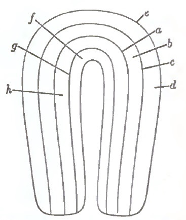
Diagram showing Muscular Strata of Uterus, as divided for clinical purposes.
If we run one line between the serous and another between the mucous membrane and the central line, as in the diagram, other strata with intervening spaces will be indicated. a would represent the centre stratum of the wall; b, the space immediately outside of that; c, a stratum still farther out; e, the subserous; and d, a deeper one. When we look at the inner layers of fibres, we find f situated immediately beneath the mucous membrane; g, farther out; and h, next the median line. The nucleus of a tumor may be first manifested in any of the strata or spaces marked by these lines, and its position with reference to the central line will, to a great extent, govern the direction it takes during development. A tumor the nucleus of which is situated in line a will, as it develops, press the muscular fibres equally in every direction, and when large, the prominence caused by pressure of the tumor would be equal in the uterine cavity and on the peritoneal surface. In marked contrast to this, when the nucleus is at f the growing tumor presses the mucous membrane before it until it becomes pendulous, and then the name of polypus is given to it; or if the origin is at e, the serous membrane is pressed before it, and the tumor is called subserous. When the nucleus is at d, the tumor elevates the serous membrane and becomes a prominent hemispherical protuberance. It is also called a subserous tumor, although situated some distance from the membrane. When a tumor takes its origin at g the mucous membrane is crowded before it, and a marked prominence into the cavity of the uterus is observed. This is the submucous tumor. These illustrations are intended to call the attention of the student to the fact that practically these tumors spring from any one or all the fibrous strata of the uterus instead of only the central, submucous, and subserous layers, and that it is profitable, on account of the difference in their effects upon the shape and functions of the uterus, to study them in this aspect of their growth.
ETIOLOGY.—While we know many of the conditions under which fibrous tumors exist, we have really very little, if any, definite and reliable information as to their causes, either remote or proximate. We know that they occur much more frequently near the time when the uterus begins to undergo senile degeneration, although they do originate in earlier years. They very seldom, if ever, are observed in the foetus or child, nor is it common for them to commence growing after the menopause. Women belonging to the African race are the most frequent subjects of these tumors.
The married or single status does not seem to have any effect in predisposing to these tumors. We do not know what physiological or pathological states of the uterus or other organs predispose to them. There is probably no tumor in the body strictly analogous in structure, mode of origin, supply, or development to the fibroid tumor of the uterus. There is no other organ in the body that undergoes analogous normal trophic changes. The vast multiplication of tissue that takes place in the uterus during gestation, and the more rapid but equally great changes toward degeneration or atrophy, would naturally suggest pathological possibilities of a peculiar nature. The rhythmical changes of menstruation are like no other functional condition. They too involve the processes of hypertrophy and atrophy. When the menstrual and generative changes are normal every part of the body of the uterus is simultaneously and proportionately hypertrophied and atrophied. Local derangements of these processes of hypertrophy and degeneration must sometimes occur, probably from defective or excessive innervation of loculi in the fibrous structure. Congestion or hyperæmia may thus result, and consequently very great influence be exerted upon the nutrition of the parts concerned after the deposit has begun; its presence increases the hyperæmia and thus perpetuates its growth indefinitely.
CLINICAL HISTORY.—Probably the earliest, most frequent, and constant symptoms connected with fibrous tumors of the uterus are hemorrhage and leucorrhoea. They are both the result of active or arterial hyperæmia, and doubtless come from the endometrium. Polypi, submucous, and intramural tumors are more likely to give rise to these two symptoms. The nearer the mucous membrane, and the greater that membrane is expanded, the greater the amount of hemorrhage and leucorrhoea, and, as a counter-fact, the nearer the serous membrane, the less the amount of these two discharges. While this statement in reference to the effects of the proximity of the tumor to the two membranes is usually true, it is not always so.
Hemorrhage is sometimes not very great, but at others it is appalling, and constitutes an imperative reason for the employment of desperate remedies. The hemorrhage is usually first noticed in connection with the menstrual flow, and it may even be confined to the periods: sometimes it extends over the whole of the interval. The leucorrhoea is generally constant, and sometimes thin and watery, especially after the hemorrhagic paroxysm has subsided, and at others it is constituted mainly of mucus with the débris of the mucous membrane and blood-corpuscles.
Other symptoms are pelvic pressure, vesical and rectal, with tenesmus, distension, and dysmenorrhoea. The pelvic pressure and tenesmus are observed early in the development of the growth, and may be relieved as the tumor becomes large enough to rise out of the pelvic cavity. The abdominal distension of course comes later. Solid tumors do not often attain to such a size as to cause great abdominal distension. The fibro-cystic generally are inconvenient, if not fatal, from this cause.
The above are the more direct and common symptoms. A less frequent yet important effect and symptom is oedema of the lower extremities from pressure upon the venous trunk passing through the pelvis. In rare cases this symptom is aggravated to a degree constituting phlegmasia alba dolens. As the tumor rises and enlarges the pressure may embarrass or interrupt the function of any or all the abdominal viscera.
In many cases none of these symptoms present themselves to an inconvenient degree, and the tumor is discovered by accident. Again, we meet with cases in which the symptoms are formidable for a time, and then entirely subside, leaving the patient free from suffering the balance of her lifetime. While this subsidence may take place at any time during the growth of the tumor, it is very apt to take place at the menopause.
The clinical history of the fibrous tumor may be very much modified by the intervention of various circumstances. As organized bodies they are subject to those affecting the organs of the body. We must regard them as adventitious growths acted upon by organs in a state of disease and reacting in turn upon them. They may become inflamed, undergo suppuration and gangrene, and produce symptomatic fever, hectic fever, prostration, gastric, hepatic, and nervous derangement in a degree sufficient to prove fatal.
When situated near the mucous membrane, nature sometimes turns these organic changes into a means of cure by destroying the portions of the capsule near the uterine cavity and permitting the pus or gangrenous material to escape. They are also subject to pressure from the development of other tumors, and either disappear, become inflamed and adherent, or cause great trouble to adjacent organs. Their clinical history is sometimes modified by complication with pregnancy.
This complication is rare, because the uterus in most cases, on account of the effects produced upon its circulation, nerve-supply, and mucous membrane especially, will not retain the ovum, and conception does not take place. The uterus being more vascular, and subject to congestions that affect the placental attachment injuriously, miscarriages are likely to occur. It is also morbidly sensitive to the pressure of the ovum, while the mucous membrane is rendered incapable of decidual changes. The retentive power of the uterus is further interfered with from the irregularity of its growth: the fibres where the tumor exists, being under a morbid influence, cannot partake of the regular hypertrophy necessary to normal gestation. There is something of uniformity in the circumstances under which the coexistence of pregnancy and fibrous tumor is observed. The nearer the tumor is situated to the mucous membrane, the less likelihood of pregnancy—the more remote, the greater the tolerance of pregnancy. Tumors that occupy the wall of the corporal portion are conducive of sterility. Those in the cervical portion of the corporal and the cervical zone are more likely to be accompanied with pregnancy than those situated in other parts of the organ. While the reader will find these statements borne out by his experience as general facts, he will also discover that pregnancy is occasionally compatible with almost any form, variety, or position of tumor. When this complication occurs, it does not generally influence the process of gestation or the condition of the tumor. The main symptoms depending on it are those caused by pressure. When small this is not very considerable.
Complication with labor generally gives rise to more apprehension than difficulty. Most of the cases of labor terminate spontaneously and happily, and the others are generally within reach of the less destructive modes of delivery. Labor more frequently decidedly affects the growth of the tumor, in the majority of cases causing its disappearance during the process of involution. The cervical polypi affect labor less, and are less affected by labor, than any other variety of the tumor. If small, they are sometimes merely pressed to one side or into the hollow of the sacrum, and the head passes by them; if a polypus is large, the head of the foetus carries it before it beyond the vulva, where it remains until the child is expelled, when it may recede into the vagina.
DIAGNOSIS.—The history usually includes hypersecretion, hemorrhage, pressure, and enlargement. These, while suggestive, are not conclusive, hence physical examination becomes indispensable to accuracy. The methods of examination vary with the size of the tumor. It is generally near the truth to say that the uterus is enlarged, and may be shown to be so by the introduction of the sound; yet the cavity is not always enlarged, and it is often so tortuous that the ordinary sound may be arrested before reaching the fundus. The sound, therefore, should in such condition be flexible. The fine whalebone or the sound of Jenks will generally pass obstructions caused by tortuosities. The most skilled and dexterous use of the inflexible sound is often delusive. We may generally determine the size by bimanual examination—one finger in the vagina or rectum while the hand is passed down into the pelvis from above. The uterus of normal size cannot be felt with any distinctness from above in this way, while an enlargement of 50 per cent. may be thus determined. The finger below will sometimes recognize the pressure from above when the upper hand will not feel the fundus distinctly. Small tumors of the uterus may be mistaken for many other conditions, and the converse. If one is situated in the posterior wall, it may be mistaken for retroflexion. We may make the distinction by means of the inflexible sound and the finger in the rectum. If the case is one of retroversion, the finger in the rectum will pass behind it and overlap it above. If a retro-uterine tumor is in the cul-de-sac, the finger will not reach above the uterus. If the case is one of retroflexion, a strongly bent sound may be made to enter it, especially if the fundus is slightly raised by the finger in the rectum. If there is a tumor in the posterior wall, the sound with slight flexion will pass above it; which is clearly ascertained by the finger in the rectum. When the sound is introduced in the case of retroflexion, the fundus may be elevated to its proper position by turning the sound upon its axis. In making these examinations with the sound the finger should be made to co-operate with it by being kept in the rectum. A small tumor in the anterior wall may be distinguished from anteflexion by the sound passing upward instead of forward, or into the part lying on the bladder. When a small tumor is intra-uterine, the uterus will occupy its natural position, with the mouth directed slightly backward; and if the polypus is large, the cervix can be moved forward with considerable difficulty. A flexible sound, especially the thin whalebone, may sometimes be made to partially or wholly surround it, and its size or connections be determined. But the diagnosis may be more definitely made out by dilating the cervical cavity and introducing the finger. The difference between a polypus and an intramural submucous tumor may be determined in this way. In the case of a polypus the finger will pass around it, while if the tumor is intramural or submucous the finger will be arrested at the point of attachment. A polypus or intramural submucous tumor presenting at the os externum may sometimes be mistaken for a partial inversion. Such a mistake may be prevented by using the sound. In the case of a tumor the flexible sound will pass to more than the normal depth. In one of inversion the sound will pass very much less or not at all. When a polypus has escaped from the mouth of the uterus and occupies the vagina, the sound will pass beyond it into the enlarged uterus, whereas in complete inversion it cannot be passed into the uterus in any direction. We cannot rely upon consistence or shape as marks of distinction in these two conditions. When the tumor rises above the pelvic brim and is not very large it generally displaces the os from its normal position. If in the front wall, the os will be too far back; if in the posterior, it will be displaced forward. In the former, when a sound is introduced, it will pass backward and upward; in the latter, the sound will pass forward and upward. In both cases the bimanual examination will enable us to determine that the tumor above the pelvis is continuous with or attached to the uterus. With the hands in this position, if we move the uterus the tumor will move with it, and vice versâ. Tumors of this size are usually more or less uneven in their outline, and of greater consistence than the uterus when enlarged from other causes. Tumors of this size may be generally distinguished from the pregnant uterus by the history of pregnancy, by the consistence, and by the size of the cervix. When pregnancy and a tumor are associated, this may be determined by a part of the enlargement being very hard and other parts quite elastic, and by auscultation. I need not caution the reader against the use of the sound where there is any suspicion of pregnancy. When a doubt exists, we should await the progress of the case until pregnancy becomes obvious. We may generally determine whether a tumor is uninuclear by the fact that a single tumor is nearly round, when if there are several points of origin it will be irregular and nodular.
When the tumor is large enough to nearly or quite fill up the abdominal cavity, the flexible sound may be made to pass a great distance into it. It is not often that a solid tumor grows large enough to fill the abdominal cavity. Before it grows to such dimensions it generally undergoes cystic degeneration. When the tumor is solid, generally its very great hardness, and often its irregular shape, will distinguish it from other abdominal tumors. The condition with which I have seen these tumors most frequently confounded is enlargement of the liver or spleen. In the South and West an enormously enlarged spleen is not infrequently met with. It sometimes spreads over the whole anterior part of the abdomen, completely covering the intestines. Less frequently the liver is found similarly enlarged. In this condition the organ becomes greatly indurated, and sometimes nodular. The distinguishing features of these enlargements are—first, that the abdomen does not present the prominent rotundity it does when filled by a growth; second, that somewhere in the extent of abdominal surface by careful manipulation the edge may be discovered and the fingers be made to sink beneath and grasp it; third, percussion will elicit general deep resonance, in some parts quite obvious, and in others less so. In the case of tumor none of these signs will be present. Again, the enlarged liver or spleen, while it may reach to the brim of the pelvis, does not reach into that cavity far enough to be recognized by the finger in the vagina, while the tumor does.
Sometimes inflammatory effusions form indurated masses in the abdomen that are mistaken for fibrous tumors. These of course have the history of inflammation, are generally if not always tender, and yield obvious intestinal resonance upon percussion. The large fibro-cystic tumor may be mistaken for pregnancy, ovarian tumor, cystic degeneration of the kidney, and omental tumors. Pregnancy can generally be established by absence of the menses, by the shape, size, consistency, and position of the cervix, together with auscultation. It may be said that in case of fibro-cystic tumor the cervix is greatly displaced in some direction, indurated, and not enlarged. In pregnancy none of these conditions prevail.
The fluctuation of the fibro-cystic tumor is more obscure than that of the ovarian tumor, and, although sometimes noticeable over a large space, it is usually more constricted in extent. There is also usually less regularity in the shape of it. In large ovarian tumors the uterine cervix is not changed in shape and size. The whole organ generally lies beneath the tumor, and the elastic sound will not pass very deeply into the cavity. If the uterus is attached to the anterior part of the tumor, which sometimes happens, the elastic sound will pass into it and the depth will not be very great. The fibro-cystic tumor may be distinguished from the enlarged encysted kidney by the facts that the kidney is traceable to one side more than the other, and it cannot be reached by the finger through the vagina or rectum. Still, if we cannot make the differentiation clear in any other way, we can generally do so by aspiration. In most cases we cannot draw the fluid from the fibro-cystic uterine tumor; in almost all cases the quantity removable in that way is small. When fluid is drawn, it usually coagulates, contains hæmatin, and none of the cells so generally found in ovarian tumors.
The fluid drawn from the kidneys presents epithelial cells, is not coagulable, certainly does not coagulate spontaneously. The abdominal cavity is sometimes more or less filled with peritoneal serum. After this is withdrawn from the peritoneal cavity the uterine attachment of the tumor may be made out by bimanual examination, as above directed, if undertaken immediately after the evacuation.
PROGNOSIS.—Less than twenty years ago the general prognosis to be made upon the discovery of a tumor of the uterus was very grave. The profession knew so little about the clinical history and diagnosis of these tumors that they were invested with many of the bad qualities of other tumors, with which they were so often confounded; and we had so little knowledge of their nature and the measures which would influence their growth that we felt an entire helplessness in the treatment of them. Fortunately, there have been many favorable changes in these respects. We understand their clinical history better, and can make a pretty clear diagnosis. We know that relatively few of them prove fatal even when left wholly to nature. Compared to all other uterine and ovarian growths, they are innocuous. Most of them are self-limited in consequence of the mode of blood-supply. A goodly number not only stop growing, but disappear without the application of any remedial measures. Then, as I shall have occasion to show, they may be often cured by the judicious administration of medicines, and the surgery for their extirpation has become a reliable resort in extreme cases. These considerations render the general prognosis of the true fibrous tumor quite hopeful. The menopause generally starves them out, and thus removes all the bad qualities they may possess.
When they lead to fatal results, they generally do so through three different conditions—viz. hemorrhage, pressure, and complicating inflammations—and probably in the order mentioned. Hemorrhage is by far the most fatal symptom. The kind of fibrous tumor accompanied with severe hemorrhage is usually the submucous variety. The submucous tumor with a broad base is the most mischievous, because it induces great hypertrophy in the vascular system of the mucous membrane especially, and also the vessels of the whole organ. A sessile submucous tumor arising from one nucleus is worse than one in the same situation with several nuclei of origin. The intracorporal polypus or pendulous tumor is almost as bad in this respect as the sessile submucous, especially if it originates at or near the fundus. Fortunately, these forms of the tumor are more amenable to the effects of medicine and more accessible to surgical treatment. The tumors located in the central stratum of fibres are next to these in mischievous qualities. The more remote the tumor is located from the mucous membrane, the less hemorrhage will attend its development.
When the tumor becomes cystic the danger from pressure is very much greater; yet the solid form becomes sometimes so large as to do much mischief from pressure upon the abdominal organs; and any of these, except perhaps the polypoid variety, may be so situated as to cause mischievous if not fatal pressure upon the pelvic organs.
It is rare, however, that the pressure in either of these cavities proves fatal, especially when the case is under intelligent management. The supervention of inflammation in the tumor, even to a moderate degree, is very apt to lead to gangrene and death from peritonitis, shock, or septicæmia. Sometimes subacute inflammation of the peritoneal surface of the tumor gives rise to serous effusion or dropsy in the abdominal cavity that proves fatal; and, as before stated, peritonitis sometimes causes adhesions which result in augmented vascularity and consequent increase of blood-supply. This condition, I believe, often changes a solid to a fibro-cystic growth, a more highly vitalized tumor, and consequently a more mischievous one.
Do these tumors ever become sarcomatous or malignant? I do not believe they have any innate tendency of that kind. Where they are found complicated with malignant growths I believe the malignancy is an independent quality, and is an invasion resulting from some cause extraneous to its organization, and in that respect is analogous to an attack on the cervix or other portions of the uterus.
The prognosis when complicated with pregnancy is of course more grave, but experience has demonstrated the practicability of complete and normal gestation. Conception will not often occur where these growths have attained any great size, but may sometimes. Of the nine cases which I have met and had an opportunity to follow, not one has been attended with abortion or premature labor. In one the pregnancy seems to have been protracted at least four weeks. The foetus was in a state of decomposition, and had probably been dead four or five weeks before labor began. What is not less remarkable also is that labor did not seem to be seriously affected in but one case, and in that the difficulty was easily overcome by turning.
Until lately there were several supposititious sources of danger at the time of confinement—viz. inefficient uterine contractions, and consequent tedious or impracticable labor, and after expulsion or artificial removal of the foetus dangerous hemorrhages from the same cause; also, the possibility of the placental connection being made at the site of the tumor, with the imperfect closure of the sinuses that was supposed to follow.
Reports of cases occurring within the last few years, while they have not completely swept away the grounds for such apprehensions, prove that the accidents so greatly feared do not in fact occur. Chadwick reports a case where the placenta was attached to the mucous membrane over the tumor, yet the placenta was spontaneously expelled and there was no considerable hemorrhage. The efficiency of the expulsive efforts were not materially affected in any of the cases I have attended. And this is what we might expect, because conception and gestation would not be perfect where there is not a sufficiency of healthy mucous membrane, upon which a normal decidua could be formed, and of fibrous structure to permit the hypertrophy of gestation.
The apprehension of obstruction from the tumor lying in such a position as to intercept the expulsion of the foetus is not often realized; for those in the cervix, either pendulous or otherwise, are pressed out of the external parts in advance of the head, while those in the body and fundus are lifted up into the abdominal cavity, where there is plenty of room. It must indeed be rare that the tumor becomes impacted in the pelvis so as to interfere with the passage of the foetus.
Neither does the puerperal condition seem to be rendered materially more dangerous in consequence of the presence of these tumors.
What effect does pregnancy have upon the growth of these tumors? It might be supposed, from the plentiful supply of blood afforded them by the growth of the vascular system of the uterus, and from the fact of their being situated in and surrounded by tissues in a state of active hypertrophy, that the tumors would grow in a corresponding degree with the uterus itself; but this is not generally, if it is ever, the case. I have not witnessed a decided increase in the size of the tumor in any of my cases. Pregnancy usually produces the opposite effect; and this can be easily understood when we remember that the tumor is subjected to great and uniform pressure, which prevents its own circulation from becoming as great as it otherwise would be; and I think this pressure often inaugurates a retromorphosis that results in the final disappearance of the tumor. Whether degeneration begins during pregnancy or not, the tumor is very apt to disappear after pregnancy and labor. In six of my own cases the tumor disappeared by a slow process of some kind after labor. Speculating as to what might be, another apprehension of danger arises out of the tumultuous excitement and terrible pressure to which it is subjected during the throes of parturition. But this apprehension is rarely if ever realized.
TREATMENT.—The treatment of fibrous tumors of the uterus consists largely of the means calculated to relieve such symptoms as endanger the life of the patient or materially affect her general health. When these are unavailing resort is had to measures calculated to get rid of the tumor. Some remedies necessary to the relief of symptoms act as very powerful curative agents; hence, while it is convenient to speak of the treatment of symptoms under one division of the subject, and the methods employed for radical cure under another, we cannot, in fact, completely separate these two branches.
Hemorrhage is by far the most important of the symptoms connected with these growths, because it is at the same time the most frequent and hazardous. It is also the symptom that leads to most suffering in consequence of depriving important organs of the blood necessary to support them in their functions. Every reasonable means should be made use of, not only to prevent fatal losses, but also to prevent moderate hemorrhage. In the outset, therefore, I would insist upon watching with great vigilance to prevent any unusual loss of blood. It is not advisable to temporize by adopting the milder and less efficient measures as being sufficient for cases not likely to prove fatal, but we should treat all hemorrhages arising from this cause with promptitude and energy. Fortunately, in many cases we can anticipate the attacks of hemorrhage, because we know when they will occur, and we are generally able to judge of their probable severity. To discharge our duty in this respect effectually, our patient should be properly provided with remedies and fully instructed how to use them. She should be made to understand that unusual hemorrhage at the menstrual period may be checked without endangering her general health. Among the remedies are—dorsal recumbency with the hips elevated, cold to the hypogastric region and cold to the dorsal spine and sacrum, ergot, and some form of tampon. The best fluid extract of ergot in drachm doses, if the stomach will bear it, is probably the most efficacious, but the fresh drug in the form of infusion is also very efficient. Full doses should be given every half hour when there is much loss, until some effect is produced upon the hemorrhage, and then continued every four hours as long as necessary. Compressed sponges saturated with the solution of sulphate of alum make the best tampons for the patient to make use of. These may be made and kept in readiness, so that they can be introduced as soon as they are found necessary. The patient or nurse can make them by taking a fine sponge, large enough to fill the vagina, passing a piece of string through the centre to aid in its removal, and then, after dipping it in the solution, winding it with twine from one end to the other, compressing it into as small a space as possible. The twine should so compress the sponge as to make it assume an elongated form. It should then be laid aside and permitted to dry. Several sponges should be thus prepared. When necessary the twine may be unwound and the sponge introduced. Its size when in the dry condition will allow of an easy passage into the vagina, where the moisture will cause it to expand, and fill up and seal the vagina so as to absolutely check the discharges. If the attending physician is present, he may tampon the vagina with pellets of cotton secured by thread and moistened with a solution of alum. The inconvenience experienced from this plug will be more than counterbalanced by the saving of blood. This form of tampon has the additional advantage of being antiseptic. I have allowed it to remain for three days, and upon removing it satisfied myself that there was no decomposition of the blood or the vaginal secretions. When the tampon is removed it will not be found difficult to wash out all the granular clots caused by its presence. It may be repeated as often as necessary, but usually, if allowed to remain forty-eight hours, the hemorrhage will not return. It may be said that for small losses this is unnecessary, but it is convenient and harmless, and will answer the purpose. In dangerous cases no one will question the propriety of its employment.
Another very important means of arresting hemorrhage which can be used by the physician when necessary is the introduction of a compressed sponge into the cervix uteri. This will temporarily act as a tampon and stimulate the uterine fibres to contraction. The free incision of the cervix, as directed by I. Baker Brown, may be tried between the times of the paroxysms of hemorrhage.
The pressure of the tumor upon the pelvic viscera is another inconvenience which calls for attention. This takes place usually at a time when the tumor has acquired a size sufficient to fill the pelvic cavity. Consequently, the elevation of the tumor above the pelvis is the remedy. This may be done sometimes by placing the patient in the knee-elbow position and pressing the growth upward. The powerful influence of atmospheric pressure called to our aid by the position and opening of the vagina is a very material auxiliary in the process of elevation. If this is not sufficient, we may pass the fingers into the rectum and elevate the tumor. I once succeeded in this operation by using an ivory-headed cane in the rectum when the fingers failed to reach high enough. If we cannot elevate the tumor by any of these means, we may introduce into the vagina or rectum a gum-elastic bag, and by means of a powerful syringe fill it with water to as great distension as the patient will bear, permit it to remain, and thus do the work more gradually.
Dysmenorrhoea is another symptom of fibrous tumors, and sometimes a very distressing one. It depends, no doubt, on the imprisonment of blood in the uterine cavity in consequence of the tortuosity of the canal causing the closure of some part of it. The remedy consists in dilating these narrow places. I know of nothing so well calculated to effect this object as the slippery-elm tent. One or more of these tents, long enough to reach the fundus uteri and of sufficient size, moistened so as to render them very flexible, may be passed up through the tortuous places with great facility. If introduced as soon as the symptom begins to manifest itself, and allowed to remain an hour or two, the relief will be pretty certain. If used once a day for four or five days before the attack, and three or four hours at a time, dysmenorrhoea may be generally avoided.
Curative Treatment.—When we broach the question of the permanent cure of these affections, we find that great difference of opinion exists among the members of the profession as to the value of medicines. One party, perhaps a majority of the profession, believe that no medicine has any direct effect upon them, and these ignore any means of permanent relief but surgical. There is, however, a respectable number of medical men who place great reliance upon the administration of certain medicines, and, if I am not greatly mistaken, recent observation has added greatly to their number. They do not, however, wholly agree as to the therapeutic processes that should be instituted, and consequently do not employ the same kind of medicines. Some gentlemen have more confidence in what I will term the sorbefacient medicines and processes of treatment. They endeavor to institute measures that will cause the absorbents to attack and remove the neoplasm in the same way that tumefactions caused by effusions are removed. This they do by friction, pressure, and the administration of the old-fashioned sorbefacient medicines. The most popular among these are the iodides, chlorides, and bromides of mercury, potassium, sodium, calcium, and ammonium. Reports may be found in books and periodical medical literature of cures by several if not all of these articles and their combinations. The late W. L. Atlee, whose experience was very extensive, had great confidence in the action of hydrochlorate of ammonia. He administered it internally, applied it externally, and used it as vaginal injections. The iodide of potassium has long enjoyed a great reputation in causing the absorption of these and other forms of tumors. There is no professional fairness in assuming that the faith in these remedies derived from the observation of their effects or the promulgation of cures from the use of sorbefacient measures are fallacious. Some of the men arrayed in favor of the opinion that cures may be effected by a patient and long-continued administration of some one of the articles I have mentioned stand high as men of honesty, accuracy of observation, and faithfulness in their records; and therefore I give full confidence to their statements. Yet I must also say that I have not witnessed the good results which I unhesitatingly believe others have seen from the sorbefacient treatment alone.
Others who expect much from medicinal treatment look to that class of medicines which cause contraction of the unstriped muscular fibres as the most promising. With these medicines they expect to diminish the supply of blood to the tumor by causing contraction of the arterioles traversing their substance, and thus disturbing their nutrition to such a degree as to stop their growth, lessen or destroy their vitality, and so render them subject to the influence of the absorbents, whereby they may be removed. Some of the more energetic of these medicines—as ergot and belladonna, for instance—often affect these growths very promptly. Ergot not only lessens the calibre of the small blood-vessels, and thus causes a diminution of their nutrition and disappearance, but it causes strong contractions in the muscular fibres of the uterine walls, which lessen more decidedly their supply of blood. It sometimes squeezes and chafes the tumor until it is disintegrated and rendered a foreign substance. The capsule finally becomes ruptured, and the tumor is expelled either piecemeal or en masse.
When properly administered, ergot frequently greatly ameliorates some of the troublesome and even dangerous symptoms of fibrous tumors of the uterus—e.g. hemorrhage and copious leucorrhoea; it often arrests their growth; in many instances it causes the absorption of the tumor, occasionally without giving the patient any inconvenience: at other times the removal of the tumor by absorption is attended by painful contractions and tenderness of the uterus; by inducing uterine contraction it causes the expulsion of the polypoid variety of the submucous tumor; in the same way it causes the disruption and discharge of the intramural tumor. There are many cases on record to substantiate every one of these propositions.
From what I consider well-authenticated sources, including the cases under my own observation and in the practice of my friends and neighbors, I have collected 136 cases of fibrous tumors treated by ergot. Of these, 25 cases were cured without giving the patients any inconvenience from painful contractions. In 46 cases the tumors were diminished in size and the hemorrhage was cured. In 27 others the hemorrhagic symptom was relieved, while the size of the tumor was not affected. In 8 other instances the tumors were broken to pieces and expelled piecemeal.
For examples of cases in which the first conditions obtained, I would refer to those cured by Hildebrandt; of the other examples, 4 were reported to me by the late J. P. White of Buffalo, N. Y., 1 each by the late Hodder of Canada and Jukes, and 11 that occurred among my immediate acquaintance and in my own practice.
Among those in which the hemorrhage was cured and a diminution of the tumor took place, 11 occurred to Hildebrandt, 2 to Chrobak, 5 to White of Buffalo, and the remainder to gentlemen upon whose veracity I have implicit reliance. The most remarkable case of which I have any knowledge was reported to me by the late G. C. Goodrich of Minneapolis, in which absorption of a large tumor took place under the administration of ergot and belladonna. I subjoin his description: "The treatment was commenced in 1870, and continued two years. The uterus filled the whole space between the ilia, and measured in the transverse diameter twelve inches and in the vertical nineteen inches—extended up under the ensiform cartilage and close up to the margin of the cartilages of the ribs. The treatment was followed by cramps in the uterus, which produced a wild enthusiasm in the mind of the patient and inspired her with strong hopes of recovery. Without consulting me she doubled the dose of medicine, which was administered internally, and as a consequence she was attacked with very strong uterine contractions and symptoms of metritis. This caused me to abandon treatment for about one month, and had it not been for the urgent determination of the patient I would not have resumed it. She insisted that as this was the first medicine which had ever affected the enlarged organ, she believed it would cure her, and promised to obey my directions if I would proceed. She so promptly and rapidly improved that I doubted if it were not a coincidence with, rather than a consequence of, the treatment. Prompted by this doubt, I abandoned the use of the ergot and belladonna and continued alterative treatment. The patient soon assured me that she no longer felt the griping pains caused by the remedy, and that the tumor was softer and larger than when she took the ergot prescription. The ergot and belladonna were again resumed, and in four months she was able to make a trip to Boston alone. While absent she continued to take the medicine. From this time she continued rapidly convalescing, and is now in the enjoyment of fine health."1
1 The author's address before the American Medical Association at its meeting in 1875.
I subjoin two cases in which the tumors were expelled piecemeal under the administration of ergot, which came under my own observation:
A woman of Sterling, Illinois, called on me December 13, 1875. She was thirty-five years old, married, and had never been pregnant. On the first of the preceding June she noticed a circumscribed hard lump two inches below and to the left of the umbilicus. She was the subject of serious uterine and sympathetic symptoms, for which she had at different times had treatment. She had profuse menorrhagia, leucorrhoea, and great sense of weight in the pelvis. Upon examination I found a hard, round, movable tumor extending up to within two inches of the umbilicus, filling up the whole of the right iliac, the hypogastric, lower half of the umbilical, and more than half of the left iliac regions. The contour of the tumor was somewhat uneven, though not distinctly nodular. The cervix was long, pointed, and thrown backward and to the left. The sound entered the small uterine mouth and passed upward, backward, and to the left five and a half inches. The diagnosis was a fibrous tumor of the right anterior wall of the uterus. I prescribed thirty drops of Squibb's fluid extract of ergot, to be taken three times a day. She went home, but did not commence taking the medicine until the 20th of December. On the 26th of December J. B. Crandall was called to see her, and describes her condition as follows: "The patient was in a state of great nervous prostration and worn out by severe pain and loss of sleep. The pains commenced soon after taking the second dose of ergot, and were excruciatingly severe for about three hours, after which they continued less severely for two days and nights. She had more or less hemorrhage from the uterus after taking the ergot. Her pulse was feeble, 110 to 120 to the minute. The skin was hot and dry, and she complained of great pain and tenderness over the uterus and lower bowels. The feet were drawn up, and the face wore a pinched and peculiar expression." Under these circumstances the doctor administered anodynes, tonics, and nourishment, to the great relief of the patient. On January 11, 1876, the patient began to pass from the vagina small masses of fibrous substance, from the size of a chestnut to that of an English walnut. The substances thus discharged were firm and gray in color, and were exceedingly fetid. This discharge continued up to the 21st of January, when the uterus was very much diminished in size, the tenderness had subsided, and the patient appeared comparatively comfortable. Up to that time she had taken but three doses of ergot—on the 20th of the preceding month—and the doctor ordered it to be resumed again. This time the ergot produced no pain, and after three or four days was discontinued. From the 21st of January there were no more pieces discharged, but up to February 1st a yellowish, thin, offensive fluid passed from the vagina in considerable quantities. On the first day of February the ergot was again ordered and continued two weeks, when, as no results ensued, it was finally dropped. Crandall states that on the 14th of February the uterus was reduced to its normal size, and on the 26th the patient was up and about her work, completely cured. He remarked, in this connection, that the first three doses of ergot taken by the patient was the cause of her recovery.2
2 This case is published in the August (1875) number of the Chicago Medical Journal and Examiner, as reported by Crandall.
Mrs. L. D. M., aged forty-seven years, had a fibroid tumor in the anterior wall of the uterus, which, with the enlarged uterus, arose to within two inches of the umbilicus. She commenced taking thirty drops of the fluid extract of ergot on the 22d of September, 1876, and was to increase gradually the dose with the object in view of causing the disruption and expulsion of the tumor. The ergot at first produced no perceptible effect until she had taken it ten days, when she began to experience the pain of contraction. The pain became so severe and continuous that it was necessary to omit it for two or three days at a time. The patient was intelligent and understood the object and mode of action of the ergot, and when the pain entirely subsided she courageously resumed it in the smaller doses, and increased again until the pains became intolerable. On the 13th of January, 1877, small pieces of the tumor showed themselves in the vaginal discharges, and by the 26th of the same month the whole of it had been discharged piecemeal. She wrote me on the 30th of January, saying, "I think I wrote one week ago to-day. At that time the tumor was passing. It continued to pass until the 26th, when, I think, the last was expelled. To-day I send you by express a portion of the last that came. I think the whole of it, including the portion I send you, would have weighed one and a half pounds. I do not believe a quart can would hold it if the whole had been preserved. It commenced to come on Saturday, and from Saturday evening to Sunday morning there was a pint or more. After that the stench was so disagreeable that we could not cleanse it; consequently we threw it away. Wednesday and Thursday it seemed to be in one continuous mass. I cannot better describe it than to say that it came like sausage-meat from a stuffer. I would cut off about four inches a day—that is, on Wednesday and Thursday. On Friday morning the last of it came away." During and for some days after the expulsion she suffered slight symptoms of septicæmia, but recovered from them, and in the course of a month afterward she visited me, when I found the uterus measured two inches and a half in depth. She then had some leucorrhoea, but was fast regaining her health. She is now perfectly well, and has passed in safety the menopause.3
3 This case—the abstract of which I have here given—was in the May (1877) number of the Archives of Clinical Surgery, N. Y.
I have known 9 cases in which the tumors were expelled piecemeal by ergot, with but 1 death. The death occurred in a patient who rode one hundred and fifty miles on a railroad train to see me with pieces of the tumor hanging from the vagina, which she would not allow her physician to remove. When she arrived I passed my fingers up into the contracted capsule and scooped out the remaining portion of the tumor. She was so exhausted, however, by the journey and the sepsis that she died three days afterward. I cannot help believing that if she had remained at home and submitted to the treatment of her physician, her life need not have been sacrificed.
The influence of ergot over the uterus has been a familiar fact to the profession for a long time. It is not long, however, since we were aware of its effects upon the muscular fibres entering into the formation of other organs. We now know that this medicine acts upon the unstriped muscular fibre wherever found, whether in the viscera or in the vessels of the body.
The fibres of the uterine walls, and the arteries supplying them with blood, both belong to this class; this fact in the formation of the uterus renders it particularly susceptible to the action of ergot. The drug acts upon the uterus4 in a threefold manner, and causes a diminished flow of blood to the morbid as well as healthy tissues in the uterine structure.
4 From the author's address before the American Medical Association, 1875.
First: the calibre of the arterial tubes is diminished by the contraction of the muscular fibres which enter into their composition. Second: the arterioles are diminished in size by compression from the contraction of the uterine muscular fibres which surround them. Third: these vessels are distorted and drawn in diverse directions by both the contraction and compression, and hence are rendered less fit for sanguineous conduits.
Another consideration of prime importance is that, under the influence of these medicines, the nutrition of fibrous tumors is interfered with, not only from diminution of blood in their tissues, but also from compression of their substance by the proper fibres of the uterus, and are therefore made more susceptible in the process of disintegration and absorption.
The great influence exerted by ergot over the circulation of the uterus is rendered more efficacious in the removal of fibrous tumors of that organ, because of the peculiar organization of the growths. It is now pretty well understood that this neoplasm is not very generously supplied with arterial blood, and that its supply is derived from numerous minute vessels instead of one or two of large calibre. From these circumstances it results that its vitality is very low, its circulation easily disturbed, and consequently its nutrition impaired.
I think we are justified from observation in assuming that the action of ergot may be graded from an almost imperceptible to a very intense degree. Probably the first degree affects the vascular supply; the second, in addition to this, causes so much contraction as to merely render the fibres tense without causing pain; and the third prompts the uterine fibres to vigorous and painful contraction.
This inference is plainly deducible, I think, from the several modes by which tumors are made to disappear under its action, as well as from direct observation of the uterine fibres.
I will now venture to call attention especially to the manner of expulsion of the polypoid and submucous intramural varieties. It will be seen that when the uterus contracts all the fibres unite in pressing the polypus through the cervical canal, which is usually already shortened, and rendered dilatable in consequence of its increased vascularity. The cervical canal dilates, and after more or less painful efforts the polypus is expelled entire, covered by the mucous membrane. This membrane is often in a state of gangrene, but so far as I have observed these cases the tumor is not broken to pieces.
A submucous intramural tumor has a thin layer of fibres separating it from the mucous membrane, and a thick and heavy layer spread over its external hemisphere. A greater part of the muscular wall is therefore applied to the outer side of the tumor. If in this position all the fibres of the uterus vigorously contract, the fibres near the mucous membrane must be overcome by the heavy layer outside. But the opposite wall plays an important part by supporting the weaker layer at the fundus of the tumor, and adding its own force in overcoming the capsule, where it usually gives way. The position of the tumor makes its escape from the concentric action of all the fibres of the uterus impossible, and every one knows that when the resistance is partially overcome the uterus is stimulated to more vigorous action, and the pains will not abate until the mass is expelled. If not too large, it is driven out without undergoing great laceration, but if its size and attachments are such as to make this impracticable, it will be broken into fragments and expelled piecemeal.
In subperitoneal tumors there is, next the uterine cavity, a thick and strong stratum of fibres, while immediately under the peritoneum the layer is very thin and comparatively weak. When the uterus is acting with vigor the former contract forcibly, and the mass becomes pedunculated; but that is all, for the tumor lies outside the field of concentric action and escapes the crushing influence to which the submucous variety is subjected. The amount of force exerted upon it is that exercised by the weaker layer of fibres in a state of conquered antagonism, and the rupture of the capsule is impossible.
In the case of a fibroid tumor situated in the central stratum of fibres the antagonism is equal at all points, and it is evident that there is no tendency to rupture of the capsule, and much less crushing influence exerted upon it than if it were situated slightly nearer the mucous membrane. This variety of the tumor, therefore, yields to ergot only as it may be starved out by diminution of its blood-supply and as the effect of pressure, which we all know are the two conditions most favorable to absorption.
Now I think we have arrived at a point in this investigation where we can draw inferences as to the forms of tumors likely to be effected by ergot in different ways, as well as those that will not be effected by it. We do not expect ergot to cause painful and efficient contractions in the healthy unimpregnated uterus; its fibres are not capable of such contraction, and it is not until the fibres have become greatly developed that they are susceptible to the impressions of ergot. In cases of early abortion its action is very unreliable, but after the fourth month of pregnancy it acts quite efficiently.
In tumors of the uterus the development of the fibrous structure is sometimes so slight that it is incapable of contraction; there may be so many nuclei of degeneration that there are not enough sound fibres left for efficient contraction. Then, where there are many small tumors developed in the uterine walls, the circulation is cut off to such a degree that they degenerate into a cartilaginoid substance, and sometimes they are infiltrated with calcareous material. In none of these cases will ergot cause any appreciable results. When, however, there are but one, two, or three nuclei of morbid growths, as they increase in size the fibres undergo the development necessary to enable them to contract with great efficiency and render them susceptible to the influence of ergot.