Volume 4 — DISEASES OF THE GENITO-URINARY SYSTEM. (continued 5)
Reading edition
In the early stage of seminal incontinence, when the nocturnal pollutions overstep the natural limits, the ejaculated fluid is unchanged. When, however, the pollutions are more frequent and diurnal discharges coexist, the semen is watery and scanty; the spermatozoids are smaller, comparatively few in number, and their movements are liable to be abolished in less than an hour, while spermatic crystals form more rapidly and more abundantly than in health. In the worst cases, or those characterized by diurnal and nocturnal pollutions and by the presence of semen in the urine, the spermatozoids are either entirely absent, or, if they are present, they are motionless, stunted, or variously deformed. In these advanced cases the ejaculated fluid, which consists principally of the secretions of the seminal vesicles and the prostate, frequently undergoes fatty degeneration, as indicated by granular epithelium, by molecular detritus, and even by oil-globules in the protoplasm of the altered zoosperms. The entire absence of spermatozoids, constituting the condition known as azoospermatorrhoea, is of infrequent occurrence.
An examination of the genital organs discloses elongation of the prepuce in nearly one-fourth of all cases; a rigid and pointed penis in one-tenth; relaxation of the scrotum in about one-eighth; irritable testes in 1 example out of every 25; varicocele in 1 case out of every 50; coldness of the genitalia in 1 case out of every 17; a feeling of heat in 1 case out of every 33; and irritability of the bladder in 1 case out of every 25. It will, moreover, be found that seminal incontinence is complicated by feebleness of erection, with precipitate ejaculation on coition, in 22 per cent. of all cases; by the occurrence of ejaculation on attempting intercourse, before penetration, simultaneously with erection, or even before erection, in 16 per cent.; and with total impotence in 5 per cent. of all cases. Prostatorrhoea is also a not infrequent complication, while urethral strictures and hyperæsthesia are nearly always present.
ETIOLOGY AND PATHOGENY.—Seminal incontinence is not a separate entity, but one of many symptoms of general or local disorders, or of both combined. In the majority of instances it must be looked upon as a neurosis, diurnal and nocturnal pollutions representing a motor neurosis with spasm of the seminal vesicles, and spermorrhagia indicating a motor neurosis with dilatation and paresis of the orifices of the ejaculatory ducts. In all of the varieties there is increased susceptibility of the cerebral and spinal genital centres to factors which in healthy persons are not productive of ill effects.
Like other nervous disorders, involuntary seminal emissions sometimes manifest themselves in several members of the same family through several generations, being the result of inherited predisposition. In this class of cases the subjects are of a nervous, excitable, or irritable temperament, somewhat anæmic, and possibly suffered during infancy from nocturnal enuresis. Among the predisposing causes the most common is indulgence in erotic fancies, which terminates in increased reflex impressibility of the centres which preside over the genital organs.
The affection is, however, usually acquired, being met with particularly in single subjects toward the termination of the second decade and between the second and third decades. Of these cases, at least nine-tenths can be traced to masturbation, while the remainder will be found to have had gonorrhoea or to have masturbated, suffered from gonorrhoea, or indulged their sexual propensities in various ways. Seminal incontinence is not common as the result of sexual coition, and it is highly probable that when married men are affected the sexual excess is engrafted upon a previously vicious habit. From a practical point of view, it is of the first importance to be aware of the fact that one or more strictures of the urethra will be found in 80 per cent. of all cases, and that decided hyperæsthesia of the prostatic portion of the urethra is present in 94 per cent. of all instances.
The rational explanation of morbid seminal emissions seems to be as follows: Under the influence of erotic ideas, masturbation, sexual excesses, or unsatisfied sexual excitement produced by dallying with women, exaggerated irritability of the genital organs is induced, and is followed by subacute or chronic inflammation and abnormal sensibility of the urethra, particularly of its prostatic division, which terminate, in cases characterized by diurnal pollutions and spermorrhagia, in relaxation and dilatation of the orifices of the ejaculatory ducts. As the natural result of the constant excitability of the terminal filaments of the nerves distributed to the prostatic urethra, these nerves are alive to the slightest impressions, act as peripheral sources of irritation, and induce permanent increased mobility or irritability of the cerebral and spinal genital centres, through which the motor nerves of the ejaculatory apparatus are thrown into action, and an emission ensues.
Seminal incontinence is an occasional accompaniment of injuries of the spine, and it is also met with during the progress of or convalescence from acute and chronic diseases which are marked by disturbances or exhaustion of the central nervous system. Thus, it may be symptomatic of phthisis, variola, typhus, progressive muscular atrophy, and incipient bulbar paralysis, ataxia, and paraplegia; while the habitual use of opium and chronic alcoholism predispose to its occurrence.
Of the local causes referable to the genitalia, by far the most important and most frequent are hyperæsthesia and inflammation of the prostatic portion of the urethra, which are generally induced by masturbation. These lesions constitute the primary source of the trouble in the large majority of cases, and tend not only to excite reflex pollutions, but to maintain the disorder by keeping the mind occupied with sexual matters. Other common local causes are found in congenital narrowing of the meatus, organic stricture of the urethra, a redundant prepuce, balanitis, and the accumulation of smegma. Among the more infrequent etiological factors may be mentioned herpes of the prepuce, congenital shortness of the frenum, spasmodic stricture, polypus of the deep urethra, spermato-cystitis, and epididymitis.
Among the remaining exciting causes of pollutions are diseases of the anus and rectum, as hemorrhoids, morbid growths, ascarides, fissures, ulcers, pruritus, and painful eruptions. The nerves of the rectum and anus being derived from the same region as those of the genitalia, it is not surprising that the ejaculatory centre should respond to an impulse transmitted from them. In habitual constipation straining at stool may also excite an emission through the consentaneous action of the muscles of the abdomen, rectum, and seminal vesicles; but this is only observed when the orifices of the ejaculatory ducts are paralyzed and patulous.
ANATOMICAL CHARACTERS.—There are no records of the morbid appearances which appertain to seminal incontinence in its early stage, but that the hyperæsthesia of the prostatic urethra depends upon chronic or subacute inflammation is rendered certain by the concomitant symptoms, by exploration with the sound, aided by the finger in the rectum, and by the results of treatment. In the advanced stage, post-mortem inspection has disclosed stricture of the urethra, injection of the mucous membrane of the deep portion of the urethra, dilatation and excoriation of the orifices of the ejaculatory ducts, and suppuration of the prostate and the seminal vesicles. The changes which occur in the nervous centres are unknown.
DIAGNOSIS.—The microscope affords the only positive mode of determining whether the fluid which is discharged from the urethra during pollutions, or constantly moistens that canal in spermorrhagia, or is expelled at stool or with the urine, or is brought away by the bulb of the explorer, is seminal in its character. Should spermatozoids be detected, there can be no doubt as to its true nature, but their absence is not an evidence that the case is not one of spermatic incontinence, since in the condition known as azoospermatorrhoea the exhausted sexual apparatus furnishes a thin, transparent, watery fluid which may be entirely devoid of fertilizing elements, and contains cylinder epithelial cells, epithelium which has undergone fatty or colloid degeneration, a few lymph-corpuscles, an abundance of fatty detritus, and a few small shining bodies which are the remains of the badly-evolved spermatozoids. Under these circumstances, the history of the case, the fact that the subject is or was a masturbator, and the associated nervous symptoms are aids in forming a diagnosis; and this is especially true of cases in which a fluid is expressed at stool, and which in the majority of instances is the altered secretion of the prostate. Under the microscope the thin, more or less milky prostatic fluid will be found to contain cylinder epithelium, numberless colorless and refracting granules of lecithin, and minute yellowish concentric amyloid concretions; and, after it has slowly dried upon the slide, crystals of phosphate of magnesium or of ammonio-magnesian phosphate will make their appearance.
Should a microscopical examination be impracticable, we may assume that the discharge which occurs during defecation in the subjects of too frequent nocturnal pollutions is an evidence of coexisting prostatorrhoea; while we may frame the rule that the flocculent sediment contained in the urine and the discharge at stool of persons suffering from both nocturnal and diurnal pollutions, and a slight continued discharge from the urethra represents semen. In the last event we may moreover assume, especially if the patient be impotent, that the orifices of the ejaculatory ducts are relaxed.
PROGNOSIS.—Nocturnal emissions are very amenable to treatment, particularly when they are kept up by appreciable local lesions, the only cases which are, as a rule, rebellious being those in which the pollutions are associated with chronic inflammation of the seminal vesicles. In expressing an opinion in a given case the physician should, however, be influenced by the severity of the signs of nervous exhaustion. If the general symptoms point to involvement of the cord alone, the prognosis is far better than when signs of cerebrasthenia are present; but the outlook is bad if, in addition to cerebral and spinal exhaustion, the patient is a sexual hypochondriac. Nocturnal pollutions occurring during the progress of acute or chronic general disorders are also, as a rule, readily checked. The prognosis in the same class of cases is, moreover, far better when the usual local lesion—namely, morbid sensibility of the prostatic urethra—has been induced by gonorrhoea rather than by masturbation; and it is also more favorable when the pollutions occur in mature years from sexual excesses than when they are due early in life to masturbation.
Even when the emissions occur during the day from trivial psychical or mechanical causes, ample experience has convinced me that the prognosis is far better than many writers would lead one to believe. These cases are, however, less tractable than those of nocturnal pollutions, but they finally recover with the exercise of a little patience. The worst outlook is when the emissions are passive, or occur without the orgasm, or during urination and defecation. In this class of cases not only are the ordinary remedies applicable to the other varieties demanded, but measures will have to be resorted to to overcome the paralyzed and dilated orifices of the ejaculatory ducts. Although the prognosis is not as favorable, I have never seen an example of spermorrhagia that did not finally yield to treatment.
TREATMENT.—Certain hygienic and moral rules must be observed in the management of all the varieties of seminal incontinence. The diet should be plain, nutritious, and digestible; the evening meal should be light and dry; and spirits and malt liquors, as well as stimulating articles of food, should be eschewed. As the morning fulness of the bladder is very liable to produce an erection, that organ should be thoroughly emptied on retiring; and as pollutions usually occur toward morning, the patient should set an alarm-clock one hour before the time at which he has generally observed that the emissions take place, in order that he may be awakened to relieve the bladder of its contents. He should also sleep upon a hair mattress without much covering. Everything calculated to induce a flow of blood to the genitalia, such as horseback exercise, driving over rough roads, and railway travelling, should be interdicted. Masturbation and sexual intercourse must be abandoned, and the subject should be informed that the enforced rest of the organs will possibly result in temporary increased frequency of the pollutions. Chaste associations should be cultivated, and erotic thoughts and desires be banished. To attain this end the mind and body should be kept pleasantly occupied by gymnastic exercises and the study of any subject which the patient may fancy. If, however, he be not in full health, or if there are commencing or marked signs of spinal or cerebral exhaustion, mental and physical exercise should be taken in moderation.
In the treatment of involuntary seminal emissions a thorough examination should be made of the genital and associated organs, with the view of detecting and getting rid of any reflex or eccentric lesions or causes which predispose to, or even excite and maintain, them in impressible subjects. If the patient has a redundant prepuce, it should be removed; if the meatus be contracted, it should be enlarged; while balanitis, herpes, hemorrhoids, rectal fissure or ulcer, or pruritus should be treated in the usual way. In not a few mild cases, particularly those dependent upon phimosis, a contracted meatus, or a stricture just behind the orifice, it will be found that operative interference is quite sufficient to bring about relief. Habitual constipation, which is met with in about one-third of all instances, demands particular attention, either by enemata of temperate water or a pill composed of one-tenth of a grain each of aloin and extract of belladonna, administered every eight hours.
In the section on the etiology and pathogeny of seminal incontinence attention is called to the fact that hyperæsthesia of the prostatic urethra is nearly always present. While it is undoubtedly true that the genital nervous centres may be highly impressible without the intervention of hyperæmia, inflammation, and abnormal sensibility of the prostatic urethra, it is none the less true that those lesions are the most constant and most important of all the causes which excite and maintain the disorder, especially in masturbators, in whom, moreover, strictures may be looked for in about eight-tenths of all cases. As a rule, the coarctations will be formed just behind the meatus, but others may be present posteriorly. Be this as it may, a knowledge of their existence is of the first importance, as they aggravate the morbid condition of the prostatic urethra and serve to keep up a peripheral source of spinal neurasthenia.
For the detection of a stricture the exploratory or acorn-headed soft bougie should be resorted to, as it is the only instrument with which coarctations of large calibre and granular patches can be accurately defined, and with which abnormal discharges can be withdrawn for minute examination. One being selected which fills the meatus, it is warmed and well oiled, and inserted as far as the bladder. Should its introduction be arrested, smaller sizes are successively employed until one will pass without difficulty. On its withdrawal the abrupt shoulder of the bulb coming in contact with the posterior face of the stricture imparts to the touch a sensation as if it had jumped over a band, while a granular patch conveys the impression of a limited roughness of the canal. Hyperæsthesia of the urethra is readily determined by the nickel-plated steel bougie, and its existence should never be based upon the passage of the soft explorer alone, as the latter is productive of far more pain than the former. In conducting these examinations a contracted meatus or a stricture just behind the orifice should first be divided, in order that the instruments for exploration may correspond to the normal calibre of the urethra. Unless this point receives attention the examination will be likely to prove valueless. Should one or more strictures be present, the case must be referred to a surgeon.
From the preceding considerations it follows that the treatment, whether it be local or general, must at the outset be of a calming and sedative nature, the end in view in the great majority of instances being to overcome the exaggerated irritability of the genital nervous centres and the abnormal sensibility of the deep urethra. By the indiscriminate employment of strychnia, cantharides, phosphorus, and cold ablutions great harm is done, and the management of involuntary seminal emissions is brought into disrepute.
Of the local remedies to overcome the hyperæsthesia of the prostatic urethra, there is not one entitled to so much confidence as the nickel-plated conical steel bougie, passed at intervals of four days, and at once withdrawn for the first few insertions, after which, with the decrease of the sensibility, the intervals should be shortened, and it should be retained longer, until it is inserted every forty-eight hours and permitted to remain in the canal for a few minutes. The size of the first instrument is to be gauged by that of the meatus if it be normal, and if it be found necessary during the course of the treatment the orifice should be enlarged, in order that bougies of progressively increasing sizes may be introduced until they correspond to the full calibre or distensibility of the urethra, as indicated by the urethrameter. Unless these precautions be observed the measure will not bring about the desired result.
As a rule, the bougie will meet the indication, but in exceptional instances a small, circumscribed area of tenderness remains, which comprises the sinus pocularis, and which proves rebellious to instrumentation. Under these circumstances it becomes necessary to apply a drop or two of a solution of nitrate of silver to the spot, which is best done with a small syringe attached to a perforated bulbous explorer. The ordinary forms of porte-caustique charged with the fused nitrate are objectionable, as the remedy does not come in contact with the orifices of the ejaculatory ducts contained within the sinus pocularis, and its application cannot be properly controlled. From an ample experience I can confidently recommend the use of a thirty-grain solution, repeated every four days. Provided the patient be kept in bed for a few hours, the pain and desire to urinate will not last more than thirty minutes. When the affection proves to be more than ordinarily obstinate, flying blisters, made by pencilling cantharidial collodion first on the one side of the perineal raphé, and, after the surface has healed, on the opposite side, will prove serviceable.
In addition to these measures great assistance will be derived on retiring from the hot sitz-bath, or from a sponge or cloth dipped in water at a temperature of at least 105° F. and applied to the perineum and lower part of the spine. Cold applications are to be studiously avoided.
Of the general remedies, not a single one is comparable to bromide of potassium, which not only diminishes the reflex excitability of the cord and suspends sexual desires and the power of erection, but corrects the acidity of the urine and exerts an anæsthetic effect upon the mucous membrane of the urethra. I am in the habit of administering from three to four scruples of the salt at bedtime, and if I find that it sets up signs of bromism I diminish it for a time, and afterward promote its excretion by the kidneys by combining with it about fifteen grains of bitartrate of potassium. Should the patient be anæmic, the dose should be reduced to one drachm, and three grains of quinine along with twenty-five drops of the tincture of the chloride of iron should be ordered every eight hours. When, on the other hand, the patient is robust and plethoric or in full health, I frequently add to the bromide ten drops of veratrum viride or tincture of gelsemium, or administer the bromide in half an ounce of the infusion of digitalis.
Another remedy which diminishes the reflex mobility of the genito-spinal centre, at the same time that it reduces the secretion of the seminal fluid, is the sulphate of atropia. Given in the average dose of the one-sixtieth of a grain on retiring, so that the patient may sleep through its disagreeable action, it will be found to be an invaluable addition to the treatment.
When the bromide of potassium and atropia do not agree with the patient, I substitute the monobromide of camphor and extract of belladonna in the proportion of ten grains of the former to one-third of a grain of the latter. In the remaining anaphrodisiacs, such as lupulin, camphor, and conium, I have not the slightest confidence.
Under the plan of treatment thus outlined the majority of cases of nocturnal and diurnal pollutions recover; but if the spinal genital centre still remains too impressible, galvanization with the anode to the lumbar region and the cathode to the perineum will prove highly serviceable. When the condition is one of spermorrhagia, after the hyperæsthetic symptoms have subsided the relaxed and paralyzed orifices of the ejaculatory ducts may be restored to their normal condition by the continuous current, the negative reophore being placed in the rectum and the positive on the perineum or the lumbar vertebræ. Should galvanization fail, the induced current may be passed through a negative catheter electrode in the prostatic urethra to the anode resting on the perineum or spine; but this mode of application requires great caution, and a feeble power should be employed at the commencement. For this reason the rectal is preferable to the urethral reophore. In the absence of electrical apparatus the tonicity of the muscles of the ejaculatory ducts may be greatly improved, and even restored, by the use of the cooling sound, by the application of a thirty-grain solution of nitrate of silver, and by cold sitz-baths. In these cases half a drachm of the fluid extract of ergot after each meal, or fifteen drops of a mixture composed of six drachms of the tincture of the chloride of iron and two drachms of the tincture of cantharides, will also prove valuable. The operations of castration and excision of portions of the vas deferens need only be mentioned to be condemned.
To sum up the results of my experience in the management of seminal incontinence, I may add that the steel bougie, bromide of potassium, and atropia are especially adapted to cases of nocturnal and diurnal pollutions, and that after the hyperæsthesia has been relieved electricity, ergot, and strychnia are the most reliable agents in spermorrhagia. The end having been accomplished, moderation in sexual intercourse should be enjoined if the patient is married; continence in thought and action should be observed if he remains single; and matrimony should be advised if his circumstances and inclination warrant it. Marriage should not, however, be encouraged if the emissions are not arrested, as I have met with several cases in which the patient was rendered miserable by this act, from the fact that he deemed his case beyond all hope, as the emissions still continued.
DISPLACEMENTS OF THE UTERUS.
BY E. C. DUDLEY, A.B., M.D.
The title of this article is not to be taken in a restricted sense, inasmuch as the uterus is anatomically so connected with adjacent organs that the displacements of the uterus cannot be intelligently considered or satisfactorily presented without at the same time incidentally taking into account the displacements, causative, resultant, or concurrent, of the ovaries, Fallopian tubes, rectum, vagina, and bladder.
Normal Location and Position of the Uterus.1
1 The importance of a distinction between location and position will become apparent hereafter: by the former is meant the situation of the organ regardless of its attitude, by the latter is meant the attitude alone. To change an object from one place to another is to change its location; to turn it over or bend it upon itself is to change its position.
In the works on anatomy and gynecology which we are accustomed to consult the uterus is represented as having a straight or nearly straight canal—as lying about midway between the symphysis pubis and the hollow of the sacrum, its axis corresponding to that of the pelvic inlet. They generally agree that its position is one of slight, and only slight, anteversion; some admit that slight anteflexion may not be injurious, but most would pronounce the organ anteverted or anteflexed to a degree that would endanger health if by conjoined manipulations its anterior wall could be felt through the anterior wall of the vagina. The classical idea of the normal position of the uterus presupposes a distended bladder and rectum occupying the anterior and the posterior thirds of the pelvic cavity. Such an arrangement would leave for the uterus only the intermediate space, and would constitute a condition seldom or never realized in health.
Suppose a straight line coincident with the vesico-vaginal wall (Fig. 1) to be continued through the cervix to the sacrum. This line represents approximately the antero-posterior diameter of the pelvis. The length of the vesico-vaginal wall is two and a half inches, and, supposing the cervix to be just midway between the symphysis and the sacrum, the distance from its posterior wall to the sacrum must also be two and a half inches. Add to the sum of these two parts of this antero-posterior diameter one inch for the cervix, and the antero-posterior diameter of the pelvis becomes six inches instead of the normal four and one-third; which proves that the cervix must normally be much nearer to the hollow of the sacrum than to the symphysis. Since the length of the vesico-vaginal wall plus the diameter of the cervix measures three and one-half inches, it follows that the distance from the posterior wall of the cervix to the hollow of the sacrum must be the difference between four and one-third and three and one-half inches, or five-sixths of an inch.
FIG. 1.
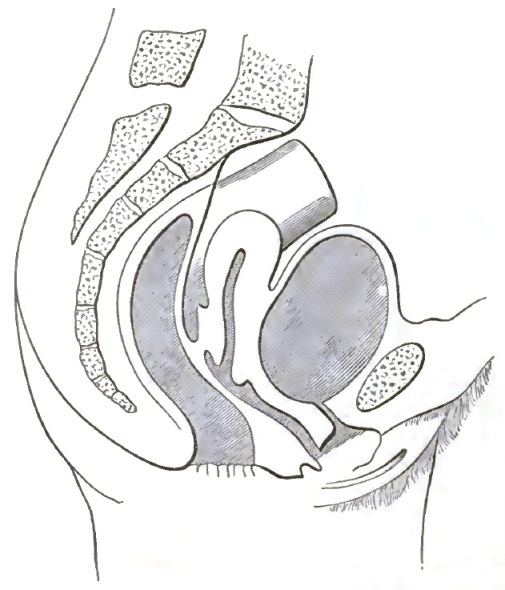
The Classical Representation of the Pelvic Organs.
Again, suppose the uterus (Fig. 1) to be carried bodily upward and backward, its axis remaining the same, until the cervix reach its normal position near the hollow of the sacrum; then would the body of the uterus impinge upon the bony sacrum. It is therefore clear that the anteversion must be the normal position, because the uterus and sacrum would otherwise occupy the same space.
Fig. 2 represents, according to Schultze,2 the location and position of the virgin uterus and its surroundings, the bladder, rectum, and vagina being empty and collapsed. The angle of about 90° which the cervix forms with the vagina measures the forward inclination of the cervix, but is subject to slight variations in consequence of the physiological movements of the uterus. The body is furthermore bent forward upon the cervix, so that its anterior surface rests upon the empty bladder. The angle of the normal anteflexion, according to careful measurements by Schultze, is about 48°; Fritsch says that 90° is the physiological limit. This question will be further considered under the subject of pathological anteflexions.
2 Archiv für Gynäkologie, 1875, Band viii. p. 134, and Lageveranderungen der Gebarmutter, Berlin, 1881.
Ely Van de Warker makes a full and critical study of the normal movements of the unimpregnated uterus in the N. Y. Medical Journal, xxi. p. 337, and of the normal position and movements of the unimpregnated uterus in the American Journal of Obstetrics, xi. p. 314. His conclusions substantially agree with those of Schultze.
Frank P. Foster (American Journal of Obstetrics, xiii. p. 30) presents a valuable paper giving a résumé of the literature, with original observations, in which he takes exceptions in part to the views of Schultze.
FIG. 2.
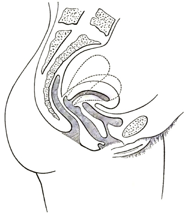
The Correct Representation of the Pelvic Organs.
Normal Movements of the Uterus.
Strictly, the uterus can have no absolutely normal position or location, because it has a certain normal range of movements which depend to some extent upon respiration, intra-abdominal forces, and locomotion, but more especially upon the varying quantity of material in the rectum and bladder. Its normal position, then, varies within the limits of its normal movements. If the body of the uterus rest upon the bladder, it must rise as the bladder becomes distended, and, conversely, if the urine be drawn through a catheter while the woman is lying on her back, the uterus, notwithstanding the opposing influence of its own weight, immediately follows the receding wall of the bladder and returns through an angle of 45°, or possibly even 90°, to its accustomed position. The dotted lines in Fig. 2 indicate the degree of version and flexion consequent upon the varying quantity of fluid in the bladder.
The full rectum forces the uterus in the opposite direction, toward the symphysis, and thereby counteracts the influence of the bladder. This anterior movement is, however, somewhat limited, and is confined to the cervical portion, except when the body has been forced back into close proximity with the rectum by the over-distended bladder.
Normal Supports of the Uterus.
The uterus is maintained in its normal position and location by the following agents:
a. The uterine ligaments;
b. The pelvic floor.3
3 For a description of the female pelvic floor see Hart's Atlas.
a. Physiologically, these ligaments are relaxed; the state of tension would be pathological; they do not fix the uterus; they only tend to limit its movements to their normal range. Backward displacement of the body is resisted by the round ligaments, backward displacement of the cervix by the utero-vesical ligaments and by the vesico-vaginal wall. Forward and downward displacements are resisted by the utero-sacral ligaments, and excessive lateral motion by the broad ligaments. This restraining power is doubtless greater in the utero-sacral than in any of the other ligaments.
b. The pelvic floor, which is the chief support of the uterus, is divided into two segments, the pubic and the sacral. The pubic segment4 is composed of bladder, urethra, anterior vaginal wall, and bladder peritoneum. It is attached in front to the symphysis pubis and laterally to the anterior bony walls of the pelvis. The sacral segment5 is composed of rectum, perineum, posterior vaginal wall, and strong tendinous and muscular tissue. It is attached to the coccyx, to the sacrum, and to the posterior wall of the bony pelvis.
4 Hart and Barbour's Manual of Gynecology.
5 Ibid.
Permeating the pelvic floor in all directions, entering into the composition of its single parts, binding them together, and sending its processes to the bony pelvis, is the pelvic connective tissue, upon the integrity of which depends the integrity of the pelvic floor as a uterine support. Its pernicious influence as a pathological factor will be considered hereafter. The old idea that the uterus is supported by the vaginal walls or by the perineum or by the uterine ligaments is obsolete; they are important parts of the pubic and sacral segments, and as such contribute their share, but the pelvic floor as a whole supports the uterus. The various uterine supports are to a great extent the seat of motor influence. They consequently not only resist excessive movement, but also serve to return the organ from its physiological migrations.
DEFINITION AND NOMENCLATURE OF DISPLACEMENTS.—In the foregoing pages the normal location, position, movements, and supports of the uterus have been defined. Those conditions are pathological which induce changes to positions or locations beyond the defined limits, or which so fix the organ that its normal movements are prevented. The displacements are divided into mal-locations and malpositions.
The mal-locations in which the entire uterus occupies a place outside its normal limits are as follows: ascent, retro-location, ante-location, lateral location, descent.
The malpositions are determined by excessive change in the inclination of the uterine axis. They are further divided into flexions, in which the organ is bent upon itself in an abnormal degree, manner, or direction; and versions, in which the axis of the unflexed uterus inclines in an abnormal degree or direction. The malpositions are retroversion, retroflexion, lateral version, lateral flexion, anteversion, anteflexion.
SYMPTOMS AND DIAGNOSIS IN GENERAL.—Each variety of displacement may be indicated by its own group of symptoms and physical signs. These will be presented in the study of the special lesions. To avoid repetition, those symptoms and signs which pertain to no special displacement, but which belong to all alike, will be mentioned at once. They may arise either from the displacement itself or from its possible complications, of which the following are examples: Metritis, ovaritis, salpingitis, atresia and stenosis, cystitis, vesical catarrh, rectitis, rectal catarrh, peri-uterine cellulitis and peritonitis, uterine catarrh, tumors, cicatrices, etc.
Uterine displacement may be a cause or an effect of associated complications, or together with them it may be a concurrent result of some common cause, or it may have had primarily no pathological connection with them. The symptoms of displacement refer to the pelvic organs or to the nervous system. Among the symptoms which refer to the pelvic organs are—difficulty in walking and standing; pelvic pain, more or less constant; dysmenorrhoea, menorrhagia, sterility, frequent abortion, constipation, painful or difficult defecation, dysuria, polyuria, tenesmus, etc. Among the symptoms which refer to the nervous system are—neuralgia in various parts, paralysis, hysteria, nervous dyspepsia, anæmia, chlorosis, spinal irritation, etc.
The final diagnosis must always depend upon direct examination of the uterus itself. The first division of the above group of symptoms is not likely to escape notice as indicative of displacement, but the nervous symptoms are constantly disregarded or treated without reference to their possible pelvic origin. The frequent dependence of these nervous phenomena upon displacement is proved by their persistence in many cases after ordinary treatment, by their prompt disappearance upon permanent replacement and retention of the uterus by mechanical means, and by their equally prompt recurrence upon removal of the support. The presence, therefore, of the second division of the group or any part thereof, even though the first be absent, will justify, may even necessitate, a careful investigation into the state of the pelvic organs.
That examination which results only in giving the name to a special variety of displacement, and does not include the complicating lesions, would not furnish a sufficient guide to the therapeutic indications, and is therefore inadequate. The successful treatment, for instance, of an anteflexion dependent upon inflammation of the utero-sacral ligaments must include the removal of the inflammation.
An important prerequisite to examination is the absence of material in the rectum and bladder. The full rectum distorts the vaginal walls, deprives the examiner of the space necessary for the introduction of the speculum, and throws the uterus out of its accustomed position. Much more troublesome is the presence of even a small quantity of urine in the bladder, because it causes the patient to render the abdominal muscles tense when the hand is placed over the lower portion of the abdomen for bimanual palpation, and makes it impossible to engage the uterus between the hand and the examining finger. The distended bladder by pushing the uterus upward and backward makes bimanual palpation almost useless. It is not surprising that conflicting opinions are common, when one day the patient is examined with rectum and bladder full, another day empty; one day in the dorsal, another in Sims's or the knee-chest position; one day with the cylindrical or bivalve speculum, another day with Sims's or Simon's.
For digital examination the dorsal position is preferred: the patient should be drawn close to the edge of a bed, or preferably a table, the thighs being flexed, the feet about fifteen inches apart, and the knees widely separated. The examiner should stand facing the patient, never at the side. The index finger of the left6 hand, lubricated with vaseline or oil, then slowly advances over the perineum into the vagina, noting the condition of the perineum, the presence or absence of cicatrices or of sub-involution of the vagina or perineum, the capacity of the vagina, the condition, size, and direction of the cervix, its distance from the sacrum and vulva, its mobility or fixation. Now, for the first time, the right hand is pressed well down behind the pubes, and the uterus is engaged between it and the examining finger. (See Figs. 16 and 17.) In this way the examiner may determine more accurately the position, location, and size of the entire organ; may detect the possible presence of complicating tumors, both inflammatory and non-inflammatory; may also note, if possible, the location and condition of the ovaries, which, especially in the posterior displacements, are liable to be prolapsed and excessively sensitive, and to constitute, therefore, a most intractable complication. The index finger sweeps around the cervix in search of tender places which may be the result of former cellulitis or the expression of some neurosis. Above all, the digital examination requires a light, gentle, delicate touch.
6 The left-hand method of examination is incomparably superior to the right. The palmar surface of the index finger is more easily directed toward the left side of the pelvis, which is especially subject to disease. Its tactile sense is more acute and more easily educated. The stronger right hand should be free to palpate the surface of the abdomen in conjoined manipulation.
In exploring the uterine cavity to learn its position the fine silver-wire probe of Emmet—not the sound—should be used. The uterus, if freely movable, is liable to be thrown out of its accustomed position by the heavier, unyielding sound. The sound also causes much more pain and exposes the patient to great danger of cellulitis. The frequent lighting and relighting of pelvic inflammation by injudicious slight manipulations of the uterus doubtless led Emmet to the utterance of a prophecy which ought to become classical: "A great advance in the treatment of the diseases of women will be made whenever practitioners become so impressed with the significance of cellulitis as to apprehend its existence in every case. The successful operator in this branch of surgery will always be on the lookout for the existence of cellulitis, and take measures to guard against its occurrence."
When the probe or the sound is used without the speculum, the patient should be on the back and the index finger of the left hand should be used as a guide. The bivalve and cylindrical specula are almost useless in explorations of the interior of the uterus. The exploration is most effectually and gently made with Sims's speculum, the patient being in the left latero-prone position. In some cases the probe cannot be passed by any other method.
Ascent of the Uterus.
This mal-location may result from traction above or from pressure below. The organ may be drawn upward and backward by shortening of the utero-sacral ligaments, which results from inflammation and which usually induces a troublesome form of anteflexion. The enlarged pregnant uterus sometimes becomes attached by adhesive inflammation to a portion of the peritoneum in one of the higher zones of the pelvis or in the abdomen, and the organ may consequently remain fixed in its elevated position after involution. A tumor connected with the uterus or its appendages which has grown too large to be retained in the pelvis may, upon rising into the abdomen, drag the uterus with it. Pressure below may come from excessive distension of the rectum or bladder, or from a large accumulation of menstrual fluid in the vagina, or from a tumor originating in any portion of the pelvis below the level of the uterus. In diagnosis, prognosis, and treatment this displacement is wholly subordinate to the more significant lesions of which it is only the incidental result.
Retro-location of the Uterus.
The uterus may be forced back into a post-normal location by the presence of a tumor in front or by the distended bladder, or it may be drawn back and fixed by peritoneal adhesions. Retro-location is liable to induce vesical irritation by putting the vesico-vaginal wall on the stretch and thereby dragging on the neck of the bladder. This intractable symptom is sometimes relieved by Emmet's buttonhole operation of urethrotomy, for an account of which see section on Anteflexion. This operation would obviously be applicable also for the relief of the same symptom when caused by ascent of the uterus.
Ante-location of the Uterus.
The causes of this displacement are similar to those which produce retro-location; they are—distension of the rectum, post-uterine hæmatocele, post-uterine tumors, and peritoneal adhesions. Ante-location often causes vesical irritation, consequent upon the invasion by the uterus of that space which belongs to the bladder.
Lateral Locations of the Uterus.
The entire uterus is often displaced to the right or the left by a tumor or by an inflammatory exudate. The latter occurs as a product of cellulitis, usually in the left broad ligament, and crowds the organ toward the opposite side of the pelvis. After resolution the ligament, shortened by inflammatory contraction, draws the uterus to the affected side and fixes it there. Lateral displacement from this cause often accompanies laceration of the cervix, the cellulitis having occurred on the side corresponding to the laceration.
Descent or Prolapse of the Uterus.
The nature of this displacement is clearly indicated by its name. It is convenient to distinguish three degrees of descent: In the first the organ is displaced downward and forward until sufficient space has been gained between the cervix and the sacrum to permit the body to turn back into extreme retroversion; in the second the cervix descends to the vulva; in the third the uterus protrudes partially or wholly through the vulva, constituting a condition sometimes called procidentia.
ETIOLOGY AND CLINICAL HISTORY.—Descent may be the result of any or all of the following causes: I. Pressure from above; II. Weakening of the supports; III. Increased weight of the uterus; IV. Traction from below. Either of the above conditions being the primary cause, the others singly or combined may result.
I. Pressure from above may depend upon the presence of a pelvic or abdominal tumor, ascites, fecal accumulations, tight or heavy clothing, etc.
II. The uterine supports may be weakened and relaxed in consequence of subinvolution, senile atrophy, abnormally large pelvis, increased weight of the uterus, pressure from above, traction from below, etc.
III. Increased weight of the uterus may be caused by congestion, subinvolution, hypertrophy, hyperplasia, pregnancy, fluid in the endometrium, uterine tumors, etc.
IV. Traction from below may be due to vaginal cicatrices, abnormally short vagina, falling of the pelvic floor, etc.
Obviously, descent of the vesico- and recto-vaginal walls, or, more comprehensively, the sacral and pubic segments of the pelvic floor, involves also concurrent descent of the uterus. Descent of the vagina, therefore, must be studied in connection with the descent of the uterus. Excessive descent of the vaginal walls usually originates with parturition.
In labor the anterior wall of the vagina is so depressed, stretched, and shortened by the advancing head that during and after the second stage the anterior lip of the cervix may be seen behind the urethra. If the puerperium progress favorably, with prompt involution of the uterus, vagina, perineum, and peritoneum, the relaxation of the vesico-vaginal wall and of the utero-sacral supports disappears and the uterus resumes its normal multiparous location and position.7 But if the enlarged uterus remain in the long axis of the vagina, with its fundus incarcerated in the hollow of the sacrum between the utero-sacral ligaments, and with its sacral supports so stretched that they cannot recover their contractile power, and with involution of all the pelvic organs arrested, the descent may not only persist, but may even progress with constantly increasing cystocele to the third degree of prolapse. The downward influence of the above conditions may be materially increased by rupture of the perineum, and consequent prolapse of the recto-vaginal wall into a pouch called rectocele.
7 The anteflexion of the multiparous uterus is less than that of the virgin.
In the great majority of cases of complete prolapse the posterior vaginal wall in its descent is peeled off from the rectum, leaving the latter in its normal position. In rare instances the lower portion of the rectum is also found to have extruded in extreme rectocele, making a pouch below and in front of the anus, where fecal matter may accumulate and remain in hard scybalæ.
Obviously, complete prolapse of the uterus is only an incident to the prolapse of the pelvic floor. The whole mechanism is in all respects analogous to that of hernia. The extruded mass drags after it a peritoneal sac, which, hernia-like, contains small intestine. This sac forces its way to the pelvic outlet and extrudes through the vulva, having the inverted vagina for its covering.
FIG. 3.
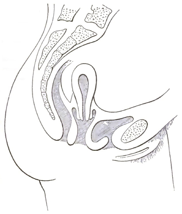
First Degree of Prolapse of the Post-partum Uterus. The posterior vaginal wall has been changed from its normal forward direction to a vertical direction by perineal rupture and anterior displacement of the cervix; the vesico-vaginal wall descends in cystocele, becomes hypertrophied, and drags the heavy uterus after it. The descending uterus carries with it a reduplication of the vaginal walls.
In descent of the first degree the location of the uterus is either changed to a lower level, the position remaining normal, or, as is more common, the cervix having moved nearer to the symphysis and the organ turns back into retroversion. In a given case suppose the vaginal walls from some cause to have become relaxed and to have settled to a lower level in the pelvis. As an associated fact the uterus to which these walls are attached must then also occupy a place correspondingly nearer to the vulva—i.e. the location of the uterus has changed, so that space enough intervenes between it and the hollow of the sacrum for the former to turn back into the position of retroversion or retroflexion. If, on the contrary, the descending uterus still maintains its normal anteversion and anteflexion, it must occupy space which belongs to the bladder. The vesical irritation consequent upon this mal-location has generally been ascribed to the anteversion and anteflexion, which are therefore oftentimes wrongly pronounced pathological. The prompt relief which follows permanent replacement of the organ in the normal location, even though in so doing its anteposition be exaggerated, proves that the symptoms depend upon the mal-location, not upon the anteposition. The importance of a clear distinction, therefore, between location and position becomes apparent. Vesical irritation, moreover, is sometimes caused by the dragging of the uterus upon the neck of the bladder. This traction occurs not only in ascent, but also when the organ descends below a certain level.
FIG. 4.
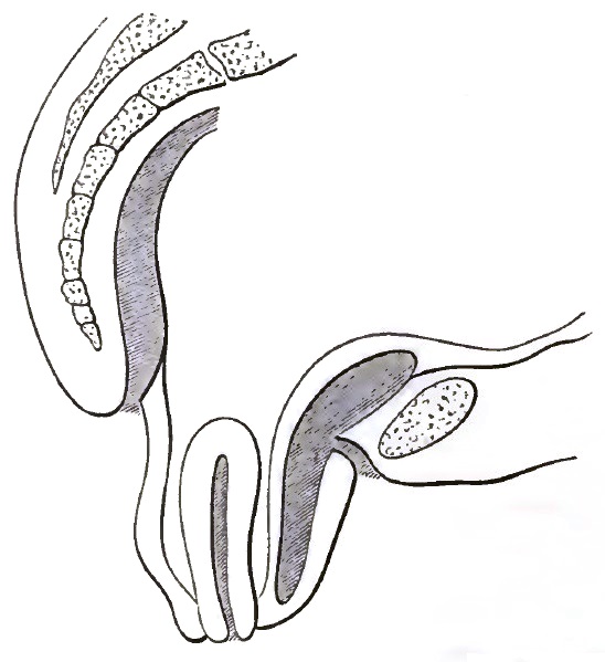
Showing Extreme Descent of the Uterus and of the Pelvic Floor, and the Hernial Character of the Lesion.
In the foregoing paragraphs traction due to the falling pelvic floor has been discussed as a cause of descent. The impairment of the uterine supports may, however, be such that instead of falling and dragging the uterus after them, they simply permit it to descend along the vaginal canal by the force of its own weight, and to carry with it the reduplicated vaginal walls. This influence is generally enforced by the increased weight of the diseased organ. The vagina more readily becomes a track for the descending uterus when from any cause the normal forward direction of the vaginal canal changes toward the vertical: this change may occur either as the result of a forward displacement of its upper extremity, involving anteposition of the cervix, or of a retro-displacement of its lower extremity in consequence of rupture or subinvolution of the perineum. (See Fig. 3.) Descent in the track of the vagina is obviously combined with some degree of retroversion, because the axes of the uterus and vagina then correspond.
The PATHOLOGICAL ANATOMY may involve all the displaced organs. The circulation throughout the pelvis is impeded by traction upon the vessels, and the entire pelvic contents therefore become the subject of venous congestion, with consequences disastrous to local innervation and nutrition.
The ovaries may suffer concurrent displacement, with resulting inflammatory and cystic enlargement. The peritoneum which enters into the formation of the uterine ligaments and of the pelvic floor is dragged along with the uterus.
The vagina is hypertrophied and swollen. Its mucous membrane becomes the seat of acute vaginitis and chronic catarrh. In the third degree of descent the exposed vagina, no longer lubricated by the normal secretions of the uterus, becomes dry, parchment-like, oedematous, eroded, and ulcerated. Sometimes the cul-de-sac of Douglas is distended by downward pressure of the intestines, by a small tumor, or by ascitic fluid, and a consequent hernial sac may protrude into the vagina through some portion of the posterior vaginal fornix. The anterior fornix is subject to a similar accident. These conditions are designated enterocele vaginalis, anterior and posterior.
The rectum and bladder are subject to inflammation and chronic catarrh, and the bladder especially to concurrent descent. The uterus may be enlarged from any one or all of a variety of causes—congestion, subinvolution, hypertrophy, and hyperplasia. Its cervix is often the seat of extreme erosion or so-called ulceration. The endometrium, in order to relieve the organ of its surplus blood, gives forth an excessive secretion of mucus, which upon being increased in quantity becomes vitiated in quality. This is termed uterine catarrh. The enlargement of the uterus often pertains more to the cervix than to the body, especially in prolapse of the second and third degrees. An explanation of this may be found in Figs. 5 and 6.
FIG. 5. FIG. 6.
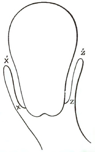
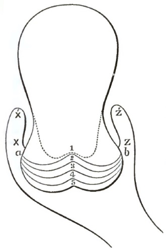
Descent of the Virgin Uterus into the Vaginal Canal, showing the Reduplicated Vaginal Walls. The utero-vaginal attachment, points X and Z, appears to be at X' and Z'. The apparent increase of length in the vaginal portion of the cervix due to the reduplication is measured by the distance from X and Z to X' and Z'. Descent of the Uterus, showing Excessive Circular Enlargement of the Lacerated Cervix, consequent upon Reduplication of the Vaginal Walls and Out-rolling of Intracervical Tissues. The divided fragments of the os externum are at a and b. The curved lines forming the angles 1, 2, 3, 4, and 5 indicate the gradual process of the eversion. The angle of the laceration at point 1 has been forced by the swelling and out-rolling of the mucous and submucous tissues of the cervix to point 5. The apparent os externum is at point 5. The utero-vaginal attachment X and Z seems to be at X' and Z'. The vaginal portion of the cervix therefore appears much larger and longer than it actually is.
Apparent elongation and disproportionate circular enlargement of the cervix are conditions which almost every standard author wrongly calls hypertrophic elongation and circular hypertrophy. The question of elongation is easily settled by placing the patient in the knee-chest position. Then the uterus by its own weight falls toward the diaphragm, the vagina unfolds, and the apparent utero-vaginal attachment X' Z' (Figs. 5 and 6) disappears, disclosing the actual attachment, X Z. Further, the point of the sound, passed into the bladder while the cervix is exposed by Sims's speculum, may be placed against the anterior wall of the cervix at Z, which would be impossible if the attachment were at Z'.
The comparatively small amount of hypertrophy in disproportionate circular enlargement is proved by the operation of trachelorraphy or by bringing the points a and b (Fig. 6) together with uterine tenacula, the organ being exposed by Sims's speculum. Then the out-rolled intracervical mucous tissues are rolled back, the proper diameter of the cervix is restored, and a laceration on one or both sides, extending past the vaginal attachment, becomes apparent.
Hypertrophy or hyperplasia usually causes a nearly symmetrical enlargement of the entire organ. At any rate, those cases in which the reduplication of the vaginal walls does not almost entirely explain the great elongation so called, or in which great disproportionate circular enlargement has not been caused by laceration of the cervix, are the rare exceptions. The great merit of having secured general assent to the foregoing proposition, and of having given to the subject a new and right direction, must be accorded to Emmet. The cervix now is seldom amputated except for malignant disease.
Congestion of the uterus consequent upon obstruction in the stretched and displaced veins is often so extreme as to induce a state analogous to erection. Measurements by the probe just before and a few minutes after replacement generally show an appreciable decrease in the length of the uterine canal. If the prolapse has been of the third degree, the difference may amount to one or even two inches. It is important not to confound the enlargement of congestion with increase in the solid constituents of the organ.
SYMPTOMS AND COURSE.—A dragging sensation and pelvic and abdominal pain are generally present. Rectocele and cystocele and rectal and vesical catarrh often cause painful and severe functional disturbances of the rectum and bladder. In descent of the third degree excoriations of the exposed vagina and cervix sometimes cause extreme suffering. The course is ordinarily chronic, but attacks of acute vaginitis and pelvic peritonitis are not uncommon. The peritonitis sometimes effects a spontaneous cure by peritoneal adhesions which fasten the uterus in an elevated position and hold it permanently. The symptoms of descent may be so severe as to necessitate absolute rest in bed. In other cases they are often attended with very little discomfort.
DIAGNOSIS is by inspection, palpation, and exploration. The prolapsed uterus may be distinguished from cystocele, rectocele, inverted uterus, and fibroid tumor by the presence of the os externum. The sound may be passed through the urethra into the cystocele, and the finger through the anus into the rectocele. The length of the uterus may be determined by the sound, the size, shape, position, extent of descent, and difficulty of replacement by conjoined manipulation.
PROPHYLAXIS.—This requires such measures during labor as may be necessary to prevent long and powerful pressure upon the pelvic floor. After labor any injury to the perineum should be promptly repaired. The vagina should be kept clean by irrigations. The urine, if necessary, should be regularly drawn and the bowels moved daily without straining. If conditions be present likely to induce subinvolution—such, for example, as pelvic inflammation or laceration of the cervix—they should receive treatment at the proper time. Undue relaxation of the pelvic floor necessitates a more prolonged rest in bed, the use of astringent douches, and the application of a pessary when the patient resumes the upright position.
TREATMENT.—The first indication is replacement, which in the first and second degree of descent is not difficult unless the uterus be held down by cicatrices or by a tumor. Complicating pelvic cellulitis and peritonitis may render replacement dangerous or impossible, and may for a time contraindicate all direct treatment. Replacement of the organs from the third degree of prolapse is accomplished in the inverse order of their descent: first, the posterior vaginal wall, then the uterus, and last the anterior vaginal wall. Not infrequently the completely prolapsed uterus and pelvic floor, hernia-like, become strangulated. Then taxis will usually suffice if supplemented by hot applications, elastic pressure, anodynes, and the knee-chest position. Should these fail anæsthesia may be required.
Undue pressure from above should if possible be removed. The clothing should be loose, and the weight of the skirts supported from the shoulders either by straps or preferably by buttoning them upon a waist made for the purpose. This waist is a good substitute for the corset, which under all circumstances and in all its forms is injurious. Increased uterine weight from subinvolution or congestion is to be overcome by appropriate means. Enlargement of the uterus when due to hypertrophy or hyperplasia is generally incurable. Amputation of the cervix for what was formerly considered circular hypertrophy and hypertrophic elongation is now seldom or never required for the purpose of decreasing uterine weight. Amputation except for malignant disease has given place to the operation of trachelorraphy. Tumors exerting pressure above or traction below should if possible be removed. Regulation of the bowels and general tonics are usually necessary. The knee-chest position assumed several times a day causes the uterus to gravitate toward the diaphragm, and thereby gives temporary rest to the overburdened supports. While in this position the patient should separate the labia, so that the air may rush in and the vagina become expanded. The measures enumerated above, together with rigid care of the diet and of such other hygienic requirements as the individual case may demand, are essential as adjuvants to the more special treatment which almost every case requires.
In exceptional cases of sudden descent, even to the third degree, replacement alone is sometimes followed by permanent relief; but if the descent has been gradual it always recurs immediately after replacement. Measures are therefore required for the maintenance of the uterus in its normal location and position. This indication is fulfilled by pessaries and by operations.
Pessaries.—The function of the pessary is not only to maintain the uterus on the health level in its normal location, but also, if possible, in its normal position, which requires the cervix to be about one inch from the sacrum. The cervix being thus placed, the organ cannot turn back into retroversion, because in so doing the fundus would encounter the sacrum. The direction of least resistance would then be forward into the normal anterior position. The application of the pessary is then based upon the general proposition that if the cervix be normally placed the body of the uterus will in the absence of complications take care of itself. Since the vagina at its upper extremity is attached to the cervix, displacement of the latter is clearly impossible if the upper extremity of the vagina be sustained in its normal location. The pessary restores and maintains the relations of the relaxed vaginal walls by crowding the posterior vaginal cul-de-sac backward into the hollow of the sacrum. It thereby also holds the attached cervix within a proper distance of the sacrum. The Hodge pessary or some modifications thereof fulfils this purpose in ordinary cases more satisfactorily than any other.
FIG. 7.
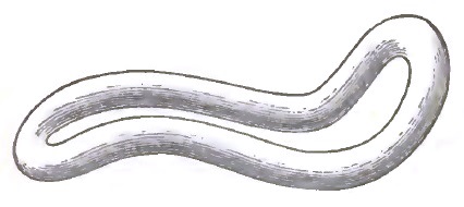
The Emmet Curves.
FIG. 8.
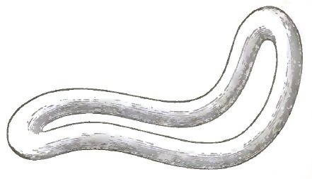
The Albert Smith Curves.
The curves of the pessary demand careful attention in its application. When the uterus is below the normal level, the broad ligaments are necessarily rendered more tense than natural, and the blood-vessels, more especially the veins, which are looped one upon the other, and which traverse these ligaments to and from the uterus, are made to collapse. This causes venous congestion and consequent increase in weight of the uterus—a condition favorable to malposition, uterine catarrh, and pathological changes in structure. A pessary which will raise the uterus to the health level clearly fulfils an indication. A pessary which raises it above the health level renders the broad ligaments tense and reproduces a condition which it was designed to relieve. Maintenance of the uterus upon the health level depends largely upon the curves of the pessary. The accompanying cuts illustrate the shape and curve of the Hodge pessary as modified by Emmet and Albert Smith. Fig. 7 represents the curve of Emmet, and Fig. 8 that of Albert Smith. For convenience let us characterize that curve which rests in the posterior vaginal cul-de-sac as the uterine curve, and that which occupies that part of the vagina adjacent to the pubis the pubic curve. The acuteness and length of the uterine curve determine the height to which the pessary will lift the uterus. The longer and more acute the curve, the higher the uterus will be lifted, and vice versâ. The smaller curve of the Emmet modification will answer the average indication more nearly than the sharper curve of the Albert Smith modification, which may lift the uterus too high. The pubic should generally be proportioned to the uterine curve; that is, the greater the uterine, the greater the pubic curve. A pessary properly adjusted in all other respects may, by pressure upon the urethra and neck of the bladder, create vesical tenesmus and urethral irritation. This calls for increase in the pubic curve. The pubic curve may, however, be so great that the lower part of the pessary occupies the centre of the vulva, where it may create irritation. For this condition lessening of the pubic curve is the remedy. The pessary should not be so wide as to distend the vagina. Its length should be measured by the distance from the lower extremity of the symphysis pubis to the posterior vaginal cul-de-sac, less the thickness of the finger. If properly adjusted it should sustain the pelvic floor in its normal relations and the uterus in stable equilibrium.
The uterus in the first and second degrees of descent is usually either retroverted or retroflexed. The reader is therefore referred to the remarks on the application of pessaries in the treatment of these displacements.
In advance prolapse dependent upon extensive injuries to the perineum and other parts of the pelvic floor, and usually associated with extreme subinvolution of all the pelvic organs, the axis of the vagina is often changed from its forward oblique to the vertical direction. (See Fig. 3.) The downward traction of the prolapsing cystocele and rectocele upon the fornix of the vagina may then be so great that the pessary is inadequate to maintain in place the upper extremity of the vagina. The cervix then moves forward, the corpus turns back, and the whole uterus easily descends in a vertical direction along the prolapsing walls of the vagina to the second or third degree of prolapse. In this condition pessaries which disappear within the vagina are liable to be forced out with the prolapsing pelvic floor, or if retained seldom maintain the uterus in position. In such cases the various cup pessaries which are supplied with external attachments and abdominal belts are often used, but they are inadequate, because they either so fix the uterus as to prevent its normal movements, or they hold it in such unstable equilibrium that it may assume any one of the various malpositions, anterior, posterior, or lateral; and they are open to the further serious objection of constantly reminding the patient of their presence. As an expedient the uterus may sometimes be held within the pelvis by means of a large Albert Smith pessary with extreme uterine and pubic curves. The rational treatment, however, requires first an operation on the anterior vaginal wall to restore the fornix of the vagina to its normal place in the hollow of the sacrum, and with it the attached cervix; and second, an operation at the vaginal outlet to bring the posterior wall in contact with the anterior, and thereby to restore the lower extremity of the vagina to its normal place under the pubis.
ANTERIOR ELYTRORRHAPHY.—Numerous operations on the vaginal walls have been devised for the purpose of narrowing the vagina, and thus preventing descent along the vaginal canal, but they are temporary in their results, because, as long as the direction of the vagina remains vertical, its walls again become dilated by the prolapsing uterus and the former condition is re-established. The operation to be effective is performed as follows: A Sims's speculum of long blade, perforated at its extreme end, to which the cervix has been attached by a piece of silver wire, passing through the perforation and the posterior lip, is introduced, the patient being in Sims's position. The cervix is thereby drawn by the point of the speculum far back into the hollow of the sacrum. The author finds this preferable to the method described by Emmet, who has the cervix held back by a sponge probang in the hand of an assistant. The space in the anterior part of the pelvis is now so increased that the uterus readily falls forward into decided anteversion. While the uterus is thus held in position by its attachment to the blade of the speculum, the operator with two uterine tenacula finds in the loose vaginal tissue on either side of the cervix two points which can be brought together in front of the cervix. Then at each of the two lateral points a surface is denuded with the curved scissors about one-half inch square, and in front of the cervix a surface an inch long by half an inch wide across the anterior vaginal wall close to the uterine attachment. A No. 26 silver-wire suture is then passed, as shown in Fig. 9, and twisted as shown in Fig. 10, so as to secure the lateral denuded surfaces in contact with the larger surface in front of the cervix.
FIG. 9.
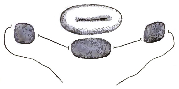
The First Suture before Twisting in Emmet's Operation for Procidentia (Emmet).
FIG. 10.
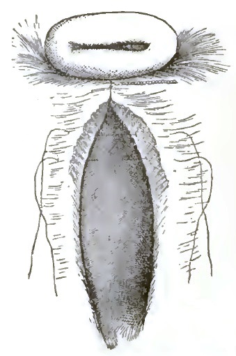
Folds on the Anterior Vaginal Wall formed after Twisting the First Suture (Emmet).
Inasmuch as the operation often fails at the point of the first suture, the author has usually introduced two or three of this kind instead of one. Two longitudinal folds are now formed on the anterior vaginal wall, which serve as guides for denuding and turning in the remaining redundant tissue by a line of sutures, which should extend forward along the centre of the vesico-vaginal wall until the folds are lost in the vaginal surface near the neck of the bladder. Sometimes the redundant tissue about the urethra cannot be disposed of by turning it in from side to side. Then it is desirable to make a crescentic denudation across the lower portion of the vagina, its concavity being on the uterine side, and to unite the margins below to those above by means of a curved line of sutures. The completed operation is shown in Fig. 11.
FIG. 11.
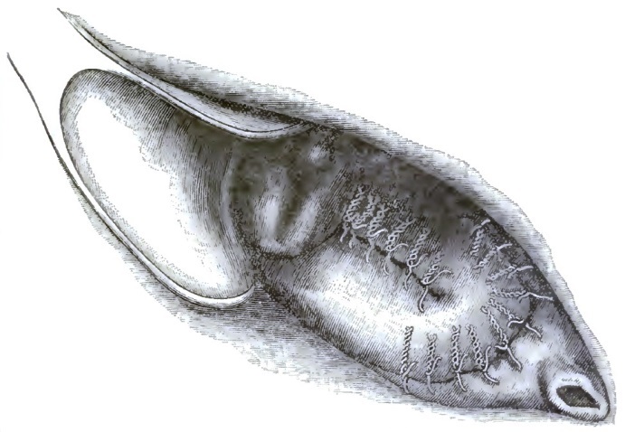
Emmet's Operation for Procidentia and Urethrocele completed. Sims's Speculum, Left Latero-prone Position (Emmet).
The after-treatment requires the self-retaining Sims's sigmoid catheter in the urethra for a week or frequent catheterization, absolute rest in bed, hot-water vaginal douches, regulation of the bowels, and the removal of the sutures on the twelfth day. After the completion of the operation the cervix is maintained near the hollow of the sacrum, and the organ remains normally anteverted and anteflexed, making an acute angle with the vesico-vaginal wall, which has now been restored to its normal direction and length. Unfortunately, it is not unusual to abandon the patient after this operation, in the vain hope that the uterus and anterior vaginal wall will maintain their normal relations without the support of the perineum and posterior vaginal wall. This is a great mistake, because the cystocele and procidentia almost always completely reappear within a few months. Anterior elytrorrhaphy, therefore, is simply one of the steps in the treatment.
PERINEORRHAPHY.—This is the name usually applied to the repair of the ruptured perineum, but the scope of the operation has been extended to include also the surgical treatment of rectocele and relaxation of the posterior vaginal wall. The most scientific operation yet devised is the one proposed by Emmet,8 which is performed as follows: The patient being etherized and in the lithotomy position, the operator seizes with a tenaculum the crest of the rectocele or posterior vaginal wall at a point which can be drawn forward without undue traction—point a. With another tenaculum the lowest caruncle or vestige of the hymen (point b), and with another the posterior commissure of the vulva (point c), are hooked up. The triangle included between these points defines one-half of the surface to be denuded. The three tenacula are now placed in the hands of assistants, the sides of the triangle are made tense by traction, and the included surface denuded. The tenaculum at c is then removed, and the middle point of the line a b is caught and drawn toward the interior of the vagina in the direction of the vaginal sulcus on that side, and the sutures are introduced, as in Fig. 13. The same thing is then repeated on the other side, and the sutures are all tightened, forming a line of union running back into each sulcus, as shown in Fig. 14.
8 Trans. Am. Gynæcological Society, 1883; Principles and Practice of Gynecology, 3d ed.
FIG. 12.
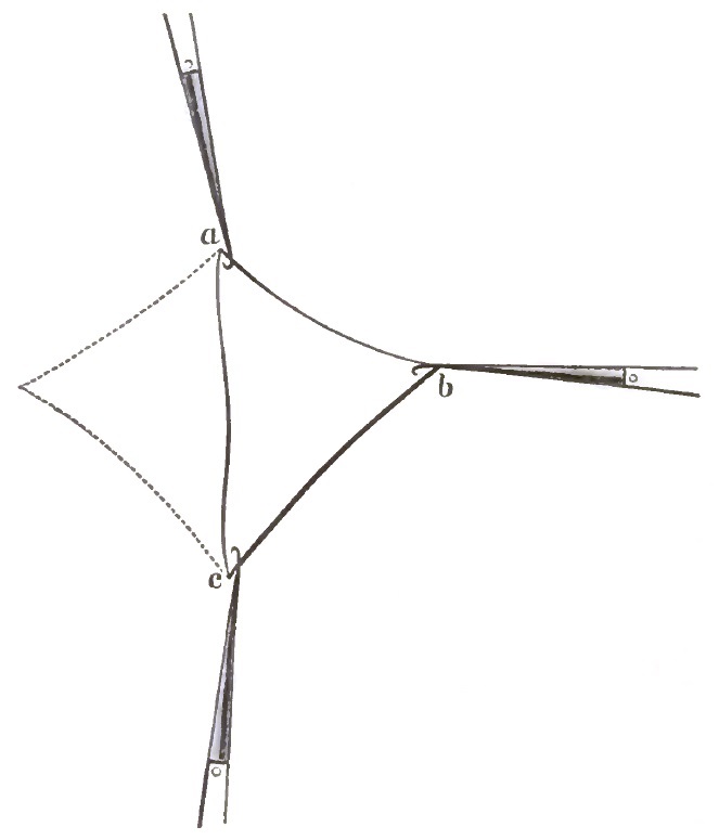
a is at the crest of the rectocele; b at the caruncle just within the labium; and c at the posterior commissure. The cut represents that half of the surface to be denuded which is on the operator's right. The dotted lines represent the other half, on the left.
FIG. 13.
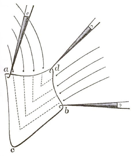
The Sutures in Place. When secured they will unite a d with b d, and lift the perineum up in contact with the anterior vaginal wall.
FIG. 14.
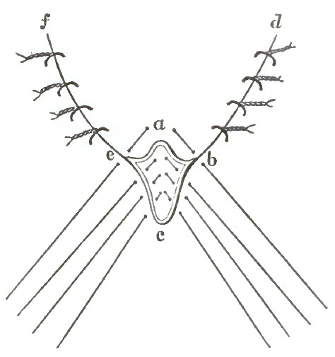
All the Vaginal Sutures Twisted. One suture, including the crest of the rectocele and the labium majus on either side, and three superficial external sutures, are yet to be secured. The lines a d and d b, Fig. 13, have been brought into coincidence by means of the sutures, and now form the line of union d b. The tissues between the lines a c and c b, Fig. 13, have been so lifted up and are so held under the line of union d b that the line c b, Fig. 13, has been reduced to c b, Fig. 14, which makes the external portion of the wound insignificant in extent.
The essential part of the operation inside the vagina almost always succeeds, but the external part of the rupture at the posterior commissure often fails to unite; furthermore, the operation as described by Emmet does not overcome the patulous condition of the introitus vaginæ in case of great relaxation of the vagina. The author has sought to obviate the first of these difficulties by the use of deep silver sutures instead of the superficial ones described by Emmet. They should be introduced before tightening the vaginal sutures, and should be passed far around in the posterior vaginal wall, their points of entrance and exit being the same as for the three lower unsecured superficial external sutures in Fig. 14. The second difficulty may be overcome by further denuding a triangular surface in the vaginal sulcus on each side, the base of the triangle corresponding to the line a b, Fig. 12, and its apex being in the vaginal sulcus at a distance corresponding to the degree of relaxation. This increases the length of the lines of union running into the sulci represented by d b and e f, Fig. 14. In the vaginal portion of the wound silk or catgut is preferable to silver, the latter being difficult to remove.
Emmet is entitled to great credit for having given to the profession an operation which brings the posterior vaginal walls up against the anterior more perfectly than any other, and which, being mostly inside of the vagina, is therefore followed by very little of the pain during convalescence which formerly rendered perineorrhaphy one of the most trying operations in gynecology. The operation furthermore has demonstrated the former teachings relative to the direction of perineal rupture9 and the tissues involved to be incorrect, or at least inadequate.
9 At the meeting of the American Medical Association in June, 1883, the author presented a paper describing the transverse laceration of the perineum and its operative treatment, which was published with illustrations in the transactions by the journal of the Association, Dec. 22, 1883. This communication referred only to the recent rupture and the immediate operation.
Retroversion.
Retroversion is that position of the uterus in which the fundus is posterior to the axis of the pelvic inlet. If the cervix be in its normal place near the sacrum, retroversion is scarcely possible, because it is prevented by the proximity of the over-arching sacrum. (See Fig. 2.) The first degree of prolapse must therefore precede any considerable backward turning of the uterus. When the cervix has been displaced downward and forward so far that its distance from the sacrum is equal to or greater than the length of the uterus, retroversion to any extent becomes possible. (See Figs. 3 and 16.)
ETIOLOGY AND HISTORY.—From the above it follows that the causes of commencing retroversion must be identical with the causes of the first degree of prolapse. After the puerperium the relaxation of the supports and the weight of the organ may persist, and spontaneous replacement may be prevented by the pressure and weight of the intestines upon the anterior surface. Every act of defecation forces the cervix forward and downward, and the uterus, being in the axis of the vagina, and having therefore little support below, must depend upon the subinvoluted peritoneal suspensory ligaments and pelvic fascia, which are inadequate. This condition is very often induced by abortions, with resulting increased weight and relaxation of the vaginal walls. Local peritonitis and cellulitis may permanently fix the corpus in its retroverted position by cicatricial bands and adhesions.
SYMPTOMS AND COURSE.—The displacement and its complications usually cause bearing-down sensations, a feeling of heaviness in the pelvis, exhaustion upon walking and standing, especially the latter, and constipation. After the puerperium the extreme engorgement of the pelvic organs often produces uterine hemorrhage, which should not be confounded with the returning menstruation. Especially after abortion the hemorrhage often persists for a long time unless cured by treatment. Gradual or sudden replacement may occur spontaneously, or the causes may continue active, and even be enforced by cystocele and rectocele. The displacement may also be complicated by disease and displacement of the ovaries. Organic disease of the uterine walls may induce a superadded retroflexion. The heavy organ may descend along the relaxed subinvoluted vaginal walls even to complete procidentia.
DIAGNOSIS AND PROGNOSIS.—The symptoms outlined in the preceding paragraph indicate the probability of displacement, but the diagnosis depends upon direct examination of the uterus. Conjoined manipulation and the probe will usually show the retroverted organ with the cervix displaced toward the pubes and with the corpus in the hollow of the sacrum. The introduction of the probe is contraindicated by cellulitis and peritonitis. In certain cases of anteflexion, as represented in Fig. 23, the cervix is bent forward in the vaginal axis as in retroversion. The condition is in reality one of retroversion of the cervix with high anteflexion of the corpus, which may usually be detected by careful conjoined examination. The prognosis with treatment is generally favorable both for speedy relief and ultimate recovery.
TREATMENT.—As in descent, the treatment consists in removing cellulitis, peritonitis, and other complications, in the use of pessaries, and in operations on the anterior and posterior vaginal walls if needed. Inasmuch as the treatment corresponds to that of retroflexion, it will be presented under that subject.
Retroflexion.
ETIOLOGY AND PATHOLOGY.—Retroflexion is that displacement in which the organ is bent backward upon itself. It usually results from, and is associated with, retroversion, but for convenience the double displacement will be termed retroflexion. It may be caused by the great weight of the corpus, the soft flexible state of the uterine walls during and after involution, intra-abdominal forces, downward pressure during defecation, tight clothing, and not commonly by the obstetric bandage.
The ovaries, unless fixed elsewhere by adhesions, are displaced with, and held down on either side of, the corpus, sometimes enlarged from inflammation, often adherent, and always extremely sensitive. Chronic metritis, cellulitis, and peritonitis, with adhesions more or less firm, are usually present, and not infrequently as the result of gonorrhoea, abortion, or injudicious treatment. Peritoneal adhesions between the corpus and the cul-de-sac of Douglas sometimes make replacement impossible. In rare cases the displacement is congenital.
FIG. 15.
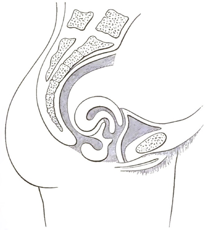
Extreme Retroflexion, with Hypertrophy of the Corpus, which impinges upon the rectum and compresses the recto-vaginal wall.
SYMPTOMS AND COURSE.—Among the most pronounced symptoms are profuse uterine catarrh, menstrual disorders, sterility, abortion, weakness, pain in the back, painful defecation, rectal tenesmus, the symptoms of pelvic inflammation, neurasthenia, and other nervous symptoms. The uterine catarrh is due to an effort on the part of the engorged pelvic organs to relieve themselves by an exaggerated secretion of mucus from the uterus, which upon being increased in quantity becomes vitiated in quality, and therefore pathological. Menorrhagia and abortion may also result from congestion. Dysmenorrhoea and sterility result from the general anæmic condition and from the inflammatory complications, and from the obstruction in the uterine canal or in the blood-vessels at the angle of flexure. (See Pathology of Anteflexion.) The rectal symptoms are caused by the pressure of the corpus uteri upon the rectum, which gives the sensation to the patient of an overloaded bowel.
Should pregnancy occur, the rapid growth of the uterus may induce spontaneous reposition at about the fourth month, when the fundus rises out of the pelvis, but if the corpus be incarcerated under the sacral promontory from adhesions or from any other cause, the uterus will, unless manually replaced, relieve itself by abortion.
Abdominal pains, nervous dyspepsia, and neuralgia in distant parts of the body are often present; indeed, the nervous symptoms may be of the most exaggerated character, and may comprise all that is implied by the word hysteria in its most comprehensive signification.
DIAGNOSIS.—Digital touch discloses the cervix low in the pelvis, and the fundus uteri is felt through the posterior vaginal wall in the cul-de-sac of Douglas. Conjoined manipulation with the index finger of the left hand, first in the vagina and then in the rectum, and the right hand over the hypogastric region, will show the size, form, consistency, and location of the uterus, the degree of the flexure, and the difficulty of replacement. An inflammatory exudate or hæmatocele, posterior to the uterus, or a fibroid in the posterior uterine wall, may be mistaken for the retroflexed corpus. The probe will always verify the diagnosis, but if there be great tenderness with fixation in the cul-de-sac of Douglas, treatment should be directed against the inflamed condition, and the final diagnosis made by repeated examinations or after the disappearance of the inflammation. Great and lasting injury is often done in the attempt to complete the diagnosis at the first examination. The presence of a fibroid in the posterior uterine wall with post-uterine inflammation is a serious complication both in diagnosis and treatment. If the rectum be overloaded with fecal matter, the diagnosis should be deferred. The displacement is distinguished from the presence of an ovary or small ovarian tumor in the pouch of Douglas by careful bimanual examination and by the probe.
TREATMENT OF RETROVERSION AND RETROFLEXION.—The objects of treatment are replacement and retention of the uterus. The obstacles to replacement are cellulitis, peritonitis, and fixation of the uterus, and these complications often require weeks, and in severe cases months, of treatment preparatory to replacement. Some of the general therapeutic suggestions under the subject of descent are also applicable to the retro-positions. Rest, massage, careful regulation of the bowels, feeding, and general tonics are essential. For the inflammation small blisters over the inguinal regions frequently repeated, and the daily application of the cotton and glycerin plug to the cervix, and dry cupping over the sacrum, are most efficacious. The glycerin may be combined with alum, tannin, chloral hydrate, or iodoform. Thymoline in small quantities partially destroys the disagreeable iodoform odor. The most useful and essential topical application is the hot-water vaginal douche, but its use will be followed by failure and disappointment if it be applied in the ordinary way. The following is quoted from a paper by the author which was published in the Chicago Medical Gazette, Jan. 1, 1880:
"Ordinary Method of Application. "Proper Method of Application.
"I. Ordinarily, the douche is applied with the patient in the sitting posture, so that the injected water cannot fill the vagina and bathe the cervix uteri, but, on the contrary, returns along the tube of the syringe as fast as it flows in. "I. It should invariably be given with the patient lying on the back, with the shoulders low, the knees drawn up, and the hips elevated on a bed-pan, so that the outlet of the vagina may be above every other part of it. Then the vagina will be kept continually overflowing while the douche is being given.
"II. The patient is seldom impressed with the importance of regularity in its administration. "II. It should be given at least twice every day, morning and evening, and generally the length of each application should not be less than twenty minutes.
"III. The temperature is ordinarily not specified or heeded. "III. The temperature should be as high as the patient can endure without distress. It may be increased from day to day, from 100° or 105° to 115° or 120° Fahr.
"IV. Ordinarily, the patient abandons its use after a short time." "IV. Its use, in the majority of cases, should be continued for months at least, and sometimes for two or three years. Perseverance is of prime importance."
"A satisfactory substitute for the bed-pan may be made as follows: Place two chairs at the side of an ordinary bed with space enough between them to admit a bucket; place a large pillow at the extreme side of the bed nearest the chairs; spread an ordinary rubber sheet over the pillow, so that one end of the sheet may fall into the bucket below in the form of a trough. The douche may then be given with the patient's hips drawn well out over the edge of the bed and resting on the pillow, and with one foot on each chair; the water will then find its way along the rubber trough into the bucket below." The Davidson syringe, which has an interrupted current, is preferable to any of the fountain syringes.
As the tenderness disappears the cotton plugs may be increased in quantity, and thereby made to serve as temporary support for the uterus until a more permanent pessary can be substituted. The sluggish circulation in the pelvis and torpid condition of the bowels may be much relieved by the daily application of the wet pack. A small flannel sheet folded lengthwise to the width of two feet, dipped in very hot water, and dried by passing it through a wringer, is wound about the hips and covered by another dry one. At the end of a half hour, during which time the patient maintains the recumbent position, the sheets are removed. When the tenderness has been sufficiently reduced, gentle attempts at replacement may be made every day or two by conjoined manipulation. The patient's tolerance of manipulation may thus be observed and the way prepared for complete replacement and permanent retention after the subsidence of the inflammation.
In retroversion and retroflexion always replace the uterus before adjusting the pessary, otherwise the instrument will press upon the sensitive uterus, when one of three unfortunate results must occur: (1) The pessary may not be tolerated on account of pain; (2) the pessary may be forced down by pressure from above so near to the vulva that it will fail to do the least good; (3) the uterus, finding it impossible to hold its position against the pessary, instead of taking its proper position will often be bent over it in exaggerated retroflexion, with the cervix between the pessary and the pubes and the body between the pessary and the sacrum, or the whole organ may slip off to one side of the instrument into a malposition more serious than the one for which relief is sought. The safest and most effective method of replacement is by conjoined manipulation, as represented in Figs. 16 and 17. The dotted lines in the former indicate the gradual elevation of the corpus out of the hollow of the sacrum to the pelvic brim, where it may be anteverted by the fingers of the right hand pressed well down behind its posterior wall. During the process of anteversion the index finger of the left hand in the anterior fornix of the vagina presses the cervix back to its place in the hollow of the sacrum, as in Fig. 17. Efficient reposition of the uterus is very often impossible without anæsthesia.
FIG. 16.
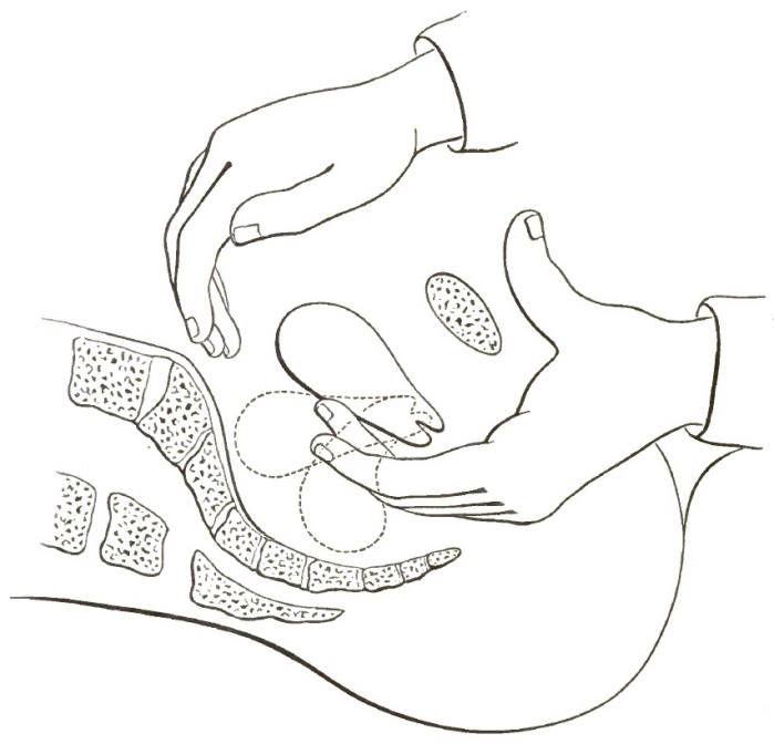
Commencing Reposition of the Retroverted or Retroflexed Uterus by Conjoined Manipulation (modified from Schultze).
FIG. 17.
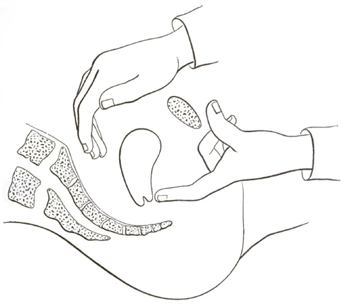
Completed Reposition of the Retroverted or Retroflexed Uterus by Conjoined Manipulation (modified from Schultze).
The replacement is not usually accomplished by drawing the fundus forward and pushing the cervix back directly in the median line. In most cases the fundus sweeps around the arc of a circle on the left side of the pelvis, and the cervix on the right. This is owing to the greater frequency of cellulitis on the left side, and consequent shortening of the left broad ligament. After replacement the organ is to be held in position by a suitable pessary.
Bimanual replacement has two great advantages over the more familiar methods of the sound or repositor: first, it is more effective and more permanent; second, the lever action of the sound or repositor, by which the operator may unwittingly use an undue and dangerous amount of force, is avoided in the use of the hands, through which the operation is not only constantly under his control, but also within his appreciation.
Inasmuch as the pessary fulfils its indications by sustaining the pelvic floor, and thereby holding the cervix in the hollow of the sacrum, the same general principles, and in fact the same pessaries, which are applicable to prolapse apply also to retroversion and retroflexion. Indeed, the first step in the genesis of the retro-positions has been shown to be prolapse. The student is therefore referred to the general remarks on the adjustment of pessaries for prolapse.
The operations of elytrorraphy and perineorraphy, especially the latter, already described in the treatment of descent, are often of the utmost importance in the treatment of the posterior displacements, and should therefore be carefully studied in this connection.
In the adjustment of the pessary it is desirable, if possible, to avoid direct pressure upon any part of the uterus. Pessaries designed to prop up the body of the uterus by pressure upon the posterior wall to correct the posterior malpositions, and upon the anterior wall to correct the anterior malpositions, are very liable to induce metritis and perimetritis, and are therefore generally unsafe. In certain cases, however, the vaginal walls, especially the posterior, may be so relaxed from subinvolution and other causes that the instrument, though very long, fails to maintain the cervix in its normal place. Under such conditions a pessary may be required to act directly upon the uterus. The Schultze's sleigh pessary represented in Fig. 19 fulfils this indication. Schultze's figure-of-eight pessary, or a long Albert Smith pessary with its uterine curve made so extreme as to bring the upper part of the instrument in front of the cervix instead of behind, answers the same purpose.
FIG. 18.
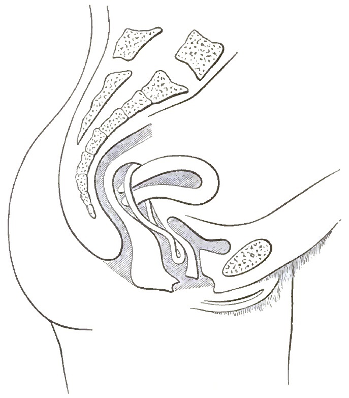
Showing the Pelvic Organs sustained by the Emmet Pessary alter reposition of the prolapsed, retroverted or retroflexed uterus.
Thomas's retroflexion pessary, with its bulbous upper extremity, is a long narrow instrument of extreme uterine curve. It lifts the uterus very high, and is specially applicable in cases of great relaxation of the pelvic floor and of complicating prolapse of the ovaries (Fig. 21). The bulbous portion is sometimes made of soft rubber.
A properly-adjusted pessary gives to the patient no consciousness of its presence. If the instrument cause pain it should be removed and search made for the tender places; it should then, if possible, be remoulded into such shape that it will not exert pressure upon them. Often a slight indentation at some point will enable the patient to wear it with comfort.
Sometimes when the corpus has been firmly bound back by peritoneal adhesions they may be broken up by very forcible conjoined manipulation under ether, but the operation is dangerous, and should therefore be undertaken only by an expert operator. In place of this operation Lawson Tait has proposed to open the abdomen, break the adhesions, and stitch the fundus uteri to the abdominal wound. This operation in the hands of such an operator as Tait is probably not more dangerous than breaking up firm adhesions by forcible conjoined manipulation.
FIG. 19.
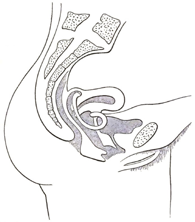
Schultze's Sleigh Pessary in place, as adjusted for prolapse, retroversion, or retroflexion with great relaxation of the vaginal walls (after Schultze).
In certain cases in which replacement is impracticable or impossible on account of inflammation or adhesions a soft rubber ring may be inserted, and will often give decided relief by lifting the uterus and pelvic floor nearer to the health level. In the treatment of all displacements coition should be forbidden or permitted only with great moderation, and the pessary should be kept clean by copious daily applications of the vaginal douche. Every three or four weeks the instrument should be removed and the pelvic organs carefully examined.
It should be urged that no man can safely apply the pessary until he has fully appreciated its indications and contraindications. Few practitioners possess naturally the mechanical skill necessary to its proper adjustment. Of this thousands of unfortunate women bear witness. Its dangers in inefficient hands are in striking contrast with its usefulness when judiciously employed.
Many cases of displacement, both anterior and posterior, are so complicated by prolapsed and adherent ovaries, by advanced disease of the ovaries and Fallopian tubes, and by peritoneal adhesions, that not only replacement, but even palliation, is impossible; then, as a final resort, the activity of the pelvic organs, both physiologically and pathologically, may be put at rest by the removal of the ovaries and Fallopian tubes.
FIG. 20. FIG. 21.
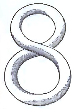
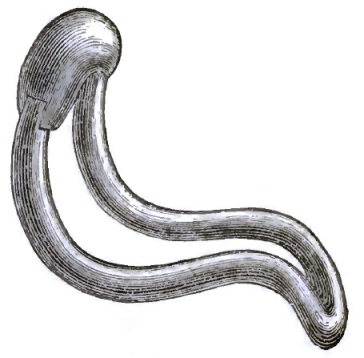
Front View of Schultze's Figure-of-Eight Pessary. The upper opening is intended to hold the cervix. This pessary has the uterine and pubic curves, as in Figs. 7 and 8. Thomas's Retroflexion Pessary.
William Alexander of Liverpool has devised an ingenious operation of shortening the round ligaments for the radical cure of descent and of the posterior displacements. He reports twenty-two cases of the operation in his own practice and several more in the practice of other surgeons, with almost uniform success in completely curing the displacements. The operation, although new, gives promise of a brilliant and successful future.
Lateral Versions and Flexions.
The lateral malpositions which often complicate retroversion and retroflexion are usually the result of inflammation in a broad ligament or in the uterus itself, or in both. Their treatment is that of the causative inflammation, and follows the general principles which have been laid down for the treatment of other versions and flexions.
Pathological Anteversion.
Sometimes the physiological angle of flexure becomes obliterated in consequence of chronic metritis, resulting in permanent straightening of the uterus, and the cervix becomes elevated and fixed above, or the corpus depressed and fixed below, the normal level. This constitutes pathological anteversion (Fig. 22).
FIG. 22.
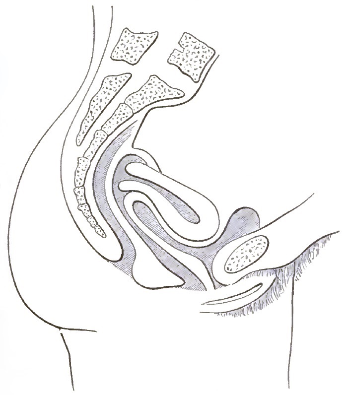
Pathological Anteversion.
ETIOLOGY.—The exaggerated anteversion of early pregnancy is physiological, the exaggerated anteversion of the uterus in chronic metritis is pathological. Elevation of the cervix and depression of the corpus may be induced by peritoneal adhesions. Increased weight from a mural fibroid may also depress the corpus.
The SYMPTOMS are due to the pelvic inflammations already mentioned and other complications. The increased weight of the uterus, which is usually hypertrophied from metritis, generally causes a dragging sensation, especially if the organ be also prolapsed. The enlarged corpus occupying the territory of the bladder often induces persistent vesical irritation or even cystitis. Menorrhagia, when present, is the result of the metritis or a fibroid rather than of the displacement per se.
DIAGNOSIS AND PROGNOSIS.—The displacement is recognized by digital touch, which discloses the anterior wall of the uterus parallel to the anterior wall of the vagina, with the fundus close to the symphysis and the cervix elevated. Conjoined examination will show the size, shape, hardness, and degree of fixation. Exaggerated anteversion of the healthy uterus is not necessarily pathological in its results. This is illustrated by the anteversion of early pregnancy. The prognosis is therefore good if the causes can be removed.
TREATMENT.—Inasmuch as exaggerated anteversion is the position taken by the uterus in chronic metritis, it follows that the treatment is often that of chronic metritis. For the treatment of metritis, perimetritis, fibroids, menorrhagia, etc. the reader is referred to the special literature of those subjects. Irritable bladder, which is often a mechanical result of the displacement and enlargement, may sometimes be relieved by means of an Albert Smith or Hodge pessary, which lifts the organ to a higher level away from the bladder. In thus elevating the uterus the anteversion may be rather increased than diminished, which proves that the symptoms were dependent not upon the anteposition, but rather upon descent and antelocation. Should the parts be too sensitive to tolerate the hard-rubber pessary or a flexible rubber ring, the daily application of medicated pledgets of cotton will give support to the uterus and decrease the tenderness until the more permanent instrument can be worn. The numerous anteversion pessaries designed to elevate the corpus by direct pressure on the anterior wall of the uterus generally irritate the organ, and thereby aggravate the inflammatory complications. They are therefore to be used with extreme caution.
Pathological Anteflexion.
DEFINITION.—The normal forward bending of the corpus upon the cervix uteri when the bladder is empty makes an angle of which the approximate physiological limits are between 45° and 90°: the flexure would generally be pathological if less than 45° or more than 90°. Furthermore, if the flexure, whether it be normal or abnormal in extent, does not disappear upon filling the bladder, but remains constant under all conditions, the rigidity makes the flexure pathological. Anteflexion is therefore pathological if the mobility at the angle of flexure is increased or diminished or absent.
ETIOLOGY AND PATHOLOGY.—Anteflexion may be congenital or acquired. By congenital is meant not defective foetal development, but failure of the immature child uterus to develop at puberty, a failure which usually pertains alike to the uterus, Fallopian tubes, ovaries, and vagina. In congenital anteflexion the uterus is bent upon itself almost double, the body and cervix both pointing in the direction of the pelvic outlet, with the cervix somewhat elongated and situated in the long axis of the vagina. (See Fig. 23.)
FIG. 23.
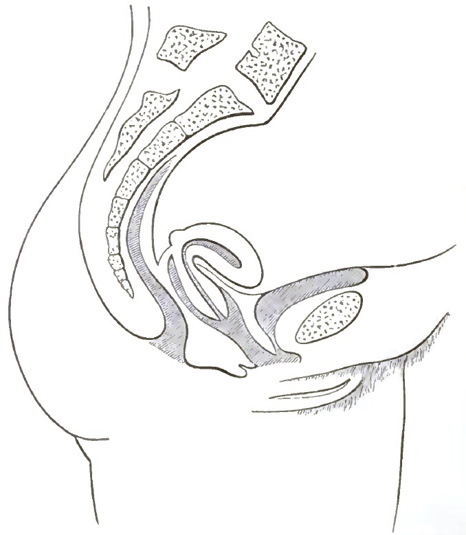
Congenital Anteflexion. Both cervix and body are flexed forward.
Acquired anteflexion may be simply an exaggeration of the normal flexure, due either to increased weight of the corpus from the presence of the uterine fibroid near the fundus or to unequal growth of the uterine walls or to unequal involution. A very frequent cause of anteflexion is thickening of the posterior wall of the uterus from the products of inflammation, and a corresponding atrophy of the anterior wall from prolonged pressure at the angle of flexure. Post-uterine cellulitis and peritonitis involving the utero-sacral ligaments is a frequent and discouraging complication. Sometimes the inflamed ligaments contract and drag the anteflexed uterus upward and backward, where it may be permanently fixed by peritoneal adhesions. (See Fig. 24.)
FIG. 24.
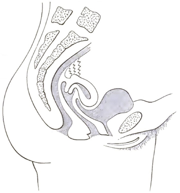
Anteflexion with Post-uterine Fixation.
A constriction of the uterine canal at the point of flexure may, by confining the secretions above, produce inflammation in the body of the uterus, Fallopian tubes, and ovaries analogous to the cystitis, ureteritis, pyelitis, and nephritis which follow stricture of the male urethra. The peri-uterine inflammations, having the relation either of cause or effect of the flexure, often bind the pelvic organs together in a mass of exudate, with resulting failure of nutrition, nerve-irritation, and constant pain, which sometimes render the patient's life miserable and useless.
SYMPTOMS AND COURSE.—The numerous symptoms due to the inflammatory and other complications should not be confounded with those of the displacement. The symptoms of anteflexion are polyuria and dysuria, dysmenorrhoea and sterility.