Volume 4 — DISEASES OF THE GENITO-URINARY SYSTEM. (continued 12)
Reading edition
Other causes of displacements are uterine and ovarian tumors, pelvic deformities, and fecal impactions of the rectum. Of the various displacements of the bladder it is needless to speak in much detail. The most important is the downward one. Various degrees of this are found up to complete cystocele, most commonly associated with prolapsus uteri. The bladder naturally sags inferiorly as age advances, and by far the most potent agent in causing this to become pathological is repeated pregnancy and injuries during labor.
It is a well-known fact that the first stage of vesicle prolapsus is apt to be marked by as great discomfort as the third, for after a while the organ seems to become accustomed to its altered relations. The treatment of this condition is difficult. The bladder should be replaced and kept there. As this usually necessitates the reposition and maintenance of the displaced uterus, it is extremely difficult, and in case of existing adhesions it is impossible. A great variety of mechanical means have been tried to furnish an artificial support to keep the parts in position. If the bladder alone is prolapsed, the pessary used for anteversion of the uterus will sometimes answer. The instruments devised by Thomas, Grailly Hewett, and myself are most commonly used.
Acute Urethritis; Inflammation of the Urethra.
This affection may be simple or gonorrhoeal, and it is often difficult to tell the one from the other. There is a difference in history when we can get correct testimony from the patient. Simple urethritis usually comes on gradually, and is often preceded by symptoms of uterine or vesical disease, while gonorrhoea comes on rather abruptly, and is preceded or attended by acute vaginitis and vulvitis. The chief symptom is painful urination. Sharp scalding is produced by the urine passing over the tender surface. There is often a frequent desire to urinate, but not so urgent as in cystitis. In some cases the urine is retained for a long time, evidently from a dread of the pain caused in passing it. In quite a number of cases I have noticed hemorrhage, the source of blood being evidenced from the fact that it was not intimately mixed with the urine, and after micturition it oozed from the meatus urinarius.
An examination of the parts will show signs of inflammation about the meatus, with or without the same condition of the vulva. Occasionally there is a discharge seen coming from the urethra, but if the parts have been recently bathed this may not be apparent. Introducing the finger into the vagina and pressing upon the urethra from above downward will cause a discharge, unless the patient has passed water immediately before. The appearance of the discharge corresponds to that of gonorrhoea in its various stages.
Cystitis, which is liable to be confounded with urethritis, may be excluded by using the catheter, and, after letting urine flow for a time, collecting the remainder for examination. The mucous membrane, as seen through the endoscope, is of a deep red, with pus or mucus lodged in its folds. The instrument cannot be used in all cases, owing to the acute tenderness of the parts. Bleeding is very likely to occur in the examination, simply from the contact of the endoscope.
The TREATMENT of acute urethritis, whether specific or not, may be conducted on the same principles as that of gonorrhoea in the male, using the same constitutional remedies, local baths, etc. This will suffice in most cases of acute disease, but when it assumes the subacute form from the beginning, then the use of injections becomes necessary. I have seen much benefit derived from douching the urethra with water as hot as the patient could bear it. For this purpose I use a catheter made like the fluted roller of a crimping-machine. The catheter conveys the water to the rounded point of the instrument. Behind the point of the catheter, where the grooves terminate, there is a perforation in each groove through which the water returns. By this arrangement the water, as it flows back through the grooves, is brought in contact with every portion of the mucous membrane. The instrument is passed up to the neck of the bladder, and a fountain syringe attached to it, and the water as it flows away is caught in a cup.
The injection of solutions of nitrate of silver and sulphate of zinc will often prove useful. It must be borne in mind that the female urethra will not hold more than ten or fifteen drops, and if more is used it will enter the bladder, even where very slight force is employed while injecting. I use a large syringe, placing the nozzle over (not in) the meatus, and inject slowly and without force a small quantity. When the case is of long standing, and the neck of the bladder appears to be involved also, I use a weak injection of one or two grains of nitrate of silver to the fluidounce, and inject it through the urethra with force enough to enter the bladder, and let it remain there, to be passed off when the patient urinates. In old cases which began by a severe acute attack, and where the walls of the urethra are very much thickened and the canal contracted, dilatation with bougies does much good. While the bougie is passed once or twice a week, I apply to the vaginal portion of the urethra oleate of mercury or the unguentum hydrargyri. This will often suffice to stop the gleety discharge, as well as remove the thickening of the urethral walls.
Inflammation of the Urethral Glands.
These glands rarely, if ever, take on inflammation primarily, but vulvitis and vaginitis, especially if gonorrhoeal, often extend into them. When they do become inflamed, the disease usually remains without any tendency to subside. More than that, when a gonorrhoea affects these glands the inflammation will remain there after all traces of the disease have left the vagina, vulva, and urethra, and in time the discharge from these glands will light up the original vaginitis and vulvitis again. The symptoms of this inflammation are not diagnostic. The physical signs are the swelling and redness around the mouths of ducts which are located just within the labiæ of the meatus urinarius. This give a general redness to the meatus. By pressure made upon the urethra from above downward a purulent discharge from the ducts will be produced and can be seen escaping. The only effective treatment is to lay open the glands their whole length. They run upward in the posterior wall of the urethra, so that by passing a fine probe-pointed scissors they can be laid open on the vaginal surface. Care should be taken to prevent the incision from reuniting, and if the inflammation does not promptly subside applications should be made, as in the ordinary treatment of inflammation.
Another very troublesome affection of the urethra which usually results from urethritis is granular erosion, as it is called. The mucous membrane is covered with young, imperfectly-developed epithelium; the papillæ are hypertrophied and extremely sensitive. This gives rise to the most excruciating pain during micturition, and generally keeps up a distressing tenesmus. This disease is rarely seen except among old people. The diagnosis is made from the history and appearance of the urethra. The treatment is cauterization of the whole surface. The milder washes and injections do not accomplish much. Pure carbolic acid may be tried first, brushing it over the surface and repeating it in eight or ten days. This is the least painful application, and generally answers very well. When it fails a solution of nitrate of silver (one drachm to the fluidounce) should be used. In obstinate cases it is desirable, before using strong caustics, to dilate the urethra, and then touch it with a 50 per cent. solution of carbolic acid.
Circumscribed and Subacute Urethritis.
Among the inflammatory affections of the female urethra there are mild forms which fall short of well-marked urethritis. Indeed, some of these attacks amount to little more than congestion or slight catarrh. In others circumscribed patches of the urethra become inflamed, the rest of the canal remaining normal.
The cause of this affection is generally some inflammation of other pelvic organs, such as cellulitis. In one case it occurred in a saleswoman who had been upon her feet many days from early morning until late at night. I found several small ecchymoses on several parts of the mucous membrane with zones of inflammation around them. The long-continued passive congestion had caused some of the small vessels to rupture, and the small blood-clots started the inflammatory process.
These cases tend to recovery if the patient is placed under favorable conditions. If there is much pain, and if the trouble appears to be tending to become chronic, mild injections may be employed.
Dilatation of the Urethra.
Dilatation of the whole urethra is not so common as dilatation of a portion of it. Even when the whole canal is larger than it should be, it is not, as a rule, uniformly so. In general, the urethral walls and the urethro-vaginal septum are usually enlarged, relaxed, and flabby. After a considerable time they may become indurated by infiltration or hyperplasia of the connective tissue. The mucous membrane is usually soft and loosely adherent to the subjacent tissues. Beneath the membrane there are sometimes masses of enlarged veins which give a dark bluish appearance to the parts. If the meatus be distended like the rest of the urethra, the mucous membrane with the large veins beneath it may protrude and form a tumor or tumors, which have quite the appearance of rectal hemorrhoids. This is especially so when the veins are large and numerous and the mucous membrane thin, so that the color of the veins can be seen through it. On the other hand, if the meatus remains normal in size, nothing will be seen by the examiner until the catheter or sound is passed into the urethra, when the distended or distensible condition of the canal will be detected. The dilatation can be easily detected, even when the meatus is normal in size, by observing that the sound can be moved about in the urethra, conveying the same impression obtained when the sound passes into the bladder. By making a digital examination of the vagina the enlarged urethra can be felt, and it is usually elastic and compressible. Through Sims's speculum the abnormal fulness or bulging of the anterior vaginal wall can be plainly seen and distinguished from displacement of the urethra. The points of difference between dilatation and displacement will be brought out more in detail farther on.
When the dilatation has existed for any length of time, the mucous membrane is usually hyperæmic, and sometimes catarrhal, secreting a muco-purulent material, which may be seen escaping from the meatus or lodged in the folds of the membrane, where it can be seen through the endoscope. When the mucous membrane is prolapsed and forms a tumor outside of the meatus, it soon becomes fissured and ulcerated, and consequently very tender and painful. This condition is produced by the retarded circulation, chafing, and the irritation from exposure to the air and the urine passing over it.
Dilatation of the anterior or lower third is the rarest of all forms of urethral dilatation, and occurs usually as a consequence of some enlargement or swelling of the mucous membrane, neoplasm of the urethra, or mechanical dilatation. The dilatation may or may not include the meatus. In rare cases it does not at first, but in time the enlarged mucous membrane slowly, sometimes rapidly, dilates the orifice. The general appearances of the parts are the same as those of which I have spoken under the head of dilatation of the whole urethra. When the dilatation is due to any new growth in the urethra, the tumor can be seen on inspecting the parts.
I have only seen one case where the lower end of the urethra was dilated without any recognizable cause for it. This was a single lady, thirty-five years of age, a school-teacher. She had displacement of the uterus and catarrh of the cervical canal, for which she consulted me. She had no trouble with her urinary organs. While examining the uterus I noticed that the meatus urinarius was peculiarly formed. In place of the concentric corrugations of the mucous membrane which form the closed meatus, the orifice was funnel-shaped and lay open when the labia minora were separated. About half an inch of the lower end of the urethra admitted a No. 21 (Eng.) sound. The remainder of the urethra was normal, and there were no signs of disease about the mucous membrane of the dilated portion. I could obtain no history which pointed to the origin of the trouble, and it caused no discomfort to the patient.
Dilatation of the posterior or upper third occurs in connection with other pathological conditions, such as prolapsus of the bladder and urethra. On this account we will defer what is to be said on this subject until we come to dislocations of the urethra.
Dilatation of the middle third of the urethra is more common than that of any other portion of the canal. In this form the anterior wall of the urethra maintains its normal position, but the central position, being distended, settles down, so that in time the urethra, in place of being a straight or slightly curved canal, becomes triangular, the upper wall being the base, and the central portion of the wall (that is, midway between the neck of the bladder and the meatus) the apex. A sac or cavity is thus formed in the central portion of the urethra.
In the earlier stages of this affection the urethra in front and behind the pouch is really or apparently contracted; but as the disease progresses the upper part of the canal and the neck of the bladder become dislocated downward, and finally the upper portion of the urethra becomes also dilated to some extent.
There is in this as in the other forms of urethral dilatation frequent urination, usually more marked, but, unlike the others, there is difficulty in passing water. This frequency of urinating, and the straining efforts necessary to do so, affect the bladder, producing irritation, and in time hypertrophy of its walls. Cystitis also follows in the order of morbid developments; but whether that comes from the frequent and difficult urination, or from extension of the inflammation from the urethra to the bladder, is a question.
ETIOLOGY.—The hyperæmia of the urethra which occurs in pregnancy, and which tends to produce over-distension of the veins, favors dilatation of the whole urethra. There is an apparent increase of tissue in the walls of the urethra during utero-gestation, and the dilatability of the canal is often increased also. Now, this condition of the parts disappears during the involution which takes place after delivery; but when from any cause the process of involution is interrupted, the enlarged vessels and relaxed condition of the urethral walls remain and sometimes increase. When to this state of the parts a catarrh of the mucous membrane is added, the enlargement of the membrane by swelling still further increases the calibre of the canal.
The dilatation caused by the passage of calculi may remain permanently, and the same may be said of the use of large sounds. Neoplasms obstructing the meatus or stricture at that point may so obstruct the escape of the urine as to cause dilatation at all points above. This is no doubt one of the most important and frequent causes of dilatation.
I have already stated that dilatation of the lower third of the urethra is rare, and is usually due to inflammation of the mucous membrane at that point or to abnormal growths, the distension remaining after the causes that produced it have been removed. This and mechanical dilatation from any cause cover the etiology of this form of the trouble. Baker-Brown says that the meatus is always dilated when there is stone in the bladder.
Regarding dilatation of the upper third of the urethra, I am inclined to believe that it occurs in consequence of a partial prolapsus of the bladder and the upper end of the urethra. The displacement of these parts implies a relaxation of the tissues, caused originally, it may be, by injuries during confinement, and the prolapsus permits an unusual pressure of the urine upon the upper end of the urethra, and dilatation is the result. On the other hand, the prolapsus and accompanying relaxation of the urethral walls may be sufficient to cause the dilatation. In all the cases that I have critically examined there has been displacement as well as dilatation, and the whole trouble could invariably be traced to childbearing or anteversion of the uterus.
One cause of dilatation of the middle third of the urethra (urethrocele) has been sufficiently dwelt upon in Bozeman's description of the pathology of that affection—that is, narrowing of the lower end of the urethra. This does not explain the etiology of all cases, however, for I have seen this form of dilatation where there was no stricture or hypertrophy of the lower end of the urethra. In such cases I have traced the cause to childbirth, during which the posterior wall of the urethra had been pushed downward and contused, while the upper remained in its normal position. The relaxation caused by this over-stretching of the urethral wall formed a small pocket in the central portion, which gradually dilated more and more by the pressure of the urine until the urethrocele was fully developed. This explanation of the cause may be rather hypothetical, but, so far as my observations go, it agrees with the facts found in those cases which cannot be accounted for by Bozeman's views on the pathology of this affection.
SYMPTOMATOLOGY.—The symptoms vary according to the extent of the dilatation, the portion of the urethra involved, and the condition of the mucous membrane. When the whole urethra is dilated the only symptom present may be frequent urination. When there is inflammation or prolapsus of the mucous membrane, then pain will be caused by passing water, and the desire to do so will be more urgent and frequent. The patient may also be annoyed by a slight loss of control of the water, under the pressure of lifting heavy weights, coughing, or the like.
Dilatation of the lower third of the urethra does not cause any derangement of function, unless accompanied with inflammation or ulceration; then there will be frequent urination possibly, and painful urination certainly. The symptoms in this form of dilatation are less marked than in the other varieties.
When the trouble is located in the upper third of the urethra, the symptoms are sometimes very distressing. In addition to the frequent—it may be constant—desire to pass water, the patient is tormented with partial incontinence. Coughing, laughing, sneezing, stooping to lift anything, a jar on stepping from the curbstone in crossing the street, causes an escape of urine. This distresses the patient very greatly. From the constant wetting of the external parts they become inflamed, unless very great care is taken to keep them dry and clean. In some of these cases the mortification of mind is sometimes more distressing than the physical suffering.
The symptoms occuring in dilatation of the middle portion of the urethra are the same as those already given, with the addition of a slight mechanical obstruction which causes difficult urination; that is, more voluntary effort is necessary on the part of the patient to empty the bladder. The forcing, straining efforts made by some of these patients while urinating are even greater than the mechanical obstruction appears to account for. This may be due to the accumulation of urine in the urethra, which excites extra reflex action in the bladder and urethra out of proportion to the obstruction. This is the only way that we can account for the difficult urination and muscular hypertrophy found in those cases in which there is no great obstruction from stricture.
The constitutional symptoms arising from these urethral troubles are the same as those produced by urethritis, and are not peculiar to this class of affections. In fact, the symptoms here given may all be produced by other pathological conditions, and consequently cannot alone guide to a correct diagnosis. The true character of the trouble can only be discovered by physical exploration.
DIAGNOSIS.—A digital examination by the vagina will detect the increased space occupied by the urethra. The canal encroaches upon the anterior vaginal wall, and feels like a ridge extending from the meatus to the neck of the bladder. This elevation or thickening of the urethra is elastic and compressible in recent cases; in those of long standing the tissues are firm to the touch, but still the canal is compressible. The extent of the dilatation, if general or located in the lower parts, can be measured by the size of the sound that can be easily passed. If at the middle or upper portions, an ordinary female catheter or sound may be used to explore it. By introducing that instrument and pressing it first against the anterior wall and then upon the posterior, the distance between the two can be approximately made out. While the catheter or sound is in the urethra the finger should be introduced into the vagina to ascertain the thickness of the urethral wall. This will differentiate between dilatation and hypertrophy.
When the meatus is dilated and the mucous membrane and enlarged vessels are prolapsed, care is necessary to distinguish that condition from urethral neoplasm. This can be done by observing that in prolapsus the opening is situated either at the upper side or in the centre of the protruding mass, whereas in abnormal growths of the urethra the meatus surrounds the tumor or its pedicle. More than that, by making pressure the distended vessels can be reduced in size and the prolapsed membrane pushed up into the canal. This cannot usually be accomplished with tumors.
PROGNOSIS.—There is no natural tendency to recovery in these affections. If left alone they generally get worse. Recovery under treatment depends upon the location of the dilatation and the duration of the trouble. The conditions upon which an unfavorable prognosis is to be based are—bladder complications, inflammation or ulceration near the neck of the bladder, great varicosity of the veins, and fatty degeneration of the muscular tissue. In the absence of all these complications a complete recovery may be expected. In all cases great relief can be secured by treatment and the patient guarded from getting worse.
TREATMENT.—In the management of all forms of urethral dilatation attention should be given to any inflammation of the mucous membrane that may exist, employing the usual treatment. When there is a relaxed and prolapsed condition of the mucous membrane, astringents should be used. Tannic acid or alum will answer well. When these fail, the redundant membrane should be retrenched, either by touching it with the thermo-cautery or excising a portion with the scissors. In employing the cautery for this purpose the long pointed tip of the instrument should be used, and, having protected one side of the urethra with the speculum, cauterize a narrow strip of the membrane parallel to the axis of the canal. Two or more of these cauterizations may be made at points equidistant on the circumference of the urethra. By operating in this way pieces of normal membrane are left between the portions cauterized, which prevents stricture from occurring after healing—a misfortune which is sure to follow if the mucous membrane is destroyed by cauterization all around.
In excising the prolapsed portion I prefer to remove one or more V-shaped portions on opposite sides and bring the edges together by sutures. This is preferable to clipping off the whole of the protruding mass, because the cicatrices left are less likely to give after trouble.
When the dilatation is caused by varicose veins it may be well to follow the example of Gustave Simon. He exposed the vessels by cutting through the vaginal wall, ligated the largest, and arrested the hemorrhage from the smaller ones by applying liquor ferri perchloridi. He repeated this operation several times on the same patient, who experienced little or no inconvenience from the proceeding and made a good recovery.
Dilatation of the lower third of the urethra is usually secondary to some other trouble, as I have already stated; and all that is necessary to do for such cases is to remove the cause and treat any inflammation that may exist. The dilatation will then disappear; and if it does not, but little if any trouble will be caused by it.
The treatment of dilatation of the upper third consists simply in supporting the parts. This can be effectually done by using the pessary already recommended for the relief of prolapsus of the bladder. It may be necessary to have the instrument so formed as to bring the pressure where it is required. This is done by placing the pessary in position and observing what change of form, if any, is necessary, and then directing the instrument-maker to make the alteration. If the parts are well supported in this way, recovery will follow unless atrophy of the muscular wall has previously taken place. Even then the patient can be kept comfortable by wearing the pessary. If there is urethritis present, it may be necessary to remove that before using the pessary; otherwise the pressure of the instrument may cause pain and aggravate the inflammation.
In dilatation of the middle third Bozeman has proposed to make an opening into the most dependent part of the urethra through the vaginal wall, and maintaining it until all inflammation has been relieved, and then closing the opening by the usual plastic operation. By this means the urethra is perfectly drained of urine and the products of inflammation which accumulated there before. This, with appropriate cleansing and topical applications, soon restores the mucous membrane to its normal condition, and the removal of the redundant tissue during the operation of closing the opening effectually cures the whole trouble. This treatment is admirably adapted to marked cases of long standing, and should be employed. By using the thermo-cautery to make the opening the operation is easily performed. In recent cases of less magnitude I have obtained satisfactory results by dilating the lower part of the urethra and supporting the dilated portion either with a pessary or a tampon of marine lint. This permits the urethra to keep itself empty, and then, by frequently washing it out and applying such remedies as will cure the urethritis, recovery will sometimes follow.
Dislocations of the Urethra.
This is one of the affections most frequently met with in practice. I have found very few cases recorded in medical literature. This neglect of the subject by authors is perhaps due to the fact that in many cases of displacement of the urethra the bladder is also dislocated, and the whole trouble is described under the head of vesicocele or cystocele. Now, it is true that displacement of the two occurs together, but either may take place alone.
The extent of displacement varies exceedingly, but I shall describe only the partial and the complete. A clear comprehension of these two degrees will cover all intermediate forms. In partial displacement downward the upper two-thirds of the urethra are prolapsed, so that the direction of that portion of the canal is backward, instead of curving upward, as in the normal condition. In complete prolapsus the urethra runs from the meatus (which is in its normal position) backward, and rests upon the perineum, or in extreme cases, accompanied with prolapsus of the bladder and uterus, its direction is backward and downward, the position of the vesical end of the urethra being below the level of the meatus. In this degree of displacement the urethra and bladder can be seen presenting at the vulva or lying between the labia minora. The urethra is usually shortened considerably when the prolapsus is marked.
ETIOLOGY.—Utero-gestation and delivery are the most important causes of this affection. In the advanced months of pregnancy I have observed that while the bladder rose above the pubes the urethra was pushed slightly downward by the settling of the enlarged uterus into the pelvis. In such cases when labor occurs the head of the child dislocates the urethra still more by pushing it still farther down. This process I have often watched in forceps delivery. When there is a partial prolapsus of the urethra existing before labor, the urethra and anterior vaginal wall are forced down before the advancing head, and that, too, while the attendant is making counter-pressure to prevent it. The displacement produced in this way is often restored during convalescence if proper care be taken to push the parts back into place and the patient is kept at rest until the tissues regain their tonicity. But in many cases the trouble is overlooked, and by permitting the patient to get up and be on her feet while there is still prolapsus it will slowly increase until the dislocation is complete. This will surely be the case if there is any loss of perineum. Indeed, rupture of the perineum is an accident which permits the urethra to descend from its place. The perineum supports the vaginal walls, which in turn support the urethra; and if it be lost, even in part, the vaginal walls become relaxed, or perhaps never regain their tonicity after delivery, and, settling down more and more, carry the urethra with them.
SYMPTOMATOLOGY.—The symptoms arising from displacement of the urethra are much the same as those found in dilatation and other urethral diseases. I need not, therefore, repeat them in detail. Suffice it to say that in dislocation of the upper portion of the canal there is, in addition to frequent urination, a partial loss of control of the bladder. Under the extra pressure of coughing, for example, the urine will escape. This loss of control does not exist, as a rule, in complete displacement. On the contrary, there is usually difficult urination, which requires increased voluntary efforts to empty the bladder. In all degrees of displacement the symptoms are increased in the erect position, and are markedly relieved on the patient's lying down.
DIAGNOSIS.—An examination of the vagina, either by touch or speculum, will reveal the downward projection of part or all of the urethra, which will show that there is either dilatation or prolapsus. The change in the direction of the canal will be shown by passing the sound, and dilatation can be excluded by observing that the urethra grasps the instrument firmly at all points. In dislocation of the upper two-thirds of the urethra the sound passes in the normal direction, but is arrested at a half or three-quarters of an inch from the meatus; but by pushing up the vaginal wall and the urethra the sound will then pass into the bladder. When the prolapsus is complete the instrument passes in easily, but takes a downward and backward direction.
PROGNOSIS.—Uncomplicated displacement of the urethra can be remedied in the great majority of cases. By placing the parts in proper position, and holding them there, the relaxed tissues will usually contract sufficiently to support themselves. Should they fail to do so, the patient can at least be made comfortable by wearing some supporter.
TREATMENT.—When the displacement of the urethra is caused by any other trouble, such as defective perineum or prolapsus uteri, then these things should first be attended to. Should there be urethritis, that also should receive appropriate treatment. But the chief indication is to retain the urethra in place; and this can be easily accomplished by using the pessary which has been recommended for supporting the prolapsed bladder. Prolapsus of the upper part of the urethra can be relieved in this way quite satisfactorily. When the whole urethra is displaced, this pessary, while it supports the upper part, will still permit the middle portion of the urethra to settle down. This difficulty may be overcome by making the anterior portion of the pessary long enough to engage in the introitus vulvæ, and in that way keep the whole canal where it should be. Should this cause the patient much discomfort, a tampon of marine lint should be used to keep the parts in position until some restoration of the parts is obtained, and then the pessary will complete the treatment.
Prolapsus or Inversion of the Urethral Mucous Membrane.
The prolapse may be limited to one side or extend all around the canal. The size and extent of the protrusion vary considerably. If the meatus is of full size, the prolapsed portion will usually preserve its natural color for a time; but after a little, from chafing when wet with urine, and especially if not kept clean, it will become red and oedematous. When the meatus is small these changes occur sooner and in a more marked degree, because the prolapsed portion is partially strangulated. The longer the membrane remains exposed the more sensitive it becomes, and the frequency of urination and pain attending it increase. It also becomes very tender and painful to the touch. In marked cases the ordinary movements of the body irritate the parts, and in that way render walking painful.
These are symptoms that closely resemble those of irritable growths at the meatus urinarius, and, so far as history is concerned, it is not easy to make a differential diagnosis. To do this it is necessary to make a local examination. The physical signs and the points in the diagnosis between this affection and other diseases have been given briefly but sufficiently under the head of Dilatations of the Urethra, and need not be repeated here.
The causes of prolapsus of the urethral mucous membrane are numerous, but those that are best known are long-continued congestion of the membrane, urethral and cystic irritation causing frequent urination and vesical tenesmus. Chlorotic and greatly debilitated women are said to be predisposed to it, as also old prostitutes. The few cases that I have seen were in women over fifty years of age, and all of them were weak, nervous patients who had suffered from some organic disease or functional derangement of the urinary organs.
PROGNOSIS.—This disease does not yield promptly to mild treatment, unless it is seen early in its progress; and if it does yield to mild, soothing, and astringent applications, it is liable to return. But in case there is no other disease present that tends to keep it up, it can usually be cured by surgical means.
TREATMENT.—When a case is first seen it is well to remove any inflammation or other complicating conditions. The prolapsed membrane should be replaced, and the patient kept quiet in bed to favor the retention of the parts in situ. Astringents, such as tannic acid, alum, or persulphate of iron in a weak solution, should also be used. Should these fail, the prolapsed portion of the membrane should be removed. The methods of doing this (by excision and the thermo-cautery) have already been described.
Stricture of the Urethra.
PATHOLOGY.—Obstruction of the urethra by narrowing of its calibre is a much less common affection in the female than in the male. Still, it occurs sufficiently often to demand attention. There are some facts in the pathology of urethral stricture peculiar to women which we will first notice. Passing over congenital narrowing of the urethra by simply saying that such a malformation has been known, we find that stricture is developed in the female, as in the male, by the deposit of inflammatory products beneath the mucous membrane, which by gradual contraction constricts the canal. Ulceration of the membrane in a marked degree produces the same results. The inflammation and ulceration which end in the formation of stricture are usually specific in character, but the same may follow from the too free use of caustics and injuries during childbirth. Stricture may also be produced by bands of scar-tissue formed in the anterior vaginal wall and stretching across the urethra. Contraction of the whole canal occasionally occurs in cases of vesico-vaginal fistula of long standing. There the narrowing is simply the result of disuse. The form of stricture that most frequently comes under observation is a contraction of the meatus urinarius, produced in many cases by the too liberal use of caustics in the treatment of abnormal growths at the lower end of the urethra, or from vulvitis. This form of stricture is the least troublesome and is easily relieved. When due to the results of former urethritis or peri-urethritis, the walls of the urethra are thickened and indurated at the point of the stricture, and there is usually subacute urethritis, sometimes ulceration. In those cases where the calibre of the canal is diminished by cicatrices of the vaginal walls, and in general contraction of the urethra in vesico-vaginal fistula of long standing, the mucous membrane may be perfectly normal.
SYMPTOMATOLOGY.—Frequent and difficult urination are the chief troubles caused by stricture of the urethra. The stream becomes smaller, and may be twisted or flat, but this is rarely observed. Patients, as a rule, only notice that they require to urinate more frequently, and that they have to make more voluntary efforts to empty the bladder than were necessary before. In almost all cases of stricture the subject has at some previous time suffered an injury at childbirth, urethritis, or something to which the origin of the stricture can be traced. The previous history of cases in which stricture is suspected will aid in settling the diagnosis and etiology.
DIAGNOSIS.—A digital examination by the vagina will reveal thickening and induration if the stricture is due to that cause. Cicatrices of the vaginal wall compressing the urethra can be detected in the same way. The use of the sound will determine the location of the stricture and the extent to which the canal is contracted. When the stricture is at the meatus it can be found with facility; but when it is located higher up the largest sound that can be introduced without force should be passed up to the point of stricture. This will localize it; then by using a sound that will pass through it the extent of the constriction will thus be ascertained.
The affections which are liable to be mistaken for stricture are retention of urine or difficult urination from pressure on the urethra by the displaced gravid uterus, pelvic tumors, and dislocations of the urethra. The former can be excluded by a vaginal examination, and the latter can also be detected by the sound, used as directed while discussing the diagnosis of the dilatations.
PROGNOSIS.—Stricture of the urethra usually yields very promptly to treatment, so that the prognosis is good. The only exceptions are where the stricture has existed in a marked degree long enough to cause dilatation of the ureters and disease of the kidneys. Chronic cystitis or urethritis, occurring as a result of the stricture or coincident with it, may so complicate matters as to make recovery slow or even impossible. In cases where the whole urethra is contracted because of the existence of a vesico-vaginal fistula of long standing, it is extremely difficult to restore the tissues of the urethral walls to their normal state.
TREATMENT.—The treatment of stricture will depend upon its location and cause. If it is situated at the meatus, it can be divided by the urethrotome or forcibly stretched with the dilator. When due to bands of scar-tissue in the vagina, they should be divided at several points and the urethra dilated by repeatedly passing the sound. When it is owing to deposition of the products of inflammation in the submucous tissue, forcible and rapid dilatation, as practised on the male subject, will answer well if the proper cases are selected for this form of treatment. Dilatation should be made carefully, with a view to breaking up the constricting tissue without lacerating the mucous membrane. To do this it is not necessary to dilate the urethra to any great extent. As soon as the stricture has given way dilatation should be suspended.
Incising the stricture from within outward, according to the method commended by surgeons for the cure of stricture in the male, will no doubt answer a good purpose. In fact, I am inclined to believe that this plan of treating this affection is the best, but my own experience with this operation on the female urethra is not sufficient to warrant my speaking positively.
In contraction of the whole urethra arising from disuse in cases of vesico-vaginal fistula gradual dilatation with graduated sounds answers very well. This should be attended to before closing the opening in the bladder. In all cases attention should be given to any inflammation that may accompany the stricture or follow the treatment. It is well also to keep such patients under observation, and pass the sound from time to time to see if there is any tendency of the stricture to return. The brilliant results obtained in the treatment of stricture in the male with electrolysis by Robert Newman should warrant a more extended trial of this method.
Stricture at the Junction of the Urethra and Bladder.
This form or location of stricture is, so far as I know, peculiar to women, and its influence on the function of the bladder has not been clearly pointed out. In fact, no distinction has been made between the pathology or clinical history of stricture at the upper end of the urethra and elsewhere in the canal. At least, I am not aware that writers on this subject have mentioned this form of stricture. My own observations have been limited, but sufficient, I think, to warrant me in saying that stricture does occur at the junction of the bladder and urethra, and that it behaves differently from ordinary stricture at other parts of the canal.
The causes are the same which give rise to stricture elsewhere; hence nothing requires to be said on this point. The point of most importance is the fact that stricture at this part of the urethra will cause difficult urination out of proportion to the extent of the narrowing of the canal. Contraction of the canal in a slight degree will cause great difficulty in urination, and frequently retention. This is contrary to the history of stricture of the urethra at other points. In such cases there is no retention of urine until the stricture closes the canal, or very nearly so; but I have seen retention in cases of stricture at the neck of the bladder while a medium-sized catheter could be passed with ease, thus showing that the narrowing of the canal was not alone the cause of the deranged function. It is possible that the original stricture causes spasmodic contraction, or in some way disturbs the normal action of that portion of the canal which performs the function of a sphincter vesicæ.
The symptoms presented in this form of stricture are difficult urination and in some cases complete retention. I have also noticed, in one case, that there was a frequent desire to urinate, but that was accounted for by a slight catarrh of the bladder. These symptoms are such as occur in other conditions, such as atrophy and paralysis of the bladder, obstruction of the urethra from tumors, calculi, the pressure of the displaced uterus, and prolapsus of the bladder.
In this form of stricture there are thickening and induration of the neck of the bladder, which may be detected by digital examination of the vagina. The sound will also reveal a narrowing of the canal at the vesical neck, but the contraction may not be marked. Our main reliance must be placed upon the exclusion of all other conditions which can produce the same symptoms. Pressure upon the urethra and prolapsus of the bladder can be excluded by an examination of the pelvic organs, and the use of the sound will show anything like complete obstruction of the canal.
Having excluded the possible existence of either of these conditions, the only two affections which are to be confounded with this form of stricture are atrophy and paralysis of the bladder. To distinguish these from the stricture, the catheter should be passed when the bladder is well distended, and the character of the flow of urine watched, when it will be observed that in stricture the urine comes away with the usual force. The bladder contracts normally and with its natural vigor, and sends the urine out in a well-sustained stream through the catheter, if there is only stricture. On the other hand, in paralysis and atrophy the stream is slow and without force—so much so that voluntary effort or the pressure of the hand on the abdomen is sometimes necessary to empty the bladder. This is especially so when the catheter is used while the patient is in the recumbent position. Finally, the diagnosis may be confirmed by testing the dilatability of the urethra. This can be done by passing a dilator along the urethra and gently testing the resistance of the walls of the canal. There is a slight yielding at all points except at the stricture, where a decided resistance is met.
Regarding the management of stricture at the junction of the urethra and bladder, I am obliged to say that my experience has not yet been sufficient to enable me to speak definitely. Rapid and free dilatation is not sufficient to effect a cure; at least it has failed in one case. Division of the stricture by incision suggests itself, but I am confident that that operation would be unsatisfactory, because of the great irritation which always occurs when there is a solution of continuity at this point. My practice, therefore, has been to produce slow and gradual dilatation by the use of graduated sounds, and the application of oleate of mercury or iodine to the anterior vaginal wall at the site of the stricture. More extended observation may develop other and better methods of treatment, but for the present that is all that I have to offer on this subject.
DISEASES OF THE VAGINA AND VULVA.
BY EDWARD W. JENKS, M.D., LL.D.
DISEASES OF THE VAGINA.
The subject will be considered in the following order: Anatomy, Vaginitis, Atresia, Prolapsus Vaginæ, Cicatrices, Double Vagina, Growths, and Vaginismus.
Anatomy.
The vagina is a musculo-membranous canal extending from the neck of the uterus—which it embraces—to the vulva. It is usually attached to the uterine neck at a point midway between the os internum and the os externum. This canal is composed of three layers or coats: the outer one is of fibrous and elastic tissue; the middle, of unstriped muscular fibre and fibre-cell; the inner coat or lining is mucous membrane, composed of connective tissue and elastic fibre and covered with squamous epithelium. The outer and middle coats spread out at the upper portion of the perineum, making the perineal septum, and attach themselves to the ischio-pubic rami. One of the peculiarities of the middle coat is that during utero-gestation it becomes much hypertrophied like the same structure in the uterus, and following labor undergoes a similar process of involution. The inner or lining coat extends to the fourchette.
Savage1 has described the general form of the vagina as similar to that which would be assumed by a flexible tube if shortened to nearly half its length by a cord passed from end to end through one of its sides. The ridge thus formed is called the anterior column of the vagina, and marks the vesico-vaginal septum; it is about two inches long, while the posterior wall or posterior column is twice that length. The anterior column or cord causes the investing mucous membrane to be puckered and thrown into folds or rugæ which run transversely toward the posterior column. "This mucous membrane is studded with papillæ which are covered with pavement epithelium. The papillæ of the vagina, which were first fully described by Franz Kilian, were regarded by him as having for their function the transmission of sensation. He represents them as being thread-like and filiform."2
1 Anatomy of the Female Pelvic Organs, London, 1870.
2 Thomas on Diseases of Women, Philada., 1880.
Anatomists have differed regarding the existence of muciparous glands in the folds of the vaginal mucous membrane, some asserting that they are present, and others being equally positive that there are none. Notwithstanding this lack of uniformity, the fact that some have discovered muciparous follicles, while others have failed, enables recent writers to state that there is no doubt of their existence.
The vagina is lined with mucous membrane and covered with pavement epithelium, studded with projecting filiform papillæ. This membrane lies in folds, between which are numerous muciparous follicles.
Vaginitis.
DEFINITION.—Vaginitis is a term used to designate inflammation of the mucous membrane of the vagina.
SYNONYMS.—Colpitis, Elythritis.
VARIETIES.—Three distinct varieties of vaginitis are met with—viz. simple, specific, and granular.
ETIOLOGY.—Predisposing Causes.—Young girls are not unfrequently the subjects of vaginitis in consequence of want of cleanliness, exposure to cold, ascarides migrating from the rectum into the vagina, or the introduction of foreign substances. It also frequently appears in consequence of smallpox, measles, and scarlatina. In adults it may be caused by exposure to cold or wet, more particularly at or near a menstrual period. The insertion of a sponge into the vagina, as is not uncommon for the purpose of topical medication or uterine support, acts as an irritant if allowed to remain a few days, which may cause severe inflammation. Pessaries, irritating vaginal injections, gonorrhoeal infection, certain conditions of the urine, as in diabetes, acrid uterine discharges, childbirth—more particularly if there has been retention of putrefying secretions—and chemical agents used in treatment of uterine diseases, are sometimes causes. Uterine discharges which cause vaginitis are not generally irritating until they reach the vulva, where by exposure to the air they become changed, first causing vulvitis, and next inflammation of the vaginal mucous membrane.
Some women have slight attacks of vaginitis after each menstrual period, but they are generally slight and soon subside; others will have attacks after each coition or after great physical exertion, but with such patients the disease is not severe, and usually passes off without any signs remaining. It is quite common among prostitutes, independent of specific causes, in consequence of excessive coition. Chronic vaginitis or vaginal leucorrhoea is not uncommon with newly-married women in consequence of excess or awkwardness in coition.
Granular vaginitis is generally caused by pregnancy, but occasionally it seems to be produced by simple or specific vaginal inflammation. A strumous diathesis or a disordered state of the blood, as in phthisis or other constitutional disorders, are predisposing causes.
Mention has been made by some writers of diphtheritic and senile vaginitis. Diphtheritic inflammation of the vagina is sometimes seen during epidemics of the disease or among puerperal women in crowded lying-in hospitals. Senile vaginitis is occasionally met with in women after the climacteric period. Its cause is wholly in consequence of the physiological retrogressive processes incident to the change of life. The epithelium is shed in patches, and, according to Hildebrandt, the raw surfaces adhere, causing contraction of the vagina.
SYMPTOMATOLOGY, COURSE, DURATION, PATHOLOGY, TERMINATION, AND COMPLICATIONS.—The subjective symptoms of the three varieties of vaginitis which have been mentioned are nearly identical, but in their physical signs a marked difference is perceptible. In the outset there is a sense of heat and burning in the vagina, a feeling of pain and weight in the perineum, and a frequent desire to urinate. The passage of urine causes pain and a feeling of scalding in the urethra. It is believed by many authorities that the sense of scalding is more pronounced in the specific variety. Not unfrequently there are backache and pain radiating down the thighs into the hips, along the spine, and into the head. Sometimes, with the other symptoms mentioned, there will be a decided febrile disturbance, chilliness alternating with heat, a rapid pulse, and a foul tongue. With such symptoms the thermometer will show an elevated temperature. Coincident with the beginning of pain and irritation the patient has an itching sensation, which sometimes becomes intolerable, and is generally worse at night when she is warm in bed. Emmet states that some cases are so severe as to require anæsthetics before relief can be obtained. After the lapse of from twenty-four to seventy-two hours these symptoms subside, and there is a profuse purulent discharge, yellowish or greenish in appearance and of an offensive odor. In many cases the discharge is of so acrid a character that it excoriates the vulva and surrounding parts. Walking, or even standing, is often painful, particularly the former, owing to the attrition of the inflamed or excoriated surfaces.
A physical examination causes pain, and if the inflammation has extended to the vulva, urethra, or the vulvo-vaginal glands, it will often produce intense suffering. When the vaginitis is acute, the labia are swollen, the vagina assumes a more or less intense red color in place of the light or pale rose-color of health; it will also be swollen, and at the beginning seem unnaturally dry, but very soon, although still red, it will be covered with a yellowish or greenish-yellow, muco-purulent discharge of an offensive odor. By careful examination with the speculum the vaginal canal will be seen to have a congested appearance, with abraded points, and sometimes follicular ulceration will be found. Generally, the appearance of thick mucus within the os uteri indicates an extension of the inflammatory process into the cervical canal.
Sometimes in gonorrhoeal vaginitis the full force of the disease seems to be chiefly expended in the urethra; when this is the case, and patients complain of intense scalding in passing urine, a finger pressed against the anterior vaginal wall will usually cause pus to exude from the urethral canal.
The duration of vaginitis depends largely upon the treatment. If appropriate treatment is begun early in the course of the disease, a cure can be effected in two or three weeks. On the other hand, it may continue an indefinite length of time or assume a chronic form, constituting a catarrhal condition of the vaginal mucous membrane, or vaginal leucorrhoea.
Sometimes inflammation of the lining of the vagina, more especially specific vaginitis, extends beyond the cervix into the cavity of the uterus, along the Fallopian tubes to the ovaries and to the pelvic peritoneum, or it may travel along the mucous membrane until it reaches the lining of the bladder, causing a cystitis, or in a similar manner involve the vulvo-vaginal glands.
It is not unusual after all the signs of a vaginitis have entirely disappeared that the inflammation recurs without any apparent exciting cause, but wholly in consequence of a diseased condition of the mucous lining of the cervix uteri, designated cervical endometritis, chronic inflammation, or uterine catarrh. In consequence of this there is an increased and changed secretion, which acts as an irritant and causes vaginitis. These recurrent attacks of vaginitis can be prevented only by a successful treatment of the cervical disease.
Chronic vaginitis or vaginal catarrh occurs after repeated attacks of the acute form in persons of a strumous diathesis, and from uterine disorders, such as catarrh, displacements, or polypi of the uterus.
Vaginal catarrh from any cause may lead to other difficulties; thus, if it is the primary affection it may lead to catarrh of the uterus and of the Fallopian tubes. Its long continuance with or without the co-existence of uterine disorders may lead to relaxation and subsequent prolapsus of the vaginal walls.
In the beginning of vaginitis, as in inflammations of mucous membranes elsewhere, the vaginal lining becomes first very vascular, presenting a congested and swollen appearance, with a diminution in the quantity of normal secretion; but within a few days portions of the epithelium are cast off, leaving abraded spots which sometimes ulcerate and become covered with exudation. Occasionally complete casts of the epithelial lining of the vagina are desquamated. In lieu of the natural secretions, within thirty-six hours after the inception of the disease the vagina is filled with an acrid, foul-smelling muco- or sero-purulent fluid, having the appearance of unhealthy pus. The discharge consists of serum, numerous epithelium cells, pus-corpuscles, blood-globules, and infusorial animalculæ designated Trichomanas vaginalis, and mucus. When an attack is very severe a true phlegmonous inflammation is often developed in consequence of the submucous cellular tissue first becoming involved.
In specific vaginitis it not infrequently occurs that the disease is confined to the vaginal cul-de-sac—a fact which, according to Guérin,3 explains how sometimes apparently healthy women communicate gonorrhoea to the male.
3 Mal. des Organes génitaux, Paris, 1864.
In granular vaginitis the mucous membrane extending throughout the entire canal and over the neck of the uterus is covered with numerous minute elevations or granulations of about the size and shape of half a millet-seed. Thomas says: "This variety of the disease appears to bear about the same relation to simple vaginitis that follicular vulvitis does to the purulent form of that affection."4 The same author mentions having seen a patient with granular vaginitis so striking in its features that the family physician believed it to be malignant disease developing, until convinced to the contrary.
4 Thomas on Diseases of Women, 5th ed., p. 219.
Simple acute vaginitis frequently causes and remains associated with vulvitis, urethritis, and less frequently endometritis, salpingitis, and pelvic peritonitis. The chronic form is not unfrequently complicated with uterine catarrh. Acute specific vaginitis is often complicated with buboes from inflammation of the femoral and inguinal glands and inflammation and abscess of the vulvo-vaginal glands. This variety more frequently than the others is liable to give rise to violent urethritis, cystitis, salpingitis, ovaritis, and pelvic peritonitis.
DIAGNOSIS.—If one is familiar with the symptoms which have been mentioned, the diagnosis of vaginitis is not a difficult task; but it is sometimes not only difficult, but quite impossible, to determine whether a case is one of simple inflammation or of gonorrhoeal contagion.
The symptoms which are most liable to lead one to decide that a case is specific are their severity, the sudden development of virulency, the scalding micturition, urethritis with pus in the urethra, the greenish-yellow discharge of a foul odor, the very irritating quality of this causing gonorrhoeal ophthalmia if applied to the conjunctiva or gonorrhea in the male following coition; the occurrence of buboes, inflammation of the vulvo-vaginal glands, peritonitis, and salpingitis. We meet with cases where it is extremely difficult to decide as to the nature of the disease, and especially when we have every reason for believing that the subject herself is chaste; on the other hand, the mere fact of a woman infecting her husband and causing him to have a urethral discharge is not always sufficient proof of her having gonorrhoea, as it is well established that certain forms of leucorrhoea will produce such a result. It is not necessary for us always to express an opinion of the character of the disease, even when convinced that it is specific, but it is always our duty "to lean to the side of charity when the question is one of chastity."5
5 Edis, Diseases of Women, Philada., 1882.
PROGNOSIS.—If appropriate treatment is instituted, the disease will usually subside in the course of a few weeks, or it will assume a chronic form, lasting indefinitely.
Acute vaginitis causes more pain and actual suffering than the chronic variety, but is less rebellious to means of cure. Simple vaginitis, of itself, cannot be considered a grave disease, but the consequences may prove of a most serious character—viz. extension of the inflammation to the bladder, uterus, Fallopian tubes, ovaries, and peritoneum.
Specific vaginitis is more virulent than the other varieties, and consequently there is more tendency to the extension of inflammation than with them. Sterility is not infrequently a sequel of specific vaginitis in consequence of contiguous parts, more especially the Fallopian tubes, being implicated in the disease. Such patients, even long after the acute symptoms have passed, are unfavorable subjects for surgical operations, even of a trivial character.
TREATMENT.—The treatment of acute vaginitis is the same in the different varieties. From the commencement of the attack until the severest symptoms have subsided patients should rest in a recumbent position, walking and coition being forbidden. If the inflammation is severe, with febrile symptoms and a furred tongue, saline laxatives, cooling drinks, and a non-stimulating diet should be prescribed. If pain exists, anodynes of some kind should be given. The best mode of administering anodynes is by means of rectal suppositories. Warm hip-baths every six or eight hours for the first twenty-four hours of the disease ought to be employed, and at the same time quite warm water should be thrown into the vagina with a syringe; this is beneficial in curing the disease and contributing to the patient's comfort.
A much better mode of irrigating the inflamed parts is as follows: The patient is to be placed on her back with her hips slightly elevated over a bed-pan, and then by means of a syringe a stream of warm or hot water should be thrown into the vagina for fifteen to thirty minutes. It has been advised by Emmet that the temperature of the water should be raised rapidly from blood-heat to 110° F., or as hot as the patient can well bear. By elevating the hips venous congestion is considerably lessened through gravitation of the blood, and, the hot water causing contraction of the blood-vessels, the mucous membrane will present a blanched appearance. The vagina becomes distended by the weight of water, and somewhat with air, by reason of position, so that with the hips elevated the injection comes in contact with every portion of the vaginal mucous membrane.
In addition to hot water or after its use, other injections are useful, as a decoction of flaxseed alone, or one of the following remedies, either in the decoction of flaxseed or in water: viz. borax, bicarbonate of sodium, hyposulphite of sodium, chlorate of potassium (drachm j ad pint j), or permanganate of potassium (gr. viij ad pint j). Hydrate of chloral and fluid extract of eucalyptus, either alone or combined, have proved useful quite a number of times in my own practice.
Mild attacks will usually subside in a few days without further treatment than has already been mentioned; but in severe cases, when the disease has got under full headway before treatment is begun, more heroic measures become necessary, especially in specific or granular vaginitis, where there is itching and a greenish offensive discharge. The vagina should be exposed by means of a speculum, the mucous membrane thoroughly dried by the use of absorbent cotton, and a solution of nitrate of silver (gr. xl ad fluidounce j) be applied to every part of the inflamed vagina. Wherever it is applied the mucous membrane presents a whitened appearance. If the vulva is involved, the same application should be made to it. After the parts thus treated become dry a piece of soft linen or a small roll of absorbent cotton should be thoroughly smeared with vaseline or soaked with carbolized glycerin, and inserted within the vagina. The pain caused by the nitrate of silver is usually better borne than the intense itching which it takes the place of. After the lapse of eighteen to twenty-four hours the linen or cotton can be removed and an injection of carbolic acid drachm ss, sulphate of zinc and borax each drachm j, in a quart of warm water, is to be used three times a day for two or three days; then a weaker solution of nitrate of silver is applied and the tampon inserted as before. This is to be followed the next day by the carbolized injection, and three days later a weaker solution of nitrate of silver is applied. The alternate use of these remedies is to be continued until the mucous membrane appears pale, and the discharge instead of being a greenish-yellow is white, when it should be discontinued, and borax alone or combined with hyposulphite of sodium is to be used as an injection; and immediately after the injection the tampon is inserted, or instead of the injection tannin dissolved in glycerin is to be painted over the vaginal walls and followed by the tampon.
The cure of vaginitis in many instances is obtained by securing rest to the parts. One of the chief objects of the tampon is to give rest to the inflamed walls by keeping them apart, rather than to make it the medium of a topical application. Some gynecologists instead of using a tampon insert one of Sims's glass vaginal dilators to keep the walls from coming in contact, directing that it shall be worn most of the time and that the patient shall rest in the recumbent posture.
The treatment of chronic vaginitis or vaginal leucorrhoea, when caused by acute vaginitis alone, should be essentially the same as in the latter after the severest symptoms have subsided, as clinically the distinction between acute and chronic vaginitis is one of degree.
Generally, vaginal leucorrhoea is an accompaniment of other affections, notably uterine diseases, and hence a consideration of its treatment and its complications would necessarily include everything pertaining to the therapeutics of leucorrhoea.
Atresia.
DEFINITION.—The term atresia ([Greek: a] privative, and [Greek: trêsis], perforation) means, in its literal sense, an imperforate condition or an entire absence of an orifice or a canal, but custom has sanctioned a more liberal use of the word; thus, atresia is the term sometimes made use of to designate a partial obliteration of a canal; e.g. atresia vaginæ, which means literally an absence or obliteration of the vagina, is also applied to a partial imperforation of the canal; hence atresia of the vagina, like that of any other portion of the generative passages, may be either complete or incomplete.
Atresia of the vulva cannot in a strict sense be considered under the head of vaginal malformations or disease, but it seems quite necessary in writing of occlusion of the vagina not to omit a consideration of similar conditions of the vulva. The writer of this article, therefore, has followed the lead of most medical authors in including vulvar under the head of vaginal atresia.
Atresia Vulvæ.
The labia majora may be adherent, and for a long time no suspicion arise of the condition, as such adhesion does not prevent the exit of menstrual blood; but, on the other hand, it does sometimes interfere with micturition, and then calculi are formed, which require surgical interference for their removal. The adhesion of the labia minora, like the same condition of the greater lips, is usually the result of accident or disease, giving rise to the same difficulties in voiding urine. Unlike adhesion of the labia majora, adhesion of the lesser lips may cause retention and accumulation of the menstrual blood. Atresia of either the greater or lesser lips may be consequent upon smallpox, measles, scarlatina, or any constitutional or local disorder that can cause inflammation of these mucous surfaces. Such occurrences are, without doubt, more common in infancy and childhood. This affection is occasionally found to be congenital, and is due to a simple agglutination of the contiguous mucous surfaces of the labia. The nurse in washing the child sometimes discovers that the vulvar orifice is closed, and it is thus brought to the notice of the physician.
Atresia Hymenalis, or Imperforate Hymen.
Although included under the head of Vulvar Atresia, this will be considered chiefly in connection with atresia of the vagina. This is a congenital condition of more frequent occurrence than the other forms of vulvar atresia.
SYMPTOMS.—If the age of puberty has been attained and the subject has all the symptoms of menstruation excepting the characteristic sanguineous flow, an imperforate condition of the genital canal is suspected. Monthly pain of a bearing-down character in the hypogastric region, and pain in the back and thighs or uterine colic, are among the symptoms. At such times the abdomen may become tender and tympanitic, the pulse more frequent, and slight febrile reaction with nausea and vomiting may occur.
These symptoms closely resemble those of an attack of peritonitis, but usually, after a few days of great distress, they gradually disappear. After a lapse of three or four weeks they again return with increased severity. The girl's general health is impaired, the appetite is poor, there is constant nausea and sometimes vomiting, the bowels are constipated, the eyes lose their brilliancy, the skin presents a dirty appearance and is often covered with an eruption. Headache is almost constant. The abdomen is often very prominent from intestinal tympanitis. Later the lower extremities become oedematous, and there are indications of septicæmia, and great constitutional disturbance. The gradual accumulation of menstrual fluid, first filling and then distending the uterus and vagina, causes a gradual enlargement of the abdomen, often giving rise to a suspicion of pregnancy.
FIG. 26.
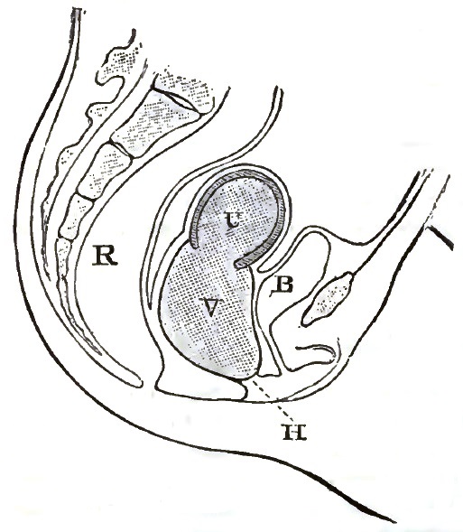
Hæmatometra.—Imperforate Hymen, causing distension of uterus and vagina: H. Hymen; V, Vagina; U, Uterus; B, Bladder; R, Rectum.
DIAGNOSIS.—If there is an accumulation of menstrual fluid in consequence of an imperforate hymen; the latter can be observed as an elastic tumor of a red color protruding outwardly between the labia. A rectal examination is necessary in order to complete the diagnosis, as by this means the presence of menstrual fluid is determined, for if it be present in sufficient quantity to distend the hymen a finger in the rectum can detect fluctuation in the vagina.
If there is no escape of the menstrual fluid beyond the vulva on account of an imperforate hymen, the vagina first becomes gradually distended, then the uterus, and finally the Fallopian tubes. As this distension increases, fluid may be forced beyond the fimbriæ of the tubes into the peritoneal cavity, or, instead, one of the tubes may rupture from the pressure within. In other instances the uterus itself ruptures from over-distension and thinning of its walls. Cases are on record where, the accumulation increasing for years, the uterus has become distended to the size attained in the latter months of pregnancy; under such circumstances its walls as well as the walls of the Fallopian tubes become thinned.
PROGNOSIS.—The physician should be careful and guarded in his prognosis. The health may become much impaired, and sometimes this is the case prior to the cause being ascertained. The chief dangers are in connection with the accumulation of menstrual fluid, such as its discharge at the fimbriated extremity of the tubes, or rupture of the tubes or uterus, and consequent escape of the fluid into the peritoneal cavity. There is also great danger in incising the hymen to permit the exit of the fluid, as will be shown under the head of Treatment. Therefore the longer has been the retention, the greater is the liability of rupture and danger in treatment.
TREATMENT.—As this is of necessity surgical, but brief allusion will be made to it. A simple incision of the hymen will permit the escape of the fluid, but the admission of air by this means is liable to cause sudden contraction of the uterus and a reflex escape of the fluid at the fimbriated extremity of the Fallopian tubes, with all the severe consequences of an intra-peritoneal hemorrhage.
The admission of air is liable to cause decomposition of retained fluid, and this in time produces septicæmia. Further, the sudden admission of air where there has been none before is liable to cause inflammation of the lining membrane of the uterus and tubes, resulting in septic peritonitis. To avoid such risks as have been enumerated two plans are recommended by authors—one being a slow draining away of the menstrual fluid and the other its rapid evacuation and washing out of the uterus and vagina. Graily Hewitt makes an opening of a valvular character in the hymen, permitting only a slow escape of the fluid. Others use a small trocar and draw off the fluid slowly, and at different times if there is a large quantity.
The aspirator is to be preferred to the trocar for emptying the vagina, and of late years has been more generally used; either instrument, but especially the former, permits of the discharge of the fluid at different times, and in such quantities as the physician may desire, without the admission of air. The rapid evacuation is best represented by Emmet's mode of procedure. He first cuts the protruding membrane sufficiently to admit the index finger, and tears the tissues enough to allow the fluid to escape rapidly, and then washes out the vagina and uterus with warm water, after which he introduces a glass plug for the purpose of dilatation and to prevent the action of air upon the parts.
Atresia Vaginæ.
Atresia of the vagina may be congenital or accidental, and, like atresia of any other portion of the genital canal, may be partial or complete. In complete congenital atresia of the vagina an examination per rectum with the index finger fails to discover the fluctuation of menstrual fluid, as in atresia from imperforate hymen, but in its place can usually be felt what seems like a hard fibrous cord. If, however, this cannot be discovered, no doubt remains of entire absence of the vagina. Sometimes the cord can be felt a portion of the distance, which indicates that there is a corresponding portion of an undilated vagina.
In case of complete congenital atresia of the vagina an operation should be avoided, unless there is an accumulation of menstrual fluid or a uterus can be distinctly felt by rectal and vesical examination, or the patient is suffering from the absence of menstruation. To these may possibly be added instances, as mentioned by Thomas, where there exists an imperative necessity for sexual intercourse. Where there is no menstrual molimen or distension of the uterus cannot be detected, and there is non-development of the uterus and ovaries, as shown by the condition of the external organs, surgical interference should be indefinitely postponed.
Accidental atresia of the vagina may be produced by causes heretofore mentioned. When the canal, which has previously been pervious, is entirely obliterated from any cause, an operation becomes, as a rule, an imperative necessity by reason of the accumulation of menstrual fluid and consequent distension of the uterus and Fallopian tubes.
In partial or incomplete atresia it frequently happens that a sinuous canal remains which serves as a guide to the surgeon.
The reader is referred to systematic treatises on surgical diseases of women for the details of the various modes of operating for these affections.
Prolapsus Vaginæ.
Displacements of the vagina are usually secondary, either in consequence of relaxation of the walls or of some form of uterine displacement. Prolapsus of the vagina is usually associated with prolapsus of the uterus, yet it may exist independently. It may be present for some time without prolapse of the uterus, or exceptionally it may be the exciting cause.
DEFINITION.—When the tonicity of the vaginal walls is from any cause impaired and they protrude downward in the direction of the vulva, the condition is called prolapsus.
SYNONYMS AND CLASSIFICATION.—Owing to the anatomical arrangement, it is impossible, with one exception, for any form of prolapsus of the vagina to occur without the coincident prolapse of some viscera. The single exception is the rare occurrence of prolapsus of the posterior wall without the rectum being similarly displaced. These displacements of the viscera with prolapsus of the vagina are commonly described by medical writers as vaginal herniæ, of which there are three different forms, as follows: cystocele vaginalis, rectocele vaginalis, and enterocele vaginalis or hernia vaginalis posterior.
ETIOLOGY.—The causes of displacements of the vagina and the different varieties of vaginal herniæ can very properly be considered together, as they are identical. Laceration of the perineum, an enfeebled condition of the vaginal structure, and a retarded involution of the vagina and uterus in consequence of pregnancy or childbirth are the most frequent causes. Other occasional causes may be mentioned, as former distension of the vagina from repeated childbirths or by tumors, and senile atrophy.
PATHOLOGY.—Following childbirth, the vagina, like the uterus, undergoes a process of involution, but if this is retarded from any cause the vagina is rendered more capacious, its tonicity is impaired, and the uterus, being heavy, crowds down upon it and causes it to be displaced. If the vaginal sphincters or the posterior wall are torn or enfeebled or the perineum lacerated, in addition to the presence of a heavy uterus, prolapsus of the vagina, associated with some form of vaginal hernia, is quite sure to follow.
There is a condition which acts as a common cause in producing vaginal and uterine displacements that has failed to receive on the part of medical authors the notice it deserves—namely, a relaxed condition of the vaginal walls and the perineum, in which there may be observed, in many instances, all of the disturbances caused by a laceration, and yet a careful examination fails to reveal where any tearing has taken place. The continuance of this excessive relaxation and atony of the vaginal walls and the perineum for a long time after parturition is, doubtless, due to subinvolution.
SYMPTOMATOLOGY AND COURSE.—The patient will complain of a bearing-down sensation in the vagina, with a sense of fulness and heat in that locality, sometimes extending to the vulva. These symptoms are aggravated by any muscular exertion, particularly by walking. A physical examination will show the presence of an elastic, globular tumor between the labia. In case it protrudes beyond the vulva, it is not unusual to find scattered over its mucous surface excoriated patches of various sizes. Sometimes these become ulcerated. In other instances the tumor has a smooth, shining appearance. Where there is simply prolapsus of the vagina without the coexistence of a hernia, it will, as a rule, be found that it is the posterior wall. If there is a prolapsus of either the anterior or posterior wall with a hernia, there will be additional symptoms to those above mentioned, which will be referred to in connection with cystocele and rectocele.
Cystocele Vaginalis, or Cysto-Vaginal Hernia.
This is sometimes designated as prolapsus of the bladder, and consists of a descent of the bladder and the anterior wall of the vagina, the two being closely adherent to each other. In consequence of such a descent a pouch is formed which becomes filled with urine. The pouch is in the outset quite small, but gradually becomes larger, so that it is not unusual for one to become of sufficient size to protrude beyond the vulva. In consequence of the pouching of the bladder only a portion of the urine is evacuated by the effort of micturition, and, remaining in the bladder, it decomposes, causing cystitis or vesical catarrh.
The SYMPTOMS are a frequent desire to urinate, with tenesmus and scalding; there is also a sense of heat and pain in the bladder. There is usually more or less ropy mucus discharged with the urine. If a uterine sound or catheter is passed into the bladder with its point downward, and can be felt protruding into the pouch, there remains no doubt as to the case being one of cystocele vaginalis.
Rectocele Vaginalis, or Recto-Vaginal Hernia.
This consists in a protrusion inward of the posterior vaginal wall and a pouch of the rectum, which is carried with it. The tendency to rectocele is seen in the natural bulging of the rectum caused by its expansion just above the sphincter ani. This is more readily perceptible in cases where the perineum has been torn. If from perineal laceration or any cause the posterior wall of the vagina fails to give adequate support to the anterior wall of the rectum, the bulging just mentioned increases, forming a pouch which becomes filled with fecal matter. The bowel becomes more distended with feces, which usually accumulate and harden, and, acting as an irritant, produce tenesmus with mucous discharges. The venous circulation being interfered with, hemorrhoids are common, adding to the patient's suffering.
On examination a tumor is found, sometimes as large as a man's fist, which can be felt projecting from the posterior vaginal wall and over the perineum; sometimes it is soft and compressible, while at other times it is quite solid, depending on the absence or presence of hardened feces. To leave no room for doubt in diagnosticating a case of rectocele, the rectum should be explored with the index finger.
Enterocele Vaginalis, or Entero-Vaginal Hernia.
This consists in a portion of small intestine dilating the cul-de-sac so that the peritoneum is carried down with the intestine between the vagina and rectum as far as the perineum, sometimes forming an elastic tumor at the vulva. The chief dangers arising from this form of vaginal hernia are from its being strangulated or lacerated during childbirth.
Enterocele vaginalis is not frequently met with, but it is important for the physician to know that such a condition is possible and difficult to differentiate from some forms of vaginal tumor. A thorough and careful rectal examination is requisite for diagnosis. An enterocele has the peculiar elastic feeling of a tumor distended with air, a tympanitic resonance on percussion, and a peristaltic movement. If there remains any room for doubt, aspiration with the smallest needle will enable the physician to perfect his diagnosis, for if the needle enter the intestine it is not in any sense a dangerous procedure.
TREATMENT.—The treatment of prolapsus and hernia of the vagina is similar to that of prolapsus of the womb.
If a prolapsus of the vagina has existed but a brief period or has come on suddenly, it should be immediately reduced and proper measures taken to prevent its recurrence. To accomplish this the patient should assume the genu-pectoral position, while the physician with well-oiled fingers restores the parts to their normal position. The patient should then lie upon her back with the hips elevated; astringent vaginal injections ought to be used every four or six hours; and quiet secured or discomfort or pain relieved by opiates. Sudden displacements of the vagina not being of frequent occurrence, the physician more frequently meets with cases of long standing which have come on gradually and slowly.
Attention to the general health is an important requisite: with this in view tonics should be prescribed in many cases, the bowels regulated by means of proper diet or if necessary by medicine, and the bladder more frequently evacuated than in health. Astringent injections are fully as useful in cases of long-standing displacements of the vagina as in those of more recent occurrence; among those more generally used are solutions of tannin, sulphate of zinc, or alum (drachm iv ad pint j). Sea-bathing and injections of sea-water into the vagina are beneficial. It is sometimes more convenient to make topical applications with vaginal suppositories containing one of the astringents just mentioned.
Where cystocele exists it is important that the bladder be completely emptied when the patient urinates; to accomplish this she may assume the genu-pectoral position, and at the same time push the tumor up into the vagina. If after this urine remains in the bladder, a catheter should be employed.
If in any form of vaginal displacement the means which have been alluded to fail, then some form of support or some surgical procedure will be necessary. In very fleshy women considerable benefit is sometimes obtained by means of an abdominal band with a perineal pad attached to it. Pessaries, which have been heretofore quite generally depended upon, are now considered as of secondary importance. Sometimes, however, when the hernia is not of great size or when associated with uterine displacement, a pessary proves of service. A Hodge's pessary with a cross-bar, or the one devised by Skene of Brooklyn, will often prove of great benefit in cystocele. For either cystocele or rectocele the most serviceable form of pessary is one like Cutter's or McIntosh's cup pessary, which is retained within the vagina and supported in position by external attachments. To effect a radical cure in either cystocele or rectocele, especially in the latter, some surgical procedure generally becomes requisite.
Of the different operations which have secured the general approval of gynecologists, the most common is perineorrhaphy: this is the name given to the operation for a torn perineum. Another operation sometimes performed with success is colporrhaphy or elytrorrhaphy, which consists of lessening the calibre of the vagina by removing a portion of the mucous membrane and bringing the edges of the wound together by sutures. This can be performed on either the anterior or posterior wall, depending on which seems to demand it the most; and if the operation on one wall is not likely to be sufficient, it should be made on both. Not unfrequently the most perfect success can be attained by a surgical procedure designated as colpo-perineorrhaphy, which combines the two operations that have been mentioned. Full descriptions of these different operations and the best modes of performing them can be found in all late standard works on surgical gynecology.
Cicatrices.
Cicatrices of the vagina may occur in consequence of lacerations or injuries received in childbirth, surgical operations, wounds from accident, or the use of caustics about the uterus. If any of the causes named excite inflammation, there may be more or less sloughing of the parts, and, as healing must take place by granulation, cicatrices of various dimensions are formed. These cicatrices may be sufficient to cause partial or complete atresia, or they may be merely in the form of projections or bands, dragging the uterus out of its normal position or interfering with its natural mobility, and cause dyspareunia and other discomforts.
Recently, since attention has been directed to the reflex symptoms produced by cicatricial tissue in the neck of the uterus, there has been a growing belief that similar symptoms are often caused by cicatrices in the vagina. Thus it is the opinion of some who have investigated this subject that many cases of remote neuralgia and other nervous disturbances may often be caused in this way.6
6 Vide Skene on "Cicatrices of the Cervix Uteri and Vagina," Amer. Gynæc. Soc., vol. i., 1876.
TREATMENT.—This is of necessity surgical, although some cases can be successfully treated without having recourse to cutting operations, but are treated by pressure. One method is to tampon the vagina with cotton or marine lint previously saturated with carbolized glycerin. The tampon can be left in position four or five days, when the vagina may be washed out and again tamponed. Another method of treating with pressure is by means of a Sims's dilator, either worn continuously or a few hours at a time. Generally a quicker and more effectual mode of treatment is to nick the bands with scissors or a knife in several places sufficiently for the vagina to assume its natural shape, and then insert the dilator. In some instances it is advisable to cut away portions of the adventitious membrane. On account of the tendency to hemorrhage after operations in the vagina the physician should avoid cutting more than is requisite, and must use a finger as a guide in cutting, to inform him when he has cut sufficiently.
If there is considerable hemorrhage it may be necessary to use a styptic, but usually the glass dilator, by putting the walls on the stretch and by pressure, will check the bleeding. It is important that the dilator be worn for several hours each day after the nicking, for fear that there will again be contraction. After each removal of the dilator the vagina should be syringed out with warm carbolized water or a very weak solution of permanganate of potassium (gr. ss ad fluidounce ij), that no septic matter may be retained and so that healing of the cuts may be more rapid.
Double Vagina.
Among the congenital deformities occasionally met with is a vagina divided by a longitudinal septum, constituting a duplex or double vagina. The septum is not always so situated as to make the passages of equal size, nor does it invariably divide the canal through its entire length. It is stated by most writers on the subject that usually with a double vagina there will also be a double uterus. The author has met with only two cases of duplex vagina, neither of which was associated with a double uterus. The treatment is of necessity surgical, and consists in dividing the partition with scissors, and inserting a tampon with some styptic or a Sims's dilator for the arrest of the bleeding which invariably occurs from cutting operations in the vagina. If there is persistent hemorrhage, a galvano- or thermo-cautery may be used.
Growths in the Vagina.
New formations of any kind are not of frequent occurrence in this locality. They consist almost exclusively of cystic tumors, fibroid tumors, papillary excrescences or vegetations, sarcomata, epithelioma, and carcinoma.
Cystic Tumors of the Vagina
are sometimes observed, but are by no means common. Their origin and nature has not seemed to be well understood. Hugier and Guérin are of the opinion that they are caused by the mucous follicles being obstructed. In this view they are sustained by Preuschen.7
7 "Die Cysten die Vagina," Centralblatt für Med., 1871, p. 775.
Sinéty remarks that there are two varieties of vaginal cysts—one superficial and the other profound. The superficial are developed in the mucous membrane, are small in size, and contain fluid which is watery or clear and glairy. The profound cysts are developed in the vaginal walls, and are of various dimensions, from the size of a walnut to an orange, and capable of attaining to much greater dimensions than is possible for the superficial variety. Their contents vary greatly; sometimes clear, mucous, and ropy, in other cases they are colored brownish or chocolate.
Cysts of the vagina are not to be confounded with those of the vulva or those which develop in the vulvo-vaginal glands, nor are they as common.
TREATMENT.—Cysts of the vagina can often be cured by laying them freely open with a bistoury and wiping out the cavity with tincture of iodine, carbolic acid, or a solution of nitrate of silver. The tincture of iodine preferred by the author is Churchill's or a saturation tincture, either being much more effective than the simple tincture. Nitric acid and the actual cautery are mentioned by Barnes as having been used for destroying vaginal cysts. Entire removal of these formations can be effected by cutting into or through the mucous membrane and dissecting them out in the same manner as they are removed from other localities.
Fibrous and Sarcomatous Tumors.
Fibrous or fibroid tumors are by no means as common in the vagina as in the uterus. It has been observed that they are frequently but not invariably associated with the latter. They are developed in the muscular or fibrous structure of the vagina in the same manner as similar formations in the muscular tissue of the uterus.
Some authorities assert that they frequently have the point of departure from the uterus, and then descend little by little between the walls of the vagina.
Sarcomatous tumors are developed in the same tissues and similarly to fibrous growths of the vagina. They are, however, of less frequent occurrence. They sometimes appear primarily in the vagina, but more frequently are consecutive to sarcoma of the uterus.
It is a difficult and often impossible task to make out the differential diagnosis of sarcomatous and fibrous growths in the vagina except by means of the microscope. The symptoms of each are similar to those which indicate sarcomatous and fibrous growths of the uterus, it being accompanied by profuse leucorrhoea, more or less sanious, and occasional hemorrhage. If tumors acquire much size, they interfere with the functions of the rectum and bladder, and cause pain and discomfort by their pressure in the pelvis; sexual intercourse is difficult, frequently painful, and followed by a flow of blood.
DIAGNOSIS.—If of a large size, diagnosis is easily made. Uterine tumors and prolapsed uteri have been mistaken for vaginal growths. By using a uterine probe and inserting a finger in the rectum there need be no error in these respects. By careful examination there is little difficulty in diagnosis.
TREATMENT.—This consists of removal by the knife, scissors, écraseur, or galvano- or thermo-cautery. If there are reasons for believing that a tumor is sarcomatous, it is important that every particle be removed. For this purpose scissors or the galvano- or thermo-cautery are preferable to the ordinary écraseur, which by its action crushes and bruises tissues, and is liable to draw into the chain or wire and crush off more than the operator desires. Serious accidents, such as opening into the peritoneal cavity or the bladder, have occurred in this way in the practice of distinguished and experienced surgeons.
Papillary growths and vegetations in the vagina will receive merely a brief allusion, as they are rarely seen even in the practice of gynecologists. They are not commonly limited to the vagina, but are of more frequent occurrence about the vulva and on the cervix uteri. Vegetations of considerable size sometimes develop in consequence of pregnancy or of granular vaginitis. Sometimes papillary growths within the vagina assume a cauliflower shape with well-defined stalks, or about the ostium vaginæ they may take the form of condylomata. These formations may be confounded with epithelioma.
Treatment consists of removal by scissors or with the thermo- or galvano-cautery, and to guard against hemorrhage some styptic and a vaginal tampon will be required.
Cancer of the Vagina.
Carcinoma or epithelioma rarely occurs as a primary affection in the vagina; it is generally secondary, extending from the neck of the uterus. The author has met with only three cases which were primary cancer.
In a recent work Kustner8 has collected statistics of twenty-two cases of primitive cancer of the vagina. The result of the analysis of these observations is, that nearly always the posterior wall is first affected in primary cancer, while in secondary cancer the anterior wall is the first to be attacked.
8 "Ueber den Primären Scheidenkrebs," Arch. f. Gyn., t. ix. p. 279.
The symptoms after the disease is somewhat advanced are similar to uterine cancer—viz. a sanious, watery discharge of an offensive odor or sometimes a veritable hemorrhage. There is no pain peculiar to or pathognomonic of the disease. It is not until infiltration causes pressure on nerves or there is considerable ulceration that pain is experienced; in either of these conditions the sufferings are often excruciating. Occasionally in women of advanced age, in consequence of cancerous infiltration before ulceration has occurred, the vagina is found to be contracted and there is roughness and induration of the walls.
Epithelioma generally occurs in young women. The early symptoms are pain and hemorrhage following coition. A digital examination will show the friable nature of the formation and an indurated base: the examination will cause blood to flow. In the early part of this stage, before there has been much ulceration, the disease is sometimes mistaken for syphilis and the growths for syphilitic condylomata. It is not an uncommon occurrence for the disease to propagate itself by contact, the opposite wall from which it primarily appeared becoming in this way affected. Later, deeper tissues are infiltrated, the bladder or rectum becomes implicated, ulceration occurs, and subsequently perforation. The progress and terminations are similar to uterine cancer.
TREATMENT.—In carcinoma there seems to be no opportunity for anything more than a palliative course of treatment. Medicine or surgery is here of but little avail. If epithelioma be detected sufficiently early, there is some hope of cure, but this lies only in complete removal. For this purpose the knife or scissors or the galvano- or thermo-cautery can be used. When there is much hemorrhage, some styptic, like the perchloride of iron, should be applied, or the cautery or curette may be of service. Unfortunately, the physician is seldom consulted early enough—prior to the cellular tissue being too much infiltrated—for the thorough eradication of the disease.