Volume 2 / Part 2 — CHAPTER XV.
Reading edition
NERVOUS SYSTEM.
Origin of the Nervous System.
One of the most important recent embryological discoveries is the fact that the central nervous system, in all the Metazoa in which it is fully established, is (with a few doubtful exceptions) derived from the primitive epiblast[152]. As we have already seen that the epiblast represents to a large extent the primitive epidermis, the fact of the nervous system being derived from the epiblast implies that the functions of the central nervous system, which were originally taken by the whole skin, became gradually concentrated in a special part of the skin which was step by step removed from the surface, and has finally become in the higher types a well-defined organ imbedded in the subdermal tissues.
Before considering in detail the comparative development of the nervous system, it will be convenient shortly to review the present state of our knowledge on the general process of its evolution.
This process may be studied either embryologically, or by a comparison of the various stages in its evolution preserved in living forms. Both the methods have led to important results.
The embryological evidence shews that the ganglion-cells of the central part of the nervous system are originally derived from the simple undifferentiated epithelial cells of the surface of the body, while the central nervous system itself has arisen from the concentration of such cells in special tracts. In the Chordata at any rate the nerves arise as outgrowths of the central organ.
Another important fact shewn by embryology is that the central nervous system, and percipient portions of the organs of special sense, especially of optic organs, are often formed from the same part of the primitive epidermis. Thus the retina of the Vertebrate eye is formed from the two lateral lobes of the primitive fore-brain.
The same is true for the compound eyes of some Crustacea. The supraœsophageal ganglia of these animals are formed in the embryo from two thickened patches of the epiblast of the procephalic lobes. These thickened patches become gradually detached from the surface, remaining covered by a layer of epidermis. They then constitute the supraœsophageal ganglia; but they form not only the ganglia, but also the retinulæ of the eye—the parts in fact which correspond to the rods and cones in our own retina. The accessory parts of these organs of special sense, viz. the crystalline lens of the Vertebrate eye, and the corneal lenses and crystalline cones of the Crustacean eye, are independently formed from the epiblast after the separation of the part which becomes the central nervous system.
In the Acraspedote Medusæ the rudimentary central nervous system has the form of isolated rings, composed of sense-cells prolonged into nervous fibres, surrounding the stalks of tentacle-like organs, at the ends of which are placed the sense-organs.
This close connection between certain organs of special sense and ganglia is probably to be explained by supposing that the two sets of structures actually originated pari passu.
We may picture the process as being somewhat as follows:—
It is probable that in simple ancestral organisms the whole body was sensitive to light, but that with the appearance of pigment-cells in certain parts of the body, the sensitiveness to light became localised to the areas where the pigment-cells were present. Since, however, it was necessary that stimuli received by such organs should be communicated to other parts of the body, some of the epidermic cells in the neighbourhood of the pigment-spots, which were at first only sensitive in the same manner as other cells of the epidermis, became gradually differentiated into special nerve-cells. As to the details of this differentiation embryology does not as yet throw any great light; but from the study of comparative anatomy there are grounds for thinking that it was somewhat as follows:—Cells placed on the surface sent protoplasmic processes of a nervous nature inwards, which came into connection with nervous processes from similar cells placed in other parts of the body. The cells with such processes then became removed from the surface, forming a deeper layer of the epidermis below the sensitive cells of the organ of vision. With the latter cells they remained connected by protoplasmic filaments, and thus they came to form a thickening of the epidermis underneath the organ of vision, the cells of which received their stimuli from those of the organ of vision, and transmitted the stimuli so received to other parts of the body. Such a thickening would obviously be the rudiment of a central nervous system, and is in fact very similar to the rudimentary ganglia of the Acraspeda mentioned above. It is easy to see by what steps it might become larger and more important, and might gradually travel inwards, remaining connected with the sense-organ at the surface by protoplasmic filaments, which would then constitute nerves. The rudimentary eye would at first merely consist of cells sensitive to light, and of ganglion-cells connected with them; while at a later period optical structures, constituting a lens capable of throwing an image of external objects upon it, would be developed, and so convert the whole structure into a true organ of vision. It has thus come about that, in the development of the individual, the retina is often first formed in connection with the central nervous system, while the lenses of the eye are independently evolved from the epidermis at a later period.
A series of forms of the Cœlenterata and Platyelminthes affords us examples of various stages in the differentiation of a central nervous system[153].
In sea-anemones (Hertwigs, No. 321) there are, for instance, no organs of special sense, and no definite central nervous system. There are, however, scattered throughout the skin, and also throughout the lining of the digestive tract, a number of specially modified epithelial cells, which are no doubt delicate organs of sense. They are provided at their free extremity with a long hair, and are prolonged on their inner side into fine processes which penetrate into the deeper part of the epithelial layer of the skin or digestive wall. They eventually join a fine network of protoplasmic fibres which forms a special layer immediately within the epithelium. The fibres of this network are no doubt essentially nervous. In addition to fibres there are, moreover, present in the network cells of the same character as the multipolar ganglion-cells in the nervous system of Vertebrates, and some of these cells are characterised by sending a process into the superjacent epithelium. Such cells are obviously intermediate between neuro-epithelial cells and ganglion-cells; and it is probable that the nerve-cells are, in fact, sense-cells which have travelled inwards and lost their epithelial character.
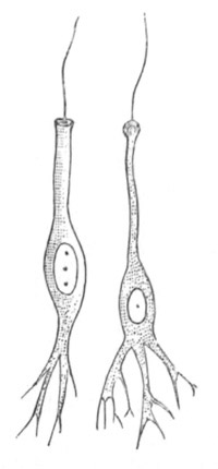
Fig. 236. Neuro-epithelial sense-cells of Aurelia aurita. (From Lankester; after Schäfer.)
In the Craspedote Medusæ (Hertwigs, No. 320) the differentiation of the nervous system is carried somewhat further. There is here a definite double ring, placed at the insertion of the velum, and usually connected with sense-organs. The two parts of the ring belong respectively to the epithelial layers on the upper and lower surfaces of the velum, and are not separated from these layers; they are formed of fine nerve-fibres and ganglion-cells. The epithelium above the nerve rings contains sense-cells (fig. 237) with a stiff hair at their free extremity, and a nervous prolongation at the opposite end, which joins the nerve-fibres of the ring. Between such cells and true ganglion-cells an intermediate type of cell has been found (fig. 237 B) which sends a process upwards amongst the epithelial cells, but does not reach the surface. Such cells, as the Hertwigs have pointed out, are clearly sense-cells partially transformed into ganglion-cells.
A still higher type of nervous system has been met with amongst some primitive Nemertines (Hubrecht, No. 323), consisting of a pair of large cephalic ganglia, and two well-developed lateral ganglionic cords placed close beneath the epidermis. These cords, instead of giving off definite nerves, as in animals with a fully differentiated nervous system, are connected with a continuous subdermal nervous plexus.
The features of the embryology and the anatomy of the nervous system, to which attention has just been called, point to the following general conclusions as to the evolution of the nervous system.
(1) The nervous system of the higher Metazoa appears to have been evolved in the course of a long series of generations from a differentiation of some of the superficial epithelial cells of the body, though it is possible that some parts of the system may have been formed by a differentiation of the alimentary epithelium.
(2) An early feature in the differentiation consisted in the growth of a series of delicate processes of the inner ends of certain epithelial cells, which became at the same time especially differentiated as sense-cells (figs. 236 and 237).
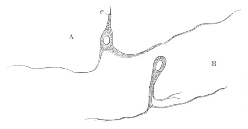
Fig. 237. Isolated cells belonging to the upper nerve-ring of Carmarina hastata. (After O. and R. Hertwig.) A. Neuro-epithelial sense-cell. c. sense-hair. B. Transitional cell between a neuro-epithelial cell and a ganglion-cell.
(3) These processes gave rise to a subepithelial nervous plexus, in which ganglion-cells, formed from sense-cells which travelled inwards and lost their epithelial character (fig. 237 B), soon formed an important part.
(4) Local differentiations of the nervous network, which was no doubt distributed over the whole body, took place partly in the formation of organs of special sense, and partly in other ways, and such differentiations gave rise to a central nervous system. The central nervous system was at first continuous with the epidermis, but became separated from it and travelled inwards.
(5) Nerves, such as we find them in the higher types, originated from special differentiations of the nervous network, radiating from the parts of the central nervous system.
The following points amongst others are still very obscure:—
(1) The steps by which the protoplasmic processes from the primitive epidermic cells became united together so as to form a network of nerve-fibres, placing the various parts of the body in nervous communication. (2) The process by which nerves became connected with muscles, so that a stimulus received by a nerve-cell could be communicated to and cause a contraction in a muscle.
It is probable, as stated in the above summary, that the nervous network took its origin from processes of the sense-cells. The processes of the different cells probably first met and then fused together, and, becoming more arborescent, finally gave rise to a complicated network.
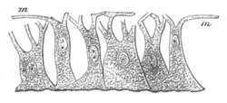
Fig. 238. Myo-epithelial cells of Hydra. (From Gegenbaur; after Kleinenberg.) m. contractile fibres; processes of cells.
The primitive relations between the nervous network and the muscular system are matters of pure speculation. The primitive muscular cells consist of epithelial cells with muscular processes (fig. 238), but the branches of the nervous network have not been traced into connection with the muscles in any Cœlenterata except Ctenophora. In the higher types a continuity between nerves and muscles in the form of motorial end plates has been widely observed. Even in the case of the Cœlenterata it is quite clear from Romanes’ experiments that stimuli received by the nerves are capable of being transmitted to the muscles, and that there must therefore be some connection between nerves and muscles. How did this connection originate?
Epithelial cells with muscular processes (fig. 238) were discovered by Kleinenberg (No. 324) in Hydra before epithelial cells with nervous processes were known, and Kleinenberg pointed out that Hydra shewed the possibility of nervous and muscular tissues existing without a central nervous system, and suggested that the epithelial part of the myoepithelial cells was a sense-organ, and that the connecting part between this and the contractile processes was a rudimentary nerve. He further supposed that in the subsequent evolution of these elements the epithelial part of the cell became a ganglion-cell, while the part connecting this with the muscular tail became prolonged so as to form a true nerve. The discovery of neuro-epithelial cells existing side by side with myoepithelial cells demonstrates that this theory must in part be abandoned, and that some other explanation must be given of the continuity between nerves and muscles. The hypothetical explanation which most obviously suggests itself is that of fusion.
It seems quite possible that many of the epithelial cells of the epidermis and walls of the alimentary tract were originally provided with processes, the protoplasm of which, like that of the Protozoa, carried on the functions of nerves and muscles at the same time, and that these processes united amongst themselves into a network. Such cells would be very similar to Kleinenberg’s neuro-muscular cells. By a subsequent differentiation some of the cells forming this network may have become specially contractile, the epithelial parts of the cells ceasing to have a nervous function, and other cells may have lost their contractility and become solely nervous. In this way we should get neuro-epithelial cells and myoepithelial cells both differentiated from the primitive network, and the connection between the two would also be explained. This hypothesis fits in moreover very well with the condition of the neuro-muscular system as we find it in the Cœlenterata.
Bibliography.
Origin of the Nervous System.
(316) F. M. Balfour. “Address to the Department of Anat. and Physiol. of the British Association.” 1880. (317) C. Claus. “Studien üb. Polypen u. Quallen d. Adria. 1. Acalephen, Discomedusen,” Denk. d. math.-naturwiss. Classe d. k. Akad. Wiss. Wien, Vol. XXXVIII. 1877. (318) Th. Eimer. Zoologische Studien a. Capri. 1. Ueber Beroë ovatus. Ein Beitrag z. Anat. d. Rippenquallen. Leipzig, 1873. (319) V. Hensen. “Zur Entwicklung d. Nervensystems.” Virchow’s Archiv, Vol. XXX. 1864. (320) O. and R. Hertwig. Das Nervensystem u. d. Sinnesorgane d. Medusen. Leipzig, 1878. (321) O. and R. Hertwig. “Die Actinien anat. u. histol. mit besond. Berücksichtigung d. Nervenmuskelsystem untersucht.” Jenaische Zeit., Vol. XIII. 1879. (322) R. Hertwig. “Ueb. d. Bau d. Ctenophoren.” Jenaische Zeitschrift, Vol. XIV. 1880. (323) A. W. Hubrecht. “The Peripheral Nervous System in Palæo- and Schizonemertini, one of the layers of the body-wall.” Quart. J. of Micr. Science, Vol. XX. 1880. (324) N. Kleinenberg. Hydra, eine anatomisch-entwicklungsgeschichtliche Untersuchung. Leipzig, 1872. (325) A. Kowalevsky. “Embryologische Studien an Würmern u. Arthropoden.” Mém. Acad. Pétersbourg, Series VII., Vol. XVI. 1871. (326) E. A. Schäfer. “Observations on the nervous system of Aurelia aurita.” Phil. Trans. 1878.
Nervous system of the Invertebrata. Our knowledge of the development of the central nervous system is still very imperfect in the case of many Invertebrate groups. In the Echinodermata and some of the Chætopoda it is never detached from the epidermis, and in such cases its origin is clear without embryological evidence.
In the majority of groups the central nervous system may be reduced to the type of a pair of cephalic ganglia, continued posteriorly into two cords provided with nerve-cells, which may coalesce ventrally or be more or less widely separated, and be unsegmented or segmented. Various additional visceral ganglia may be added, and in different instances parts of the system may be much reduced, or peculiarly modified. The nervous system of the Platyelminthes (when present), of the Rotifera, Brachiopoda, Polyzoa (?), the Mollusca, the Chætopoda, the Discophora, the Gephyrea, the Tracheata, and the Crustacea, the various small Arthropodan phyla (Pœcilopoda, Pycnognida, Tardigrada, &c.), the Chætognatha (?), and the Myzostomea, probably belongs to this type.
The nervous system of the Echinodermata cannot be reduced to this form; nor in the present state of our knowledge can that of the Nematelminthes or Enteropneusta.
It is only in the case of members of the former set of groups that any adequate observations have yet been made on the development of the nervous system, and even in the case of these groups observations which have any claim to completeness are confined to certain members of the Chætopoda, the Arthropoda and the Mollusca. An account of imperfect observations on other forms, where such have been made, will be found in the systematic part of this work.
Chætopoda. We are indebted to Kleinenberg (No. 329) for the most detailed account which we have of the development of the central nervous system in the Chætopoda.
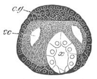
Fig. 239. Section through the head of a young embryo of Lumbricus trapezoides. (After Kleinenberg.) c.g. cephalic ganglion; cc. cephalic portion of the body cavity; x. œsophagus.
The supraœsophageal ganglion with the œsophageal commissure developes independently of the ventral cord. It arises as an unpaired thickening of the epiblast, close to the dorsal side of the œsophagus at the front end of the head (fig. 239), which becomes separated from the epiblast, and extends obliquely backwards and downwards in a somewhat arched form; its lower extremities being somewhat swollen. The inner portion of this curved rudiment becomes converted into commissural nerve-fibres, while the cells of the outer and upper portion assume the characters of ganglion-cells. The commissural fibres are continued downwards to meet the ventral chord, but their junction with the latter structure is not effected till late in embryonic life.
The ventral cord is formed by the coalescence of a pair of linear cords, the development of which takes place from before backwards, so that when their anterior part is well developed their posterior part is hardly differentiated. These cords arise, one on each side of a ventral ciliated furrow, first as a single row of epiblast cells, and subsequently as several rows (fig. 240, Vg). While still united to the external epiblast, they extend themselves below the cells lining the ventral furrow, and unite into a single nervous band, which however exhibits its double origin by its bilobed section. Before the two cords unite, the groove between them becomes somewhat deep, but subsequently shallows out and disappears. The nervous band, before separating from the epiblast, exhibits, in correspondence with the mesoblastic segments, alternate swellings and constrictions. The former become the ganglia, and the latter the connecting trunks.
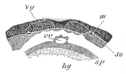
Fig. 240. Section through part of the ventral wall of the trunk of an embryo of Lumbricus trapezoides. (After Kleinenberg.) m. longitudinal muscles; so. somatic mesoblast; sp. splanchnic mesoblast; hy. hypoblast; Vg. ventral nerve-cord; vv. ventral vessel.
As soon as the cord becomes free from the epiblast, it becomes surrounded by a sheath, formed of somatic mesoblast. In each of the ganglionic enlargements there next appears on the dorsal surface a pair of areas of punctiform material, the substance of which soon differentiates itself into nerve-fibres. These areas, by uniting from side to side, give rise to the transverse commissures, and also by a linear coalescence to the longitudinal commissures of the cord. The cellular parts of the band surrounding them become converted into a ganglionic covering of the cord.
In each ganglion the cells of this ganglionic investment penetrate as a median septum into the cord. A fissure is next formed, dividing this septum into two; it is subsequently continued for the whole length of the cord.
Arthropoda. In the Tracheata and the Crustacea the development of the ventral cord is in the main similar to that in the Chætopods, while that of the supraœsophageal ganglia is as a rule somewhat more complicated. No such clear evidence of an independent development of these two parts, as in the case of the Chætopods, has as yet been produced.
The most primitive type of nervous system amongst the Tracheata is that of Peripatus, where it consists of large supraœsophageal ganglia, continuous with a pair of widely separated but large ventral cords united posteriorly above the anus. These cords have an investment of ganglion-cells for their whole length, and are imperfectly divided into ganglia corresponding in number with the feet.
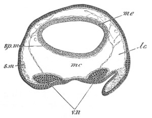
Fig. 241. Section through the trunk of an embryo of Peripatus. The embryo from which the section is taken was somewhat younger than that of fig. 242. sp.m. splanchnic mesoblast; s.m. somatic mesoblast; mc. median section of body cavity; lc. lateral section of body cavity; v.n. ventral nerve cord; me. mesenteron.
The ventral cords are formed as two separate epiblastic ridges (fig. 241, v.n), continued in front into a pair of thickenings of the procephalic lobes, which are at first independent of each other, and from which a large part of the supraœsophageal ganglia takes its origin. After the latter have become separated from the epiblast an invagination of the epiblast covering them grows into each lobe (fig. 242), and becoming constricted from the superficial epiblast, which remains as the epidermis, forms a not unimportant part of the permanent supraœsophageal ganglia.
In the Arachnida the mode of development of the nervous system is essentially the same, and the reader will find a detailed account of it for Spiders in Vol. II. pp. 447-451. The ventral cords are here formed as independent and at first widely separated strands (fig. 243, vn), which for a long time remain far apart; they are subsequently divided into ganglia and become united by transverse commissures.
The supraœsophageal ganglia are formed as two independent thickenings of the procephalic lobes (fig. 244), which eventually separate from the superficial skin. There is formed however in each of them a semicircular groove (fig. 244, gr) lined by the superficial epiblast, which becomes detached from the skin, and is involuted to form part of the ganglia.
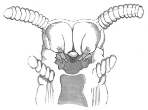
Fig. 242. Head of an embryo Peripatus. (From Moseley.) The figure shews the jaws (mandibles), and close to them epiblastic involutions, which grow into the supraœsophageal ganglia. The antennæ, oral cavity, and oral papillæ are also shewn.
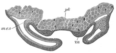
Fig. 243. Transverse section through the Ventral plate of Agelena labyrinthica. The ventral cords have begun to be formed as thickenings of the epiblast, and the limbs are established. me.s. mesoblastic somite; vn. ventral nerve-cord; yk. yolk.
A similar mode of formation of both the ventral cords and the supraœsophageal ganglia obtains in Insects (fig. 245). The ventral cords are however much less widely separated than in Spiders, and early unite in the median line. In the supraœsophageal ganglia the invaginated epiblast has in Lepidoptera (Hatschek) the form of a pit on the dorsal border of the antennæ.
Hatschek states that there takes place an invagination of a median part of the skin between the two ventral cords, for the details of which I must refer the reader to Vol. II. p. 410. He has made more or less similar statements for the earthworm, but his observations in both instances are open to serious doubt.
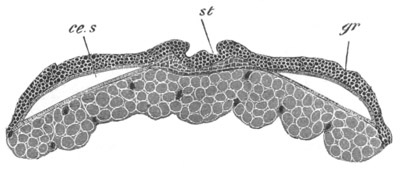
Fig. 244. Section through the procephalic lobes of an embryo of Agelena labyrinthica. st. stomadæum; gr. section through semicircular groove in procephalic lobe; ce.s. cephalic section of body cavity.
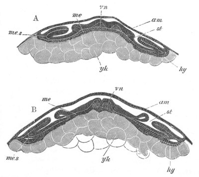
Fig. 245. Two transverse sections through the embryo of Hydrophilus. (After Kowalevsky.) A. Transverse section through an embryo in the region of one of the stigmata. B. Transverse section through an older embryo. vn. ventral nerve-cord; am. amnion and serous membrane; me. mesoblast; me.s. somatic mesoblast; hy. hypoblast (?); yk. yolk-cells (true hypoblast); st. stigma of trachea.
Full details as to the development of the nervous system in the Crustacea are still wanting; a fairly complete account of what is known on the subject is given in Vol. II. pp. 521-2. It appears that the ventral cord may either arise as an unpaired thickening of the epiblast (Isopoda), marked however by a shallow median furrow, or from two cords which eventually coalesce[154]. It is not certain how far the supraœsophageal ganglia are usually in the first instance continuous with the ventral cord. In Astacus, the early stages of which have been elaborately investigated by Reichenbach (No. 331), they are stated to be so; the supraœsophageal ganglia are moreover described by this author as having a somewhat complicated origin. Five elements enter into their composition. There is first formed a pair of pits on the procephalic lobes, which become very deep during the Nauplius stage, and are continuous with a pair of epiblastic ridges which pass round the mouth, and join the ventral cords just described. The walls of the pits are believed to form a part of the embryonic ganglia which gives rise to the retina as well as to the optic ganglia. The ridges form the remainder of the ganglia and the œsophageal commissures; while the fifth element is supplied by a median invagination in front of the mouth, which appears at a much later date than the other parts.
In the Isopoda supraœsophageal ganglia are stated to arise as thickenings of the procephalic lobes, which become eventually detached from the epidermis.
The ventral cord is at first unsegmented, but soon becomes partially divided by a series of constrictions into a number of ganglia, corresponding with the segments. The development of the commissural and ganglionic portions takes place much as in the Chætopoda.
The Gephyrea approach closely the types so far dealt with, but the ventral cord in the Inermia is formed as an unpaired thickening of the epiblast. In Echiurus, as has been shewn by Hatschek in an interesting paper on the larva of this species, published since the appearance of the first volume, there is a pair of ventral cords[155]. In correspondence with a general segmentation of the body, which is subsequently lost, these cords become segmented. The two cords unite in the median line, and Hatschek, in accordance with his general view on this subject, states that their junction is effected by means of a median cord of invaginated epiblast. The segmentation of the cords subsequently becomes lost. The supraœsophageal ganglia arise as an unpaired median thickening of the procephalic lobe. No traces of segmentation in the ventral cord have been observed by Spengel in Bonellia, and the supraœsophageal ganglion is formed in this genus as an unpaired band.
In all the groups above considered the nervous system clearly presents the same type of development with various modifications.
It is formed of two parts, viz. (1) the supraœsophageal ganglia, and (2) the ventral cord.
In the simpler forms, Chætopoda and Gephyrea, the supraœsophageal ganglia are usually stated to be formed as an unpaired thickening at the apex of the præoral lobe, which in most cases becomes subsequently bilobed.
In the Arthropoda the unpaired præoral lobe of the Chætopoda is replaced by the so-called procephalic lobes, which are themselves bilobed; and the supraœsophageal ganglia are formed of two independent halves; further complications in development are also generally found.
There is not as yet sufficient evidence to decide whether the supraœsophageal ganglia were primitively developed continuously with, or independently of, the ventral cords.
The ventral cord appears in the embryo as two independent unsegmented strands, although in a few cases (some Crustacea and Gephyrea) these cords, by an abbreviation in development, arise as an unpaired median thickening of the epiblast.
The form of nervous system of the Chætopoda, Arthropoda, and Gephyrea is clearly therefore to be derived, as was first pointed out by Gegenbaur, from a more or less similar type to that now found in the Nemertines; and as suggested in the chapter on larval forms (vide p. 378) may perhaps be derived from the elongation of a circular ring, of which the anterior end has become developed into the supraœsophageal ganglia, the lateral parts into the two lateral strands, while the posterior part persists in some forms in the junction of the ventral cords above the anus (Enopla and Peripatus).
Mollusca. While study of the anatomy of the nervous system of the Mollusca, especially of certain primitive genera (Chiton, Haliotis, Fissurella, &c.) leaves little doubt that it is formed on the same type as that of the groups just spoken of, the development, so far as our imperfect knowledge enables us to make definite statements on the subject, is somewhat abnormal[156].
In the Gasteropoda and Pteropoda the supraœsophageal ganglia appear most probably to be developed either as paired thickenings of the epiblast of the velar area, or as invaginated pits of the velar area, which become detached from the surface, and then become solid (Hyaleacea and Limax). In either case the supraœsophageal ganglia appear to be developed quite independently of the pedal ganglia. The latter, as might be anticipated, are earlier in their development and more constant than the various visceral ganglia; and, if the views above expressed are correct, are homologous with the ventral cord of the Chætopods and Arthropods. Their actual development is very imperfectly known.
The most precise statements on the subject, viz. those of Bobretzky and Fol, would lead us to suppose that they arise in the mesoblast, but it seems more probable that they are formed as thickenings of the sides of the foot.
In the Cephalopods all the ganglia are stated to be differentiated in the mesoblast (Lankester, Bobretzky).
Hatschek[157] has recently given a detailed description of the development of the supraœsophageal and pedal ganglia of Teredo. He finds that the former ganglia arise as an unpaired thickening of the epiblast in the centre of the velar area, and the latter as an unpaired thickening of the epiblast of the ventral side of the body between the mouth and the anus. The two ganglia would thus seem to be disconnected with each other in their development.
(327) F. M. Balfour. “Notes on the development of the Araneina.” Quart. J. of Micr. Science, Vol. XX. 1880. (328) B. Hatschek. “Beitr. z. Entwicklung d. Lepidopteren.” Jenaische Zeitschrift, Vol. XI. 1877. (329) N. Kleinenberg. “The development of the Earthworm, Lumbricus Trapezoides.” Quart. J. of Micr. Science, Vol. XIX. 1879. (330) A. Kowalevsky. “Embryologische Studien an Würmern u. Arthropoden.” Mém. Acad. Pétersbourg, Series VIII., Vol. XVI. 1871. (331) H. Reichenbach. “Die Embryonalanlage u. erste Entwick. d. Flusskrebses.” Zeit. f. wiss. Zool., Vol. XXIX. 1877.
The Central Nervous System of the Vertebrata[158].
The formation of the cerebrospinal axis of the Chordata from the medullary plate has already been treated at length (pp. 301-304). Before entering into the consideration of the morphological value of the various parts of this cord, it will be convenient to describe the more important features of its ontogeny. For this purpose the two parts into which the nervous axis becomes at an early period divided, viz. the spinal cord and the brain, may be dealt with separately.
The Spinal Cord, shortly after the closure of the medullary canal, has, in all the true Vertebrata, the form of an oval tube; the walls of which are of a fairly uniform thickness, and are composed of several rows of elongated cells. This cord, as development proceeds, usually becomes vertically prolonged in transverse section, and the central canal which it contains also becomes vertically elongated. The variations in shape of the spinal canal are very great at different periods and in different parts of the body, and an attempt to chronicle them would appear, in the present state of our knowledge, to be quite valueless[159]. fig. 117, in which the spinal cord of the chick of the third day is shewn in transverse section, illustrates the character of the cord at the stage just described. Up to this time the walls of the spinal canal have exhibited an uniform structure. A series of changes now however takes place, which results in the differentiation (1) of the epithelium of the central canal, (2) of the grey matter of the cord, and (3) of the external coating of white matter.
The relative time at which each of these parts becomes developed is not constant in the different forms.
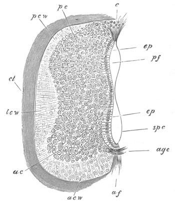
Fig. 246. Section through the spinal cord of a seven days’ Chick. pcw. dorsal white column; lcw. lateral white column; acw. ventral white column; c. dorsal tissue filling up the part where the dorsal fissure will be formed; pc. dorsal grey cornu; ac. anterior grey cornu; ep. epithelial cells; agc. anterior commissure; pf. dorsal part of spinal canal; spc. ventral part of spinal canal; af. anterior fissure.
The white matter is apparently the result of a differentiation of the outermost parts of the superficial cells of the cord into longitudinal nerve-fibres, which remain for a long period without a medullary sheath. These fibres appear in transverse sections as small dots. The white matter forms a transparent investment of the grey matter and would seem to contain neither nuclei nor cells[160]. The white matter may from the first form only two masses, one on each side, forming a layer on the ventral and lateral parts of the spinal cord but not extending to the dorsal surface (Elasmobranchii, fig. 185, W); or it may form four patches, viz. an anterior and a posterior white column on each side, which lie on a level with the origin of the anterior and posterior nerve-roots (the Fowl, Human embryo, etc.). In whichever of these forms the white matter appears, it is always, at first, a layer of extreme tenuity, which rapidly increases in thickness in the subsequent stages, and extends so as gradually to cover the whole cord (fig. 246).
The anterior white commissure is formed very shortly after the first appearance of the white matter. The grey matter and the central epithelium are formed by a differentiation of the main mass of the spinal cord. The outer cells lose their epithelial-like arrangement, and, becoming prolonged into fibres, give rise to the grey matter, while the innermost cells retain their primitive arrangement, and constitute the epithelium of the canal. The process of formation of the grey matter would appear to proceed from without inwards, so that some of the cells, which have, on the formation of the grey matter, an epithelial-like arrangement, subsequently become converted into true nerve-cells.
As has already been mentioned, the central epithelium of the nervous system probably corresponds with the so-called epidermic layer of the epiblast.
The grey matter soon becomes prolonged dorsally and ventrally into the posterior and anterior horns. Its fibres may especially be traced in two directions:—(1) round the anterior end of the spinal canal, immediately outside its epithelium and so to the grey matter on the opposite side, forming in this way an anterior grey commissure, through which a decussation of the fibres from the opposite sides is effected: (2) dorsalwards along the outside of the lateral walls of the canal.
There is at this period no trace of the ventral or dorsal fissure, and the shape of the central canal is not very different to what it was at an earlier period. This condition of the spinal cord is especially instructive, as it is very nearly that which is permanent in Amphioxus.
The next event of importance is the formation of the ventral or anterior fissure. This owes its origin to a downgrowth of the anterior horns of the cord on each side of the middle line. The two downgrowths enclose between them a somewhat linear space—the anterior fissure—which increases in depth in the succeeding stages (fig. 246, af).
The dorsal or posterior fissure is formed at a later period than the anterior, and accompanies the atrophy of the dorsal section of the embryonically large canal of the spinal cord.
The exact mode of its formation appears to me to be still involved in some obscurity.
In the Elements of Embryology the development of the posterior fissure was described in the following way:
“On the seventh day the most important event is the formation of the posterior fissure.
“This is brought about by the absorption of the roof of the posterior of the two parts into which the neural canal has become divided.
“Between the posterior horns of the cord, the epithelium forming the roof of the, so to speak, posterior canal is along the middle line covered neither by grey nor by white matter, and on the seventh day is partially absorbed, thus transforming the canal into a wedge-shaped fissure, whose mouth however is seen in section to be partially closed by a triangular clump of elongated cells (fig. 246, c). Below this mass of cells the fissure is open. It is separated from the ‘true spinal canal’ by a very narrow space along which the side walls have coalesced. In the lumbar and sacral regions the two still communicate.
“We thus find, as was first pointed out by Lockhart Clarke, that the anterior and posterior fissures of the spinal cord are, morphologically speaking, entirely different. The anterior fissure is merely the space left between two lateral downward growths of the cord, while the posterior fissure is part of the original neural canal separated from the rest of the cavity (which goes to form the true spinal canal) by a median coalescence of the side walls.”
I confess that I have some doubts as to the complete accuracy of the above statement.
Kölliker gives a full account of the gradual atrophy of the central canal; but I do not fully understand his statements with reference to the formation of the posterior fissure, which in fact appears to be only incidentally mentioned. It would seem from his account that a shallow and somewhat wide dorsal fissure is formed to start with, in the human embryo, by two projections of the posterior white horns. On the atrophy of the central canal this furrow becomes narrowed, but Kölliker does not definitely state how it becomes deepened so as to give rise to the permanent dorsal fissure.
It seems to me probable, though further investigations on the point are still required, that the dorsal fissure is a direct result of the atrophy of the dorsal part of the central canal of the spinal cord.
The walls of the canal coalesce dorsally, and the coalescence gradually extends ventralwards, so as finally to reduce the central canal to a minute tube, formed of the ventral part of the original canal. The epithelial wall formed by the coalesced walls on the dorsal side of the canal is gradually absorbed.
The epithelium of the central canal, at the period when its atrophy commences, is not covered dorsally either by grey or white matter, so that, with the gradual reduction of the dorsal part of the canal, and the absorption of the epithelial wall formed by the fusion of its two sides, a fissure between the two halves of the spinal cord becomes formed. This fissure is the posterior or dorsal fissure. In the process of its formation the white matter of the dorsal horns becomes prolonged so as to line its walls; and shortly after its formation the dorsal grey commissure makes its appearance, which is not improbably derived from part of the epithelium of the original central canal.
Development of the Brain.
The brain is formed from the anterior portion of the medullary plate. When the medullary plate first becomes differentiated it is not possible to distinguish between the region of the brain and that of the spinal cord. The brain region is however usually very early indicated by a widening of the medullary plate, but does not become sharply marked off from the region of the spinal cord. In many Ichthyopsida (Elasmobranchii (fig. 28, C) and Amphibia (fig. 77, A)) the anterior dilatation gives to the medullary plate, before its sides meet to form a canal, a spatula-like form; which is either not present or less marked in Reptilia, Aves and Mammalia.
The length of the brain as compared to the spinal cord is always very great in the embryo, and in the earliest developmental periods the disproportion in the size of the brain is specially marked, owing to the full number of the somites of the trunk not having been formed. In Elasmobranchii the brain is about one-third of the whole length of the embryo at the stage immediately following the closure of the medullary canal.
The first differentiation of the brain into distinct parts is a very early occurrence, and may take place before (Mammalia) or during the closure of the medullary folds. The brain first becomes divided into two successive lobes or vesicles by a single transverse constriction, and subsequently the posterior of these again becomes divided into two, so that three lobes are formed—known as the fore- the mid- and the hind-brain; of these the hind-brain is usually the longest. In some instances a bilobed stage can hardly be recognised. This primitive division of the brain is shewn in many of the figures already given. The reader may perhaps best refer to fig. 108. On the closure of the medullary groove the lumen of the medullary canal is continued uninterruptedly through the brain, but dilates considerably in each of the cerebral vesicles.
The anterior lobe of the brain becomes converted into the cerebral hemispheres, the thalamencephalon, the primary optic vesicles, and the parts connected with them. The middle lobe becomes the optic lobes (corpora bigemina or corpora quadrigemina in Mammalia) and the crura cerebri; while the posterior lobe becomes converted into the cerebellum and medulla oblongata.
Before describing in detail the changes by which the primary vesicles of the brain become converted into the above parts, it will be convenient to say a few words about the general development of the brain.
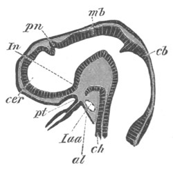
Fig. 247. Longitudinal section through the brain of a young Pristiurus embryo. cer. commencement of the cerebral hemisphere; pn. pineal gland; In. infundibulum; pt. ingrowth from mouth to form the pituitary body; mb. mid-brain; cb. cerebellum; ch. notochord; al. alimentary tract; Iaa. artery of mandibular arch.
The most striking peculiarity with reference to the general development of the brain is a curvature which appears in its axis, known as the cranial flexure. The flexure takes place through the mid-brain, and causes the fore-brain to be gradually bent downwards so that the axis of its floor forms, first, a right angle with that of the hinder part of the brain, and subsequently, as a rule, an acute angle.
During these changes the brain, in most Amniota at any rate, becomes in the first instance retort-shaped, the cerebral vesicle forming the swollen part of the retort, but subsequently the retort-shape is lost owing to the great development of the vesicle of the mid-brain, which forms the termination of the long axis of the embryo. Figs. 29, 76, and 118, are representative figures of embryos of various vertebrate forms at a period when the mid-brain forms the termination of the long axis of the body.
It is generally stated that the cranial flexure is at its maximum at the stage represented in these figures, and there can be no doubt that viewed from the exterior the cranial flexure ceases to be so marked a feature, and finally disappears as the embryo gradually grows older; but though the mid-brain ceases to form the termination of the long axis of the embryo, the flexure of the brain becomes in many forms absolutely more marked; while in other forms, though stated to diminish, it does not entirely vanish.
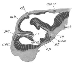
Fig. 248. Longitudinal section through the brain of Scyllium canicula at an advanced stage of development. cer. cerebral hemisphere; pn. pineal gland; op.th. optic thalamus, connected with its fellow by a commissure (the middle commissure). In front of it is seen a fold of the roof of the fore-brain, which is connected with the choroid plexus of the third ventricle; op. optic chiasma; pt. pituitary body; in. infundibulum; cb. cerebellum; au.v. passage leading from the auditory vesicle to the exterior; mel. medulla oblongata; c.in. internal carotid artery.
The general nature of the changes which take place will perhaps best be understood by a comparison of figs. 247 and 248 representing longitudinal sections at two stages through the brain of an embryo Elasmobranch. The actual cranial flexure, i.e. flexure of the floor of the brain, is obviously greater in the older of the two brains, though viewed from the exterior the axis of this brain appears to be quite straight. In the younger stage, fig. 247, the mid-brain (mb) forms the end of the long axis of the body, while in the older one the cerebral hemispheres (cer) have grown very greatly, especially forwards and dorsalwards. They have thus come to lie in front of the mid-brain, and to form the end of the long axis of the body, and have at the same time compressed the originally large thalamencephalon against the mid-brain. The same general features may be seen in fig. 250 representing a longitudinal section of the brain of an embryo fowl, and fig. 255 representing a longitudinal section of the brain of a Mammal.
The infundibulum or perhaps rather the point of origin of the optic nerves is to be regarded as the anterior termination of the axis of the base of the brain.
The cranial flexure is least marked in Cyclostomata (fig. 253), Teleostei, Ganoidei, and Amphibia, while it is very pronounced in Elasmobranchii, Reptilia, Aves, and Mammalia. In Teleostei, and still more in Cyclostomata, it permanently remains slight, owing to the small development of the cerebral hemispheres.
In addition to the cranial flexures, two other flexures make their appearance in the base of the brain. A posterior at the junction of the brain and spinal cord, and an anterior at the boundary between the cerebellum and medulla oblongata, just at the point where the pons Varolii is formed in Mammalia. The anterior of these is the most marked and constant; it is shewn in fig. 250. It arises considerably later than the main cranial flexure, and since it is turned the opposite way it assists to a considerable extent in causing the apparent straightening of the cranial axis.
Histogenetic changes[161]. The walls of the brain are at first very thin and, like those of the spinal cord, are formed of a number of ranges of spindle-shaped cells. The processes of each of these cells are stated to be continued through the whole thickness of the wall. In the floor of the hind- and mid-brain a superficial layer of delicate nerve-fibres is formed at an early period. This layer appears in the first instance on the floor and sides of the hind-brain, and very slightly, if at all, later on the floor and the sides of the mid-brain. The cells internal to the nerve-fibres become differentiated into an innermost epithelial layer lining the cavities of the ventricles, and an outer layer of grey matter.
The similarity of the primitive arrangement and histological character of the parts of the brain behind the cerebral hemispheres to that of the spinal cord is very conclusively shewn by the examination of any good series of sections. In both brain and spinal cord the white matter forms a cap on the ventral and lateral parts considerably before it extends to the dorsal surface. In the medulla the white matter does not eventually extend to the roof owing to the peculiar degeneration which that part undergoes.
In the case of the fore-brain the earliest histological changes, except possibly in Mammals, take place on the same general plan as those of the remainder of the central nervous system[162]; but though the general plan is the same, yet the early histological distinction between the fore-brain, and the mid- and hind-brain is more marked than the distinction between the latter and the spinal cord.
On the floor and sides of the thalamencephalon, and apparently the whole of the hemispheres of the lower types, there is formed, somewhat later than in the remainder of the brain, a very delicate layer of white matter. The inner part of the wall, which still remains comparatively thin, is not at first clearly divided into an epithelial and nervous layer. This distinction soon however becomes more or less apparent, though it is not so marked as in most other parts of the brain; and it appears that in the subsequent growth the greater part of the original epithelial layer becomes converted into nervous tissue.
In Mammals the same plan of differentiation would seem to be followed, though somewhat less obviously than in the lower types. The walls of the hemispheres become first divided (Kölliker) into a superficial thinner layer of rounded elements, and a deeper and thicker epithelial layer, and between these the fibres of the crura cerebri soon interpose themselves. At a slightly later period a thin superficial layer of white matter, homologous with that of the remainder of the brain, becomes established.
The inner layer, together with the fibres from the crura cerebri, gives rise to the major part of the white matter of the hemispheres and to the epithelium lining the lateral ventricles.
The outer layer of rounded cells becomes divided into (1) a superficial part with comparatively few cells, which, together with its coating of white matter, forms the cortical part of the grey matter, and (2) a deeper layer with numerous cells which forms the main mass of the grey matter of the hemispheres.
The development of the several parts of the brain will now be described.
The hind-brain. The hind-brain is at first an elongated, funnel-shaped tube, the walls of which are of a nearly uniform thickness, though the roof and floor are somewhat thinner than the sides. It forms a direct continuation of the spinal cord, into which it passes without any sharp line of demarcation. The ventricle it contains is known as the fourth ventricle.
The sides become in the chick marked by a series of transverse constrictions, dividing it into lobes, which are somewhat indefinite in number. The first of these remains permanent, and its roof gives rise to the cerebellum. It is uncertain whether the other constrictions have any morphological significance. More or less similar constrictions are present in Teleostei. In Elasmobranchii the medulla presents on its inner face at a late period a series of lobes corresponding with the roots of the vagus and glossopharyngeal nerves, and it is possible that the earlier constrictions may potentially correspond to so many nerve-roots.
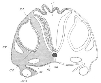
Fig. 249. Section through the hind-brain of a Chick at the end of the third day of incubation. IV. Fourth ventricle. The section shews the very thin roof and thicker sides of the ventricle. Ch. Notochord; CV. Anterior cardinal vein; CC. Involuted auditory vesicle; CC points to the end which will form the cochlear canal; RL. Recessus labyrinthi (remains of passage connecting the vesicle with the exterior); hy. Hypoblast lining the alimentary canal; AO., AOA. Aorta, and aortic arch.
Throughout the Vertebrata an anterior lobe of the hind-brain becomes very early marked off, so that the primitive hind-brain becomes divided into two regions which may be conveniently spoken of as the cerebellum (figs. 247 and 248, cb) and medulla oblongata. The floor of these regions is quite continuous and is also prolonged without any break into the floor of the mid-brain.
The posterior section of the hind-brain, which forms the medulla, undergoes changes of a somewhat complicated character. In the first place its roof becomes in front very much extended and thinned out. At the raphe, where the two lateral halves of the brain originally united, a separation, as it were, takes place, and the two sides of the brain become pushed apart, remaining united by only a very thin layer of nervous matter, consisting of a single row of flattened cells (fig. 249). As a result of this peculiar growth in the brain, the roots of the nerves of the two sides, which were originally in contact at the dorsal summit of the brain, become carried away from one another, and appear to arise at the sides of the brain.
The thin roof of the fourth ventricle is triangular, or, in Mammalia, somewhat rhomboidal in shape. The apex of the triangle is directed backwards.
At a later period the blood-vessels of the pia mater form a rich plexus over the anterior part of the thin roof of the medulla, which becomes at the same time somewhat folded. The whole structure is known as the tela vasculosa, or choroid plexus of the fourth ventricle (fig. 250, chd 4). The floor of the whole hind-brain becomes thickened, and there very soon appears on its outer surface a layer of non-medullated nerve-fibres, similar to those which first appear on the spinal cord. They are continuous with a similar layer of fibres on the floor of the mid-brain, where they constitute the crura cerebri. On the ventral floor of the medulla is a shallow continuation of the anterior fissure of the spinal cord.
In Elasmobranchii and many Teleostei the restiform tracts are well developed, and are anteriorly continued into the cerebellum, of which they form the peduncles. Near their junction with the cerebellum they form prominent bodies, which are regarded by Miklucho-Maclay as representing the true cerebellum of Elasmobranchii.
In Elasmobranchii a dorsal pair of ridges projects into the cavity of the fourth ventricle, corresponding apparently with the fasciculi teretes of the Mammalia.
In Mammalia there develop, subsequently to the longitudinal fibres already spoken of, first the olivary bodies of the ventral side of the medulla, and at a still later period the pyramids. The fasciculi teretes in the cavity of the fourth ventricle are developed shortly before the pyramids.
When the hind-brain becomes divided into two regions the roof of the anterior part does not become thinned out like that of the posterior, but on the contrary, becomes somewhat thickened and forms a band-like structure roofing over the anterior part of the fourth ventricle (fig. 247 and fig. 253, cb).
This is a rudiment of the cerebellum, and in all Craniate Vertebrates it at first presents this simple structure and insignificant size. In Cyclostomata, Amphibia and many Reptilia this condition is permanent. In Elasmobranchii, on the other hand, the cerebellum assumes in the course of development a greater and greater prominence (fig. 248, cb), and eventually overlaps both the optic lobes in front and the medulla behind. In the later embryonic stages it exhibits in surface-views the appearance of a median constriction, and the portion of the ventricle contained in it is prolonged into two lateral outgrowths.
Miklucho-Maclay, from his observations on the brains of adult Elasmobranchii, was led to regard what is here called the cerebellum as identical with the mid-brain, and the true mid-brain as part of the thalamencephalon. Miklucho-Maclay was no doubt misled by the large size of the cerebellum, but, as we have seen, this body does not begin to be conspicuous till late in embryonic life.
The mid-brain and thalamencephalon (according to the ordinary interpretations) have in the embryo of Elasmobranchs exactly the same relations as in the embryos of other Vertebrates; so that the embryological evidence appears to me to be conclusive against Miklucho-Maclay’s view.
In Birds the cerebellum attains a very considerable development (fig. 250, cbl), consisting of a folded central lobe with an arbor vitæ, into which the fourth ventricle is prolonged. There are two small lateral lobes, apparently equivalent to the flocculi. Anteriorly the cerebellum is connected with the roof of the mid-brain by a delicate membrane, the velum medullæ anterius, or valve of Vieussens (fig. 250, vma). The pons Varolii of Mammalia is represented by a small number of transverse fibres on the floor of the hind-brain immediately below the cerebellum.
In Mammalia the cerebellum attains a still greater development. The median lobe or vermiform process is first developed. In the higher Mammalia the lateral parts forming the hemispheres of the cerebellum become formed as swellings at the sides at a considerably later period, and are hardly developed in the Monotremata and Marsupialia.
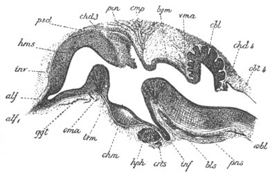
Fig. 250. Longitudinal section through the brain of a Chick of ten days. (After Mihalkovics.) hms. cerebral hemispheres; alf. olfactory lobe; alf1. olfactory nerve; ggt. corpus striatum; oma. anterior commissure; chd3. choroid plexus of the third ventricle; pin. pineal gland; cmp. posterior commissure; trm. lamina terminalis; chm. optic chiasma; inf. infundibulum; hph. pituitary body; bgm. commissure of Sylvius (roof of iter a tertio ad quartum ventriculum); vma. velum medullæ anterius (valve of Vieussens); cbl. cerebellum; chd4. choroid plexus of the fourth ventricle; obl4. roof of fourth ventricle; obl. medulla oblongata; pns. commissural part of medulla; inv. sheath of brain; bls. basilar artery; crts. internal carotid.
The cerebellum is connected with the roof of the mid-brain in front and with the choroid plexus of the fourth ventricle behind by delicate membranous structures, known as the velum medullæ anterius (valve of Vieussens) and the velum medullæ posterius.
The pons Varolii is formed on the ventral side of the floor of the cerebellar region as a bundle of transverse fibres at about the same time as the olivary bodies.
The mid-brain. The changes undergone by the mid-brain are simpler than those of any other part of the brain. We have already seen that the mid-brain, on the appearance of the cranial flexure, forms an unpaired vesicle with a vaulted roof and curved floor, at the front end of the long axis of the body (fig. 118, MB). It is at this period in most Vertebrates relatively much larger than in the adult; and it is only in the Teleostei that it more or less retains in the adult its embryonic proportions.
The cavity of the mid-brain, greatly reduced in size in the higher forms, is known as the iter a tertio ad quartum ventriculum, or aqueductus Sylvii.
The roof of the mid-brain is sharply constricted off from the divisions of the brain in front of and behind it, but these constrictions do not extend to the floor.
In some Vertebrates the region of the mid-brain is stated to undergo hardly any further development. In the Axolotl it remains according to Stieda[163] as a simple tube with nearly uniformly thick walls. In the majority of forms it undergoes, however, a more complicated development.
In Elasmobranchs the sides become thickened to form the optic lobes, which are soon separated by a median longitudinal groove. The floor becomes thickened to form the crura cerebri. The primitive simple median cavity becomes imperfectly divided into a median portion below, and two lateral diverticula in the optic lobes.
In Teleostei the changes, resulting in the formation of (1) a pair of longitudinal ridges projecting from the roof into the cavity of the iter, constituting the fornix of Gottsche, and (2) of the two swellings on the floor, forming the tori semicirculares, are more complicated, but have not been satisfactorily worked out. In Bombinator and the Anura generally the changes are of the same nature as those in Elasmobranchii, except that the prolongations of the ventricle into the optic lobes are still further constricted off from the median portion, which forms the true iter.
In Reptilia and Aves the development of the mid-brain takes place on the same type as in Elasmobranchii and the Anura. In Birds the optic lobes are pushed very much aside, and the roof of the iter is greatly thinned out. In Mammalia the sides of the mid-brain give rise to two pairs of prominences—the corpora quadrigemina—instead of the two optic lobes of other Vertebrata. The prominences, which do not contain prolongations of the iter, become first visible on the appearance of an oblique transverse furrow, while the anterior pair alone are separated by a longitudinal furrow. In the later stages of development the longitudinal furrow is continued so as to bisect the posterior pair.
The floor, which is bounded posteriorly by the pons Varolii, becomes the crura cerebri. The corpora geniculata interna also belong to this division of the brain.
Fore-brain. In its earliest condition the fore-brain forms a single vesicle without a trace of separate divisions, but very early it buds off the optic vesicles, whose history is described with that of the eye.
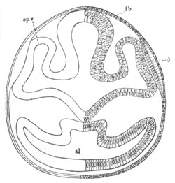
Fig. 251. Section through the front part of the head of a Lepidosteus embryo on the seventh day after impregnation. al. alimentary tract; fb. thalamencephalon; l. lens of eye; op.v. optic vesicle. The mesoblast is not represented.
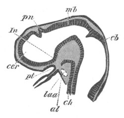
Fig. 252. Longitudinal section through the brain of a young Pristiurus embryo. cer. commencement of cerebral hemisphere; pn. pineal gland; In. infundibulum; pt. ingrowth of mouth to form the pituitary body; mb. mid-brain; cb. cerebellum; ch. notochord; al. alimentary tract; Iaa. artery of mandibular arch.
The optic vesicles become gradually constricted off from the fore-brain in a direction obliquely backwards and downwards. They remain, however, attached to it at the anterior extremity of the base of the fore-brain (fig. 251, op.v.). While the above changes are taking place in the optic vesicles the anterior part of the fore-brain becomes prolonged, and at the same time somewhat dilated. At first there is no sharp boundary between the primitive fore-brain and its anterior prolongation, but there shortly appears a constriction which passes from above obliquely forwards and downwards. This constriction is shallow at first, but soon becomes much deeper, leaving however the cavities of the two divisions of the fore-brain united ventrally by a somewhat wide canal (fig. 252).
Of these two divisions the posterior becomes the thalamencephalon, while the anterior and larger division (cer) forms the rudiment of the cerebral hemispheres and olfactory lobes. For a considerable period this rudiment remains perfectly simple, and exhibits no signs, either externally or internally, of a longitudinal constriction dividing it into two lobes.
From the above description it may be concluded that the rudiment of the cerebral hemispheres is contained in the original fore-brain. In spite however of their great importance in all the Craniata, it is probable that the hemispheres were either not present as distinct structures, or only imperfectly separated from the thalamencephalon, in the primitive vertebrate stock.
The thalamencephalon. The thalamencephalon varies so slightly in structure throughout the Vertebrate series that a general description will suffice for all the types.
It forms at first a simple vesicle, the walls of which are of a nearly uniform thickness and formed of the usual spindle-shaped cells.
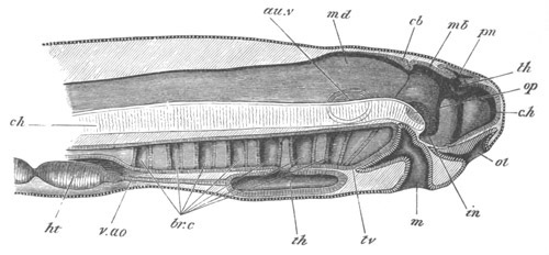
Fig. 253. Diagrammatic vertical section through the head of a larva of Petromyzon. The larva had been hatched three days, and was 4.8 mm. in length. The optic and auditory vesicles are supposed to be seen through the tissues. c.h. cerebral hemisphere; th. optic thalamus; in. infundibulum; pn. pineal gland; mb. mid-brain; cb. cerebellum; md. medulla oblongata; au.v. auditory vesicle; op. optic vesicle; ol. olfactory pit; m. mouth; br.c. branchial pouches; th. thyroid involution; v.ao. ventral aorta; ht. ventricle of heart; ch. notochord.
The cavity it contains is known as the third ventricle. Anteriorly it opens widely into the cerebral rudiment, and posteriorly into the ventricle of the mid-brain. The opening into the cerebral rudiment becomes the foramen of Munro.
For convenience of description I shall divide it into three regions, viz. (1) the floor, (2) the sides, and (3) the roof.
The floor becomes divided into two parts, an anterior part, giving origin to the optic nerves, in which is formed the optic chiasma; and a posterior part, which becomes produced into an at first inconspicuous prominence—the rudiment of the infundibulum (fig. 252, In). This comes in contact with an involution from the mouth, which gives rise to the pituitary body (fig. 252, pt), the development of which will be dealt with separately.
In the later stages of development the infundibulum becomes gradually prolonged, and forms an elongated diverticulum of the third ventricle, the apex of which is in contact with the pituitary body (figs. 252, 254, in, and figs. 250 and 255, inf).
Along the sides of the infundibulum run the commissural fibres connecting the floor of the mid-brain with the cerebrum.
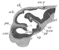
Fig. 254. Longitudinal section through the brain of Scyllium canicula at an advanced stage of development. cer. cerebral hemisphere; pn. pineal gland; op. th. optic thalamus, connected with its fellow by a commissure (the middle commissure). In front of it is seen a fold of the roof of the fore-brain, which is the choroid plexus of the third ventricle; op. optic chiasma; pt. pituitary body; in. infundibulum; cb. cerebellum; au.v. passage leading from the auditory vesicle to the exterior; mel. medulla oblongata; c.in. internal carotid artery.
In its later stages the infundibular region presents considerable variations in the different vertebrate types. In Fishes it generally remains very large, and permanently forms a marked diverticulum of the floor of the thalamencephalon. In Elasmobranchii the distal end becomes divided into three lobes—a median and two lateral. The lateral lobes appear to become the sacci vasculosi of the adult.
In Teleostei peculiar bodies known as the lobi inferiores (hypoaria) make their appearance at the sides of the infundibulum. They appear to correspond in position with the tuber cinereum of Mammalia[164]. In Birds, Reptiles, and Amphibia the lower part of the embryonic infundibulum becomes atrophied and reduced to a mere finger-like process—the processus infundibuli.
In Mammalia the posterior part of the primitive infundibulum becomes the corpus albicans, which is double in Man and the higher Apes; the ventral part of the posterior wall forms the tuber cinereum. Laterally, at the junction of the optic thalami and infundibulum, there are placed the fibres of the crura cerebri, which are probably derived from the walls of the infundibulum. A special process grows out from the base of the infundibulum, which undergoes peculiar changes, and becomes intimately united with the pituitary body; in which connection it will be more fully described.
The sides of the thalamencephalon become very early thickened to form the optic thalami, which constitute the most important section of the thalamencephalon. They are separated, in Mammalia at all events, on their inner aspect from the infundibular region by a somewhat S-shaped groove, known as the sulcus of Munro, which ends in the foramen of Munro. They also become in Mammalia secondarily united by a transverse commissure, the grey or middle commissure, which passes across the cavity of the third ventricle. This commissure is probably homologous with, and derived from, a commissural band in the roof of the thalamencephalon, placed immediately in front of the pineal gland which is well developed in Elasmobranchii (fig. 254).
The roof undergoes more complicated changes. It becomes divided, on the appearance of the pineal gland as a small papilliform outgrowth (the development of which is dealt with separately), into two regions—a longer anterior in front of the pineal gland and a shorter posterior. The anterior region becomes at an early period excessively thin, and at a later period, when the roof of the thalamencephalon is shortened by the approach of the cerebral hemispheres to the mid-brain, it becomes (vide figs. 250 and 255, chd 3, and 254) considerably folded, while at the same time a vascular plexus is formed in the pia mater above it. On the accomplishment of these changes it is known as the tela choroidea of the third ventricle.
In the roof of the third ventricle behind the pineal gland there appear in Elasmobranchii, the Sauropsida and Mammalia transverse commissural fibres, forming a structure known as the posterior commissure, which connects together the two optic thalami.
The most remarkable organ in the roof of the thalamencephalon is the pineal gland, which is developed in most Vertebrates as a simple papilliform outgrowth of the roof, and is at first composed of cells similar to those of the other parts of the central nervous system (figs. 250, 252, 254 and 255, pn or pin). In the lower Vertebrata it is directed forwards, but in Mammalia, and to some extent in Aves, it is directed backwards.
In Amphibia it is described by Götte (No. 296) as being a product of the point where the roof of the brain remains latest attached to the external skin.
The figure which Götte gives to prove this does not appear to me fully to bear out his conclusion; which if true is very important. Although I directed my attention specially to this point, I could find no indication in Elasmobranchii of a process similar to that described by Götte, and his observations have not as yet been confirmed for other Vertebrates. Götte compares the pineal gland to the long-persisting pore which leads into the cavity of the brain in the embryo of Amphioxus, and we might add the Ascidians, and, should his facts be confirmed, the conclusion he draws from them would appear to be well founded.
The later stages in the development of the pineal gland in different Vertebrates have not in all cases been fully worked out[165].
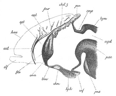
Fig. 255. Longitudinal vertical section through the anterior part of the brain of an embryo rabbit of four centimetres. (After Mihalkovics.) The section passes through the median line so that the cerebral hemispheres are not cut; their position is however indicated in outline. spt. septum lucidum formed by the coalescence of the inner walls of part of the cerebral hemispheres; cna. anterior commissure; frx. vertical pillars of the fornix; cal. genu of corpus callosum; trm. lamina terminalis; hms. cerebral hemispheres; olf. olfactory lobes; acl. artery of corpus callosum; fmr. position of foramen of Munro; chd3. choroid plexus of third ventricle; pin. pineal gland; cmp. posterior commissure; bgm. lamina uniting the lobes of the mid-brain; chm. optic chiasma; hph. pituitary body; inf. infundibulum; pns. pons Varolii; pde. cerebral peduncles; agd. iter.
In Elasmobranchii the pineal gland becomes in time very long, and extends far forwards over the roof of the cerebral hemispheres (fig. 254 pn). Its distal extremity dilates somewhat, and in the adult the whole organ forms (Ehlers, No. 337) an elongated tube, enlarged at its free extremity, and opening at its base into the brain. The enlarged extremity may either be lodged in a cavity in the cartilage of the cranium (Acanthias), or be placed outside the cranium (Raja).
In Petromyzon its form is very different. It arises (fig. 253 pn) as a sack-like diverticulum of the thalamencephalon extending at first both backwards and forwards. In the Ammocœte the walls of this sack are deeply infolded.
The embryonic form of the pineal gland in Amphibia is very much like that which remains permanent in Elasmobranchii; the stalk connecting the enlarged terminal portion with the brain soon however becomes solid and very thin except at its proximal extremity. The enlarged portion also becomes solid, and is placed in the adult externally to the skull, where it forms a mass originally described by Stieda as the cerebral gland.
In Birds the primitive outgrowth to form the pineal gland becomes, according to Mihalkovics, deeply indented by vascular connective tissue ingrowths, so that it assumes a dendritic structure (fig. 250 pin).
The proximal extremity attached to the roof of the thalamencephalon forms a special section, known as the infra-pineal process. The central lumen of the free part of the gland finally atrophies, but the branches still remain hollow. The infra-pineal process becomes reduced to a narrow stalk, connecting the branched portion of the body with the brain. The branched terminal portion and the stalk obviously correspond with the vesicle and distal part of the stalk of the types already described. In Mammalia the development of the pineal gland is, according to Mihalkovics, generally similar to that of Birds. The original outgrowth becomes branched, but the follicles or lobes to which the branching gives rise eventually become solid (fig. 255 pin). An infra-pineal process is developed comparatively late, and is not sharply separated from the roof of the brain.
No satisfactory suggestions have yet been offered as to the nature of the pineal gland, unless the view of Götte be regarded as such. It appears to possess in all forms an epithelial structure, but, except at the base of the stalk (infra-pineal process) in Mammalia, in the wall of which there are nerve-fibres, no nervous structures are present in it in the adult state.
The pituitary body. Although the pituitary body is not properly a nervous structure, yet from its intimate connection with the brain it will be convenient to describe its development here. The pituitary body is in fact an organ derived from the epiblast of the stomodæum. This fact has been demonstrated for Mammalia, Aves, Amphibia and Elasmobranchii, and may be accepted as holding good for all the Craniata[166]. The epiblast in the angle formed by the cranial flexure becomes involuted to form the cavity of the mouth. This cavity is bordered on its posterior surface by the front wall of the alimentary tract, and on its anterior by the base of the fore-brain. Its uppermost end does not at first become markedly constricted off from the remainder, but is nevertheless the rudiment of the pituitary body.
Fig. 256 represents a transverse section through the head of an Elasmobranch embryo, in which, owing to the cranial flexure, the fore part of the head is cut longitudinally and horizontally, and the section passes through both the fore-brain (fb) and the hind-brain. Close to the base of the fore-brain are seen the mouth (m), and the pituitary involution from this (pt). In contact with the pituitary involution is the blind anterior termination of the throat (al) which a little way back opens to the exterior by the first visceral cleft (1 v.c.). This figure alone suffices to demonstrate the correctness of the above account of the pituitary body; but its truth is still further confirmed by fig. 252; in which the mouth involution (pt) is in contact with, but still separated from, the front end of the alimentary tract. Very shortly after the septum between the mouth and throat becomes pierced, and the two are placed in communication, the pituitary involution becomes very partially constricted off from the mouth involution, though still in direct communication with it. In later stages the pituitary involution becomes longer and is dilated terminally; while the passage connecting it with the mouth becomes narrower and narrower, and is finally reduced to a solid cord, which in its turn disappears.
Before the connection between the pituitary vesicle and the mouth is obliterated the cartilaginous cranium becomes developed, and it may then be seen that the infundibulum projects through the pituitary space to come into close juxtaposition with the pituitary body.
After the pituitary vesicle has lost its connection with the mouth it lies just in front of the infundibulum (figs. 250 and 255 hph and fig. 254 pt); and soon becomes surrounded by vascular mesoblast, which grows in and divides it into a number of branching tubes. In many forms the cavity of the vesicle completely disappears, and the branches become for the most part solid [Cyclostomata and some Mammalia (the rabbit), Elasmobranchii, Teleostei and Amphibia]. In Reptilia, Aves and most Mammalia the lumen of the organ is more or less retained (W. Müller, No. 344).
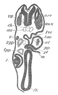
Fig. 256. Transverse section through the front part of the head of a young Pristiurus embryo. The section, owing to the cranial flexure, cuts both the fore- and the hind-brain. It shews the premandibular and mandibular head cavities 1pp and 2pp, etc. The section is moreover somewhat oblique from side to side. fb. fore-brain; l. lens of eye; m. mouth; pt. upper end of mouth, forming pituitary involution; 1ao. mandibular aortic arch; 1pp. and 2pp. first and second head cavities; 1vc. first visceral cleft; V. fifth nerve; aun. auditory nerve; VII. seventh nerve; aa. roots of dorsal aorta; acv. anterior cardinal vein; ch. notochord.
Although in the majority of the Vertebrata there is a close connection between the pituitary body and the infundibulum, there is no actual fusion between the two. In Mammalia the case is different. The part of the infundibulum which lies at the hinder end of the pituitary body is at first a simple finger-like process of the brain (fig. 255 inf), but its end becomes swollen, and the lumen in this part becomes obliterated. Its cells, originally similar to those of the other parts of the nervous system and even (Kölliker) containing differentiated nerve-fibres, partly atrophy, and partly assume an indifferent form, while at the same time there grow in amongst them numerous vascular and connective-tissue elements. The process of the infundibulum thus metamorphosed becomes inseparably connected with the true pituitary body, of which it is usually described as the posterior lobe. The part of the infundibulum which undergoes this change is very probably homologous with the saccus vasculosus of Fishes.
The true nature of the pituitary body has not yet been made out. It is clearly a rudimentary organ in existing craniate Vertebrates, and its development indicates that when functional it was probably a sense organ opening into the mouth, its blind end reaching to the base of the brain. No similar organ has as yet been found in Amphioxus, but it seems possible perhaps to identify it with the peculiar ciliated sack placed at the opening of the pharynx in the Tunicata, the development of which was described at p. 18. If the suggestion is correct, the division of the body into lobes in existing Vertebrata must be regarded as a step towards a retrogressive metamorphosis.
Another possible view is to regard the pituitary body as a glandular structure which originally opened into the mouth in the lower Chordata, but which has in all existing forms ceased to be functional. The intimate relation of the organ to the brain appears to me opposed to this view of its nature, while on the other hand its permanent structure is more easily explained on this view than on that previously stated. In the Ascidians a glandular organ has been described by Lacaze Duthiers[167] in juxtaposition to the ciliated sack, and it is possible that this organ as well as the ciliated sack may be related to the pituitary body. In view of this possibility further investigations ought to be carried out in order to determine whether the whole pituitary body is derived from the oral involution, or whether there may not be a nervous part and a glandular part of the organ.
The Cerebral Hemispheres. It will be convenient to treat separately the development of the cerebral hemispheres proper, and that of the olfactory lobes.
Although the cerebral hemispheres vary more than any other part of the brain, they are nevertheless developed from the unpaired cerebral rudiment in a nearly similar manner throughout the series of Vertebrata.
In the cerebral rudiment two parts may be distinguished, viz. the floor and the roof. The former gives rise to the ganglia at the base of the hemispheres—corpora striata, etc.—the latter to the hemispheres proper.
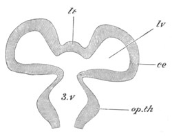
Fig. 257. Diagrammatic longitudinal horizontal section through the fore-brain. 3.v. third ventricle; lv. lateral ventricle; lt. lamina terminalis; ce. cerebral hemisphere; op.th. optic thalamus.
The first change which takes place consists in the roof growing out into two lobes, between which a shallow median constriction makes its appearance (fig. 257). The two lobes thus formed are the rudiments of the two hemispheres. The cavity of each of them opens by a widish aperture into the vestibule at the base of the cerebral rudiment, which again opens directly into the cavity of the third ventricle (3 v). The Y-shaped aperture thus formed, which leads from the cerebral hemispheres into the third ventricle, is the foramen of Munro. The cavity (lv) in each of the rudimentary hemispheres is a lateral ventricle. The part of the cerebrum which lies between the two hemispheres, and passes forwards from the roof of the third ventricle round the end of the brain to the optic chiasma, is the rudiment of the lamina terminalis (figs. 257 lt and 255 trm). Up to this point the development of the cerebrum is similar in all Vertebrata, but in some forms it practically does not proceed much further.
In Elasmobranchii, although the cerebrum reaches a considerable size (fig. 254 cer), and grows some way backwards over the thalamencephalon, yet it is not in many forms divided into two distinct lobes, but its paired nature is only marked by a shallow constriction on the surface. The lamina terminalis in the later stages of development grows backwards as a thick median septum which completely separates the two lateral ventricles[168] (fig. 263).
There are, it may be mentioned, considerable variations in the structure of the cerebrum in Elasmobranchii into which it is not however within the scope of this work to enter.
In the Teleostei the vesicles of the cerebral hemispheres appear at first to have a wide lumen, but it subsequently becomes almost or quite obliterated, and the cerebral rudiment forms a small bilobed nearly solid body. In Petromyzon (fig. 253 ch) the cerebral rudiment is at first an unpaired anterior vesicle, which subsequently becomes bilobed in the normal manner. The walls of the hemispheres become much thickened, but the lateral ventricles persist.
In all the higher Vertebrates the division of the cerebral rudiment into two distinct hemispheres is quite complete, and with the deepening of the furrow between the two hemispheres the lamina terminalis is carried backwards till it forms a thin layer bounding the third ventricle anteriorly, while the lateral ventricles open directly into the third ventricle.
In Amphibians the two hemispheres become united together immediately in front of the lamina terminalis by commissural fibres, forming the anterior commissure. They also send out anteriorly two solid prolongations, usually spoken of as the olfactory lobes, which subsequently fuse together.
In all Reptilia and Aves there is formed an anterior commissure, and in the higher members of the group, especially Aves (fig. 250), the hemispheres may obtain a considerable development. Their outer walls are much thickened, while their inner walls become very thin; and a well-developed ganglionic mass, equivalent to the corpus striatum, is formed at their base.
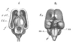
Fig. 258. Brain of a three months’ human embryo: natural size. (From Kölliker.) 1. From above with the dorsal part of hemispheres and mid-brain removed; 2. From below. f. anterior part of cut wall of the hemisphere; f´. cornu ammonis; tho. optic thalamus; cst. corpus striatum; to. optic tract; cm. corpora mammillaria; p. pons Varolii.
The cerebral hemispheres undergo in Mammalia the most complicated development. The primitive unpaired cerebral rudiment becomes, as in lower Vertebrates, bilobed, and at the same time divided by the ingrowth of a septum of connective tissue into two distinct hemispheres (figs. 260 and 261 f and 258 1). From this septum is formed the falx cerebri and other parts.
The hemispheres contain at first very large cavities, communicating by a wide foramen of Munro with the third ventricle (fig. 260). They grow rapidly in size, and extend, especially backwards, and gradually cover the thalamencephalon and the mid-brain (fig. 258 1, f). The foramen of Munro becomes very much narrowed and reduced to a mere slit.
The walls are originally nearly uniformly thick, but the floor becomes thickened on each side, and gives rise to the corpus striatum (figs. 260 and 261 st). The corpus striatum projects upwards into each lateral ventricle, giving to it a somewhat semilunar form, the two horns of which constitute the permanent anterior and descending cornua of the lateral ventricles (fig. 262 st).
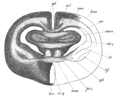
Fig. 259. Transverse section through the brain of a rabbit of five centimetres. (After Mihalkovics.) The section passes through nearly the posterior border of the septum lucidum, immediately in front of the foramen of Munro. hms. cerebral hemispheres; cal. corpus callosum; amm. cornu ammonis (hippocampus major); cms. superior commissure of the cornua ammonis; spt. septum lucidum; frx 2. vertical fibres of the fornix; cma. anterior commissure; trm. lamina terminalis; str. corpus striatum; ltf. nucleus lenticularis of corpus striatum; vtr 1. lateral ventricle; vtr 3. third ventricle; ipl. slit between cerebral hemispheres.
With the further growth of the hemisphere the corpus striatum loses its primitive relations to the descending cornu. The reduction in size of the foramen of Munro above mentioned is, to a large extent, caused by the growth of the corpora striata.
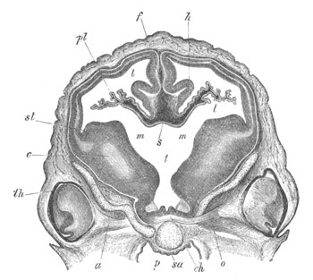
Fig. 260. Transverse section through the brain of a sheep’s embryo of 2.7 cm. in length. (From Kölliker.) The section passes through the level of the foramen of Munro. st. corpus striatum; m. foramen of Munro; t. third ventricle; pl. choroid plexus of lateral ventricle; f. falx cerebri; th. anterior part of optic thalamus; ch. optic chiasma; o. optic nerve; c. fibres of the cerebral peduncles; h. cornu ammonis; p. pharynx; sa. presphenoid bone; a. orbitosphenoid bone; s. points to part of the roof of the brain at the junction between the roof of the third ventricle and the lamina terminalis; l. lateral ventricle.
The corpora striata are united at their posterior border with the optic thalami. In the later stages of development the area of contact between these two pairs of ganglia increases to an immense extent (fig. 261), and the boundary between them becomes somewhat obscure, so that the sharp distinction which exists in the embryo between the thalamencephalon and cerebral hemispheres becomes lost. This change is usually (Mihalkovics, Kölliker) attributed to a fusion between the corpora striata and optic thalami, but it has recently been attributed by Schwalbe (No. 349), with more probability, to a growth of the original surface of contact, and an accompanying change in the relations of the parts.
The outer wall of the hemispheres gradually thickens, while the inner wall becomes thinner. In the latter, two curved folds, projecting towards the interior of the lateral ventricle, become formed. These folds extend from the foramen of Munro along nearly the whole of what afterwards becomes the descending cornu of the lateral ventricle.
The upper fold becomes the hippocampus major (cornu ammonis) (figs. 259 amm, 260 and 261 h, and 262 am). When the rudiment of the descending cornu has become transformed into a simple process of the lateral ventricle the hippocampus major forms a prominence upon its floor.
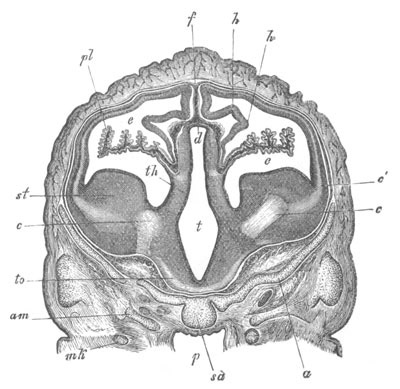
.
Fig. 261. Transverse section through the brain of a sheep’s embryo of 2.7 cm. in length. (From Kölliker.) The section is taken a short distance behind the section represented in fig. 260, and passes through the posterior part of the hemispheres and the third ventricle. st. corpus striatum; th. optic thalamus; to. optic tract; t. third ventricle; d. roof of third ventricle; c. fibres of cerebral peduncles; c´. divergence of these fibres into the walls of the hemispheres; e. lateral ventricle with choroid plexus pl; h cornu ammonis; f. primitive falx; am. alisphenoid; a. orbitosphenoid; sa. presphenoid; p. pharynx; mk. Meckel’s cartilage.
The wall of the lower fold becomes very thin, and a vascular plexus, derived from the connective-tissue septum between the hemispheres, and similar to that of the roof of the third ventricle, is formed outside it. It constitutes a fold projecting far into the cavity of the lateral ventricle, and together with the vascular connective tissue in it gives rise to the choroid plexus of the lateral ventricle (figs. 260 and 261 pl).
It is clear from the above description that a marginal fissure leading into the cavity of the lateral ventricle does not exist in the sense often implied in works on human anatomy, in that the epithelium covering the choroid plexus, which forms the true wall of the brain, is a continuous membrane. The epithelium of the choroid plexus of the lateral ventricle is quite independent of that of the choroid plexus of the third ventricle, though at the foramen of Munro the roof of the third ventricle is of course continuous with the inner wall of the lateral ventricle (fig. 260 s). The vascular elements of the two plexuses form however a continuous structure.
The most characteristic parts of the Mammalian cerebrum are the commissures connecting the two hemispheres. These commissures are (1) the anterior commissure, (2) the fornix, and (3) the corpus callosum, the two latter being peculiar to Mammalia.
By the fusion of the inner walls of the hemispheres in front of the lamina terminalis a solid septum is formed, known as the septum lucidum, continuous behind with the lamina terminalis, and below with the corpora striata (figs. 255 and 259 spt). It is by a series of differentiations within this septum that the above commissures originate. In Man there is a closed cavity left in the septum known as the fifth ventricle, which has however no communication with the true ventricles of the brain.
In the septum lucidum there become first formed, below, the transverse fibres of the anterior commissure (fig. 255 and fig. 259 cma), and in the upper part the vertical fibres of the fornix (fig. 255 and fig. 259 frx 2). The vertical fibres meet above the foramen of Munro, and thence diverge backwards, as the posterior pillars, to lose themselves in the cornu ammonis (fig. 259 amm). Ventrally they are continued, as the descending or anterior pillars of the fornix, into the corpus albicans, and thence into the optic thalami.
The corpus callosum is not formed till after the anterior commissure and fornix. It arises in the upper part of the region (septum lucidum) formed by the fusion of the lateral walls of the hemispheres (figs. 255 and 259 cal), and at first only its curved anterior portion—the genu or rostrum—is developed. This portion is alone found in Monotremes and Marsupials. The posterior portion, which is present in all the Monodelphia, is gradually formed as the hemispheres are prolonged further backwards.
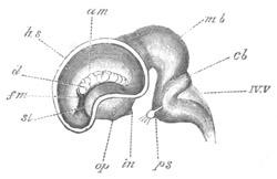
Fig. 262. Lateral view of the brain of a calf embryo of 5 cm. (After Mihalkovics.) The outer wall of the hemisphere is removed, so as to give a view of the interior of the left lateral ventricle. hs. cut wall of hemisphere; st. corpus striatum; am. hippocampus major (cornu ammonis); d. choroid plexus of lateral ventricle; fm. foramen of Munro; op. optic tract; in. infundibulum; mb. mid-brain; ch. cerebellum; IV.V. roof of fourth ventricle; ps. pons Varolii, close to which is the fifth nerve with Gasserian ganglion.
Primitively the Mammalian cerebrum, like that of the lower Vertebrata, is quite smooth. In many of the Mammalia, Monotremata, Insectivora, etc., this condition is nearly retained through life, while in the majority of Mammalia a more or less complicated system of fissures is developed on the surface. The most important, and first formed, of these is the Sylvian fissure. It arises at the time when the hemispheres, owing to their growth in front of and behind the corpora striata, have assumed a somewhat bean-shaped form. At the root of the hemispheres—the hilus of the bean—there is formed a shallow depression, which constitutes the first trace of the Sylvian fissure. The part of the brain lying in this fissure is known as the island of Reil.
The olfactory lobes. The olfactory lobes, or rhinencephala, are secondary outgrowths of the cerebral hemispheres, and contain prolongations of the lateral ventricles, but may however be solid in the adult state. According to Marshall they develop in Birds and Elasmobranchs and presumably other forms later than the olfactory nerves, so that the olfactory region of the hemispheres is indicated before the appearance of the olfactory lobes.
In most Vertebrates the olfactory lobes arise at a fairly early stage of development from the under and anterior part of the hemispheres (fig. 250 olf). In Elasmobranchs they arise, not from the base, but from the lateral parts of the brain (fig. 263), and become subsequently divided into a bulbous portion and a stalk. They vary considerably in their structure in the adult.
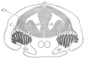
Fig. 263. Section through the brain and olfactory organ of an embryo of Scyllium. (Modified from figures by Marshall and myself.) ch. cerebral hemispheres; ol.v. olfactory vesicle; olf. olfactory pit; Sch. Schneiderian folds; I. olfactory nerve. The reference line has been accidentally taken through the nerve to the brain; pn. anterior prolongation of pineal gland.
In Amphibia the solid anterior prolongations of the cerebral hemispheres already spoken of are usually regarded as the olfactory lobes, but according to Götte, whose view appears to me well founded, small papillæ, situated at the base of these prolongations, from which olfactory nerves spring, and which contain a process of the lateral ventricle, should properly be regarded as the olfactory lobes. These papillæ arise prior to the solid anterior prolongations of the hemispheres.
In Birds the olfactory lobes are small. In the chick they arise (Marshall) on the seventh day of incubation.
General conclusions as to the Central Nervous System.
It has been shewn above that both the brain and spinal cord are primitively composed of a uniform wall of epithelial cells, and that the first differentiation results in the formation of an external layer of white matter, a middle layer of grey matter (ganglion cells), and an inner epithelial layer. This primitive histological arrangement, which in many parts of the brain at any rate, is only to be observed in the early developmental stages, has a simple phylogenetic explanation.
As has been already explained in an earlier part of this chapter the central nervous system was originally a differentiated part of the superficial epidermis.
This differentiation (as may be concluded from the character of the nervous system in the Cœlenterata and Echinodermata) consisted in the conversion of the inner ends of the epithelial cells into nerve-fibres; that is to say, that the first differentiation resulted in the formation of a layer of white matter on the inner side of the epidermis. The next stage was the separation of a deeper layer of the epidermis as a layer of ganglion cells from the superficial epithelial layer, i.e. the formation of a middle layer of ganglion cells and an outer epithelial layer. Thus, phylogenetically, the same three layers as those which first make their appearance in the ontogeny of the vertebrate nervous system became successively differentiated, and in both cases they are clearly placed in the same positions, because the central canal of the vertebrate nervous system, as formed by an involution, is at the true outer surface, and the external part of the cord is at the true inner surface.
It is probable that a very sharp distinction between the white and grey matter is a feature acquired in the higher Vertebrata, since in Amphioxus there is no such sharp separation; though the nerve-fibres are mainly situated externally and the nerve-cells internally.
As already stated in Chapter XII. the primitive division of the nervous axis was probably not into brain and spinal cord, but into (1) a fore-brain, representing the ganglion of the præoral lobe, and (2) the posterior part of the nervous axis, consisting of the mid- and hind-brains and the spinal cord. This view of the division of the central nervous system fits in fairly satisfactorily with the facts of development. The fore-brain is, histologically, more distinct from the posterior part of the nervous system than the posterior parts are from each other; the front end of the notochord forms the boundary between these two parts of the central nervous system (vide fig. 253), ending as it does at the front termination of the floor of the mid-brain, and finally, the nerves of the fore-brain have a different character to those of the mid- and hind-brain.
This primitive division of the central nervous system is lost in all the true Vertebrata, and in its place there is a secondary division—corresponding with the secondary vertebrate head—into a brain and spinal cord. The brain, as it is established in these forms, is again divided into a fore-brain, a mid-brain and a hind-brain. The fore-brain is, as we have already seen, the original ganglion of the præoral lobe. The mid-brain appears to be the lobe, or ganglion, of the third pair of nerves (first pair of segmental nerves), while the hind-brain is a more complex structure, each section of which (perhaps indicated by the constrictions which often appear at an early stage of development) giving rise to a pair of segmental nerves is, roughly speaking, homologous with the whole mid-brain.
The type of differentiation of each of the primitively simple vesicles forming the fore-, the mid- and the hind-brains is very uniform throughout the Vertebrate series, but it is highly instructive to notice the great variations in the relative importance of the parts of the brain in the different types. This is especially striking in the case of the fore-brain, where the cerebral hemispheres, which on embryological grounds we may conclude to have been hardly differentiated as distinct parts of the fore-brain in the most primitive types now extinct, gradually become more and more prominent, till in the highest Mammalia they constitute a more important section of the brain than the whole of the remaining parts put together.