Volume 2 — CHAPTER VIII. (continued 2)
Reading edition
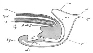
Fig. 124. Diagrammatic longitudinal section through the posterior end of an embryo Bird at the time of the formation of the Allantois. ep. epiblast; Sp.c. spinal canal; ch. notochord; n.e. neurenteric canal; hy. hypoblast; p.a.g. postanal gut; pr. remains of primitive streak folded in on the ventral side; al. allantois; me. mesoblast; an. point where anus will be formed; p.c. perivisceral cavity; am. amnion; so. somatopleure; sp. splanchnopleure.
Before dealing with the later history of the fœtal membranes, it will be convenient to complete the history of the yolk-sack.
Yolk-Sack. The origin of the area opaca has already been described. It rapidly extends over the yolk underneath the vitelline membrane; and is composed of epiblast and of the hypoblast of the germinal wall continuous with that of the area pellucida, which on the fourth day takes the form of a more or less complete layer of columnar cells[69]. Between the epiblast and hypoblast there is a layer of mesoblast, which does not extend as far as the two other layers. The yolk is completely surrounded by the seventh day.
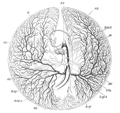
Fig. 125. Diagram of the circulation of the Yolk-Sack at the end of the third day of incubation. H. heart; AA. the second, third and fourth aortic arches; the first has become obliterated in its median portion, but is continued at its proximal end as the external carotid, and at its distal end as the internal carotid; AO. dorsal aorta; L.Of.A. left vitelline artery; R.Of.A. right vitelline artery; S.T. sinus terminalis; L.Of. left vitelline vein; R.Of. right vitelline vein; S.V. sinus venosus; D.C. ductus Cuvieri; S.Ca.V. superior cardinal vein; V.Ca. inferior cardinal vein. The veins are marked in outline and the arteries are black. The whole blastoderm has been removed from the egg and is supposed to be viewed from below. Hence the left is seen on the right, and vice versâ.
Towards the end of the first day blood-vessels begin to be developed in the inner part of the mesoblast of the area opaca. Their development is completed on the second day; and the region through which they extend is known as the area vasculosa. The area vasculosa also grows round the yolk, and completely encloses it not long after the area opaca. The part of the blastoderm which thus encloses the yolk forms the yolk-sack. The splitting of the mesoblast gradually extends to the mesoblast of the yolk-sack, and eventually the somatopleure of the sack, which is continuous, it will be remembered, with the outer limb of the amnion, separates completely from the splanchnopleure; and between the two the allantois inserts itself. These features are represented in fig. 121 E, K, and L.
The circulation of the yolk-sack is most important during the third day of incubation. The arrangement of the vessels during that day is shewn in fig. 125.
The blood leaving the body of the embryo by the vitelline arteries (fig. 125, R.Of.A, L.Of.A), which are branches of the dorsal aortæ, is carried to the small vessels and capillaries of the vascular area, a small portion only being appropriated by the pellucid area.
From the vascular area part of the blood returns directly to the sinus venosus by the main lateral trunks of the vitelline veins (R.Of, L.Of), and so to the heart. During the second day these venous trunks join the body of the embryo considerably in front of, that is nearer, the head than the corresponding arterial ones. Towards the end of the third day, owing to the continued lengthening of the heart, the veins and arteries run not only parallel to each other, but almost in the same line, the points at which they respectively join and leave the body being nearly at the same distance from the head.
The rest of the blood brought by the vitelline arteries finds its way into the lateral portions of a venous trunk bounding the vascular area, which is known as the sinus terminalis, S.T., and there divides on each side into two streams. Of these, the two which, one on either side, flow backward, meet at a point about opposite to the tail of the embryo, and are conveyed along a distinct vein which, running straight forward parallel to the axis of the embryo, empties itself into the left vitelline vein. The two forward streams reaching a gap in the front part of the sinus terminalis fall into either one, or in some cases two veins, which run straight backwards parallel to the axis of the embryo, and so reach the roots of the heart. When one such vein only is present it joins the left vitelline trunk; where there are two they join the left and right vitelline trunks respectively. The left vein is always considerably larger than the right; and the latter when present rapidly gets smaller and speedily disappears. After the third day, although the vascular area goes on increasing in size until it finally all but encompasses the yolk, the prominence of the sinus terminalis becomes less and less.
The fœtal membranes and the yolk-sack may conveniently be treated of together in the description of their later changes and final fate.
On the sixth and seventh days they exhibit changes of great importance.
The amnion, at its complete closure on the fourth day, very closely invested the body of the chick: the true cavity of the amnion was then therefore very small. On the fifth day fluid begins to collect in the cavity, and raises the membrane of the amnion to some distance from the embryo. The cavity becomes still larger by the sixth day, and on the seventh day is of very considerable dimensions, the fluid increasing with it. On the sixth day Von Baer observed movements of the embryo, chiefly of the limbs; he attributes them to the stimulation of the cold air on opening the egg. By the seventh day very obvious movements begin to appear in the amnion itself; slow vermicular contractions creeping rhythmically over it. The amnion in fact begins to pulsate slowly and rhythmically, and by its pulsation the embryo is rocked to and fro in the egg. This pulsation is probably due to the contraction of involuntary muscular fibres, which seem to be present in the attenuated portion of the mesoblast, forming part of the amniotic fold. Similar movements are also seen in the allantois at a considerably later period.
The growth of the allantois has been very rapid, and it forms a flattened bag, covering the right side of the embryo, and rapidly spreading out in all directions between the primitive folds of the amnion, that is, between the amnion proper and the false amnion or serous envelope. It is filled with fluid, so that in spite of its flattened form its opposite walls are distinctly separated from each other.
The vascular area has become still further extended than on the fifth day, but with a corresponding loss in the definite character of its blood-vessels. The sinus terminalis has indeed by the end of the seventh day lost all its previous distinctness; and the vessels which brought back the blood from it to the heart are no longer to be seen.
Both the vitelline arteries and veins now pass to and from the body of the chick as single trunks, assuming more and more the appearance of being merely branches of the mesenteric vessels.
The yolk is still more fluid than on the previous day, and its bulk has (according to von Baer) increased. This can only be due to its absorbing the white of the egg, which indeed is diminishing rapidly.
During the eighth, ninth, and tenth days, the amnion does not undergo any very important changes. Its cavity is still filled with fluid, and on the eighth day its pulsations are at their height, henceforward diminishing in intensity.
The splitting of the mesoblast has now extended to the outer limit of the vascular area, i.e. over about three-quarters of the yolk-sack. The somatopleure at this point is continuous (as can be easily seen by reference to fig. 121) with the original outer fold of the amnion. It thus comes about that the further splitting of the mesoblast merely enlarges the cavity in which the allantois lies. The growth of this organ keeps pace with that of the cavity in which it is placed. Spread out over the greater part of the yolk-sack as a flattened bag filled with fluid, it now serves as the chief organ of respiration. It is indeed very vascular and a marked difference may be observed between the colour of the blood in the outgoing and the returning vessels.
The yolk now begins to diminish rapidly in bulk. The yolk-sack becomes flaccid, and on the eleventh day is thrown into a series of internal folds, abundantly supplied by large venous trunks. By this means the surface of absorption is largely increased, and the yolk is more and more rapidly taken up by the blood-vessels, and in a partially assimilated condition transferred to the body of the embryo[70].
By the eleventh day the abdominal parietes, though still much looser and less firm than the walls of the chest, may be said to be definitely established; and the loops of intestine, which have hitherto been hanging down into the somatic stalk, are henceforward confined within the cavity of the abdomen. The body of the embryo is therefore completed; but it still remains connected with its various appendages by a narrow somatic umbilicus, in which run the stalk of the allantois and the solid cord suspending the yolk-sack.
The cleavage of the mesoblast is still progressing, and the yolk is completely invested by a splanchnopleural sack.
The allantois meanwhile spreads out rapidly, and lies over the embryo close under the shell, being separated from the shell membrane by nothing more than the attenuated serous envelope, formed out of the outer primitive fold of the amnion and the remains of the vitelline membrane. With this membrane the allantois partially coalesces, and in opening an egg at the later stages of incubation, unless care be taken, the allantois is in danger of being torn in the removal of the shell-membrane. As the allantois increases in size and importance, the allantoic vessels are correspondingly developed.
On about the sixteenth day, the white having entirely disappeared, the cleavage of the mesoblast is carried right over the pole of the yolk opposite the embryo, and is thus completed (fig. 121). The yolk-sack now, like the allantois which closely wraps it all round, lies loose in a space bounded outside the body by the serous membrane, and continuous with the pleuroperitoneal cavity of the body of the embryo. Deposits of urates now become abundant in the allantoic fluid.
The loose and flaccid walls of the abdomen enclose a space which the empty intestines are far from filling, and on the nineteenth day the yolk-sack, diminished greatly in bulk but still of some considerable size, is withdrawn through the somatic stalk into the abdominal cavity, which it largely distends. Outside the embryo there now remains nothing but the highly vascular allantois and the bloodless serous membrane and amnion. The amnion, whose fluid during the later days of incubation rapidly diminishes, is continuous at the umbilicus with the body-walls of the embryo. The serous membrane (or outer primitive amniotic fold) is, by the completion of the cleavage of the mesoblast and the withdrawal of the yolk-sack, entirely separated from the embryo. The cavity of the allantois, by means of its stalk passing through the umbilicus, is of course continuous with the cloaca.
When the chick is about to be hatched it thrusts its beak through the egg-membranes and begins to breathe the air contained in the air chamber. Thereupon the pulmonary circulation becomes functionally active, and at the same time blood ceases to flow through the allantoic arteries. The allantois shrivels up, the umbilicus becomes completely closed, and the chick, piercing the shell at the broad end of the egg with repeated blows of its beak, casts off the dried remains of allantois, amnion and serous membrane, and steps out into the world.
Bibliography.
(117) K. E. von Baer. Ueb. Entwicklungsgeschichte d. Thiere. Königsberg, 1828-1837. (118) F. M. Balfour. “The development and growth of the layers of the Blastoderm,” and “On the disappearance of the Primitive Groove in the Embryo Chick.” Quart. J. of Micros. Science, Vol. XIII. 1873. (119) M. Braun. “Die Entwicklung d. Wellenpapagei’s.” Part I. Arbeit. d. zool.-zoot. Instit. Würzburg. Vol. V. 1879. (120) M. Braun. “Aus d. Entwick. d. Papageien; I. Rückenmark; II. Entwicklung d. Mesoderms; III. Die Verbindungen zwischen Rückenmark u. Darm bei Vögeln.” Verh. d. phys.-med. Ges. zu Würzburg. N. F. Bd. XIV. and XV. 1879 and 1880. (121) J. Disse. “Die Entwicklung des mittleren Keimblattes im Hühnerei.” Archiv für mikr. Anat., Vol. XV. 1878. (122) J. Disse. “Die Entstehung d. Blutes u. d. ersten Gefässe im Hühnerei.” Archiv f. mikr. Anat., Vol. XVI. 1879. (123) Fr. Durante. “Sulla struttura della macula germinativa delle uova di Gallina.” Ricerche nel Laboratorio di Anatomia della R. Università di Roma. (124) E. Dursy. Der Primitivstreif des Hühnchens. 1867. (125) M. Duval. “Etude sur la ligne primitive de l'embryon de Poulet.” Annales des Sciences Naturelles, Vol. VII. 1879. (126) M. Foster and F. M. Balfour. Elements of Embryology. Part I. London, 1874. (127) Gasser. “Der Primitivstreifen bei Vogelembryonen.” Schriften d. Gesell. zur Beförd. d. gesammten Naturwiss. zu Marburg, Vol. II. Supplement I. 1879. (128) A. Götte. “Beiträge zur Entwicklungsgeschichte d. Wirbelthiere. II. Die Bildung d. Keimblatter u. d. Blutes im Hühnerei.” Archiv für mikr. Anat., Vol. X. 1874. (129) V. Hensen. “Embryol. Mitth.” Archiv f. mikr. Anat., Vol. III. 1867. (130) W. His. Untersuch. üb. d. erste Anlage d. Wirbelthierleibes. Leipzig, 1868. (131) W. His. Unsere Körperform und das physiol. Problem ihrer Entstehung. Leipzig, 1875. (132) W. His. “Der Keimwall des Hühnereies u. d. Entstehung d. parablastischen Zellen.” Zeit. f. Anat. u. Entwicklungsgeschichte. Bd. I. 1876. (133) W. His. “Neue Untersuchungen üb. die Bildung des Hühnerembryo I.” Archiv f. Anat. u. Phys. 1877. (134) E. Klein. “Das mittlere Keimblatt in seiner Bezieh. z. Entwick. d. ers. Blutgefässe und Blutkörp. im Hühnerembryo.” Sitzungsber. Wien. Akad., Vol. LXIII. 1871. (135) A. Kölliker. Entwicklungsgeschichte d. Menschen u. d. höheren Thiere. Leipzig, 1879. (136) C. Kupffer. “Die Entsteh. d. Allantois u. d. Gastrula d. Wirbelth.” Zoolog. Anzeiger, Vol. II. 1879, pp. 520, 593, 612. (137) C. Kupffer and B. Benecke. “Photogramme z. Ontogenie d. Vögel.” Nov. Act. d. k. Leop.-Carol.-Deutschen Akad. d. Naturforscher, Vol. XLI. 1879. (138) J. Oellacher. “Untersuchungen über die Furchung u. Blatterbildung im Hühnerei.” Stricker’s Studien. 1870. (139) C. H. Pander. Beiträge z. Entwick. d. Hühnchens im Eie. Würzburg, 1817. (140) A. Rauber. “Ueber die Embryonalanlage des Hühnchens.” Centralblatt für d. medic. Wissenschaften. 1874-75. (141) A. Rauber. Ueber die Stellung des Hühnchens im Entwicklungsplan. 1876. (142) A. Rauber. “Primitivrinne und Urmund. Beiträge zur Entwicklungsgeschichte des Hühnchens.” Morphol. Jahrbuch, B. II. 1876. (143) A. Rauber. Primitivstreifen und Neurula der Wirbelthiere in normaler und pathologischer Beziehung. 1877. (144) R. Remak. Untersuch. üb. d. Entwicklung d. Wirbelthiere. Berlin, 1850-55. (145) S. L. Schenk. “Beiträge z. Lehre v. d. Organanlage im motorischen Keimblatt.” Sitz. Wien. Akad., Vol. LVII. 1860. (146) S. L. Schenk. “Beiträge z. Lehre v. Amnion.” Archiv f. mikr. Anat., Vol. VII. 1871. (147) S. L. Schenk. Lehrbuch d. vergleich. Embryol. d. Wirbelthiere. Wien, 1874. (148) S. Stricker. “Mittheil. üb. d. selbstständigen Bewegungen embryonaler Zellen.” Sitz. Wien. Akad., Vol. XLIX. 1864. (149) S. Stricker. “Beiträge zur Kenntniss des Hühnereies.” Wiener Sitzungsber., Vol. LIV. 1866. (150) H. Virchow. Ueber d. Epithel d. Dottersackes im Hühnerei. Inaug. Diss. Berlin, 1875. (151) W. Waldeyer. “Ueber die Keimblätter und den Primitivstreifen bei der Entwicklung des Hühnerembryo.” Zeitschrift für rationelle Medicin. 1869. (152) C. F. Wolff. Theoria generationis. Halæ, 1759. (153) C. F. Wolff. Ueb. d. Bildung d. Darmcanals im bebrüteten Hühnchen. Halle, 1812.