Volume 2 / Part 6 — APPENDICES.
Reading edition
APPENDIX A. SUBSTITUTION OF AXIAL FOR FOLIAR ORGANS IN PLANTS.
I append here the evidences referred to in § 190. The most numerous and striking I have met with among the Umbelliferæ. Monstrosities having the alleged implication, are frequent in the common Cow-Parsnep—so frequent that they must be familiar to botanists; and wild Angelica supplies many over-developments of like meaning. Omitting numerous cases of more or less significance, I will limit myself to two.
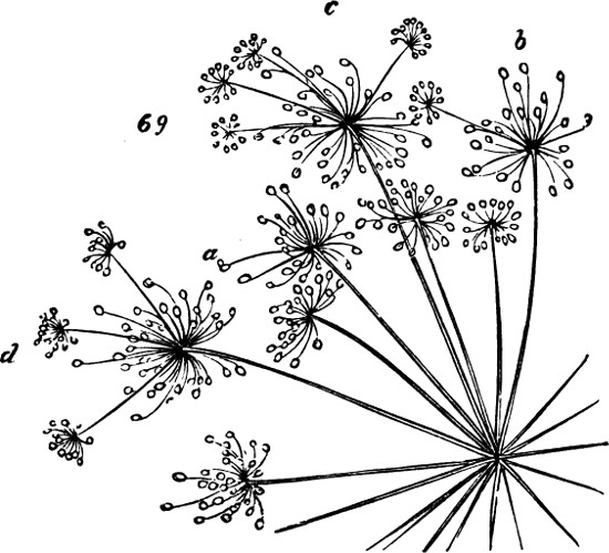
Fig. 69.
One of them is that of a terminal umbel, in which nine of the outer umbellules are variously transformed—here a single flower being made monstrous by the development of some of its members into buds; there several such malformed flowers being associated with rays that bear imperfect umbellules; and elsewhere, flowers being replaced by umbellules: some of which are perfect, and others imperfect only in the shortness of the flower-stalks. The annexed Fig. 69, representing in a somewhat conventionalized way, a part of the dried specimen, will give an idea of this Angelica. At a is shown a single flower partially changed; in the umbellule marked b, one of the rays bears a secondary umbellule; and there may be seen at c and d, several such over-developments.
But the most conclusive instance is that of a Cow-Parsnep, in which a single terminal umbel, besides the transformations already mentioned, exhibits higher degrees of such transformations.[68] The components of this complex growth are;—three central umbellules, abnormal only in minor points; one umbellule, external to these, which is partially changed into an umbel; one rather more out of the centre, which is so far metamorphosed as to be more an umbel than an umbellule: nine peripheral clusters formed by the development of umbellules into umbels, some of which are partially compounded still further. Examined in detail, these structures present the following facts:—1. The innermost umbellule is normal, save in having a peripheral flower of which one member (apparently a petal) is transformed into a flower-bud. 2. The next umbellule, not quite so central, has one of its peripheral flowers made monstrous by the growth of a bud from the base of the calyx. 3. The third of the central umbellules has two abnormal outer flowers. One of them carries a flower-bud on its edge, in place of a foliar member. The other is half flower and half umbellule: being composed of three petals, three stamens, and five flower-buds growing where the other petals and stamens should grow. 4. Outside of these umbellules comes one of the mixed clusters. Its five central flowers are normal. Surrounding these are several flowers transformed in different degrees: one having a stamen partially changed into a flower bud. And then, at the periphery of this mixed cluster, come three complete umbellules and an incomplete one in which some petals and stamens of the original flower remain. 5. A mixed cluster, in which the umbel-structure predominates, stands next. Its three central flowers are normal. Surrounding them are five flowers over-developed in various ways, like those already described. And on its periphery are seven complete umbellules in place of flowers; besides an incomplete umbellule that contains traces of the original flower, one of them being a petal imperfectly twisted up into a bud. 6. Of the nine external clusters, in which the development of simple into compound umbels is most decided, nearly all present anomalies. Three of them have each a central flower untransformed; and in others, the central umbellule is composed of two, three, or four flowers. 7. But the most remarkable fact is, that in sundry of these peripheral clusters, resulting from the metamorphosis of simple umbels into compound umbels, the like metamorphosis is carried a stage higher. Some of the component rays, are themselves the bearers of compound umbels instead of simple umbels. In Fig. 70, a portion of the dried specimen is represented. Two of the central umbellules are marked a and b; those marked c and d are mixed clusters; at e and f are compound umbels replacing simple ones; and g shows one of the rays on which the over-development goes still further.
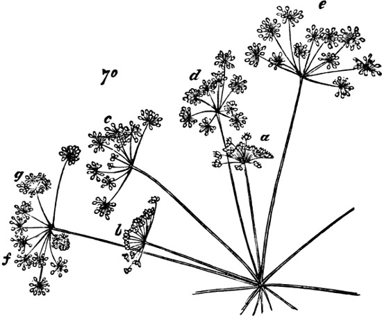
Fig. 70.
Does not this evidence, enforced as it is by much more of like kind, go far to prove that foliar organs may be developed into axial organs? Even were not the transitional forms traceable, there would still, I think, be no other legitimate interpretation of the facts last detailed. The only way of eluding the conclusion here drawn, is by assuming that where a cluster of flowers replaces a single flower, it is because the axillary buds which hypothetically belong to the several foliar organs of the flower, become developed into axes; and assuming this, is basing an hypothesis on another hypothesis that is directly at variance with facts. The foliar organs of flowers do not bear buds in their axils; and it would never have been supposed that such buds are typically present, had it not been for that mistaken conception of “type” which has led to many other errors in Biology. Goethe writes: “Now as we cannot realize the idea of a leaf apart from the node out of which it springs, or of a node without a bud, we may venture to infer,” &c. See here an example of a method of philosophizing not uncommon among the Germans. The method is this—Survey a portion of the facts, and draw from them a general conception; project this general conception back into the objective world, as a mould in which Nature casts her products; expect to find it everywhere fulfilled; and allege potential fulfilment where no actual fulfilment is visible.
If instead of imposing our ideal forms on Nature, we are content to generalize the facts as Nature presents them, we shall find no warrant for the morphological doctrine above enunciated. The only conception of type justified by the logic of science, is—that correlation of parts which remains constant under all modifications of the structure to be defined. To ascertain this, we must compare all these modifications, and note what traits are common to them. On doing so with the successive segments of a phænogamic axis, we are brought to a conclusion widely different from that of Goethe. Axillary buds are almost universally absent from the cotyledons; they are habitually present in the axils of fully-developed leaves higher up the axis; they are often absent from leaves that are close to the flower; they are nearly always absent from the bracts; absent from the sepals; absent from the petals; absent from the stamens; absent from the carpels. Thus, out of eight leading forms which folia assume, one has the axillary bud and seven are without it. With these facts before us, it seems to me not difficult to “realize the idea” “of a node without a bud.” If we are not possessed by a foregone conclusion, the evidence will lead us to infer, that each node bears a foliar appendage and may bear an axillary bud.
Even, however, were it granted that the typical segment of a Phænogam includes an axillary bud, which must be regarded as always potentially present, no legitimate counter-interpretation of the monstrosities above described could thence be drawn. If when an umbellule is developed in place of a flower, the explanation is, that its component rays are axillary to the foliar organs of the flower superseded; we may fairly require that these foliar organs to which they are axillary, shall be shown. But there are none. In the last specimen figured, the inner rays of each such umbellule are without them; most of the outer rays are also without them; and in one cluster, only a single ray has a bract at its point of origin. There is a rejoinder ready, however: the foliar organs are said to be suppressed. Though Goethe could not “realize the idea” “of a node without a bud,” those who accept his typical form appear to find no difficulty in realizing the idea of an axillary bud without anything to which it is axillary. But letting this pass, suppose we ask what is the warrant for this assumed suppression. Axillary buds normally occur where the nutrition is high enough to produce fully-developed leaves; and when axillary buds are demonstrably present in flowers, they accompany foliar organs that are more leaf-like than usual—always greener if not always larger. That is to say, the normal and the abnormal axillary buds, are alike the concomitants of foliar organs coloured by that chlorophyll which habitually favours foliar development. How, then, can it be supposed that when, out of a flower there is developed a cluster of flower-bearing rays, the implied excess of nutrition causes the foliar organs to abort? It is true that very generally in a branched inflorescence, the bracts of the several flower-branches are very small (their smallness being probably due to that defective supply of certain chlorophyll-forming matters, which is the proximate cause of flowering); and it is true that, under these conditions, a flowering axis of considerable size, for the development of which chlorophyll is less needful, grows from the axil of a dwarfed leaf. But the inference that the foliar organ may therefore be entirely suppressed, seems to me irreconcilable with the fact, that the foliar organ is always developed to some extent before the axillary bud appears. Until it has been shown that in some cases a lateral bud first appears, and a foliar organ afterwards grows out beneath it, to form its axil, the conception of an axillary bud of which the foliar organ is suppressed, will remain at variance with the established truths of development.
The above originally formed a portion of § 190. I have transferred it to the Appendix, partly because it contains too much detail to render it fit for the general argument, and partly because the interpretations being open to some question, it seemed undesirable to risk compromising that argument by including them. The criticisms passed upon these interpretations have not, however, sufficed to convince me of their incorrectness. Unfortunately, I have since had no opportunity of verifying the above statements by microscopic examinations, as I had intended.
Though unable to enforce the inference drawn by further facts more minutely looked into, I may add some arguments based on facts that are well known. One of these is the fact that the so-called axillary bud is not universally axillary—is not universally seated in the angle made by the axis and an appended foliar organ. In certain plants the axillary bud is placed far above the node, half-way between it and the succeeding node. So that not only may a segment of a phæenogamic axis be without the axillary bud, but the axillary bud, when present, may be removed from that place in which, according to Goethe, it necessarily exists. Another fact not congruous with the current doctrine, is the common occurrence of “adventitious” buds—the buds that are put out from roots and from old stems or branches bare of leaves. The name under which they are thus classed, is meant to imply that they may be left out of consideration. Those, however, who have not got a theory to save by putting anomalies out of sight, may be inclined to think that the occurrence of buds where they are avowedly unconnected with nodes, and are axillary to nothing, tells very much against the assumption that every bud implies a node and a corresponding foliar organ. And they may also see that the development of these adventitious buds at places where there is excess of nutritive materials, favours the view above set forth. For if a bud thus arises at a place where it is not morphologically accounted for, simply because there happens to be at that place an abundance of unorganized protoplasm; then, clearly, it is likely that if the mass of protoplasm from which a leaf would usually arise, is greatly increased in mass by excess of nutrition, it may develop into an axis instead of a leaf.
Many years after this work was published, I discovered among my papers a memorandum which unfortunately I had overlooked, containing further evidence in support of the foregoing conclusion. With the omission of an error concerning the species of plant, I reproduce this memorandum just as it stood:—
“I found at Dieppe, July 1, 1860, in a garden near the sea a sample of cultivated wild flower (I thought it was grown as an ornamental flower) in which some of the single flowers of the umbel were developed into groups of flowers thus:—
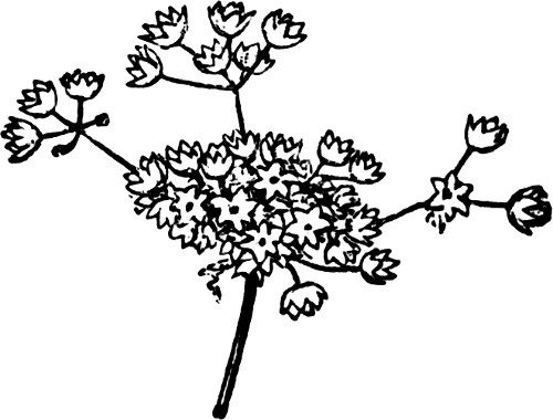
“In the case where the transformation was fully effected the umbellule had six flowers, answering to the six petals of the original flowers. In other cases the transformation was incomplete. There were instances where but two of the petals were developed into flowers; and the other petals remained unchanged. Others in which three were developed; and others where four were developed. In some cases, too, the development of a petal into a flower was imperfect, in the absence of the flower-stalk—the flowers were sessile in the place where the petals would have been. In one case there was an imperfect flower sessile; another imperfect flower on a short stalk; and three perfect flowers on long stalks.
“I was in some doubt whether the petals or the stamens were developed. In cases of imperfect transformation the petals at the base of the umbellule seemed to stand in the position of calyx or involucrum, giving the idea that the stamens were developed into flowers. But in the case where there were six flowers developed there were no petals at the base.
“That it was a matter of extra nutrition was shown by this:—
“1. That they were cultivated as garden flowers.
“2. That where there was one perfectly developed umbellule, it was the only one in the umbel.
“3. That where there were three umbellules they were all imperfect.
“4. That in this imperfect umbellule the perfect flowers were on long stalks and the imperfect ones sessile.
“5. That the umbellules were on stalks both longer and thicker than those of single flowers.”
[Concerning the foregoing argument at large an expert writes:—“The abnormalities you describe certainly show that an axis may arise abnormally in the place of a normal leaf-structure, and every modern botanist would be in agreement with you in your criticism of the older form of the doctrine of axillary buds. I think we are largely emancipated from the dextrous juggling with the arrangements and relations of organs which used to pass current as morphology.
“You have quoted sufficient evidence in the text (§ 190) to establish the conclusion that no sharp line can be drawn between axes and leaf-structure; and a very great deal more could be added in the same sense. Petioles for instance, exist which the most highly trained histological observer could not distinguish from stems.
“But I must demur to the suggestion that the replacement of one by the other is primarily a question of nutrition. We are as ignorant as ever of the proximate cause of the production of a leaf or a shoot at a certain spot in meristematic tissue.”
To this last remark I had at first made only the reply that the plants exhibiting the abnormalities were in all cases excessively luxuriant in their growths; but to this I am now able to add a more definite reply. The expert from whom I have just quoted, had read this appendix before there had been made to it the above addition describing the flower from Dieppe; and I was not myself aware, until I came to read over this addition, what clear evidence it contains that extra nutrition was the cause of these transformations of foliar structures into axial structures; but the above paragraphs 1, 2, 3, 4, 5, contain different evidences conspiring to prove this.]
APPENDIX B. A CRITICISM ON PROF. OWEN’S THEORY OF THE VERTEBRATE SKELETON.
[From the British & Foreign Medico-Chirurgical Review for Oct., 1858.]
I. On the Archetype and Homologies of the Vertebrate Skeleton. By Richard Owen, F.R.S.—London, 1848. pp. 172.
II. Principes d’Ostéologie Comparée, ou Recherches sur l’Archétype et les Homologies du Squelette Vertébré. Par Richard Owen.—Paris.
Principles of Comparative Osteology; or, Researches on the Archetype and the Homologies of the Vertebrate Skeleton. By Richard Owen.
III. On the Nature of Limbs. A Discourse delivered on Friday, February 9, at an Evening Meeting of the Royal Institution of Great Britain. By Richard Owen, F.R.S.—London, 1849. pp. 119.
Judging whether another proves his position is a widely different thing from proving your own. To establish a general law requires an extensive knowledge of the phenomena to be generalized; but to decide whether an alleged general law is established by the evidence assigned, requires merely an adequate reasoning faculty. Especially is such a decision easy where the premises do not warrant the conclusion. It may be dangerous for one who has but little previous acquaintance with the facts, to say that a generalization is demonstrated; seeing that the argument may be one-sided: there may be many facts unknown to him which disprove it. But it is not dangerous to give a negative verdict when the alleged demonstration is manifestly insufficient. If the data put before him do not bear out the inference, it is competent for every logical reader to say so.
From this standpoint, then, we venture to criticize some of Professor Owen’s osteological theories. For his knowledge of comparative osteology we have the highest respect. We believe that no living man has so wide and detailed an acquaintance with the bony structure of the Vertebrata. Indeed, there probably has never been any one whose information on the subject was so nearly exhaustive. Moreover, we confess that nearly all we know of this department of biology has been learnt from his lectures and writings. We pretend to no independent investigations, but merely to such knowledge of the phenomena as he has furnished us with. Our position, then, is such that, had Professor Owen simply enunciated his generalizations, we should have accepted them on his authority. But he has brought forward evidence to prove them. By so doing he has tacitly appealed to the judgments of his readers and hearers—has practically said, “Here are the facts; do they not warrant these conclusions?” And all we propose to do, is to consider whether the conclusions are warranted by the facts brought forward.
Let us first limit the scope of our criticisms. On that division of comparative osteology which deals with what Professor Owen distinguishes as “special homologies,” we do not propose to enter. That the wing of a bird is framed upon bones essentially parallel to those of a mammal’s fore-limb; that the cannon-bone of a horse’s leg answers to the middle metacarpal of the human hand; that various bones in the skull of a fish are homologous with bones in the skull of a man—these and countless similar facts, we take to be well established. It may be, indeed, that the doctrine of special homologies is at present carried too far. It may be that, just as the sweeping generalization at one time favoured, that the embryonic phases of the higher animals represent the adult forms of lower ones, has been found untrue in a literal sense, and is acceptable only in a qualified sense; so the sweeping generalization that the skeletons of all vertebrate animals consist of homologous parts, will have to undergo some modification. But that this generalization is substantially true, all comparative anatomists agree.
The doctrine which we are here to consider, is quite a separate one—that of “general homologies.” The truth or falsity of this may be decided on quite apart from that of the other. Whether certain bones in one vertebrate animal’s skeleton correspond with certain bones in another’s, or in every other’s, is one question; and whether the skeleton of every vertebrate animal is divisible into a series of segments, each of which is modelled after the same type, is another question. While the first is answered in the affirmative, the last may be answered in the negative; and we propose to give reasons why it should be answered in the negative.
In so far as his theory of the skeleton is concerned, Professor Owen is an avowed disciple of Plato. At the conclusion of his Archetype and Homologies of the Vertebrate Skeleton, he quotes approvingly the Platonic hypothesis of ἰδέαι, “a sort of models, or moulds in which matter is cast, and which regularly produce the same number and diversity of species.” The vertebrate form in general (see diagram of the Archetypus), or else the form of each kind of vertebrate animal (see p. 172, where this seems implied), Professor Owen conceives to exist as an “idea”—an “archetypal exemplar on which it has pleased the Creator to frame certain of his living creatures.” Whether Professor Owen holds that the typical vertebra also exists as an “idea,” is not so certain. From the title given to his figure of the “ideal typical vertebra,” it would seem that he does; and at p. 40 of his Nature of Limbs, and indeed throughout his general argument, this supposition is implied. But on the last two pages of the Archetype and Homologies, it is distinctly alleged that “the repetition of similar segments in a vertebral column, and of similar elements in a vertebral segment, is analogous to the repetition of similar crystals as the result of polarizing force in the growth of an inorganic body;” it is pointed out that, “as we descend the scale of animal life, the forms of the repeated parts of the skeleton approach more and more to geometrical figures;” and it is inferred that “the Platonic ἰδέα or specific organizing principle or force, would seem to be in antagonism with the general polarizing force, and to subdue and mould it in subserviency to the exigencies of the resulting specific form.” If Professor Owen’s doctrine is to be understood as expressed in these closing paragraphs of his Archetype and Homologies—if he considers that “the ἰδέα” “which produces the diversity of form belonging to living bodies of the same materials,” is met by the “counter-operation” of “the polarizing force pervading all space,” which produces “the similarity of forms, the repetition of parts, the signs of unity of organization,” and which is “subdued” as we ascend “in the scale of being;” then we may pass on with the remark that the hypothesis is too cumbrous and involved to have much vraisemblance. If, on the other hand, Professor Owen holds, as every reader would suppose from the general tenor of his reasonings, that not only does there exist an archetypal or ideal vertebrate skeleton, but that there also exists an archetypal or ideal vertebra; then he carries the Platonic hypothesis much further than Plato does. Plato’s argument, that before any species of object was created it must have existed as an idea of the Creative Intelligence, and that hence all objects of such species must be copies of this original idea, is tenable enough from the anthropomorphic point of view. But while those who, with Plato, think fit to base their theory of creation upon the analogy of a carpenter designing and making a table, must yield assent to Plato’s inference, they are by no means committed to Professor Owen’s expansion of it. To say that before creating a vertebrate animal, God must have had the conception of one, does not involve saying that God gratuitously bound himself to make a vertebrate animal out of segments all moulded after one pattern. As there is no conceivable advantage in this alleged adhesion to a fundamental pattern—as, for the fulfilment of the intended ends, it is not only needless, but often, as Professor Owen argues, less appropriate than some other construction would be (see Nature of Limbs, pp. 39, 40), to suppose the creative processes thus regulated, is not a little startling. Even those whose conceptions are so anthropomorphic as to think they honour the Creator by calling him “the Great Artificer,” will scarcely ascribe to him a proceeding which, in a human artificer, they would consider a not very worthy exercise of ingenuity.
But whichever of these alternatives Professor Owen contends for—whether the typical vertebra is that more or less crystalline figure which osseous matter ever tends to assume in spite of “the ἰδέα or organizing principle,” or whether the typical vertebra is itself an “ἰδέα or organizing principle”—there is alike implied the belief that the typical vertebra has an abstract existence apart from actual vertebræ. It is a form which, in every endo-skeleton, strives to embody itself in matter—a form which is potentially present in each vertebra; which is manifested in each vertebra with more or less clearness; but which, in consequence of antagonizing forces, is nowhere completely realized. Apart from the philosophy of this hypothesis, let us here examine the evidence which is thought to justify it.
And first as to the essential constituents of the “ideal typical vertebra.” Exclusive of “diverging appendages” which it “may also support,” “it consists in its typical completeness of the following elements and parts”:—A centrum round which the rest are arranged in a somewhat radiate manner; above it two neurapophyses—converging as they ascend, and forming with the centrum a trianguloid space containing the neural axis; a neural spine, surmounting the two neurapophyses, and with them completing the neural arch; below the centrum two hæmapophyses and a hæmal spine, forming a hæmal arch similar to the neural arch above, and enclosing the hæmal axis; two pleurapophyses radiating horizontally from the sides of the centrum; and two parapophyses diverging from the centrum below the pleurapophyses. “These,” says Professor Owen, “being usually developed from distinct and independent centres, I have termed ‘autogenous elements.’” The remaining elements, which he classes as “exogenous,” because they “shoot out as continuations from some of the preceding elements,” are the diapophyses diverging from the upper part of the centrum as the parapophyses do below, and the zygapophyses which grow out of the distal ends of the neurapophyses and hæmapophyses.
If, now, these are the constituents of the vertebrate segment “in its typical completeness;” and if the vertebrate skeleton consists of a succession of such segments; we ought to have in these constituents, representatives of all the elements of the vertebrate skeleton—at any rate, all its essential elements. Are we then to conclude that the “diverging appendages,” which Professor Owen regards as rudimental limbs, and from certain of which he considers actual limbs to be developed, are typically less important than some of the above-specified exogenous parts—say the zygapophyses?
That the meaning of this question may be understood, it will be needful briefly to state Professor Owen’s theory of The Nature of Limbs; and such criticisms as we have to make on it must be included in the parenthesis. In the first place, he aims to show that the scapular and pelvic arches, giving insertion to the fore and hind limbs respectively, are displaced and modified hæmal arches, originally belonging in the one case to the occipital vertebra, and in the other case to some trunk-vertebra not specified. In support of this assumption of displacement, carried in some cases to the extent of twenty-seven vertebræ, Professor Owen cites certain acknowledged displacements which occur in the human skeleton to the extent of half a vertebra—a somewhat slender justification. But for proof that such a displacement has taken place in the scapular arch, he chiefly relies on the fact that in fishes, the pectoral fins, which are the homologues of the fore-limbs, are directly articulated to certain bones at the back of the head, which he alleges are parts of the occipital vertebra. This appeal to the class of fishes is avowedly made on the principle that these lowest of the Vertebrata approach closest to archetypal regularity, and may therefore be expected to show the original relations of the bones more nearly. Simply noting the facts that Professor Owen does not give us any transitional forms between the alleged normal position of the scapular arch in fishes, and its extraordinary displacement in the higher Vertebrata; and that he makes no reference to the embryonic phases of the higher Vertebrata, which might be expected to exhibit the progressive displacement; we go on to remark that, in the case of the pelvic arch, he abandons his principle of appealing to the lowest vertebrate forms for proof of the typical structure. In fishes, the rudimentary pelvis, widely removed from the spinal column, shows no signs of having belonged to any vertebra; and here Professor Owen instances the perennibranchiate Batrachia as exhibiting the typical structure: remarking that “mammals, birds, and reptiles show the rule of connexion, and fishes the exception.” Thus in the case of the scapular arch, the evidence afforded by fishes is held of great weight, because of their archetypal regularity; while in the case of the pelvic arch, their evidence is rejected as exceptional. But now, having, as he considers, shown that these bony frames to which the limbs are articulated are modified hæmal arches, Professor Owen points out that the hæmal arches habitually bear certain “diverging appendages;” and he aims to show that the “diverging appendages” of the scapular and pelvic arches respectively, are developed into the fore and hind limbs. There are several indirect ways in which we may test the probability of this conclusion. If these diverging appendages are “rudimental limbs”—“future possible or potential arms, legs, wings, or feet,” we may fairly expect them always to bear to the hæmal arches a relation such as the limbs do. But they by no means do this. “As the vertebræ approach the tail, these appendages are often transferred gradually from the pleurapophysis to the parapophysis, or even to the centrum and neural arch.” (Arch. and Hom., p. 93.) Again, it might naturally be assumed that in the lowest vertebrate forms, where the limbs are but little developed, they would most clearly display their alliance with the appendages, or “rudimental limbs,” by the similarity of their attachments. Instead of this, however, Professor Owen’s drawings show that whereas the appendages are habitually attached to the pleurapophyses, the limbs, in their earliest and lowest phase, alike in fishes and in the Lepidosiren, are articulated to the hæmapophyses. Most anomalous of all, however, is the process of development. When we speak of one thing as being developed out of another, we imply that the parts next to the germ are the first to appear, and the most constant. In the evolution of a tree out of a seed, there come at the outset the stem and the radicle; afterwards the branches and divergent roots; and still later the branchlets and rootlets; the remotest parts being the latest and most inconstant. If, then, a limb is developed out of a “diverging appendage” of the hæmal arch, the earliest and most constant bones should be the humerus and femur; next in order of time and constancy should come the coupled bones based on these; while the terminal groups of bones should be the last to make their appearance, and the most liable to be absent. Yet, as Professor Owen himself shows, the actual mode of development is the very reverse of this. At p. 16 of the Archetype and Homologies, he says:—
“The earlier stages in the development of all locomotive extremities are permanently retained or represented in the paired fins of fishes. First the essential part of the member, the hand or foot, appears: then the fore-arm or leg, both much shortened, flattened, and expanded, as in all fins and all embryonic rudiments of limbs: finally come the humeral and femoral segments; but this stage I have not found attained in any fish.”
That is to say, alike in ascending through the Vertebrata, generally, and in tracing up the successive phases of a mammalian embryo, the last-developed and least constant division of the limb, is that basic one by which it articulates with the hæmal arch. It seems to us that, so far from proving his hypothesis, Professor Owen’s own facts tend to show that limbs do not belong to the vertebræ at all: that they make their first appearance peripherally; that their development is centripetal; and that they become fixed to such parts of the vertebrate axis as the requirements of the case determine.
But now, ending here this digressive exposition and criticism, and granting the position that limbs “are developments of costal appendages,” let us return to the question above put—Why are not these appendages included as elements of the “ideal typical vertebra?” It cannot be because of their comparative inconstancy; for judging from the illustrative figures, they seem to be as constant as the hæmal spine, which is one of the so-called autogenous elements: in the diagram of the Archetypus, the appendage is represented as attached to every vertebrate segment of the head and trunk, which the hæmal spine is not. It cannot be from their comparative unimportance; seeing that as potential limbs they are essential parts of nearly all the Vertebrata—much more obviously so than the diapophyses are. If, as Professor Owen argues, “the divine mind which planned the archetype also foreknew all its modifications;” and if, among these modifications, the development of limbs out of diverging appendages was one intended to characterize all the higher Vertebrata; then, surely, these diverging appendages must have been parts of the “ideal typical vertebra.” Or, if the “ideal typical vertebra” is to be understood as a crystalline form in antagonism with the organizing principle; then why should not the appendages be included among its various offshoots? We do not ask this question because of its intrinsic importance. We ask it for the purpose of ascertaining Professor Owen’s method of determining what are true vertebral constituents. He presents us with a diagram of the typical vertebra, in which are included certain bones, and from which are excluded certain others. If relative constancy is the criterion, then there arises the question—What degree of constancy entitles a bone to be included? If relative importance is the criterion, there comes not only the question—What degree of importance suffices? but the further question—How is importance to be measured? If neither of these is the criterion, then what is it? And if there is no criterion, does it not follow that the selection is arbitrary?
This question serves to introduce a much wider one:—Has the “ideal typical vertebra” any essential constituents at all? It might naturally be supposed that though some bones are so rarely developed as not to seem worth including, and though some that are included are very apt to be absent, yet that certain others are invariable: forming, as it were, the basis of the ideal type. Let us see whether the facts bear out this supposition. In his “summary of modifications of corporal vertebræ” (p. 96), Professor Owen says—“The hæmal spine is much less constant as to its existence, and is subject to a much greater range of variety, when present, than its vertical homotype above, which completes the neural arch.” Again he says—“The hæmapophyses, as osseous elements of a vertebra, are less constant than the pleurapophyses.” And again—“The pleurapophyses are less constant elements than the neurapophyses.” And again—“Amongst air-breathing vertebrates the pleurapophyses of the trunk segments are present only in those species in which the septum of the heart’s ventricle is complete and imperforate, and here they are exogenous and confined to the cervical and anterior thoracic vertebræ.” And once more, both the neurapophyses and the neural spine “are absent under both histological conditions, at the end of the tail in most air-breathing vertebrates, where the segments are reduced to their central elements.” That is to say, of all the peripheral elements of the “ideal typical vertebra,” there is not one which is always present. It will be expected, however, that at any rate the centrum is constant: the bone which “forms the axis of the vertebral column, and commonly the central bond of union of the peripheral elements of the vertebrate (p. 97), is of course an invariable element. No: not even this is essential.
“The centrums do not pass beyond the primitive stage of the notochord (undivided column) in the existing lepidosiren, and they retained the like rudimental state in every fish whose remains have been found in strata earlier than the permian æra in Geology, though the number of vertebræ is frequently indicated in Devonian and Silurian ichthyolites by the fossilized neur-and hæmapophyses and their spines” (p. 96).
Indeed, Professor Owen himself remarks that “the neurapophyses are more constant as osseous or cartilaginous elements of the vertebræ than the centrums” (p. 97). Thus, then, it appears that the several elements included in the “ideal typical vertebra” have various degrees of constancy, and that no one of them is essential. There is no one part of a vertebra which invariably answers to its exemplar in the pattern-group. How does this fact consist with the hypothesis? If the Creator saw fit to make the vertebrate skeleton out of a series of segments, all formed on essentially the same model—if, for the maintenance of the type, one of these bony segments is in many cases formed out of a coalesced group of pieces, where, as Professor Owen argues, a single piece would have served as well or better; then we ought to find this typical repetition of parts uniformly manifested. Without any change of shape, it would obviously have been quite possible for every actual vertebra to have contained all the parts of the ideal one—rudimentally where they were not wanted. Even one of the terminal bones of a mammal’s tail might have been formed out of the nine autogenous pieces, united by suture but admitting of identification. As, however, there is no such uniform typical repetition of parts, it seems to us that to account for the typical repetition which does occur, by supposing the Creator to have fixed on a pattern-vertebra, is to ascribe to him the inconsistency of forming a plan and then abandoning it.
If, on the other hand, Professor Owen means that the “ideal typical vertebra” is a crystalline form in antagonism with “the idea or organizing principle;” then we might fairly expect to find it most clearly displaying its crystalline character, and its full complement of parts, in those places where the organizing principle may be presumed to have “subdued” it to the smallest extent. Yet in the Vertebrata generally, and even in Professor Owen’s Archetypus, the vertebræ of the tail, which must be considered as, if anything, less under the influence of the organizing principle than those of the trunk, do not manifest the ideal form more completely. On the contrary, as we approach the end of the tail, the successive segments not only lose their remaining typical elements, but become as uncrystalline-looking as can be conceived.
Supposing, however, that the assumption of suppressed or undeveloped elements be granted—supposing it to be consistent with the hypothesis of an “ideal typical vertebra,” that the constituent parts may severally be absent in greater or less number, sometimes leaving only a single bone to represent them all; may it not be that such parts as are present, show their respective typical natures by some constant character: say their mode of ossification?
To this question some parts of the Archetype and Homologies seem to reply, “Yes;” while others clearly answer, “No.” Criticising the opinions of Geoffrey St. Hilaire and Cuvier, who agreed in thinking that ossification from a separate centre was the test of a separate bone, and that thus there were as many elementary bones in the skeleton as there were centres of ossification, Professor Owen points out that, according to this test, the human femur, which is ossified from four centres, must be regarded as four bones; while the femur in birds and reptiles, which is ossified from a single centre, must be regarded as a single bone. Yet, on the other hand, he attaches weight to the fact that the skull of the human fœtus presents “the same ossific centres” as do those of the embryo kangaroo and the young bird. (Nature of Limbs, p. 40.) And at p. 104 of the Homologies, after giving a number of instances, he says—
“These and the like correspondences between the points of ossification of the human fœtal skeleton, and the separate bones of the adult skeletons of inferior animals, are pregnant with interest, and rank among the most striking illustrations of unity of plan in the vertebrate organization.”
It is true that on the following page he seeks to explain this seeming contradiction by distinguishing
“between those centres of ossification that have homological relations, and those that have teleological ones—i.e., between the separate points of ossification of a human bone which typify vertebral elements, often permanently distinct bones in the lower animals; and the separate points which, without such signification, facilitate the progress of osteogeny, and have for their obvious final cause the well-being of the growing animal.”
But if there are thus centres of ossification which have homological meanings, and others which have not, there arises the question—How are they always to be distinguished? Evidently independent ossification ceases to be a homological test, if there are independent ossifications that have nothing to do with the homologies. And this becomes the more evident when we learn that there are cases where neither a homological nor a teleological meaning can be given. Among various modes of ossification of the centrum, Professor Owen points out that “the body of the human atlas is sometimes ossified from two, rarely from three, distinct centres placed side by side” (p. 89); while at p. 87 he says:—“In osseous fishes I find that the centrum is usually ossified from six points.” It is clear that this mode of ossification has here no homological signification; and it would be difficult to give any teleological reason why the small centrum of a fish should have more centres of ossification than the large centrum of a mammal. The truth is, that as a criterion of the identity or individuality of a bone, mode of ossification is quite untrustworthy. Though, in his “ideal typical vertebra,” Professor Owen delineates and classifies as separate “autogenous” elements, those parts which are “usually developed from distinct and independent centres;” and though by doing so he erects this characteristic into some sort of criterion; yet his own facts show it to be no criterion. The parapophyses are classed among the autogenous elements; yet they are autogenous in fishes alone, and in these only in the trunk vertebræ, while in all air-breathing vertebrates they are, when present at all, exogenous. The neurapophyses, again, “lose their primitive individuality by various kinds and degrees of confluence:” in the tails of the higher Vertebrata they, in common with the neural spine, become exogenous. Nay, even the centrum may lose its autogenous character. Describing how, in some batrachians, “the ossification of the centrum is completed by an extension of bone from the bases of the neurapophyses, which effects also the coalescence of these with the centrum,” Professor Owen adds:—“In Pelobates fuscus and Pelobates cultripes, Müller found the entire centrum ossified from this source, without any independent points of ossification” (p. 88). That is to say, the centrum is in these cases an exogenous process of the neurapophyses. We see, then, that these so-called typical elements of vertebræ have no constant developmental character by which they can be identified. Not only are they undistinguishable by any specific test from other bones not included as vertebral elements; not only do they fail to show their typical characters by their constant presence; but, when present, they exhibit no persistent marks of individuality. The central element may be ossified from six, four, three, or two points; or it may have no separate point of ossification at all: and similarly with various of the peripheral elements. The whole group of bones forming the “ideal typical vertebra” may severally have their one or more ossific centres; or they may, as in a mammal’s tail, lose their individualities in a single bone ossified from one or two points.
Another fact which seems very difficult to reconcile with the hypothesis of an “ideal typical vertebra,” is the not infrequent presence of some of the typical elements in duplicate. Not only, as we have seen, may they severally be absent, but they may severally be present in greater number than they should be. When we see, in the ideal diagram, one centrum, two neurapophyses, two pleurapophyses, two hæmapophyses, one neural spine, and one hæmal spine, we naturally expect to find them always bearing to each other these numerical relations. Though we may not be greatly surprised by the absence of some of them, we are hardly prepared to find others multiplied. Yet such cases are common. Thus the neural spine “is double in the anterior vertebræ of some fishes” (p. 98). Again, in the abdominal region of extinct saurians, and in crocodiles, “the freely-suspended hæmapophyses are compounded of two or more overlapping bony pieces” (p. 100). Yet again, at p. 99, we read—“I have observed some of the expanded pleurapophyses in the great Testudo elephantopus ossified from two centres, and the resulting divisions continuing distinct, but united by suture.” Once more “the neurapophyses, which do not advance beyond the cartilaginous stage in the sturgeon, consist in that fish of two distinct pieces of cartilage; and the anterior pleurapophyses also consist of two or more cartilages, set end on end” (p. 91). And elsewhere referring to this structure, he says:—
“Vegetative repetition of perivertebral parts not only manifests itself in the composite neurapophyses and pleurapophyses, but in a small accessory (interneural) cartilage, at the fore and back part of the base of the neurapophysis; and by a similar (interhæmal) one at the fore and back part of most of the parapophyses” (p. 87).
Thus the neural and hæmal spines, the neurapophyses, the pleurapophyses, the hæmapophyses, may severally consist of two or more pieces. This is not all: the like is true even of the centrums.
“In Heptanchus (Squalus cinereus) the vertebral centres are feebly and vegetatively marked out by numerous slender rings of hard cartilage in the notochordal capsule, the number of vertebræ being more definitely indicated by the neurapophyses and parapophyses.... In the piked dog-fish (Acanthias) and the spotted dog-fish (Scyllium) the vertebral centres coincide in number with the neural arches” (p. 87).
Is it not strange that the pattern-vertebra should be so little adhered to, that each of its single typical pieces may be transformed into two or three?
But there are still more startling departures from the alleged type. The numerical relations of the elements vary not only in this way, but in the opposite way. A given part may be present not only in greater number than it should be, but also in less. In the tails of homocercal fishes, the centrums “are rendered by centripetal shortening and bony confluence fewer in number than the persistent, neural, and hæmal arches of that part”—that is, there is only a fraction of a centrum to each vertebra. Nay, even this is not the most heteroclite structure. Paradoxical as it may seem, there are cases in which the same vertebral element is, considered under different aspects, at once in excess and defect. Speaking of the hæmal spine, Professor Owen says:—
“The horizontal extension of this vertebral element is sometimes accompanied by a median division, or in other words, it is ossified from two lateral centres; this is seen in the development of parts of the human sternum; the same vegetative character is constant in the broader thoracic hæmal spines of birds; though, sometimes, as e.g., in the struthionidæ, ossification extends from the same lateral centre lengthwise—i.e., forwards and backwards, calcifying the connate cartilaginous homologues of halves of four or five hæmal spines, before these finally coalesce with their fellows at the median line” (p. 101).
So that the sternum of the ostrich, which according to the hypothesis, should, in its cartilaginous stage, have consisted of four or five transverse pieces, answering to the vertebral segments, and should have been ossified from four or five centres, one to each cartilaginous piece, shows not a trace of this structure; but instead, consists of two longitudinal pieces of cartilage, each ossified from one centre, and finally coalescing on the median line. These four or five hæmal spines have at the same time doubled their individualities transversely, and entirely lost them longitudinally!
There still remains to be considered the test of relative position. It might be held that, spite of all the foregoing anomalies, if the typical parts of the vertebræ always stood towards each other in the same relations—always preserved the same connexions, something like a case would be made out. Doubtless, relative position is an important point; and it is one on which Professor Owen manifestly places great dependence. In his discussion of “moot cases of special homology,” it is the general test to which he appeals. The typical natures of the alisphenoid, the mastoid, the orbito-sphenoid, the prefrontal, the malar, the squamosal, &c. he determines almost wholly by reference to the adjacent nerve-perforations and the articulations with neighbouring bones (see pp. 19 to 72): the general form of the argument being—This bone is to be classed as such or such, because it is connected thus and thus with these others, which are so and so. Moreover, by putting forth an “ideal typical vertebra,” consisting of a number of elements standing towards each other in certain definite arrangement, this persistency of relative position is manifestly alleged. The essential attribute of this group of bones, considered as a typical group, is the constancy in the connexions of its parts: change the connexions, and the type is changed. But the constancy of relative position thus tacitly asserted, and appealed to as a conclusive test in “moot cases of special homology,” is clearly negatived by Professor Owen’s own facts. For instance, in the “ideal typical vertebra,” the hæmal arch is represented as formed by the two hæmapophyses and the hæmal spine; but at p. 91 we are told that
“The contracted hæmal arch in the caudal region of the body may be formed by different elements of the typical vertebra: e.g., by the parapophyses (fishes generally); by the pleurapophyses (lepidosiren); by both parapophyses and pleurapophyses (Sudis, Lepidosteus), and by hæmapophyses, shortened and directly articulated with the centrums (reptiles and mammals).”
And further, in the thorax of reptiles, birds, and mammals, “the hæmapophyses are removed from the centrum, and are articulated to the distal ends of the pleurapophyses; the bony hoop being completed by the intercalation of the hæmal spine” (p. 82). So that there are five different ways in which the hæmal arch may be formed—four modes of attachment of the parts different from that shown in the typical diagram! Nor is this all. The pleurapophyses “may be quite detached from their proper segment, and suspended to the hæmal arch of another vertebra;” as we have already seen, the entire hæmal arch may be detached and removed to a distance, sometimes reaching the length of twenty-seven vertebræ; and, even more remarkable, the ventral fins of some fishes, which theoretically belong to the pelvic arch, are so much advanced forward as to be articulated to the scapular arch—“the ischium elongating to join the coracoid.” With these admissions it seems to us that relative position and connexions cannot be appealed to as tests of homology, nor as evidence of any original type of vertebra.
In no class of facts, then, do we find a good foundation for the hypothesis of an “ideal typical vertebra.” There is no one conceivable attribute of this archetypal form which is habitually realised by actual vertebræ. The alleged group of true vertebral elements is not distinguished in any specified way from bones not included in it. Its members have various degrees of inconstancy; are rarely all present together; and no one of them is essential. They are severally developed in no uniform way: each of them may arise either out of a separate piece of cartilage, or out of a piece continuous with that of some other element; and each may be ossified from many independent points, from one, or from none. Not only may their respective individualities be lost by absence, or by confluence with others; but they may be doubled, or tripled, or halved, or may be multiplied in one direction and lost in another. The entire group of typical elements may coalesce into one simple bone representing the whole vertebra; and even, as in the terminal piece of a bird’s tail, half-a-dozen vertebræ, with all their many elements, may become entirely lost in a single mass. Lastly, the respective elements, when present, have no fixity of relative position: sundry of them are found articulated to various others than those with which they are typically connected; they are frequently displaced and attached to neighbouring vertebræ; and they are even removed to quite remote parts of the skeleton. It seems to us that if this want of congruity with the facts does not disprove the hypothesis, no such hypothesis admits of disproof.
Unsatisfactory as is the evidence in the case of the trunk and tail vertebræ, to which we have hitherto confined ourselves, it is far worse in the case of the alleged cranial vertebræ. The mere fact that those who have contended for the vertebrate structure of the skull, have differed so astonishingly in their special interpretations of it, is enough to warrant great doubt as to the general truth of their theory. From Professor Owen’s history of the doctrine of general homology, we gather that Duméril wrote upon “la tête considérée comme une vertèbre;” that Kielmeyer, “instead of calling the skull a vertebra, said each vertebra might be called a skull;” that Oken recognized in the skull three vertebræ and a rudiment; that Professor Owen himself makes out four vertebræ; that Goethe’s idea, adopted and developed by Carus, was, that the skull is composed of six vertebræ; and that Geoffrey St. Hilaire divided it into seven. Does not the fact that different comparative anatomists have arranged the same group of bones into one, three, four, six, and seven vertebral segments, show that the mode of determination is arbitrary, and the conclusions arrived at fanciful? May we not properly entertain great doubts as to any one scheme being more valid than the others? And if out of these conflicting schemes we are asked to accept one, ought we not to accept it only on the production of some thoroughly conclusive proof—some rigorous test showing irrefragably that the others must be wrong and this alone right? Evidently where such contradictory opinions have been formed by so many competent judges, we ought, before deciding in favour of one of them, to have a clearness of demonstration much exceeding that required in any ordinary case. Let us see whether Professor Owen supplies us with any such clearness of demonstration.
To bring the first or occipital segment of the skull into correspondence with the “ideal typical vertebra,” Professor Owen argues, in the case of the fish, that the parapophyses are displaced, and wedged between the neurapophyses and the neural spine—removed from the hæmal arch and built into the upper part of the neural arch. Further, he considers that the pleurapophyses are teleologically compound. And then, in all the higher vertebrata, he alleges that the hæmal arch is separated from its centrum, taken to a distance, and transformed into the scapular arch. Add to which, he says that in mammals the displaced parapophyses are mere processes of the neurapophyses (p. 133): these vertebral elements, typically belonging to the lower part of the centrum, and in nearly all cases confluent with it, are not only removed to the far ends of elements placed above the centrum, but have become exogenous parts of them!
Conformity of the second or parietal segment of the cranium with the pattern-vertebra, is produced thus:—The petrosals are excluded as being partially-ossified sense-capsules, not forming parts of the true vertebral system, but belonging to the “splanchno-skeleton.” A centrum is artificially obtained by sawing in two the bone which serves in common as centrum to this and the preceding segment; and this though it is admitted that in fishes, where their individualities ought to be best seen, these two hypothetical centrums are not simply coalescent, but connate. Next, a similar arbitrary bisection is made of certain elements of the hæmal arches. And then, “the principle of vegetative repetition is still more manifest in this arch than in the occipital one:” each pleurapophysis is double; each hæmapophysis is double; and the hæmal spine consists of six pieces!
The interpretation of the third and fourth segments being of the same general character, need not be detailed. The only point calling for remark being, that in addition to the above various modes of getting over anomalies, we find certain bones referred to the dermo-skeleton.
Now it seems to us, that even supposing no antagonist interpretations had been given, an hypothesis reconcilable with the facts only by the aid of so many questionable devices, could not be considered satisfactory; and that when, as in this case, various comparative anatomists have contended for other interpretations, the character of this one is certainly not of a kind to warrant the rejection of the others in its favour; but rather of a kind to make us doubt the possibility of all such interpretations. The question which naturally arises is, whether by proceeding after this fashion, groups of bones might not be arranged into endless typical forms. If, when a given element was not in its place, we were at liberty to consider it as suppressed, or connate with some neighbouring element, or removed to some more or less distant position;—if, on finding a bone in excess, we might consider it, now as part of the dermo-skeleton, now as part of the splanchno-skeleton, now as transplanted from its typical position, now as resulting from vegetative repetition, and now as a bone teleologically compound (for these last two are intrinsically different, though often used by Professor Owen as equivalents);—if, in other cases, a bone might be regarded as spurious (p. 91), or again as having usurped the place of another;—if, we say, these various liberties were allowed us, we should not despair of reconciling the facts with various diagrammatic types besides that adopted by Professor Owen.
When, in 1851, we attended a course of Professor Owen’s lectures on Comparative Osteology, beginning though we did in the attitude of discipleship, our scepticism grew as we listened, and reached its climax when we came to the skull; the reduction of which to the vertebrate structure, reminded us very much of the interpretation of prophecy. The delivery, at the Royal Society, of the Croonian Lecture for 1858, in which Professor Huxley, confirming the statements of several German anatomists, has shown that the facts of embryology do not countenance Professor Owen’s views respecting the formation of the cranium, has induced us to reconsider the vertebral theory as a whole. Closer examination of Professor Owen’s doctrines, as set forth in his works, has certainly not removed the scepticism generated years ago by his lectures. On the contrary, that scepticism has deepened into disbelief. And we venture to think that the evidence above cited shows this disbelief to be warranted.
There remains the question—What general views are we to take respecting the vertebrate structure? If the hypothesis of an “ideal typical vertebra” is not justified by the facts, how are we to understand that degree of similarity which vertebræ display?
We believe the explanation is not far to seek. All that our space will here allow, is a brief indication of what seems to us the natural view of the matter.
Professor Owen, in common with other comparative anatomists, regards the divergences of individual vertebræ from the average form, as due to adaptive modifications. If here one vertebral element is largely developed, while elsewhere it is small—if now the form, now the position, now the degree of coalescence, of a given part varies; it is that the local requirements have involved this change. The entire teaching of comparative osteology implies that differences in the conditions of the respective vertebræ necessitate differences in their structures.
Now, it seems to us that the first step towards a right conception of the phenomena, is to recognize this general law in its converse application. If vertebræ are unlike in proportion to the unlikeness of their circumstances, then, by implication, they will be like in proportion to the likeness of their circumstances. While successive segments of the same skeleton, and of different skeletons, are all in some respects more or less differently acted on by incident forces, and are therefore required to be more or less different; they are all, in other respects, similarly acted on by incident forces, and are therefore required to be more or less similar. It is impossible to deny that if differences in the mechanical functions of the vertebræ involve differences in their forms; then, community in their mechanical functions, must involve community in their forms. And as we know that throughout the Vertebrata generally, and in each vertebrate animal, the vertebræ, amid all their varying circumstances, have a certain community of function, it follows necessarily that they will have a certain general resemblance—there will recur that average shape which has suggested the notion of a pattern-vertebra.
A glance at the facts at once shows their harmony with this conclusion. In an eel or a snake, where the bodily actions are such as to involve great homogeneity in the mechanical conditions of the vertebræ, the series of them is comparatively homogeneous. On the contrary, in a mammal or a bird, where there is considerable heterogeneity in their circumstances, their similarity is no longer so great. And if, instead of comparing the vertebral columns of different animals, we compare the successive vertebræ of any one animal, we recognize the same law. In the segments of an individual spine, where is there the greatest divergence from the common mechanical conditions? and where may we therefore expect to find the widest departure from the average form? Obviously at the two extremities. And accordingly it is at the two extremities that the ordinary structure is lost.
Still clearer becomes the truth of this view, when we consider the genesis of the vertebral column as displayed throughout the ascending grades of the Vertebrata. In its first embryonic stage, the spine is an undivided column of flexible substance. In the early fishes, while some of the peripheral elements of the vertebræ were marked out, the central axis was still a continuous unossified cord. And thus we have good reason for thinking that in the primitive vertebrate animal, as in the existing Amphioxus, the notochord was persistent. The production of a higher, more powerful, more active creature of the same type, by whatever method it is conceived to have taken place, involved a change in the notochordal structure. Greater muscular endowments presupposed a firmer internal fulcrum —a less yielding central axis. On the other hand, for the central axis to have become firmer while remaining continuous, would have entailed a stiffness incompatible with the creature’s movements. Hence, increasing density of the central axis necessarily went hand in hand with its segmentation: for strength, ossification was required; for flexibility, division into parts. The production of vertebræ resulting thus, there obviously would arise among them a general likeness, due to the similarity in their mechanical conditions, and more especially the muscular forces bearing on them. And then observe, lastly, that where, as in the head, the terminal position and the less space for development of muscles, entailed smaller lateral bendings, the segmentation would naturally be less decided, less regular, and would be lost as we approached the front of the head.
But, it may be replied, this hypothesis does not explain all the facts. It does not tell us why a bone whose function in a given animal requires it to be solid, is formed not of a single piece, but by the coalescence of several pieces, which in other creatures are separate; it does not account for the frequent manifestations of unity of plan in defiance of teleological requirements. This is quite true. But it is not true, as Professor Owen argues respecting such cases, that “if the principle of special adaptation fails to explain them, and we reject the idea that these correspondences are manifestations of some archetypal exemplar, on which it has pleased the Creator to frame certain of his living creatures, there remains only the alternative that the organic atoms have concurred fortuitously to produce such harmony.” This is not the only alternative: there is another, which Professor Owen has overlooked. It is a perfectly tenable supposition that all higher vertebrate forms have arisen by the superposing of adaptations upon adaptations. Either of the two antagonist cosmogonies consists with this supposition. If, on the one hand, we conceive species to have resulted from acts of special creation; then it is quite a fair assumption that to produce a higher vertebrate animal, the Creator did not begin afresh, but took a lower vertebrate animal, and so far modified its pre-existing parts as to fit them for the new requirements; in which case the original structure would show itself through the superposed modifications. If, on the other hand, we conceive species to have resulted by gradual differentiations under the influence of changed conditions; then, it would manifestly follow that the higher, heterogeneous forms, would bear traces of the lower and more homogeneous forms from which they were evolved.
Thus, besides finding that the hypothesis of an “ideal typical vertebra” is irreconcilable with the facts, we find that the facts are interpretable without gratuitous assumptions. The average community of form which vertebræ display, is explicable as resulting from natural causes. And those typical similarities which are traceable under adaptive modifications, must obviously exist if, throughout creation in general, there has gone on that continuous superposing of modifications upon modifications which goes on in every unfolding organism.
[I might with propriety have added to the foregoing criticisms, the remark that Professor Owen has indirectly conferred a great benefit by the elaborate investigations he has made with the view of establishing his hypothesis. He has himself very conclusively proved that the teleological interpretation is quite irreconcilable with the facts. In gathering together evidence in support of his own conception of archetypal forms, he has disclosed adverse evidence which I think shows his conception to be untenable. The result is that the field is left clear for the hypothesis of Evolution as the only tenable one.]
APPENDIX C.
[From the Transactions of the Linnean Society, vol. xxv.]
XV. On Circulation and the Formation of Wood in Plants. By Herbert Spencer, Esq. Communicated by George Busk, Esq., F.R.S., Sec. L.S.
Read March 1st, 1866.
Opinions respecting the functions of the vascular tissues in plants appear to make but little progress towards agreement. The supposition that these vessels and strings of partially-united cells, lined with spiral, annular, reticulated, or other frameworks, are carriers of the plant-juices, is objected to on the ground that they often contain air: as the presence of air arrests the movement of blood through arteries and veins, its presence in the ducts of stems and petioles is assumed to unfit them as channels for sap. On the other hand, that these structures have a respiratory office, as some have thought, is certainly not more tenable, since, if the presence of air in them negatives the belief that their function is to distribute liquid, the presence of liquid in them equally negatives the belief that their function is to distribute air. Nor can any better defence be made for the hypothesis which I find propounded, that these parts serve “to give strength to the parenchyma.” Tubes with fenestrated and reticulated internal skeletons have, indeed, some power of supporting the tissue through which they pass; but tubes lined with spiral threads can yield extremely little support, while tubes lined with annuli, or spirals alternating with annuli, can yield no support whatever. Though all these types of internal framework are more or less efficient for preventing closure by lateral pressure, they are some of them quite useless for holding up the mass through which the vessels pass; and the best of them are for this purpose mechanically inferior to the simple cylinder. The same quantity of matter made into a continuous tube would be more effective in giving stiffness to the cellular tissue around it.
In the absence of any feasible alternative, the hypothesis that these vessels are distributors of sap claims reconsideration. The objections are not, I think, so serious as they seem. The habitual presence of air in the ducts that traverse wood, can scarcely be held anomalous if when the wood is formed their function ceases. The canals which ramify through a Stag’s horn, contain air after the Stag’s horn is fully developed; but it is not thereby rendered doubtful whether it is the function of arteries to convey blood. Again, that air should frequently be found even in the vessels of petioles and leaves, will not appear remarkable when we call to mind the conditions to which a leaf is subject. Evaporation is going on from it. The thinner liquids pass by osmose out of the vessels into the tissues containing the liquids thickened by evaporation. And as the vessels are thus continually drained, a draught is made upon the liquid contained in the stem and roots. Suppose that this draught is unusually great, or suppose that around the roots there exists no adequate supply of moisture. A state of capillary tension must result—a tendency of the liquid to pass into the leaves resisted below by liquid cohesion. Now, had the vessels impermeable coats, only their upper extremities would under these conditions be slowly emptied. But their coats, in common with all the surrounding tissues, are permeable by air. Hence, under this state of capillary tension, air will enter; and as the upper ends of the tubes, being both smaller in diameter and less porous than the lower, will retain the liquids with greater tenacity, the air will enter the wider and more porous tubes below—the ducts of the stem and branches. Thus the entrance of air no more proves that these ducts are not sap-carriers, than does the emptiness of tropical river-beds in the dry season prove that they are not channels for water. There is, however, a difficulty which seems more serious. It is said that air, when present in these minute canals, must be a great obstacle to the movement of sap through them. The investigations of Jamin have shown that bubbles in a capillary tube resist the passage of liquid, and that their resistance becomes very great when the bubbles are numerous—reaching, in some experiments, as much as three atmospheres. Nevertheless the inference that any such resistance is offered by the air-bubbles in the vessels of a plant, is, I think, an erroneous one. What happens in a capillary tube having impervious sides, with which these experiments were made, will by no means happen in a capillary tube having pervious sides. Any pressure brought to bear on the column of liquid contained in the porous duct of a plant, must quickly cause the expulsion of a contained air-bubble through the minute openings in the coats of the duct. The greater molecular mobility of gases than liquids, implies that air will pass out far more readily than sap. Whilst, therefore, a slight tension on the column of sap will cause it to part and the air to enter, a slight pressure upon it will force out the air and reunite the divided parts of the column.
To obtain data for an opinion on this vexed question, I have lately been experimenting on the absorption of dyes by plants. So far as I can learn, experiments of this kind have most, if not all of them, been made on stems, and, as it would seem from the results, on stems so far developed as to contain all their characteristic structures. The first experiments I made myself were on such parts, and yielded evidence that served but little to elucidate matters. It was only after trying like experiments with leaves of different ages and different characters, and with undeveloped axes, as well as with axes of special kinds, that comprehensible results were reached; and it then became manifest that the appearances presented by ordinary stems when thus tested, are in a great degree misleading. Let me briefly indicate the differences.
If an adult shoot of a tree or shrub be cut off, and have its lower end placed in an alumed decoction of logwood or a dilute solution of magenta,[69] the dye will, in the course of a few hours, ascend to a distance varying according to the rate of evaporation from the leaves. On making longitudinal sections of the part traversed by it, the dye is found to have penetrated extensive tracts of the woody tissue; and on making transverse sections, the openings of the ducts appear as empty spaces in the midst of a deeply-coloured prosenchyma. It would thus seem that the liquid is carried up the denser parts of the vascular bundles; neglecting the cambium layer, neglecting the central pith, and neglecting the spiral vessels of the medullary sheath. Apparently the substance of the wood has afforded the readiest channel. When, however, we examine these appearances critically, we find reasons for doubting this conclusion. If a transverse section of the lower part, into which the dye passed first and has remained longest, be compared with a transverse section of the part which the dye has but just reached, a marked difference is visible. In the one case the whole of the dense tissue is stained; in the other case it is not. This uneven distribution of stain in the part which the dye has incompletely permeated is not at random; it admits of definite description. A tolerably regular continuous ring of colour distinguishes the outer part of the wood from the inner mass, implying a passage of liquid up the elongated cells next the cambium layer. And the inner mass is coloured more round the mouths of the pitted ducts than elsewhere: the dense tissue is darkest close to the edges of these ducts; the colour fades away gradually on receding from their edges; there is most colour where there are several ducts together; and the dense tissue which is fully dyed for some space, is that which lies between two or more ducts. These are indications that while the layer of pitted cells next the cambium has served as a channel for part of the liquid, the rest has ascended the pitted ducts, and oozed out of these into the prosenchyma around. And this conclusion is confirmed by the contrast between the appearances of the lowest part of a shoot under different conditions. For if, instead of allowing the dye time for oozing through the prosenchyma, the end of the shoot be just dipped into the dye and taken out again, we find, on making transverse sections of the part into which the dye has been rapidly taken up, that, though it has diffused to some distance round the ducts, it has left tracts of wood between the ducts uncoloured—a difference which would not exist had the ascent been through the substance of the wood. Even still stronger is the confirmation obtained by using one dye after another. If a shoot that has absorbed magenta for an hour be placed for five minutes in the logwood decoction, transverse sections of it taken at a short distance from its end show the mouths of the ducts surrounded by dark stains in the midst of the much wider red stains.
Based on these comparisons only, the inference pointed out has little weight; but its weight is increased by the results of experiments on quite young shoots, and shoots that develope very little wood. The behaviour of these corresponds perfectly with the expectation that a liquid will ascend capillary tubes in preference to simple cellular tissue or tissue not differentiated into continuous canals. The vascular bundles of the medullary sheath are here the only channels which the coloured liquid takes. In sections of the parts up to which the dye has but just reached, the spiral, fenestrated, scalariform, or other vessels contained in these bundles are alone coloured, and lower down it is only after some hours that such an exudation of dye takes place as suffices partially to colour the other substances of the bundle. Further, it is to be noted that at the terminations of shoots, where the vessels are but incompletely formed out of irregularly-joined fibrous cells which still retain their original shapes, the dye runs up the incipient vessels and does not colour in the smallest degree the surrounding tissue.
Experiments with leaves bring out parallel facts. On placing in a dye a petiole of an adult leaf of a tree, and putting it before the fire to accelerate evaporation, the dye will be found to ascend the mid-rib and veins at various rates, up even to a foot per hour. At first it is confined to the vessels; but by the time it has reached the point of the leaf, it will commonly be seen that at the lower part it has diffused itself into the sheaths of the vessels. In a quite young leaf from the same shoot, we find a much more rigorous restriction of the dye to the vessels. On making oblique sections of its petiole, mid-rib, and veins, the vessels have the appearance of groups of sharply defined coloured rods imbedded in the green prosenchyma; and this marked contrast continues with scarcely an appreciable change after plenty of time has been allowed for exudation.
The facts thus grouped and thus contrasted seem, at first sight, to imply that while they are young the coats of these ramifying canals lined with spiral or allied structures are not readily permeable, but that, becoming porous as they grow old, they allow the liquids they carry to escape with increasing facility; and hence a possible interpretation of the fact that, in the older parts, the staining of the tissue around the vessels is so rapid as to suggest that the dye has ascended directly through this tissue, whereas in the younger parts the reverse appearance necessitates the reverse conclusion. But now, is this difference determined by difference of age, or is it otherwise determined? The evidence as presented in ordinary stems and leaves shows us that the parts of the vascular system at which there is a rapid escape of dye are not simply older parts, but are parts where a deposit of woody matter is taking place. Is it, then, that the increasing permeability of the ducts, instead of being directly associated with their increasing age, is directly associated with the increasing deposit of dense substance around them?
To get proof that this last connexion is the true one, we have but to take a class of cases in which wood is formed only to a small extent. In such cases experiments show us a far more general and continued limitation of the dye to the vessels. Ordinary herbs and vegetables, when contrasted with shrubs and trees, illustrate this; as instance the petioles of Celery, or of the common Dock, and the leaves of Cabbages or Turnips. And then in very succulent plants, such as Bryophyllum calycinum, Kalanchoë rotundifolia, the various species of Crassula, Cotyledon, Kleinia, and others of like habit, the ducts of old and young leaves alike retain the dye very persistently: the concomitant in these cases being the small amount of prosenchyma around the ducts, or the small amount of deposit in it, or both. More conclusive yet is the evidence which meets us when we turn from very succulent leaves to very succulent axes. The tender young shoots of Kleinia ante-euphorbium, or Euphorbia Mauritanica, which for many inches of their lengths have scarcely any ligneous fibres, show us scarcely any escape of the coloured liquid from the vessels of the medullary sheath. So, too, is it with Stapelia Buffonia, a plant of another order, having soft swollen axes. And then we have a repetition of the like connexion of facts throughout the Cactaceæ: the most succulent showing us the smallest permeability of the vessels. In two species of Rhipsalis, in two species of Cereus, and in two species of Mammillaria, which I have tried, I have found this so. Mammillaria gracilis may be named as exemplifying the relation under its extreme form. Into one of these small spheroidal masses, the dye ascends through the large bundles of spiral or annular ducts, or cells partially united into such ducts, colouring them deeply, and leaving the feebly-marked sheath of prosenchyma, together with the surrounding watery cellular tissue, perfectly uncoloured.
The most conclusive evidence, however, is furnished by those Cactaceæ in which the transition from succulent to dense tissue takes place variably, according as local circumstances determine. Opuntia yields good examples. If a piece of it including one of the joints at which wood is beginning to form, be allowed to absorb a coloured liquid, the liquid, running up the irregular bundles of vessels and into many of their minute ramifications, is restricted to these where they pass through the parenchyma forming the mass of the stem; but near the joints the hardened tissue around the vessels is coloured. In one of these fleshy growths we get clear evidence that the escape of the dye has no immediate dependence on the age of the vessels, since, in parts of the stem that are alike in age, some of the vessels retain their contents while others do not. Nay, we even find that the younger vessels are more pervious than the older ones, if round the younger ones there is a formation of wood.
Thus, then, is confirmed the inference before drawn, that in ordinary stems the staining of the wood by an ascending coloured liquid is due, not to the passage of the coloured liquid up the substance of the wood, but to the permeability of its ducts and such of its pitted cells as are united into irregular canals. And the facts showing this, at the same time indicate with tolerable clearness the process by which wood is formed. What in these cases is seen to take place with a dye, may be fairly presumed to take place with sap. Where the dye exudes but slowly, we may infer that the sap exudes but slowly; and it is a fair inference that where the dye leaks rapidly out of the vessels, the sap does the same. Inferring, thus, that where-ever there is a considerable formation of wood there is a considerable escape of the sap, we see in the one the result of the other. The thickening of the prosenchyma is proportionate to the quantity of nutritive liquid passing into it; and this nutritive liquid passes into it from the vessels, ducts, and irregular canals it surrounds.
But an objection is made to such experiments as the foregoing, and to all the inferences drawn from them. It is said that portions of plants cut off and thus treated, have their physiological actions arrested, or so changed as may render the results misleading; and it is said that when detached shoots and leaves have their cut ends placed in solutions, the open mouths of their vessels and ducts are directly presented with the liquids to be absorbed, which does not happen in their natural states. Further, making these objections look serious, it is alleged that when solutions are absorbed through the roots, quite different results are obtained: the absorbed matters are found in the tissues and not in the vessels. Clearly, were the experiments yielding these adverse results conducted in unobjectionable ways, the conclusion implied by them would negative the conclusions above drawn. But these experiments are no less objectionable than those to which they are opposed. Such mineral matters as salts of iron, solutions of which have in some cases been supplied to the roots for their absorption, are obviously so unlike the matters ordinarily absorbed, that they are likely to interfere fatally with the physiological actions. If experiments of this kind are made by immersing the roots in a dye, there is, besides the difficulty that the mineral mordant contained by the dye is injurious to the plant, the further difficulty that the colouring matter, being seized by the substances for which it has an affinity, is left behind in the first layers of root-tissues passed through, and that the decolorized water passing up into the plant is not traceable. To be conclusive, then, an experiment on absorption through roots must be made with some solution which will not seriously interfere with the plant’s vital processes, and which will not have its distinctive element left behind. To fulfil these requirements I adopted the following method. Having imbedded a well-soaked broad-bean in moist sand, contained in an inverted cone of cardboard with its apex cut off for the radicle to come through—having placed this in a wide-mouthed dwarf bottle, partly filled with water, so that the protruding radicle dipped into the water—and having waited until the young bean had a shoot some three or more inches high, and a cluster of secondary rootlets from an inch to an inch and a-half long—I supplied for its absorption a simple decoction of logwood, which, being a vegetal matter, was not likely to do it much harm, and which, being without a mordant, would not leave its suspended colour in the first tissues passed through. To avoid any possible injury, I did not remove the plant from the bottle, but slightly raising the cone out of its neck, I poured away the water through the crevice and then poured in the logwood decoction; so that there could have been no broken end or abraded surface of a rootlet through which the decoction might enter. Being prepared with some chloride of tin as a mordant, I cut off, after some three hours, one of the lowest leaves, expecting that the application of the mordant to the cut surface would bring out the characteristic colour if the logwood decoction had risen to that height. I got no reaction, however. But after eight hours I found, on cutting off another leaf, that the vessels of its petiole were made visible as dark streaks by the colour with which they were charged—a colour differing, as was to be expected, from that of the logwood decoction, which spontaneously changes even by simple exposure. It was then too late in the day to pursue the observations; but next morning the vessels of the whole plant, as far as the petioles of its highest unfolded leaves, were full of the colouring matter; and on applying chloride of tin to the cut surfaces, the vessels assumed that purplish red which this mordant produces when directly mixed with the logwood decoction. Subsequently, when one of the cotyledons was cut open by Prof. Oliver, to whom, in company with Dr. Hooker, I showed the specimen, we found that the whole of its vascular system was filled with the decoction, which everywhere gave the characteristic reaction. And it became manifest that the liquid absorbed through the rootlets, in the central vessels of which it was similarly traceable, had part of it passed directly up the vessels of the axis, while part of it had passed through other vessels into the cotyledon, out of which, no doubt, the liquid ordinarily so carried returns charged with a supply of the stored nutriment. I have since obtained a verification by varying the method. Digging up some young plants (Marigolds happened to afford the best choice) with large masses of soil round them, placing them in water, so as gradually to detach the soil without injuring the rootlets, planting them afresh in a flower-pot full of washed sand, and then, after a few days, watering them with a logwood decoction, I found, as before, that in less than twenty-four hours the colouring matter had run up into the vessels of the leaves. Though the reaction produced by the mordant was not so strong as before, it was marked enough to be quite unquestionable.
As these experiments were so conducted that there was no access to the vessels except through the natural channels, and as the vital actions of the plants were so little interfered with that at the end of twenty-four hours they showed no traces of disturbance, I think the results must be held conclusive.
Taking it, then, as a fact that in plants possessing them the vessels and ducts are the channels through which sap is distributed, we come now to the further question—What determines the varying permeability of the walls of the vessels and ducts, and the consequent varying formation of wood? To this question I believe the true reply is—The exposure of the parts to intermittent mechanical strains, actual or potential, or both. By actual strains I of course mean those which the plant experiences in the course of its individual life. By potential strains I mean those which the form, attitude, and circumstances common to its kind involve, and which its inherited structure is adapted to meet. In plants with stems, petioles, and leaves, having tolerably constant attitudes, the increasing porosity of the tubes and consequent deposit of dense tissue takes place in anticipation of the strains to which the parts of the individual are liable, but takes place at parts which have been habitually subject to such strains in ancestral individuals. But though in such plants the tendency to repeat that distribution of dense tissue caused by mechanical actions on past generations, goes on irrespective of the mechanical actions to which the developing individual is subject, these direct actions, while they greatly aid the assumption of the typical structure, are the sole causes of those deviations in the relative thickenings of parts which distinguish the individual from others of its kind. And then, in certain irregularly growing plants, such as Cactuses and Euphorbias, where the strains fall on parts that do not correspond in successive individuals, we distinctly trace a direct relation between the degrees of strain and the rates of these changes which result in dense tissue. I will not occupy space in detailing the evidence of this relation, which is conspicuous in the orders named, but will pass to the question—What are the physical processes by which intermittent mechanical strains produce this deposit of resistant substance at places where it is needed to meet the strains? We have not to seek far for an answer. If a trunk, a bough, a shoot, or a petiole, is bent by a gust of wind, the substance of its convex side is subject to longitudinal tension: the substance of its concave side being at the same time compressed. This is the primary mechanical effect. There is, however, a secondary mechanical effect, which here chiefly concerns us. That bend by which the tissues of the convex side are stretched, also produces lateral compression of them. Buttoning on a tight glove and then closing the hand, will make this necessity clear: the leather, while it is strained along the backs of the fingers, presses with considerable force on the knuckles. It is demonstrable that the tensions of the outer layer of a mass made convex by bending, must, by composition of forces, produce at every point a resultant at right angles to the layer beneath it; that, similarly, the joint tensions of these two layers must throw a pressure on the next deeper layer; and so on. Hence, if at some little distance beneath the surface of a stem, twig, or leaf-stalk, there exist longitudinal tubes, these tubes must be squeezed each time the side of the branch they are placed on becomes convex. Modifying the illustration just drawn from the clenched hand will make this clear. When, on forcibly grasping something, the skin is drawn tightly over the back of the hand, the whitening of the knuckles shows how the blood is expelled from the vessels below the surface by the pressure of the tightened skin. If, then, the sap-vessels must be thus compressed, what will happen to the liquid they contain? It will move away along the lines of least resistance. Part, and probably the greater part, will escape lengthways from the place of greatest pressure: some of it being expelled downwards, and some of it upwards. But, at the same time, part of it will be likely to ooze through the walls of the tubes. If these walls are so perfect as to permit the passage of liquid only by osmose, it may still be inferred that the osmose will increase under pressure; and probably, under recurrent pressure, the places at which the osmotic current passes most readily will become more and more permeable, until they eventually form pores. At any rate it is manifest that where pores and slits exist, whether thus formed or formed in any other way, the escape of sap into the adjacent tissue at each bend will become easy and rapid. What further must happen? When the branch or shoot recoils, the vessels on the side that was convex, being relieved from pressure, will tend to resume their previous diameters; and will be helped to do this by the elasticity of the surrounding tissue, as well as by those spiral, annular, and allied structures which they contain. But this resumption of their previous diameters must cause an immediate rush of sap back into them. Whence will it come? Not to any considerable extent from the surrounding tissues into which part of it has been squeezed, seeing that the resistance to the return of liquid through small pores will be greater than the resistance to its return along the vessels themselves. Manifestly the sap which was thrust up and down the vessels from the place of compression will return—the quantities returning from above and from below varying, as we shall hereafter see, according to circumstances. But this is not all. From some side a greater quantity must come back than was sent away; for the amount that has escaped out of the tube into the prosenchyma has to be replaced. Thus during the time when the side of the branch or twig becomes concave, more sap returns from above or below than was expelled upwards or downwards during the previous compression. The refilled vessels, when the next bend renders their side convex, again have part of their contents forced through their parietes, and are again refilled in the same way. There is thus set up a draught of sap to the place where these intermittent strains are going on, an exudation proportionate to the frequency and intensity of the strains, and a proportionate nutrition or thickening of the wood-cells, fitting them to resist the strains. A rude idea of this action may be obtained by grasping in one hand a damp sponge, having its lower end in water, while holding a piece of blotting-paper in contact with its upper end, and then giving the sponge repeated squeezes. At each squeeze some of the water will be sent into the blotting-paper; at each relaxation the sponge will refill from below, to give another portion of its contents to the blotting-paper when again squeezed.
But how does this explanation apply to roots? If the formation of wood is due to intermittent transverse strains, such as are produced in the aërial parts of upright plants by the wind, how does it happen that woody matter is deposited in roots, where there are no lateral oscillations, no transverse strains? The answer is, that longitudinal strains also are capable of causing the effects described. It is true that perfectly straight fibres united into a bundle and pulled lengthways would not exert on one another any lateral pressure, and would not laterally compress any similarly-straight canals running along with them. But if the fibres united into a bundle are variously bent or twisted, they cannot be longitudinally strained without compressing one another and structures imbedded in them. It needs but to watch a wet rope drawn tight by a capstan, to see that an action like that which squeezes the water out of its strands, will squeeze the sap out of the vessels of a root into the surrounding tissue, as often as the root is pulled by the swaying of the plant it belongs to. Here, too, as before, the vessels will refill when the pull intermits; and so, in the roots as in the branches, this rude pumping process will produce a growth of hard tissue proportionate to the stress to be borne.