Chapter X · Diseases of Farm Animals (continued 3)
Reading edition
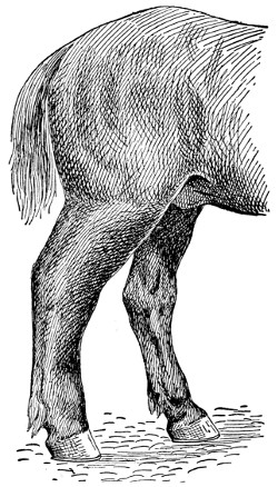
LYMPHANGITIS
This kind of inflammation is usually seen in the hind legs. It is most frequent in heavy draft horses, or in coarse plethoric individuals. It occurs most frequently after a short period of idleness.
LYMPHANGITIS.
—An inflammation of the lymphatics, usually of the hind legs. Hence the name “big legs.” It is the result of too rich feeding, and too little work in many cases on the one hand, or of overwork and insufficient food on the other. Lymphangitis often follows other diseases like distemper, influenza, or pneumonia, in which cases the system is weakened and the lymphatics in abnormal condition. It shows itself after a short period of idleness and rest. It usually begins with a chill and a rise of temperature, which may be as much as 105 degrees, depending on the intensity of the attack. One or both hind legs may show swelling and be so stiff and sore after standing during the night as to be moved only with difficulty when the horse is taken out of the stable in the morning. The horse in moving seems able to bear little or no weight on the affected leg. At the same time, the pulse is full and throbby, respiration is fast, the bowels are constipated and the appetite is lost.
In some cases the legs swell to an enormous size. If the inflammation is not relieved in a few days, the glands get badly diseased and blood poison may result. The disease, however, if taken in time, is easily treated. If it is caused by overfeeding, change this; give more exercise. When the disease is first noticed, give the horse 4 tablespoonfuls of aloes, 4 tablespoonfuls of carbonate of soda and 4 tablespoonfuls of ginger. These should be dissolved in a half pint of boiling water, then mixed with a half pint of cold water, and then given as a drench. If the pulse is fast, it may be made easier and slower by giving 20 to 30 drops of tincture of aconite, every couple of hours. A couple of tablespoonfuls of nitrate of potash in the drinking water three times a day will increase the urine. This is desirable to do in this disease. The leg should be bathed for at least a half an hour and then dried and a wash consisting of 2 tablespoonfuls of acetate of lead, 8 tablespoonfuls of tincture of opium, and a quart of water should be applied to the legs. This should be rubbed in well with the hand every hour. In from 20 to 30 hours, a great change for the better will be noticed the inflammation will have been reduced; the pain will have disappeared and the bowels will be loose and active.
From now on give general exercise at frequent periods, during the day. In cases caused by overwork or too little food or those following debilitating diseases, like influenza or distemper, the treatment should be more stimulating; therefore, nutritive foods and tonics are best. Good hay and oats and other feed of a laxative nature should be furnished.
A preparation, consisting of 4 tablespoonfuls, each, of tincture chloride of iron, tincture of gentian, and ginger in a pint of water three times a day will be found both stimulating and nourishing. If the disease has progressed so far that the legs break and show that matter is formed, wash them with warm water and follow with acetate of lead, sulphate of iron and carbolic acid. Use 2 tablespoonfuls of each in a quart of water and apply twice each day. If the swelling hangs on use Fowler’s solution of arsenic, 4 tablespoonfuls to a dose in a bran mash once a day. Continue this for four or five weeks. A salve made of 2 teaspoonfuls of iodide and 8 tablespoonfuls of vaseline should also be rubbed on the leg twice a week.
MAD DOG.
—See Hydrophobia.
MAGGOTS.
—The grubs of the ordinary flesh-flies so common about stables and houses. The adult fly deposits the minute larvæ in fresh meat, in wounds, and frequently in dirty wool. These become the maggots so well known about the farm. The distress caused by these when present in a wound is considerable, and they endanger life.
The best treatment is in line of cleanliness. Keep old wounds clean by means of antiseptic washes and tag the sheep that no filth and dirt may accumulate. If for any reason maggots are found, open the infected part and remove, if possible, both the maggots and sloughed tissue. Old sores or wounds, if they will not lend themselves to complete removal of the maggots, should be treated with a solution of carbolic acid and water. On some, turpentine can be used. Chloroform may be sprayed on, or injected into the wound with almost instant results. After the maggots are destroyed follow up the treatment with a good disinfectant until the wound has healed.
MALLENDERS.
—An eruption of the skin above the feet in horses. The disease at first is very much like eczema. In time the watery fluid dries up and the sore parts become covered with hard crusts and scabs. The sore spots should be washed with some good disinfectant and repeated frequently enough to destroy the infection. A moderate purge is advisable. See that only wholesome food is provided.
MAMMITIS.
—Inflammation of the mammary gland or udder. The disease is frequently called caked bag and garget. In the last named, the milk secretion is altered and appears as a thick or a stringy fluid. Heavy milkers are most commonly affected. The udder becomes swollen, hot and somewhat tender just before calving. The swelling may extend forward along the belly. It often gets so severe as to require treatment. It is in this sense physiological. In a few days after calving, as a rule, the swelling disappears and the normal condition is regained more quickly if the calf is allowed to suck the cow. In the first stages bloody milk is secreted and often pus is formed in one quarter or more of the udder. The udder should be carefully milked, cleaned, and, if the milk ducts are closed, it may be necessary to use a milk tube. This should be used cautiously so as not to injure the tissue of the udder and should be perfectly clean before inserting, otherwise serious inflammation may result. In bathing, use hot water for 15 to 20 minutes at a time, after which rub dry and apply an ointment made by dissolving 3 tablespoonfuls of gum camphor and 4 tablespoonfuls of fluid extract of belladonna to a pint of clean, fresh lard. This ointment should be applied three times a day.
A more serious form of the disease is known as contagious mammitis, and is due to invasion of the gland by bacteria. In cases of this kind the inflammation is more extensive and the disorder calls for more careful treatment. Since the milk contains bad bacteria, it is necessary to destroy them so as to prevent spreading of the disease. The milker should have clean hands and should wash them in a disinfecting solution before milking another cow. The milk tube may be necessary in withdrawing the milk. After the milk has been removed from the udder, inject a solution of peroxide of hydrogen or dioxygen or a solution of carbolic acid, 1 part to 50 parts of boiled water. After the solution has acted for a few minutes, it should be milked out. The external treatment for contagious mammitis should be similar to that of ordinary mammitis.
MANGE.
—See Scab in Cattle.
MILK FEVER.
—It is a remarkable fact that this disease occurs most commonly in cows which calved easily. This is explained by the fact that in such cases the os uteri remains relaxed for a greater length of time than it does in cases of difficult parturition. Milk fever generally occurs in cows which are heavy milkers, and great eaters. Keeping the animals in permanent stables, and feeding large quantities of rich food while they are giving no milk are predisposing causes.
The disease makes its appearance usually in from 24 to 48 hours after parturition. It seldom occurs after the third day, and some authors state that it has never been recognized before the starting of the milk secretion. The most salient symptoms to the average layman would, perhaps, be the anxious expression of the animal, bellowing and mounting into the manger. Later they become very weak, stagger and fall, and are unable to rise. The members are usually extended in a rigid position. A rattling or whistling noise is heard in case the larynx is paralyzed. The feet, ears and horns feel cold to the touch. When a case is going to recover we see improvement as early as the second or third day. Recovery is usually complete at the end of from two to five days.
Milk fever is one of the cases where the old maxim, an ounce of prevention is worth a pound of cure, is doubly applicable. If proper precautions were taken a large number of cases could be prevented. Give the pregnant animals daily exercise, and decrease their allowance of food.
Treatment Very Simple.
—Make the cow comfortable. Now give her a small dose of Epsom salts from one-half to one pound, depending on her size. This should be given as a drench. Animals afflicted with this ailment swallow with difficulty. Use care that the drench does not get into the lungs. Perhaps the most satisfactory medical treatment is to use what is known as the Schmidt treatment. This is nothing more than injecting into the udder a solution made by dissolving in one quart of clean boiled water 3 teaspoonfuls of iodide of potash, after stripping all milk from the udder. A very satisfactory way is to get a rubber tube, attach it to a common milking tube which is placed into the teats in turn and pour the solution into the tube by means of a funnel. By massaging the udder the solution can be worked into each quarter in a short time without difficulty.
In case iodide of potash is not available, inject air into the udder after drawing out the milk. I have known of many cases where air has been forced into the udder by means of a bicycle pump, and the animal recovered in a very short time. If the disease does not respond to the treatment with readiness, repeat in a few hours, say, anywhere from five to ten hours after. Cold water or ice on the head is advisable. The use of stimulants is also recommended. Whiskey can be given in doses of 10 to 15 tablespoonfuls and jamaica ginger 6 to 8 tablespoonfuls. Milk the cow frequently and massage the udder, bathing in hot water.
After the cow is on the way to recovery, withhold milk-stimulating foods for a few days and give some tonic like gentian and nux vomica, half and half, 2 or 3 tablespoonfuls two or three times a day.
MONDAY MORNING SICKNESS.
—See Azoturia.
NASAL GLEET.
—When a cold or simple catarrh is neglected it may run into a chronic condition giving rise to nasal gleet. A thin, bluish discharge comes from the nose; and the membranes of the nostrils, instead of being moist and pink in color, take on a leaden hue. The coat at the same time shows unthriftiness. In such cases the face and head may swell because the accumulated materials fail to pass out. When these bunches are tapped with the fingers, a dull sound is heard. Treatment consists of isolating the animals and giving them good care, nutritious food and well-ventilated quarters. A bucket, filled with boiling water, in which a half cup of turpentine is placed, and held under the nose to steam the nostrils and face, is excellent. Any sort of blanketing that will hold the steam about the head is very good to have at hand at the time. For internal treatment give a teaspoonful of sulphate of copper three times daily in a small bran mash; following this drop the copper sulphate and give 2 tablespoonfuls of Fowler’s solution of arnica twice a day in the mash. Should the bulges on the face become large, it will be necessary to open them. Often a part of the bone requires sawing out to get effective results. In these severe cases it is best to have your veterinarian make the operation.
NAVICULAR DISEASE.
—A disease of the navicular bone and the structures surrounding it. It is called “coffin joint lameness.” This bone is situated at the back and inferior part of the coffin joint, and acts as a pulley over which the flexor tendon of the foot passes. Horses with upright pasterns are most liable to it, as more weight is thrown on this joint. Those shod with calkins on their shoes, which prevent the frog from coming in contact with the ground, therefore causing a shock to this joint, are also very liable to it. Some horses have hereditary tendency to this disease. Nails penetrating too deep through the sole, or anything that will cause inflammation of this joint, is likely to produce navicular disease. The most prolific cause is bad shoeing. By degrees the inflammation in a chronic form extends to other parts, causing a shrinking of the soft parts, resulting in contraction of the foot.
The lameness may appear suddenly and perhaps immediately after the horse has been shod, and is then usually thought to be the fault of nailing on the shoe. It is likely in this case that the smith has pared the sole and frog too thin, and that the part has suffered from a bruise by the horse stepping on something hard. After a rest it may disappear, to return after the next drive. Sometimes the disease is of very slow progress in one or both fore feet. The first thing that is noticed is that the animal points its toe, and if both are affected, first one, then the other. The animal may not be lame, but it does not step out so well as it used to, and by degrees the part gets more tender, until the animal begins to go lame, and the lameness gradually gets worse. There is a form of this lameness where the animal shows stiffness and lameness when first taken out of the stable, but, after being driven for a short distance, it passes off, and after it stands for awhile it will start off lame again. If this disease lasts for some time the muscles of the chest and shoulders seem stiff and may shrink. This has been called “chest founder” by horsemen. This is brought about by the soreness of the feet. The horse is afraid to step out, giving it the appearance of being stiff; the muscles of the chest and shoulders will shrink from want of proper action, caused by the feet being sore. If there is heat and tenderness in the hollow of the heel or a redness of the sole, and an absence of any other disease of the foot or leg, we may consider with almost a certainty that it is a case of navicular or coffin joint lameness. The result is contraction of the foot.
Take off the shoes, so that the frog will rest on the ground, then poultice the feet with bran, made up with cold water if it is a recent case, but if it is of some months’ standing hot water is better than cold; put the poultices into bags made a little larger than the foot; put about two inches deep of the bran mash into the bag, then put the foot in and fill in all around as high as the fetlock, and tie the bag above the fetlock and around the ankle to keep it well on the foot. Wet this several times a day and change it once daily. Continue this for two weeks, and see that it is properly done; if not, it will be of no service. Then blister the coronet with cantharides 2 teaspoonfuls and lard 4 tablespoonfuls. Repeat in three weeks, and give the animal a long rest.
NITS.
—See Bot Flies.
NODULAR DISEASE IN SHEEP.
—Nodules resembling those of tuberculosis found in the intestines of sheep, are due to the presence of parasitic worms. Profuse diarrhœa and a pronounced anemic condition prevail. A post mortem examination of the intestines discloses the presence of numerous nodules in the intestinal walls. If the worm is present, no treatment is possible, for the reason that any medicine that would affect the worm would also affect the tissues and lead to their destruction. Prevention, therefore, is the only means of overcoming the disease. Sheep must be kept off infested pastures, and infested pastures must be plowed and given over to cultivated crops. Give lambs only clean pastures to graze over. This means crop rotation in connection with sheep husbandry. No feed that has been tramped over by infected sheep should ever be supplied to lambs or sheep not infested with the disease.
OBSTETRICS.
—Difficult parturition is common in some females. And frequently others, less bothered as a rule with any difficulty at this period, deliver their offspring only after great labor and much difficulty. When such cases occur close vigilance not only frequently hastens delivery, but often saves the life of either the mother or offspring or both.
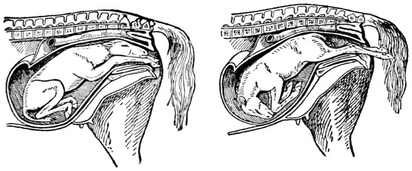
NATURAL PRESENTATION OF THE FOAL
In either of these cases delivery follows in the usual order without delay or injury to the mother.
In many instances the trouble is seated in the womb; the neck of the womb remains closed, and even though long-continued and vigorous efforts are made, the offspring does not arrive. In cases of this kind assistance can be rendered which quickly removes the difficulty. First oil the hand and forearm and work the fingers into the passage, gently pressing it open. If the womb does not yield to this treatment saturate a sponge or cloth with extract of belladonna and rub it around the neck, leaving it thus for a little while. On removing the sponge the passage will open.
Manner of Delivery.
—The natural position of the fetus at birth calls for the fore feet forward with the head resting on the knees. The fore feet, therefore, in a normal delivery, are first presented and then the head. If the fetus is not unduly large, the mother will likely force the delivery without assistance. In case the struggle is extended gentle assistance will be in order. This can be rendered by a gentle pull on the legs and head. If this does not bring the offspring, you can consider that something is wrong. However, do not be hasty, just give time. Mares usually deliver in a few minutes and cows often require an hour or so after labor begins. If you conclude that something is wrong oil the hand and arm. Shove the fetus back and ascertain, if possible, the trouble. If this examination shows dropsy of the abdomen—water in the belly—puncture the abdomen with a knife in order that the fetus may be delivered. If the trouble is with the head—water in the brain—puncture the head that the water may run out, and then remove the arm and hand. When the struggle pains come on again, give a gentle pull and delivery will follow.
Frequently the position is changed. Sometimes but one fore foot appears with the head, making it impossible to deliver the offspring. When a case like this occurs, shove the fetus back and bring the unpresented leg forward where it belongs, and then likely no further trouble will result. If the legs are in proper place but the head turned backward, it will be necessary to push the fetus back into the womb and bring the head forward in position. In case the head resists your efforts, adjust a noose over the head, and while you work with your hand inside, have an assistant gently pull on the rope, in order to draw the head into the proper position. After the head and fore feet are put in natural position, delivery will follow without further difficulty.
When all four feet appear together it is necessary to push the fore feet back into the womb just as far as it is possible to force them. This done, pull now on the hind feet and bring the fetus out, hind feet first. It is always a mistake to attempt delivery with the head first when delivery has proceeded as suggested in cases of this nature.
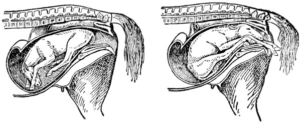
ABNORMAL PRESENTATION OF THE FOAL
Delivery is not possible in either of the cases here illustrated. Where such occur assistance must be rendered. See article on obstetrics for treatment.
Where delivery is attempted with the hind legs foremost, it is regarded as safe, provided the feet come out as they should. If any difficulty is encountered, shove the fetus back, straighten the legs, and then with the renewal of the labor struggles assist the mother by a gentle pull on the hind legs.
Another common presentation is where you feel nothing but the tail, rump and hips. Adjust the fetus for proper delivery by shoving the hind end upwards and towards the front of the womb, then slip the hand down and get hold of the foot of the hind leg and lift upwards and backwards until the legs are brought out into the passage. Now repeat the work for the other leg and the job is done.
It is always a good plan, after difficult parturition, especially when any abnormal discharge appears, to wash out the womb with warm water in which a little carbolic acid or creolin is placed. Use this daily for a few days.
PALISADE WORM.
—The worms are found in the horse in two periods of existence. The mature worms are usually found attached to the mucous membrane of the intestinal wall of the large intestine, with the head sunk deep for the purpose of sucking blood, which gives them the brown or red color. The immature are found sometimes in the same organs, in a small capsule covering, in small pellets of manure, in cavities or cysts, varying in size from a pin-head to that of a hazel nut, in the walls of the intestines, and also in the arteries and other structures of the body.
When present in the kidneys or in the arteries leading to the kidneys, or in the surrounding tissues, a horse is especially sensitive to pressure over the loins. They have been known to cause paralysis. When found in the brain, an animal, when working, suddenly begins to stagger, the eyes become fixed, and the horse shows many of the symptoms of “blind staggers.” When the large arteries of the abdomen are affected, and this is their favorable location in the circulatory system, the animal is frequently subject to colic, which often results in death. This is also the case when found in great numbers in the intestines.
From a thorough investigation of a great many cases, both before and after death, the conclusions are drawn that the parasite evolves a poisonous substance (toxin), which, in many instances, stupefies the brain or parts of the nervous system of the horse, and in that way causes coma, paralysis and death of the animal.
Prevention is the best treatment. Hay and fodder from swampy land are to be looked upon as suspicious. Pastures which are subject to overflow should be avoided. Medicinal treatment consists of a prolonged, careful use of some of the essential oils or other vermifuges. The ordinary spirits of turpentine has proved a fairly good common remedy. An ordinary animal will stand 8 tablespoonfuls of turpentine given in a pint to a quart of raw linseed oil, thoroughly mixed. If the animal is badly affected, the above dose may be given night and morning for two or three days, then omit for a week or two and repeat. The remedy should be discontinued as soon as the animal shows signs of irritation of the kidneys.
PARALYSIS.
—A loss of power over some of the muscles due to a disordered state of the brain or nerves. This may result from disease or injury or some irritation. In horses and cattle the hindquarters are not infrequently affected in this way, the result of indigestion from constipation or from attacks of colic. The animal shows weakness in one hind limb, moving it with difficulty when the opposite limb may then become affected. If the attack is very severe, the animal falls on its haunches and may not be able to rise. Temperature, pulse and respiration, all are rather normal. Treatment should be directed to remove the cause of the disease. When there is colic or constipation, give purges. A half teaspoonful of extract of nux vomica, given in a pint of milk twice a day, is very good. Pouring cold water from a height and then immediately hot water sometimes greatly strengthens the muscles and has its use in treating. Rubbing the parts with mustard stimulates them, and in some cases good results. Paralysis resulting from injury usually disappears as the part returns to its normal state.
PARASITES.
—These are living plants or animals that live temporarily or continually in the bodies of other plants or animals and draw their nourishment from their host. It is doubtful if there is a single farm animal that does not harbor parasites at nearly all times during its life. There may be many of these in the same individual at the same time. Parasites may be harmful or not, as the case may be. Parasites may be divided into two classes—plant parasites and animal parasites. The bacteria and molds are the most important among the former, whereas in the latter certain minute protozoa, certain forms of insects and certain worms are the most commonly met. Such diseases as staggers, tuberculosis, and typhoid fever are the result of bacterial diseases, while Texas fever is an example of the protozoa class; and then the insects and worms are types with which we are all acquainted. When a disease is caused by either, discussion will be found under the name of that disease.
PARTURIENT APOPLEXY.
—See Milk Fever.
PARTURITION, DIFFICULT.
—See Obstetrics.
PERITONITIS.
—An inflammation of the membrane which lines the abdominal cavity and which also invests the abdominal organs. It may be caused from some exposure to cold after some weakening disease. Some injury to the abdomen or belly may cause it, or it may start from some inflammation that has attacked the stomach, liver, intestines, or the spleen. When attacked, a slight pain is felt and the animal lies down, stretches himself, sweats freely, and moans. Then he rises, walks about somewhat, and all the time breathes heavy and shows much weakness. The pulse runs up between 75 and 100 beats a minute. In time the legs and ears get cold. A good treatment is a pint of raw linseed oil, 4 tablespoonfuls of laudanum, and 10 drops of aconite. Mix these and give as a drench. A mustard plaster for the abdomen and something hot for the back are desirable. In two hours, if the pain continues, give 4 tablespoonfuls of laudanum and 10 drops aconite in a pint of lukewarm water. Use as a drench.
PINK EYE.
—A contagious epidemic disease of the horse affecting the animal all over and particularly the membranes of the air passages. There is general debility, considerable cough, and a general discharge from the nostrils. The transparent covering of the eyeball becomes inflamed. At times the disease is very fatal, many horses succumbing to it. It is most common in the spring.
One of the symptoms is the general weakness of the animal. He hangs his head, and trembles; has little appetite and appears cold. The eyes show a watery discharge and later a stare coat. The pulse at first is weak, but quick, and later rising to 80 or 90 beats a minute. At this stage the temperature is high, around 103 to 105 degrees. The breathing is accelerated to about 50 times a minute. The bowels do not act, or act very poorly, and the urine is very scanty. In treating, first isolate the animal and disinfect the stables to prevent spreading. Any of the common disinfectants will do.
Good nursing is necessary. Keep the horse warm with blankets. Give him soft, nourishing food. The eyes should be bathed three or four times a day with hot water. A little boric acid, say, a teaspoonful to a half pint of water, is good to use as a wash for the eyes and nostrils. To keep the kidneys active and to reduce the fever, give a tablespoonful of nitrate of potash dissolved in water two or three times a day. If the horse is very weak, one-half glass of whiskey in a pint of gruel three times a day is stimulating and helpful. It is better not to give any physic of any kind. After recovery, the horse should be given little or no work. A long rest of several weeks is necessary.
PLACENTA.
—The covering of the fetus, commonly called the afterbirth. As a rule, this comes away with the birth of the offspring. Occasionally in the cow it remains attached to the walls of the uterus, and if not removed will cause trouble, if not sickness and death. Soon after the birth of the calf, if the afterbirth remains, decomposition sets in and as a result the system is more or less poisoned. The first symptoms observed are the offensive odor, the reddish discharge and the decrease in the milk flow.
If the afterbirth does not come away of itself, assistance is necessary. Do this during the first or second day, or the third day at the latest. To remove the afterbirth, tie up the cow and fasten her in a way that she cannot jump around. Now introduce the hand and arm, after careful washing and disinfecting and oiling, into the uterus and gradually and gently break the buttons or attachments from the walls of the uterus with the fingers. With patience these will come away and the whole membrane be removed. An occasional injection is advisable. Use some good disinfectant in the water, flush out thoroughly.
PLEURISY.
—This disease occurs in the chest cavity and is found inside the ribs and over the lungs. It is caused very much in the same way as inflammation of the lungs, like exposure to cold, standing in a draft, and cooling when warm. Some injury to the ribs may also cause the trouble.
In the early stages the animal is noticed to shiver, the pulse is quick and strong, and there is great pain. The breath is heavy, and this is noticed as far back as the flanks. While the animal may lie down, its disposition is to stand up most of the time. There is an inclination to cough, but this is suppressed, because of the pain occasioned by it; therefore the cough really ends in a groan rather than in a normal cough. The extremities of the body become cold.
The best treatment endeavors to prevent the disease from developing. Do just as you would in a case of inflammation of the lungs. Mustard plasters for the chest on each side are good. Keep the body well covered, including the legs and neck; have good ventilation in the stable, but keep the patient out of any draft.
As soon as the disease is noticed, mix the following in a pint of cold water, and give as a drench: Ten drops of aconite, a half teaspoonful of belladonna and two tablespoonfuls of laudanum. These should be given every two hours until the pain subsides. If the animal seems to be weak, and needs a stimulant, give 4 tablespoonfuls of spirits of niter and a half glass of whiskey. This may be given in a pint of cold water mixed with the gruel and given as a drench three or four times a day.
At the same time use the following medicine to improve the kidney action: One-fourth pound of saltpeter or nitrate of potash and one-fourth of a pound of gentian root. These are to be mixed well together and a teaspoonful given three or four times daily. Soft foods are desirable. A small amount of water should be given frequently. Small quantities at a time are preferable to large quantities at infrequent intervals.
PLEURO-PNEUMONIA.
—This is a very contagious disease of cattle introduced in this country from Europe. At one time it was a very serious menace to the cattle industry. Thanks to the very aggressive work of the United States Department of Agriculture, the disease has, so far as is known, been eradicated from this country. No cases of the disease have been reported during the past dozen years.
PNEUMONIA.
—See Inflammation of the Lungs.
POLL EVIL.
—A swelling or soreness at the top of the head. Usually it is caused by an injury, like bumping the head in a doorway, or from a bruise made by the halter or bridle. It is first noticed by a swelling or soreness, which frequently causes trouble by forming an abscess; sometimes this works down and even affects the bone. Treatment is very simple if handled in time. Remove the cause and then bathe with warm water and vinegar twice a day and apply a liniment of some kind. If the abscess is formed, it should be opened with a knife at the lowest point to remove the matter. From now on for a few days bathe the opening with warm water in which has been added some carbolic acid or creolin. If the case causes much trouble, you had better consult a veterinarian, as bad cases frequently leave the neck stiff so that the animals are not able to eat off the ground.
QUARTER CRACK.
—See Sand Crack.
QUITTOR.
—A name given to a fistulous opening upon the heels and quarters of the coronary band, and is caused by treads, pricks in shoeing, bruises, and suppurating corns. Any injury which will cause suppuration within the foot will usually cause matter to form at the coronet, and may result in quittor. The disease is indicated by a swelling upon the coronet where the hair and hoof meet, great lameness, and a discharge of thin or thick curdy pus. There may be one or a number of small openings leading down into the sensitive part of the foot. The parts surrounding the quittor swell and become hard and take on an unhealthy action and are difficult to cure, and may be permanently diseased.
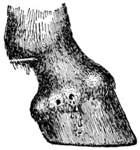
QUITTOR
Fistulous wounds on any part of the coronet are usually the result of a tread or bruise. If neglected serious trouble may result.
Clean the foot and put it into a bran poultice for several days, then remove any horn that may be pressing on the sore part. If it is at the heel remove the crust with a knife; if it is in front of the hoof rasp it thin. Then probe the opening at the top to find the depth and direction. Put a grain of bichloride of mercury into tissue paper and roll it into a cone and press it down to the bottom of the opening. Treat all the openings in the same way. Put the foot into a bag to protect it from injury and let it alone for three days, then clean out the openings and put in some more of the bichloride of mercury, and so on for two weeks, or until the parts become healthy and the hard swelling has decreased; then make up a bath of chloride of zinc one ounce, cool water one gallon; put the foot into this twice a day for twenty minutes at a time. As soon as the openings are healed blister the coronet with the following: Mix 2 teaspoonfuls of cantharides with 4 tablespoonfuls of lard; repeat in two weeks if necessary. When it is time to put on the shoe and work the horse, a bar shoe will be best. If the animal has much fever in the early stages of the disease give a dose of aloes, and follow this by giving 2 tablespoonfuls of nitrate of potassium twice a day in bran mash. Later in the disease give a teaspoonful of sulphate of iron once a day in bran mash as a tonic.
RABIES.
—See Hydrophobia.
RHEUMATISM.
—A disease which affects the muscles or joints, wandering from one part of the body to another. It affects nearly all animals, including the horse, ox, dog, hog, and sheep. Rheumatism of the muscles is usually due to catching cold, while rheumatism of the joints is often due to some micro-organism.
Stiffness, which usually comes on suddenly, is a characteristic symptom. The animal may be able to move only with great difficulty. The joints may crack when moved, the affected muscles are hard and painful to touch, the soreness may shift from one part to another; and the animal sometimes makes a quick recovery, only to be followed by another attack in a short time or perhaps never again. These symptoms may be associated with a rise in body temperature and increased pulse. The disease may last for a long time or only for a few days. In chronic cases the muscles decrease in size in the parts affected. In the dog it is very painful when caused to move and he will howl, or even howl when he thinks he is going to be moved. In sheep it seldom occurs except in young lambs. Pigs are often affected in the legs or back, sometimes becoming paralyzed in the hind legs.
Rheumatism of the Joints
usually shows very rapid swelling, increased heat, and is very painful. The animal is often so lame that it will not put any weight on the foot of the affected limb.
For horses and cows, treatment consists of local applications of alcohol 50 parts and oil of mustard 1 part, rubbing it in well; or spirits of camphor. Give at the same time internally 1 teaspoonful of potassium iodide twice daily and not to exceed 12 doses; or salicylate of soda 4 tablespoonfuls daily. Keep the animal warm and in a well-ventilated stable. Pigs or dogs, according to size, should be given from 4 to 16 grains of salol, also using the above local applications.
RINGBONE.
—A growth of bone on the pastern bone, just above the hoof. It causes lameness when it interferes with the joint or the passage of any of the tendons. Some horses are predisposed to bony diseases from the least injury, while others are not, and in selecting mares for breeding purposes the former should be rejected. This disease results from strains, bruises, or injuries to the cartilage of the joints. When the membrane of the bone or cartilage becomes inflamed there may be great lameness for several months before any enlargement takes place, and it is somewhat difficult to detect. The absence of other diseases of the foot, with some heat in the pasterns, and soreness on pressure or moving the joints indicates this disease. In other cases the enlargement may make its appearance for some time before the horse becomes lame, and in some cases it may never cause any lameness, but should always be looked upon with suspicion, as in the majority of cases it sooner or later causes lameness. Ringbone is more difficult to cure on the fore feet than on the hind ones, as the pasterns are more upright on the former than on the latter, and, besides, the horse’s fore legs have to bear two-thirds the weight of the body.
The horse should have rest, and the shoes should be removed and the foot pared level. If there is heat in the part, keep it wet with the following lotion by means of a bandage saturated with it: Acetate of lead half an ounce and water one quart. Continue this for a few days, then apply a blister composed of cantharides 2 teaspoonfuls, biniodide of mercury 1 teaspoonful and lard 8 tablespoonfuls. Rub on a third of this with the fingers. It is not necessary to cut off the hair if the blister is well rubbed in. Let it remain on for 24 hours, then wash off and rub on a little lard. Repeat every second week until three blisters have been applied. Keep the horse’s head tied while the blister is on so that he cannot get his mouth to the part. The horse should have a few months’ rest after this treatment. If it does not cure the animal it is best to have him fired by a qualified veterinarian.
RINGWORM.
—This is common in the domestic animals, especially in calves and young cattle, and is contagious. It depends upon the presence of a vegetable parasite, which develops and grows rapidly when it finds a suitable place for development. Ringworm may affect any part of the body, but its favorite seat is around the eyes, the face, ears, and neck of cattle, and sometimes the back and hindquarters.
A gray crust appears on the skin, and the hair drops out. This keeps spreading in the form of a ring until around the eyes, the side of the face, ears, or neck may be covered with it. It appears in the same way on the back, hips, and inside of the hind legs. It does not seem to affect the health of the animal, as it is found in the well-kept as well as those poorly kept.
First remove the crusts by washing with warm water in which one ounce of carbonate of potassium has been put to every quart of water. A brush should be used in washing the parts. Then use the following: Iodine 2 teaspoonfuls and vaseline 4 tablespoonfuls. Rub a little of this on with a gloved hand. Repeat in three days. Or mix carbolic acid 1 ounce with 2 ounces of alcohol and apply a little of this to the parts with a feather once or twice; this last is very effective.
ROARING.
—A disease, due to the wasting of the larynx; is characterized by loud, unnatural sounds after any violent exertion. The disease sometimes follows distemper and influenza or a local injury to the throat. Once established the disease is incurable. In its early stages repeated light blisters may help. A common blister can be made of a half teaspoonful of cantharides, a half teaspoonful of biniodide of mercury and 4 tablespoonfuls of vaseline or lard.
ROUP.
—A disease of the mucous membrane in fowls. It is of the nature of an inflammation, with a discharge from the eyes and nostrils usually accompanying. Damp and unsanitary quarters favor the development and spread of roup. It is clearly a germ disease, and, therefore, contagious. It is spread by means of infected quarters and fowls. All discharges must be destroyed by disinfection, and the diseased fowls quarantined off by themselves. The dead should be burned. Keep the quarters light and airy; admit an abundance of sunshine and fresh air. Feed wholesome, nutritious food, that the poultry stock may ward off the disease. The best treatment is that which prevents spreading to healthy fowls. If an outbreak occurs, disinfect thoroughly, liberally, and continuously. Antiseptics administered about the head will usually break up the disease. Creolin is good—say, 1 part to 100 parts of water. Kerosene is also recommended.
In a sense, roup is the result of neglected colds. The birds sneeze, and manifest their uneasiness as animals do with common colds. A teaspoonful of pure carbolic acid to each gallon of drinking water is an excellent preventive and can be provided at small cost.
SAND CRACK.
—A crack found in any part of the wall of the foot. The crack is due to over-exertion. When the hoof is dry and hard and brittle, the crack usually begins at the top and extends downward. Frequently the sensitive tissue creeps into the crack, causing pain, and from which blood frequently issues. When a crack is first seen, the feet should be poulticed with linseed meal for a few days. This will remove the inflammation and soften the hoof. The next step will be to pare out a piece of the hoof at the top, separating it completely from the coronary band a half inch or so on each side of the crack down to the quick. Fill this hole with tar. A bar shoe attached so as not to rest on the wall where the crack is located is very helpful.
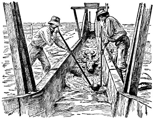
A CATTLE BATH TUB
The tank here shown is used for dipping the cattle for treatment of mange. The dipping tank is now generally used throughout the West.
SCAB IN CATTLE.
—Scab or itch, sometimes called mange of cattle, is caused by a minute mite that lives upon the surface of the skin, burrowing into it. Other animals are not attacked by this parasite, although a similar one does afflict sheep. So long as cattle are doing well on grass, no disturbance is noticed. As soon, however, as they are placed on dry food and cold weather sets in, the disease appears, and, if the cattle do poorly, develops into a very aggravating form. Old cattle are less troubled, the attacks being more frequently on calves and yearlings and two-year-olds out of condition. In the early stages the itching of the skin in the region of the neck or shoulders is first noticed. This is indicated by the animals digging at the skin with teeth and horns and the constant rubbing against posts or barbed wire or anything that may give relief at the time. The disease gradually spreads along the back, sides and outside of legs. In the early stages the coat looks rough, the skin has a scurvy appearance. In time, the hair comes off or is rubbed off, presenting bald patches of thick, glazed and wrinkled skin. After the hair comes off the parasites leave these regions, seeking other quarters and then the hair grows in again. There is a dejected and debilitated condition in animals thus afflicted and they fail rapidly in flesh. Their appetites are poor and most of their time is expended in scratching themselves.
Scab spreads rapidly through a bunch of cattle, especially if they are not thrifty, and disseminates itself through a herd in four to six weeks. The thrifty, vigorous animals resist the infection for some time, but they gradually succumb. The disease is spread by direct contact and by contact with infected quarters. While the mites will live a week or ten days in protected places, they are almost immediately destroyed by direct sunlight. As soon as the disease is discovered in a bunch of cattle, the infected animal should be isolated and the infected quarters and rubbing posts disinfected with a 5 per cent solution of carbolic acid. Infected animals should be well fed and cared for, and be salted with a mixture of 1 pound of flowers of sulphur mixed with 10 pounds of common salt. External treatment is necessary to affect a cure. If a large number of cattle are affected, a dipping wash through which the animals must swim in the dip is the best means for destroying the mites.
The most efficient remedies, considering cost, are the coal tar products advertised as dip solutions. A homemade dip that is both cheap and effective for treating a small number of animals may be made of 3 pounds of flowers of sulphur, 21⁄2 pounds of unslaked lime, 15 gallons of water. In making this unslaked lime into a thick paste, sift in the sulphur and stir well. Put this mixture in a kettle with, say, five gallons of water and boil for at least half an hour—a longer time is better. When the chocolate-looking mass settles, the clear liquid is drawn off and water enough is added to make 15 gallons. The dip will be more effective if used when warm, just a bit hotter than the normal heat of the body. After the animals are dipped, they should remain in the solution about two minutes. This will be time enough to thoroughly saturate the scabs and destroy them. A couple of ablutions are required for complete eradication. When no treatment is resorted to, the dip should be applied with a scrubbing brush, cloth or sponges and all scabs and crusts should be thoroughly saturated. Warm sunny days are preferable for this kind of work.
SEPTIC NAVEL INFECTION.
—A diseased condition at the attachment of the navel cord soon after birth. It is a good plan just after birth to apply some septic powder to the navel at the breaking point. If trouble arises, apply a solution of carbolic acid, 1 part to 20 parts of water, after using some hydrogen peroxide. A little iodoform and alum, mixed half and half, make a good dusting powder to use also.
SHEEP BOTS.
—See Bot Flies.
SIDE BONES.
—On either side of the coffin bone there is a cartilage which may in certain cases become hardened by deposits of mineral matters, which may thus lead to lameness. Side bones are situated on one or both sides of the leg and bulge above the upper portion of the hoof. They may be the result of inflamed conditions, bruises or troubles like corns or hoof cracks. Slipping on the stony pavement is a frequent cause, as well as the great weight of the bodies in heavy horses. If the wagon tongue falls on the foot at this point, the cartilage may be injured and induce the disease. The swelling is first noticed just above the hoof or near the heel. Lameness soon follows.
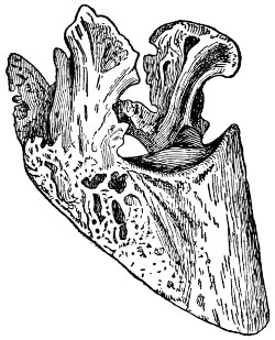
SIDE BONES
When the cartilages on either side of the foot of a horse just at the top of the hoof and close to the heel turn to bone, side bones are the result.
The treatment usually recommended for side bones consists in the free use of cold foot baths or cold water bandages for a week or more. Tincture of iodine applied to the swollen parts is very good. A blister applied after the water applications have been made for a week or so, is used by many veterinarians. The blister is made of 2 teaspoonfuls of cantharides mixed with 4 tablespoonfuls of lard. It is rubbed in well with the fingers and allowed to remain for 24 hours, when it is washed off and applied a second time the following week. These applications are continued until the lameness disappears. If this does not bring permanent relief, then firing of the injured parts and several months’ rest will be necessary.
SLOBBERING.
—Some kinds of food cause an unnatural flow of saliva. Fresh crimson clover hay is one of these. Of course the continual flow of saliva is undesirable and unpleasant. It is unnatural and should be checked as soon as possible. This can be accomplished by changing the feed and then washing the mouth out with alum water. If a change is not observed soon, give a good physic. For horses use 8 teaspoonfuls of bitter aloes, a teaspoonful of common soda and a teaspoonful of ginger. Mix these in a pint of water and give as a drench. For cattle, dissolve a pound of Epsom salts, a tablespoonful of common soda and a tablespoonful of ginger in a quart of lukewarm water and give as a drench.
SPASMODIC COLIC.
—See Colic.
SPAVIN.
—This disease, known in common language as bone spavin, is an enlargement of the hock joint similar to a ringbone about the coronary joint. It may affect the hock joint in such a way as to cement the small joints together, not causing lameness, and apparently no blemish, but the free movement of the limb is impaired. Any condition which favors sprains, such as fast driving over hard or uneven roads, unequal paring of the hoof, thus causing the weight to be unequally distributed in the joints, and severe labor in early life, or blows, bruises, or any injuries to tendons, ligaments, or joints may cause spavin. In addition to these causes may be mentioned sprains caused by jumping, galloping, or trotting animals faster than they are accustomed to; also straining by starting a heavy load, slipping on an icy surface or sliding on a bad pavement.
If the patient is examined before any bony growth has developed, inflammation will be detected on the inside of the hock joint at the junction of the cannon bone and the joint. While in the stable the horse prefers to rest the diseased leg by setting the heel on the toe of the opposite foot with the hock joint flexed. In traveling the patient is very lame when first taken out of the barn, but after traveling for a short distance goes sound. The diseased leg is not lifted clear from the ground, but nicks the toe in the middle of the stride, which is very noticeable on a pavement. A strained horse becomes very lame after being allowed to stand for even a very short time, then moved again.
Preventive treatment consists in keeping horses’ feet trimmed properly, not overworking colts while young, careful driving on hard or uneven roads, and avoiding all injuries that are liable to strain tendons, ligaments or joints of the limbs. Even after a spavin has developed it may be cured by proper treatment of the feet, and applying a fly blister. The fly blister is prepared by mixing thoroughly 4 tablespoonfuls of pulverized cantharides, 4 tablespoonfuls of biniodide of mercury and 8 ounces of lard. The hair is clipped over the spavin and the blister applied with considerable rubbing. The horse’s head should be tied so as to avoid his biting the part blistered. A second application of the blister is to be used about a month after the first. If blistering fails to cure the spavin, point-firing may be resorted to. It is necessary to “fire” rather deeply to secure good results, care being taken not to fire into a joint. After firing, a fly blister should be rubbed into the holes where the hot iron has been used.
SPAYING.
—The removal of the ovaries to prevent breeding. Cast the animal on her right side. Give an anesthetic to prevent pain. When the animal is unconscious, free the limbs sufficiently to remove any pressure from the abdomen. Now pinch up a fold of the skin in the left side, midway between the prominent bone of the haunch or pelvis, and the last rib, about 4 inches below the backbone. Make an incision in the skin 5 or 6 inches long; now do likewise with the abdominal muscles until the lining membrane of the abdominal cavity is exposed. This membrane is then punctured and an incision made as long as that in the skin and muscles. Now kneel down in close contact with the cow’s back and insert the arm, passing the hand within the brim or cavity of the pelvis. By so doing both ovaries can be secured and detached. This ended, the operation of uniting the abdominal muscles follows by means of stitches and sutures.
Great care is necessary in having the instruments boiled and washed in antiseptics, and in having the fingers, hands, and arms severely clean and well saturated with a strong antiseptic solution. The operation should be made out in the open where neither dirt nor dust are to be found. Extreme care about germs will remove much of the risk associated with the operation.
In spaying a sow, she is laid on an inclined board with the hindquarters up. The operator stands at the back of the sow. The hair is first clipped from the skin where the incision is to be made, high up in the flank and midway between the haunch and the last rib. The incision needs to be just large enough to admit the two fingers. Ovaries are located, pulled through the opening in the flank, and removed by tearing off with the fingers. The flank incision is then closed by the necessary number of stitches.
This operation is sometimes performed in mares. But being rather uncommon the process is less understood. In this case it is best to call your veterinarian or someone in the community well skilled in the operation. In all cases of spaying let severe cleanliness be the rule and practice, from the very beginning to the very end.
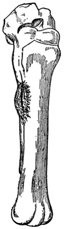
SPLINT
SPLINTS.
—Splints occur more commonly in the heavier breeds of horses than in those that are light in the bone below the knee. It is rare that splints occur anywhere except on the inside of the front cannon bone, although they are sometimes seen on the outside of both the front and hind legs. Any enlargement of the bone occurring on the inside of the leg between the knee and fetlock comes under the name of splint. The usual cause is concussion, that is, the impact of the foot on the hard road. It may be the result of other causes, such as a blow, a twisting strain or faulty conformation. Some animals are more liable to splints than others. It is, after all, to a certain extent, dependent upon heredity. At first the splint is hard to detect. If you notice a young horse going lame while doing road work, it is well to examine for splints. While working there seems to be no lameness at all, and when standing there seems to be no pain, but when put to a trot the horse shows lameness and may raise and lower his head.
If taken in time, a splint can be cured. The first thing to do with an animal suffering from a splint is to give the animal rest and place in such quarters where there is a soft floor, preferably the ground, and when so quartered one very frequently effects a complete cure. The application of cold water bandages acts well. If treatment of this sort fails, apply a blister of red iodide of mercury, 1 tablespoonful to 2 tablespoonfuls of lard. This blister should be applied with rubbing every day from two to four days, or until the area is well blistered. Then wait until the little scabs fall off, and if the animal is still lame, repeat the application of this blister. To apply the blister, clip off the hair over the enlargement and wash with vinegar to remove grease, then rub in blister with ends of fingers. Keep the animals tied short for two to four days in order to prevent rubbing or biting the leg. Four days after the last application of blister, wash carefully with warm water and soap and over it apply every day or so a little lard, to prevent drying and also to loosen the scabs.
SPRAINS.
—Injuries to the ligaments of joints, tendons, or muscles. They are caused by violence, as twisting, or from over-exertion; also sprains are often the result of overwork. If an animal is worked until tired or exhausted he is unable to use the proper muscle force, and more strain has to be borne by the ligaments, resulting in sprains, which often occur in young horses or even in old horses, when put to work after long periods of rest. Swelling, heat, soreness, and partial or complete loss of the use of the part, which is shown by the degree of lameness, characterize the disorder. Sprains are most common in the legs, at the fetlock joint, in the tendons just back and above the fetlocks, but may occur in any part.
The first and most important thing in the treatment of sprains is rest, as sprains are a long time in making a complete recovery. In the early stages, that is, before swelling has taken place, applications of cold water should be used, applications of hot water, or hot packs of water, 1,000 parts, and bichloride of mercury 1 part, are very good. This will relieve the pain and reduce the swelling. Applications of liniments are also very good. Should there be great heat and soreness in the part, it is well to use cold applications. Never blister in the early stages. A blister may be used after the swelling has gone down, and the part has become cold, from two to four weeks after the injury occurred. This should be followed by rest for some time after all lameness has disappeared.
STAGGERS.
—Staggers in horses is an affection of the brain showing itself usually in one of two forms—sleepy or stomach staggers and blind or mad staggers. In the first form the stomach is at fault. Sudden change of feed, moldy or dirty food heavy work or fast driving right after a heavy meal or severe exposure is liable to cause indigestion in the stomach and this is reflected to the brain, causing the animal to act dull or sleepy, sometimes showing symptoms of serious colic, with gas forming from the fermentation of the food, frequently resulting in death.
Blind or mad staggers is an inflammation of the brain and may affect any of the lower animals. In the beginning of this form the symptoms closely resemble those in the stomach form, but as the inflammation progresses the animal becomes blind and violent and may roll, paw, kick, wander around in a circle, usually going only one way, either to the left or right, or it may walk or run in a straight line as near as possible for hours at a time—paying no attention to injuries received in its travels. In either case the animal may be drenched once daily with a quart of raw linseed oil or a pound of Glauber salts, dissolved in water, which sometimes gives relief.
Staggers in Sheep
is mostly caused by the young stage of a tapeworm which infests sheep dogs. The dog eats the infected brain of the sheep and the sheep eats the egg of the tapeworm after it has passed through the dog. After the egg hatches in the stomach of the sheep the young worm passes through the bowels and other organs or tissues or circulates through the blood and reaches the brain, where it develops and causes an inflammation, resulting in disease. It is most common in young animals, rarely occurring in sheep after their second year.
Prevention is about the only practical way of handling this trouble. The grounds should be thoroughly drained, allowing the animals only pure, fresh water to drink. It may be necessary to change pastures for a year or two. The brains of all sheep killed and the heads of all dying with the disease should be burned.
STOMACH AND INTESTINAL WORMS IN SHEEP.
—If a box of salt is kept covered in some place frequented by the sheep, to which they are allowed to help themselves, and if said salt is saturated with spirits of turpentine in proportions of a gill to every four quarts of salt, it will wonderfully help to keep the worms from multiplying. It is well, also, to have another box of larger size, where sheep can help themselves at will, filled with tobacco stems. These stems should be cut up in inch lengths and from time to time a quantity of wheat bran should be put on top of the stems. When this is done the sheep soon instinctively learn to use tobacco, and no young intestinal worm or stomach worm, except the tapeworm, can stand the diet. This will not kill mature worms. It will only prevent the worm family multiplying to the extent of injuring the health of sheep.
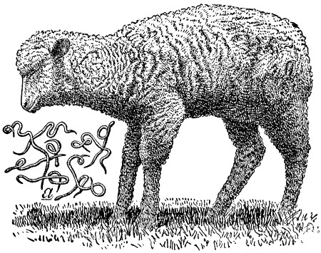
TWISTED STOMACH WORMS
A common attitude observed when sheep are afflicted with twisted stomach worms. The animal loses in flesh, and unless relief is found in time, dies. The parasite is shown in the illustration.
But no sheep owner should feel wholly satisfied by preventive treatment of stomach worms. Twice a year the whole flock should be drenched with some agent which will destroy the mature worms. There are two very inexpensive drenches which will quite effectually do this. The one is gasoline, the other coal tar creosote. The objection to gasoline is that it needs to be so extremely carefully used or sheep will be killed by it. The dose is 1 tablespoonful (never more at one dose) to a mature sheep; mix with not less than 4 tablespoonfuls of raw linseed oil (never boiled oil); then add a half pint of sweet milk. In giving, set the sheep up on its haunches and shake the liquids well together until the last minute it is administered, or the gasoline will separate and, if it enters the stomach in the unmixed form, it will seriously injure and may kill the sheep.
There is no direct vermifuge that will as effectually kill all species of worms in a sheep’s stomach and intestines as will gasoline; yet the coal tar creosote or the more refined class of sheep dips, if given after a full 12-hour fast, before the flock is turned to pasture in the spring, and again about November, will destroy a large number of the mature worms. All lambs born in April or May should be drenched about August or September following, to be certain of ridding them of worms that may later cause their death. The dose of any of the sheep dips is a dessertspoonful mixed in a full pint of water.
STONE IN BLADDER.
—See Concretions or Calculi of Urinary Organs.
STRANGLES.
—This trouble, commonly called colt distemper, affects horses, and rarely mules and donkeys. It is such an infectious disease that nearly all horses contract the disease when colts and usually remain immune to future exposures. The cause is a very small organism or germ which enters the system when a healthy colt comes in contact with a diseased one or when fed and watered in infected vessels. The seat of trouble is largely restricted to the respiratory organs, occasionally causing difficulty in breathing, owing to swelling in region of throat or to accumulations in air passages.
The symptoms start out with more or less sluggishness. The animal eats little, and does not care to take much exercise. A little watery discharge frequently appears from the eyes, and about the same time a watery discharge from the nostrils, which soon becomes thicker and more yellow in color. Usually the glands between the lower jawbones become enlarged and undergo suppuration with a rupture of them and free discharge of pus. The temperature of the animal may be slightly or very greatly increased from 103° to 105°. The pulsations may also be considerably quickened. When complications do not occur this disease usually runs its course in two weeks, leaving the animal little the worse for having passed through the affliction.
The milder forms of this disease will need little or no treatment other than careful feeding and nursing. A laxative diet, with something green, if possible, should be given. The colt should be placed in clean, airy, and comfortable quarters, but not in a draft. To hasten the suppuration of the glands a poultice of hot bran or flaxseed may be applied to that region, and as soon as softening can be detected within, puncture the gland containing abscess with a clean knife blade and allow the escape of the collection of pus. During the course of the disease the animal should not be worked and care should be taken that it be not exposed to conditions likely to produce a cold.
STRINGHALT IN HORSES.
—Stringhalt is an involuntary contraction of the muscles that bring the hind leg or legs forward. The cause of stringhalt is a deranged condition of the nerves supplying the muscles, causing the leg or legs to be brought up with a jerk. In slight cases of stringhalt it is necessary sometimes to turn the animal round from right to left, and from left to right, in order to make him show signs of stringhalt, the symptoms of the disease being exhibited as he turns one way only. This disease sometimes comes on suddenly, but generally develops slowly. It is an unsoundness, and depreciates the animal’s value and makes him unfit for hard work or fast driving. There is no sure cure for stringhalt; the animal can sometimes be relieved by giving him one ounce bromide of potassium at a dose twice a day in bran mash, and continuing it for one week, then skipping a week and giving again. It can sometimes be relieved by cutting the tendon or tendons of the affected muscles, but the operation should be performed by a qualified veterinarian.
SUNSTROKE.
—See Heat Exhaustion and Sunstroke.
SWAMP FEVER.
—This disease, by some called infectious anemia of horses, is produced by an invisible organism, which is transmissible to horses, mules, and asses. About the first symptoms noticed are a general weakness of the animal; it tires very easily and is not able to do any work. The loss of flesh is apparent in spite of the voracious appetite which the animal has at times. The appetite usually remains good until death, but the feed seems to do the animal no good. The temperature is very irregular. Some days it runs quite high, at times to 107°; again it is below normal. An animal may have several attacks of the trouble, but each succeeding attack seems to be more severe. The blood becomes thin, and the circulation impaired, and frequently there appears a swelling under the chest or abdomen, or an enlargement of one or more legs. It is quite easy to recognize the trouble, especially in the advanced stages. The slow progress at the beginning, remittent fever, progressive emaciation and anemia, unimpaired or ravenous appetite, staggering gait, and excessive urination are usually all present to a greater or less degree. Recovery takes place only when treatment is begun early and when the disease is not too acute.
In treating, absolute rest until fully recovered is one of the primary requisites, and purgatives are to be avoided. For the fever, the United States Department of Agriculture recommends an antipyretic of quinine 40 grains, acetanilide 2 drams, and powdered nux vomica 30 grains, four times daily. Cold water sponge baths and frequent copious rectal injections of cold water also aid in reducing the fever. After the fever subsides the following is recommended: Arsenious acid, 2 grams; powdered nux vomica, 28 grams; powdered cinchona bark, 85 grams; powdered gentian root, 110 grams. These should be well mixed and one-half teaspoonful given at each feed of the affected animal.
As in the case of all other infectious diseases, the healthy should be separated from the sick horses, and thorough disinfection of the infected stable, stalls, litter, and stable utensils should be used by mixing six ounces of any one of these chemicals with one gallon of water. One of the approved coal-tar sheep dips might also be used to advantage in a five per cent solution, and should be applied liberally to all parts of the stable, and sufficient lime may be added to the solution to make the disinfectant area conspicuous.
From the fact that the disease is more prevalent during wet seasons, it is always best to guard against allowing the animals to graze upon swampy land or to drink from ponds of stagnant water. The spread of the disease has been traced along creeks from one farm to another, which would suggest avoiding these places also. The draining of the low, swampy lands is especially recommended.
SWEENY.
—Wasting of the muscles covering the shoulder blade of the horse is commonly called “sweeny,” and the cause may be any strain, sprain, jerk, or bruise of the parts due to a bad fitting collar, or to awkward steps of a colt plowing for the first time, and especially when worked in the furrow. The great nerves of the shoulder are affected, and in consequence nutrition is impaired and the muscles waste away. A similar condition may affect the muscles of the hip, or of the space between the stifle and hip.
Lameness seldom is a prominent feature in shoulder sweeny. Ordinarily the wasting comes on some time after the causative injury; then the skin alone appears to cover the bone (scapula) and the animal may have little power for work. In this connection it should be remembered that wasting of the shoulder muscles also may be due to any chronic lameness or soreness of the foot, or leg, between foot and shoulder. Wasting (atrophy) of muscles occurs when the muscles for any reason are not fully exercised. It, therefore, is important to make sure whether the cause is in the foot or in the shoulder before commencing treatment.
Treatment consists in stimulating flow of blood to the poorly nourished parts, and if this can be done the muscles gradually grow in again and regain their normal development and power. An old-fashioned plan is to make incisions in the skin and then blow up the parts with air to separate the skin from the bone. This should not be done. Setons (rowels) of tape may be inserted under the skin, but they leave scars. Better treatment consists in rubbing the parts twice daily with a stimulating liniment, or blistering at intervals of three weeks with cerate of cantharides, after removing the hair. A suitable liniment may be made by mixing together four ounces of druggist’s soap liniment, one ounce each of aqua ammonia and water to make one pint.
SWINE PLAGUE.
—See Hog Cholera.
TAPE WORMS.
—The flat worms of domestic animals. They are most serious and common in sheep. Treatment is only partially satisfactory. To get any reasonable result food must be withheld for several hours before the medicine is given. Use the following: 1 teaspoonful of ethereal extract of male fern in four ounces of castor oil. It is desirable to keep the sheep inclosed, so that the ground can be disinfected after the worms are expelled, otherwise infection will occur right over again.
TETANUS.
—See Lockjaw.
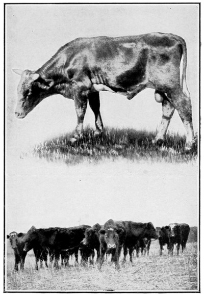
TEXAS FEVER
The annual loss to the South, because of the cattle tick, extends into many millions of dollars. Investigations show that a complete extermination can be effected at a cost of $6 per farm.
TEXAS OR TICK FEVER.
—The earliest accounts that we have of this disease date back to 1814. It was found that cattle driven from a certain district in South Carolina to other parts of the state would infect others with the disease, while they themselves seemed to be in perfect health. The disease is known by various names in the different sections of the country. It is often called red water, Spanish fever, Australian tick fever, and murain.
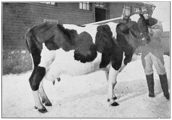
A TYPICAL CASE OF FOOT AND MOUTH DISEASE
The disease shows itself about the mouth, the feet and the teats. When an outbreak occurs all affected animals should be destroyed and all quarters thoroughly disinfected.
This is a specific fever, and is characterized by the peculiarity among animal diseases that animals which scatter the infection are apparently in good health, while those which sicken and die from it do not, as a rule, infect others.
When the cattle are brought into the infected districts they usually contract the disease during the first of the summer, and if they are adult cattle, particularly milch cows or fat cattle, nearly all die; calves are more likely to survive. The disease is one from which immunity is acquired, and, therefore, calves which recover from the disease are not again attacked, as a rule, even after they become adult.
When the disease is prevalent or scattered beyond the infected district the roads, barns and pastures are dangerous until freezing weather, when the disease disappears and cattle can be kept in the grounds or driven over the roads without catching the disease. The midwinter months is the only time that cattle can be safely driven from an infected area to a non-infected area without spreading the disease.
The Cause.
—Texas fever is caused by an organism which lives within the red-blood corpuscles and breaks them up. It is not a bacteria, but a protozoa, and belongs to the lowest forms of the animal kingdom. How it gets into the blood corpuscles is not known. The fatality is due not so much to the loss of blood corpuscles as to the difficulty which the organs have in getting rid of the waste products arising from this wholesale destruction.
The Course of the Disease.
—After a period of exposure, which may vary from 13 to 90 days, the disease first shows itself in dullness, loss of appetite and a tendency to leave the herd and lie down alone. A few days before these symptoms appear the temperature rises from 103° to 107°. There is little change in temperature until death or recovery.
Pathological Changes Observable After Death.
—The presence of small ticks on the udder or escutcheon is a very important sign in herds north of the Texas fever line. The watery condition of the blood. The spleen or milt very much enlarged, and filled with a blackish pulp. Enlargement of the liver, and its color changed to a mahogany color. The distended gall-bladder, caused by an excessive amount of bile in it.
The Cattle Tick
(Boophilus bovis) is the carrier of this disease. Its life history is quite simple. It is unable to come to maturity and reproduce its kind unless it becomes attached to the skin of cattle, whence it may obtain its food. The eggs laid on the ground by the female tick after falling off the cattle begin to develop at once. The time required for hatching varies considerably, according to the temperature. In the heat of summer about 13 days, and in the fall, under the same conditions, from four to six weeks. On pastures these little creatures soon find their way on to cattle. They attach themselves, by preference, to the tender skin on the escutcheon, the inside of the thighs, and on the base of the udder. When very numerous they may be found on various parts of the body. They remain clinging to the cattle until mature, and then fall off and lay their eggs and hatch more new ticks.
How Prevention Is Possible.
—The spread of Texas fever can be prevented by two ways—sanitary arrangements and by vaccination. Where the cattle are infected with the tick, the ticks can be killed by smearing the animals with a solution capable of killing the ticks without harming the cattle. In large herds a large vat of crude petroleum is used to immerse the cattle in. In small herds smear the cattle with a mixture of equal parts of cottonseed oil and crude petroleum.
How to rid the pastures of the tick without killing the vegetation on them has for a long time been the problem. Divide the pasture in two parts by a double parallel line of fence with a 10-foot space between, to prevent ticks from crawling across. One of these pastures is then kept free of cattle for two winters and one summer. After the second winter it will be free of ticks and ready for tickless cattle, when the other pasture is abandoned for the same time.
Vaccination is for the purpose of immunizing cattle that are brought from a non-infected district to an infected district. Calves about six to eight months old should be used, as they are more immune than adult cattle. The immunity is caused by introducing the germ into the blood in a weakened form. This may be done in two ways—by placing virulent young ticks on the calves or by artificial vaccination. When this is practiced, it should be done in two or three inoculations, as it gives better results. The intervals should be about three weeks. The amount of virulent blood should be small the first time and increased in the following treatments.
The inoculation always results in a more or less serious attack of the fever upon the animal treated. Some may die, but the proportion of deaths resulting among animals taken directly into the infected district is large to the proportion of deaths resulting from vaccination. Medical treatment for this disease has proven unsatisfactory in the acute form, although in some chronic cases some good results may have been obtained by medical treatment.
THICK LEG.
—See Lymphangitis.
THOROUGHPIN.
—An enlargement situated on the sides and upper part of the hock joint of the horse, arising from a derangement of the sheath of the back tendon. The fluid with which it is filled can be pressed from one side to the other, hence the term thoroughpin. It seldom causes lameness. For treatment mix a teaspoonful of biniodide of mercury with 4 tablespoonfuls of lard. Rub on a little with the fingers, let it remain on for 24 hours, then wash off and rub on a little lard or vaseline. Repeat the blister every third week until the enlargement disappears. The horse should have rest while under treatment.
THRUSH.
—A diseased condition of the secreting surface of the fatty frog in the foot. In severe cases the horny part often detaches from the sensitive tissue within. Bad shoeing is a common cause of the trouble, or anything else that prevents the frog from coming in contact with the ground. Lameness is sometimes associated with the disease. Treatment consists of careful cleaning, followed with linseed meal poultices if lame. After the foot is made dry, insert calomel into the little cavities. The calomel can be kept in and the dirt kept out by using paper or cloth plugs. Follow this treatment until normal condition is attained.
THUMPS.
—This disease is limited in its action to pigs. Its cause is not definitely known. It is recognized by a peculiar contraction of the diaphragm in young pigs. While the pig may eat fairly well the disturbance is associated with digestion. Such patients like to lie around and take very little exercise. The disease is more common where one kind of food like corn is fed. The old common method was to cut off the ear. The common practice now is to give a purgative so as to relieve the stomach and bowels of accumulated material. The food should be changed and from 1 to 2 tablespoonfuls of Epsom salts should be given. The jerking movement of the muscles may be relieved or stopped by using laudanum, say, four drops to 1 or 2 teaspoonfuls of aromatic spirits of ammonia in a half pint of water.
TICK FEVER.
—See Texas Fever.
TRICHINOSIS.
—A disease caused by the trichina, a minute worm that affects people, hogs and rats. People become affected with the disease from raw or partly cooked pork. These worms are killed by thorough cooking or by the process of hot pickling and curing meat products.
Hogs become affected through eating offal and rats about the slaughterhouses. Hogs that are fed on green grass and other wholesome food, free from these minute worms, are less likely to have trichinæ embedded in their flesh and muscles. Hogs do not seem to be bothered with the trichinæ, but people suffer very severely, as both soreness in the muscles and fever result.
A few days after eating the trichinæ, the worms multiply very rapidly in the digestive tract, from which they migrate to other parts of the body and work their way through the tissues. There is no remedy in way of treatment when affected. Prevention is the one cure. Inasmuch as five to ten per cent of hogs are affected, it is advisable that all pork or ham be eaten only after most thorough cooking.
TUBERCULOSIS.
—Tuberculosis is a disease resulting from the growth of tubercle bacteria in the tissues of the animal. The bacteria, or germs, of tuberculosis, usually gain entrance to the organs of the body by being taken in with the food. Sometimes they penetrate through the membranes in the throat and get into the glands of the head. Sometimes they are taken into the digestive tract, where they pass through the walls of the intestines into the lymph channels and are carried through the large lymph vessel into the blood circulation. In some cases it would seem that the bacteria get into the lungs on particles of dust that are inhaled.
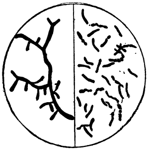
TUBERCULOSIS GERMS
These germs may be inhaled in the lungs with the air, admitted to the stomach and intestines with food and drink, or established in the flesh by inoculation through broken skin or mucous membrane.
After getting into the body, tubercle bacteria multiply in the tissues to which they have been carried and produce the changes in them which we find on the examination of an animal suffering with tuberculosis. Tuberculosis, therefore, is simply the outcome of the growth of the tubercle bacteria in the organs.
Where Tubercles Are to Be Found.
—Tuberculous areas may be found in almost any part of the infected animal, but the organs that are usually affected are the lymphatic glands, either in the throat, the bronchial glands or those about the intestines and on the liver; the lungs; the liver; the kidneys; intestines; udder and generative organs. The membrane covering the lungs (pleura), the heart (pericardium), and intestines (peritoneum), are frequently affected. It often happens that a large mass, or masses, of tuberculous tissue grow over one or more of these membranes. The most peculiar thing about bovine tuberculosis is the fact that frequently an animal will appear to be perfectly well, but when slaughtered will be found to have a large number of tuberculous areas or masses on the membranes or in its organs. The reason for this is that the diseased area is not at a vital point.
The organ or membrane affected depends upon the one to which the germ is carried. Usually animals are infected in but one organ in the beginning, and from this diseased area the germs spread through the blood vessels or lymph channels to other organs. When the diseased area is restricted to one organ or part, it is called “localized” tuberculosis, because it appears at the point where the seed or germ was first planted. When the germs spread through the circulation from this first or primary diseased area to other organs and set up new tuberculous growths, the condition is called “generalized” tuberculosis. When cattle are slaughtered for food, if they are found to be afflicted with localized tuberculosis, the flesh is considered to be fit for food, but if the disease is generalized the carcass is condemned.
The Symptoms of Tuberculosis
vary according to the location of the disease. If it is in the glands of the throat it is suggested by their enlargement. If it is in a gland about the lungs, which, because of its enlargement, presses on the œsophagus (gullet), there might be bloating. If the disease is in the lung tissue there would be, after it is sufficiently advanced, coughing and perhaps difficult breathing. If the disease is in the liver, it cannot be readily distinguished until it is far advanced. If the disease is in the udder it manifests itself usually by the organ becoming firm or hard, and when the tissues are sufficiently broken down the milk from that quarter will be changed in appearance; sometimes it is thick, containing pus, sometimes thin and watery. It is very difficult to diagnose tuberculosis from the symptoms, as many other causes may give rise to similar manifestations.
As tuberculosis is caused by a specific germ, the disease is spread by the germs escaping from the diseased animals and getting into the bodies of healthy ones. The tubercle bacteria escape from the infected animal with some one or more of the natural discharges of the body. For example, if the cow has a bad tuberculous area in the lung, the bacteria may be discharged into one of the air tubes and coughed up into the mouth. Some of them will escape with the saliva and infect mangers or pastures. Some of them may be swallowed and escape from the body with the feces. If the disease is in the udder the germs will escape with the milk. There are some observations which indicate that sometimes the bacteria will escape with the milk where the udder is not affected. After the bacteria leave the diseased animal and are left in the manger, or in the pasture, or on the surface of water in the drinking trough, they can be readily taken up by healthy cattle that eat or drink after them. If they escape with the milk, calves and pigs that are fed with it readily become infected. After the germs get into the body of the healthy animal they will multiply and produce the disease, just as the seed of a noxious weed will, if blown into a new field, germinate and produce the weed there. Tuberculosis spreads from animal to animal on the same principle that weeds spread from one field to another.
In order to prevent the spread of tuberculosis it is simply necessary to prevent healthy animals from coming in contact with the diseased ones or eating or drinking after them.
As tuberculosis cannot be readily detected by a physical examination until the disease is far advanced in the organs affected, it is necessary, in order to determine which animals have the disease, to apply some test or to find the germs of the disease in their excretions. The simplest test that has thus far been discovered is the action of tuberculin. When tuberculin is injected under the skin of the animals affected with active tuberculosis the animals respond by a rise of temperature, which follows a somewhat definite curve. By means of this test it is possible to pick out the infected individuals so that they can be separated from the healthy ones. The test should be repeated in from six months to a year in order to detect any new cases which might have developed from latent or arrested ones. We cannot always get all of the infected animals with the first test any more than we can always remove every weed from the garden by one hoeing.
The Bang Method for the Control
of tuberculosis consists in separating the animals that are infected from the well ones and keeping them for breeding purposes. The calves are removed from their dams as soon as born and fed with the milk of healthy cows, or the pasteurized milk of the infected ones. It has been found that but a small percentage of calves that are raised under proper precautions from such animals have tuberculosis. By this means a sound herd of cattle may be developed from tuberculous animals. This method was introduced by Prof. Bang of Copenhagen, and it has been found to be very effective in Denmark and other countries in Europe. It has been applied with much success in a large number of individual herds in the United States. Its success depends entirely upon the care which is taken in keeping tubercle bacteria away from the calves.
In purchasing cattle for dairy or breeding purposes it is important that they should be taken from herds that are free from tuberculosis. The sound herd is the unit to be dealt with. Animals from such herds are far more reliable than non-reactors from tuberculous herds.
TUMORS.
—Abnormal growths of tissues. There are many kinds of tumors. They are named from the kind of tissue of which they are composed, as fibrous and fatty. Just why tumors should develop is not known. Treatment is in the direction of direct removal; this means they are to be cut out with a knife. Another method is to tie a strong cord around the stem of the tumor, thus shutting off the blood supply. As soon as this is effected, there will be a sloughing away, with a sore remaining, which is to be treated as in an ordinary wound. Some tumors are burnt off with caustics. Arsenic or corrosive sublimate are commonly used, either singularly or combined. Better consult a veterinarian about the removal of tumors on valuable animals.
TUMORS IN PIGS AFTER CASTRATION.
—Bunches form on the cords of pigs after castration as a result of infection from dirty instruments or hands during the operation; or from leaving the cord too long, thus increasing the liability of its becoming infected. These tumors continue to grow, and in the worst cases attain the size of a man’s head. Cut down on a tumor the same as in a simple case of castration. Separate the skin from the tumor and then swallow up the cord with the hands. Cut the cord off as high up as possible. The wound may be healed by the use of any of the common disinfectants. A teaspoonful of carbolic acid in a quart of water may be used once daily until the pigs are healed. Pigs should be kept in a clean pen after the operation.
WARBLES.
—These are lumps in the skin of cattle, caused by grubs or warbles. A simple treatment is to cut the skin and squeeze out the grubs where the lumps are noticed. If all the grubs are killed in this way, there will be no mature flies to cause trouble later on. See article on Bot Flies.
WARTS.
—The cause of these little tumors of the skin is not definitely known. They occur on all domestic animals, appearing most frequently on horses and cattle. Pure acetic acid, dropped on the wart until it is saturated and softened, destroys in the early stages. Warts about which a small cord may be tied are most easily treated in that way. After they have sloughed off, apply a little terchloride of antimony with a feather or cotton. When the scab forms, remove it and apply the chemical again. With a couple of applications the spot will be lower than the surrounding skin. Now use an ointment, made of 4 tablespoonfuls of oxide of zinc and 8 tablespoonfuls of lard. Apply this daily until the sore spot is healed. Sometimes a form of warts suddenly appears on colts and calves and scatter themselves about the lips, nose and face. They are common and appear and disappear suddenly. No treatment is necessary.
WATER IN THE BRAIN.
—Dropsy in the brain. A condition characterized by an accumulation of fluid in the brain. The disease is either congenital or arises during the first years of life. When it occurs the best thing is to kill the young individual at once.
WATER IN THE CHEST.
—Often after a case of pleurisy a reaction comes and a very large quantity of water settles in the chest cavity, anywhere from two to four pailfuls. When the disease comes on the animal has difficulty in breathing; takes in the breath quickly. There is a constant biting at the flanks; the pulse increases to a hundred beats a minute. If you place your ear over the chest you will likely hear no sound at all. Best treatment is wholesome food, boiled flaxseed, and blisters for both sides of the chest. Use strong mustard plasters. A good medicine to use is one-fourth of a pound of saltpeter or nitrate of potash, one fourth of a pound of ground gentian and one-fourth of a pound of sulphate of iron. These should be mixed and then 1 teaspoonful given every four hours. You had better consult a veterinarian. Other complications set in so readily that help may be secured in other ways. Some veterinarians puncture the chest so as to draw off the surplus water that has accumulated.