PLATES
Reading edition
Plate 2 Blaberus craniifer, nymph. (Photograph by Jack Salmon.)
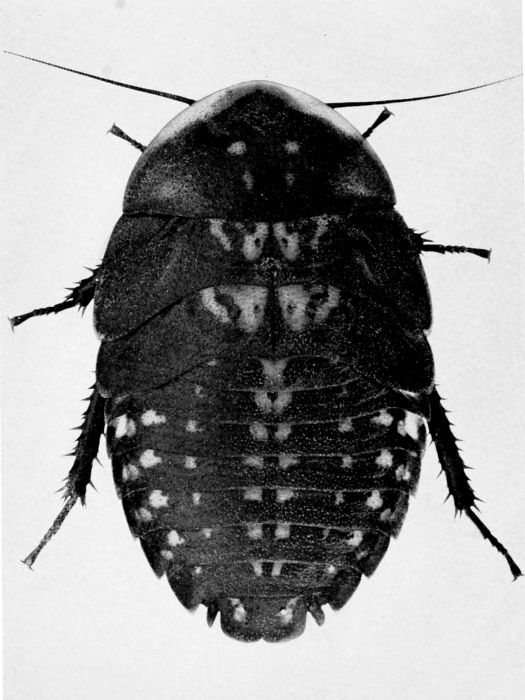
Plate 3 Blaberus giganteus, c. X 2.2. (Photograph by Jack Salmon.)
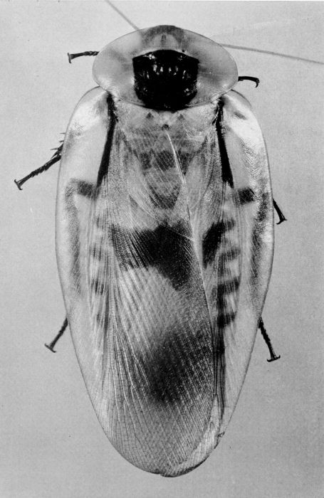
Plate 4 Blatta orientalis, c. X 3.8. A, Male. B, Female. (Photographs by Jack Salmon.)
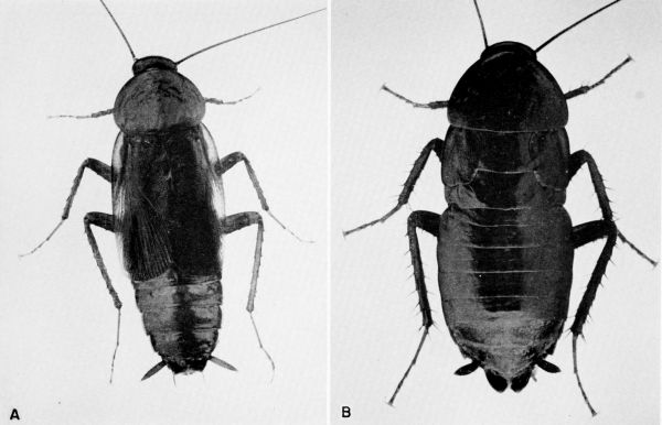
Plate 5 A-B, Blattella germanica, c. X 5.2. A, Male. B, Female. C-D, Blattella vaga, c. X 5.2. C, Male. D, Female with oötheca.
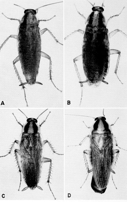
Plate 6 Byrsotria fumigata, c. X 2. A, Brachypterous male. B, Macropterous male. C, Female.
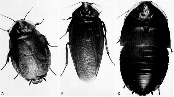
Plate 7 A and B, Cariblatta lutea minima, X 10. A, Male. B, Female with partly formed oötheca. C, Ectobius pallidus, female with completely formed oötheca, X 8. (C, From Roth and Willis [1957].)
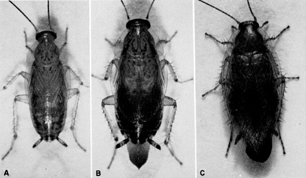
Plate 8 A, Cryptocercus punctulatus, c. X 4.6. (Photograph by Jack Salmon.) B, Panesthia australis, X 2.8.
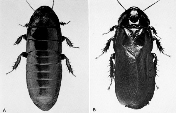
Plate 9 Cutilia sp. near sedilloti, c. X 5. A, Male. B, Female.
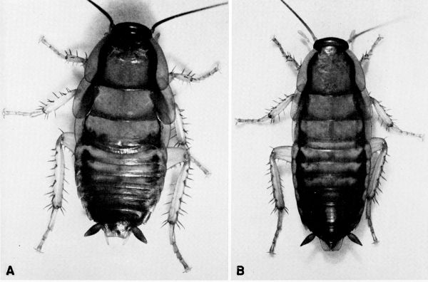
Plate 10 Diploptera punctata, c. X 5. A, Male. B, Female.
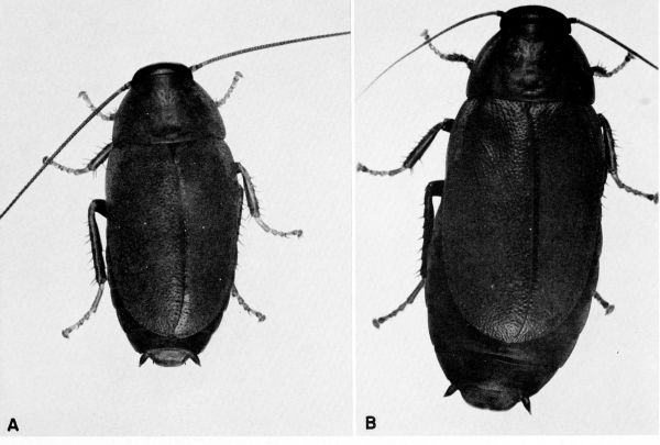
Plate 11 Eurycotis floridana, c. X 2.8. A, Male. B, Female. (Photographs by Jack Salmon.)
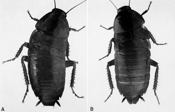
Plate 12 A-B, Gromphadorhina portentosa, c. X 1.5. A, Male nymph. B, Adult female. C, Coleolaelaps (?) sp., a mite from G. portentosa, c. X 32. (Glycerine jelly preparation and photograph of C by Dr. Barbara Stay.)
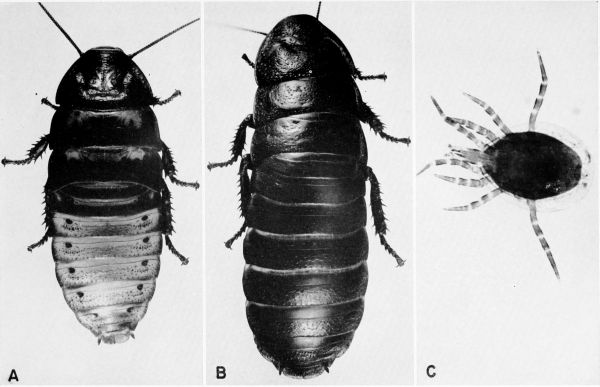
Plate 12A Ischnoptera deropeltiformis, c. X 5.3. A, Male. B, Female.
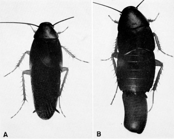
Plate 13 Leucophaea maderae, c. X 2.2. A, Male. B, Female. (Photographs by Jack Salmon.)
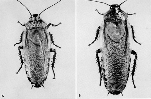
Plate 14 Nauphoeta cinerea, c. X 3.4. A, Male. B, Female.
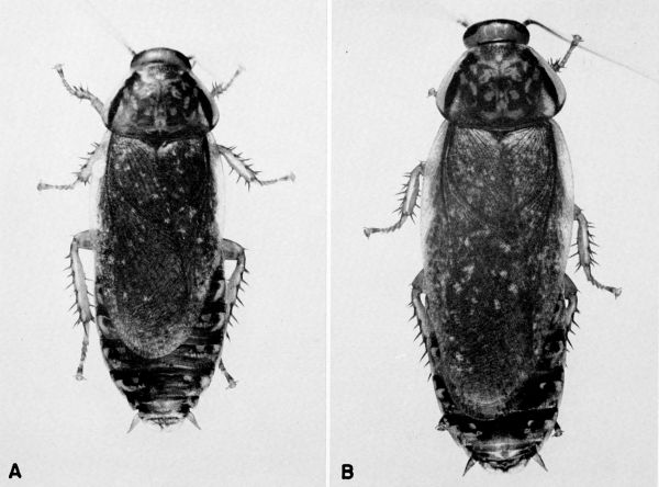
Plate 15 Neostylopyga rhombifolia, c. X 3.4. A, Male. B, Female with partially formed oötheca.
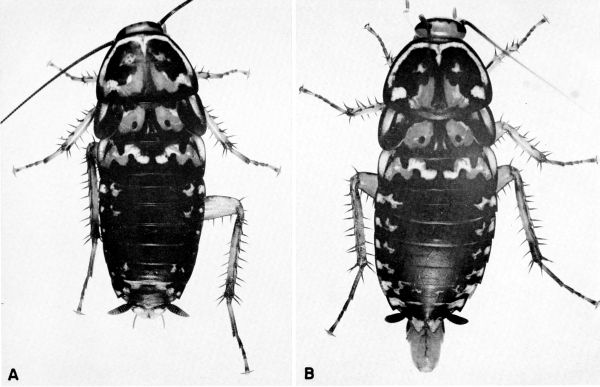
Plate 16 Panchlora nivea, X 4.5. A, Dead individual showing normal, pale green coloration. B, Dead individual showing the bright red coloration (very dark areas) characteristic of infection with Serratia marcescens. C, Living male. D, Living female.
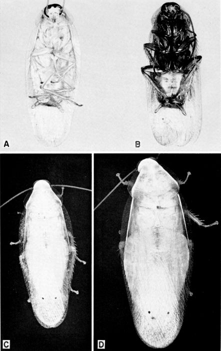
Plate 17 A, Parcoblatta pensylvanica, female with completely formed oötheca, X 4. B, Parcoblatta virginica, female with partly formed oötheca, X 7.3.
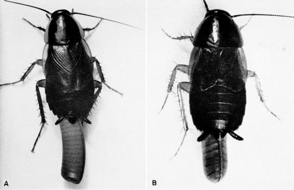
Plate 18 Parcoblatta uhleriana, c. X 5.5. A, Male. B, Female with oötheca.
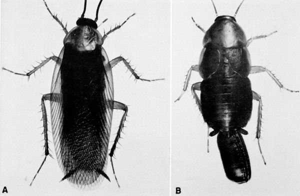
Plate 19 Periplaneta americana, c. X 3. A, Male. B, Female. (Photographs by Jack Salmon.)
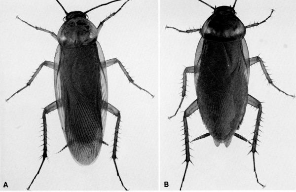
Plate 20 Periplaneta australasiae, c. X 3.2. A, Male. B, Female. (Photographs by Jack Salmon.)
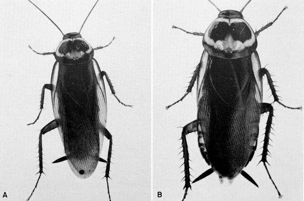
Plate 21 Periplaneta brunnea, c. X 2.9. A, Male. B, Female.
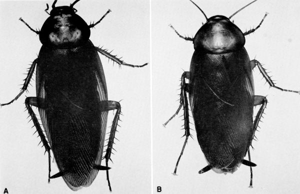
Plate 22 Periplaneta fuliginosa, c. X 2.9. A. Male. B. Female.
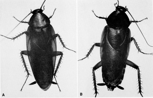
Plate 23 Platyzosteria novae seelandiae, c. X 2.9. A. Male. B, Female with oötheca.
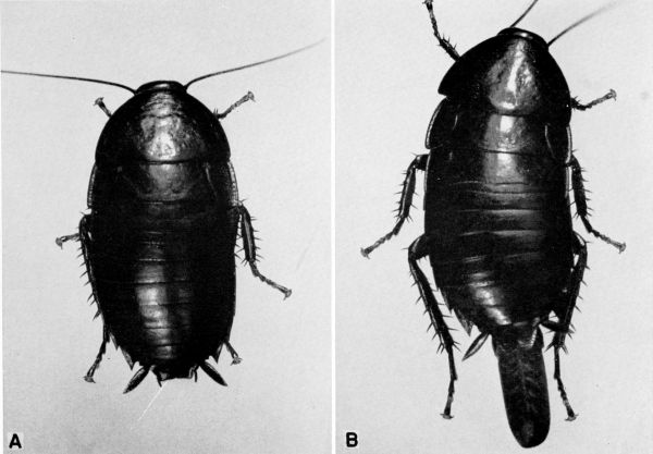
Plate 24 Pycnoscelus surinamensis, c. X 3.7. A, Male from Hawaii. B, Macropterous parthenogenetic female from Florida. C, Brachypterous nonparthenogenetic female from Hawaii. D, Late instar nymph. (Photograph of nymph D, by Jack Salmon.)
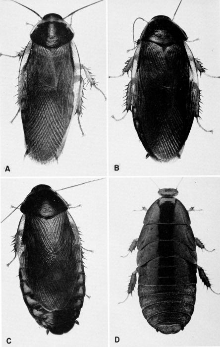
Plate 25 Supella supellectilium, c. X 6.3. A, Male. B, Female.
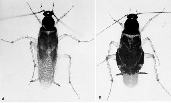
Plate 26 Bacteroids from Blattella germanica. A, Part of abdomen showing mycetocytes in fat body, X 225. B, Lobe of fat body showing 3 mycetocytes, X 750. C, Single mycetocyte; bacteroids appear hollow as result of fixation in Carnoy's fluid, X 1725. D, Smear of fat body showing bacteroids in various stages, X 1800. (All preparations and photographs through the courtesy of Dr. Marion A. Brooks.)
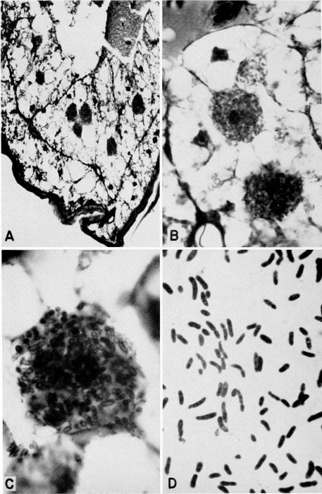
Plate 27 Fungi parasitic on cockroaches. A, Herpomyces arietinus growing on antennae, legs, body, and cerci of a nymph of Parcoblatta virginica, X 7. B, Herpomyces stylopygae on antenna of Blatta orientalis, X 35. (Reproduced from Richards and Smith [1955, 1956].) C, Herpomyces sp. [probably H. stylopygae] on antenna of B. orientalis, X 132. (Photographs B and C through the courtesy of Dr. A. G. Richards.)
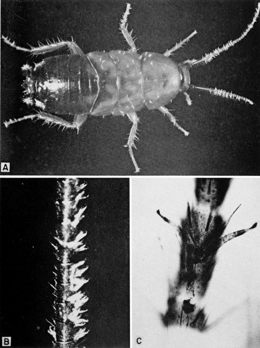
Plate 28 A-B, Gregarines (Diplocystis sp.?) from Blaberus craniifer. A, Organisms removed from intestine, X 50. B, Organisms removed from hemocoele, X 32. C, Gregarine cysts in feces of Leucophaea maderae, X 12.
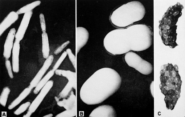
Plate 29 A, Undetermined mermithid that parasitizes Ectobius pallidus. X 9. The worm has partly emerged from the neck region of the cockroach. (Reproduced from Roth and Willis [1957].) B, Undetermined gordian worm that parasitized Eurycotis floridana shown beside its host, X 1.8. (Specimen courtesy of Dr. T. Eisner.)
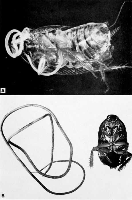
Plate 30 A, Heteropoda venatoria, a cockroach-hunting spider, slightly less than natural size, on bananas. (Reproduced from a Kodachrome transparency through the courtesy of Dr. B. J. Kaston.) B to E, Lycosa sp. (avida?) capturing and feeding on a nymph of Supella supellectilium in the laboratory, X 1.4.
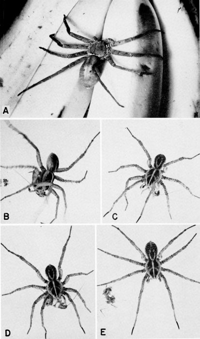
Plate 31 The centipede Scutigera coleoptrata capturing and feeding on cockroaches in the laboratory. A to E, Pursuit, capture, and eating of a nymph of Supella supellectilium, c. X 1.2. F, Centipede feeding on adult of Blattella germanica, X 1.8.
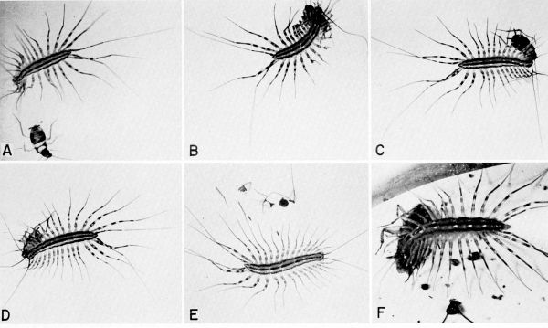
Plate 32 The mantid Hierodula tenuidentata (?) devouring a nymph of Periplaneta australasiae, c. X 1.5.
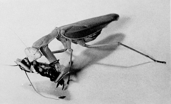
Plate 33 A, Prosevania punctata ♂ beside an oötheca of Periplaneta americana, X 5. B, Hyptia harpyoides with oötheca of Parcoblatta uhleriana from which it had emerged, X 5. C, Larva of a lampyrid beetle feeding on Parcoblatta virginica in the laboratory, X 4.
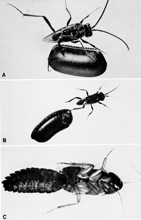
Plate 34 Chalcid parasites of cockroach eggs. A, Anastatus floridanus ovipositing into an oötheca which is still being carried by Eurycotis floridana, c. X 4. B, Comperia merceti ovipositing into an oötheca of Supella supellectilium, c. X 13. C, Tetrastichus hagenowii ovipositing into an oötheca of Periplaneta americana, c. X 10. (C from Roth and Willis [1954b].)
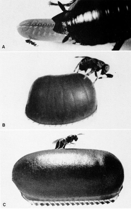
Plate 35 Ampulex compressa attacking Periplaneta sp. (presumably americana), about natural size A, The wasp finds a cockroach. B, She stings the prey in the thorax. C, She then leads the disabled cockroach (antennae clipped) to her nest. D, The wasp's egg was placed on the coxa of the cockroach's right mesothoracic leg where it hatched. E, Portion of the host's abdomen removed to show feeding larva. F, New adult wasp emerging from dead host. (Reproduced from F. X. Williams [1942] from the color paintings of the late W. Twigg-Smith, through the courtesy of F. A. Bianchi.)
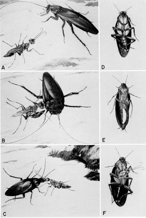
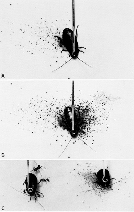
Plate 36 Chemical defense of Diploptera punctata against predators; the spray pattern is displayed on KI-starch indicator paper. A, Spray pattern after right mesothoracic leg was pinched. B, Cumulative spray pattern after left mesothoracic leg of the same insect was pinched. C, The defensive glands of the cockroach on the left had been excised, and it is under persistent attack by ants from a laboratory colony of Pogonomyrmex badius (Latreille). The intact cockroach on the right was also attacked by the ants, but it discharged a spray of quinones and repelled the attackers. (From Eisner [1958], through the courtesy of Dr. T. Eisner.)