Pl. V.
Reading edition
Megalaspis attenuata Wahlenb.
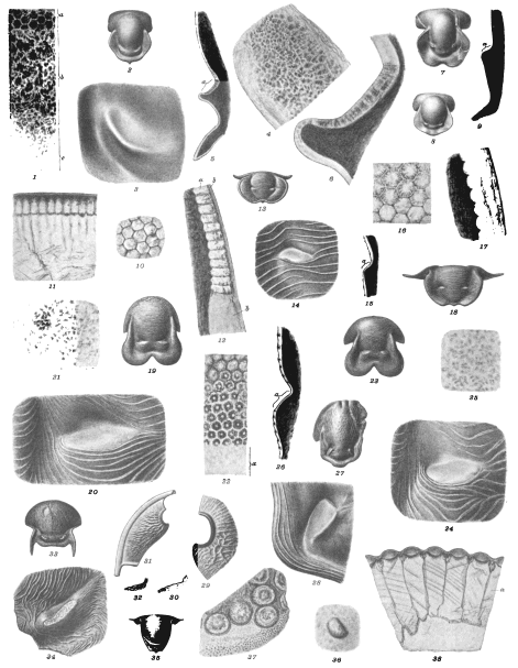
Fig. 1. Horizontal section of cephalic eye near the inferior margin 60/1. a surface of the eye, b frame with irregular or reserve prisms, c part of the cheek with normal structure. Spec. DP. » 2. Hypostoma 2/1. Spec. BM. » 3. The right macula 12/1. » 4. Part of the same in transmitted light 60/1. Spec. DR. » 5. Vertical (sagittal) section of the inferior part of the hypostoma, crossing the macula a 12/1. » 6. Vertical section of the same macula 60/1. All spec. from Westanå, Östergötland.
Megalaspis limbata Boeck p. 61.
Fig. 7. Hypostoma, mostly decorticated 2/1. Specimen from Fogelsång, Scania, the same as Brögger's original to his fig. 23.
Megalaspis planilimbata Angel. p. 61.
Fig. 8. Hypostoma, Öland, Äleklinta 2/1.
Megalaspis sp.
Fig. 9. Vertical section of macula (a) 12/1. Öland, Lower grey Orthoc. limestone.
Nileus armadillo Dalm. p. 62.
Fig. 10. Horizontal section of cephalic eye 60/1. » 11. Vertical section of the same 60/1. » 12. A similar section 60/1. a the prisms of clear calcareous spar, b the surface of the eye and the free cheek of yellow spar. » 13. The hypostoma 2/1. » 14. The right macula 12/1. » 15. Vertical section of macula (a) 12/1. All specimens from Persnäs, Öland, excepting fig. 13 from Westanå, Östergötland.
Symphysurus palpebrosus Dalman p. 62.
Fig. 16. Horizontal section of cephalic eye 60/1. » 17. Vertical section of the same 60/1. » 18. Hypostoma 2/1. From Westanå, Östergötland.
Niobe frontalis Ang. p. 63.
Fig. 19. The hypostoma 2/1. » 20. The right macula 10/1. » 21. Part of the same in transmitted light 60/1. Specimen from Kungs Norrby, Östergötland.
Niobe læviceps Ang. p. 63.
Fig. 22. Horizontal section of cephalic eye near the inferior margin, a cheek 60/1. » 23. Hypostoma 2/1. » 24. The right macula 10/1. » 23. Part of the same in transmitted light 60/1. » 26. Vertical section of macula a 12/1.
Ogygia dilatata var. Sarsi Angel. p. 63.
Fig. 27. Hypostoma 2/1. » 28. The right macula 10/1. From Christiania, Haakavik, Collection of Dr. G. Holm.
Olenus sp. p. 33.
Fig. 29. A part of the free cheek with the scallop 6/1. » 30. Profile of the scallop. Andrarum, Scania.
Parabolina spinulosa Wahlenb. p. 33.
Fig. 31. The free cheek with the scallop 3/1. » 32. Profile of the scallop. From Jemtland.
Paradoxides oelandicus Angel. p. 64.
Fig. 33. Hypostoma 2/1. » 34. The left macula 12/1. Öland, Torp.
Phacops cephalotes Barr. p. 65.
Fig. 35. The hypostoma; the shell mostly decorticated and the maculæ visible as casts on the inside. » 36. Cast of the inside of the right macula 12/1. Spec. from Lochkow, Bohemia.
Phacops quadrilineata Angel. p. 66.
Fig. 37. Horizontal section of cephalic eye near the inferior margin 30/1. The dark points lie in the outermost stratum of the cheek. » 38. Vertical section of cephalic eye 20/1. An irregularly hexaedral crystal below each lens. Specimen from Wisby a.
K. Sv. Vet. Akad. Handl., Bd 34, No 8. Lindström: Trilobites. Pl. V.
Click on image to view larger sized.
G. Liljevall del. Ljustryck af Chr. Westphal Stockholm.