Pl. III.
Reading edition
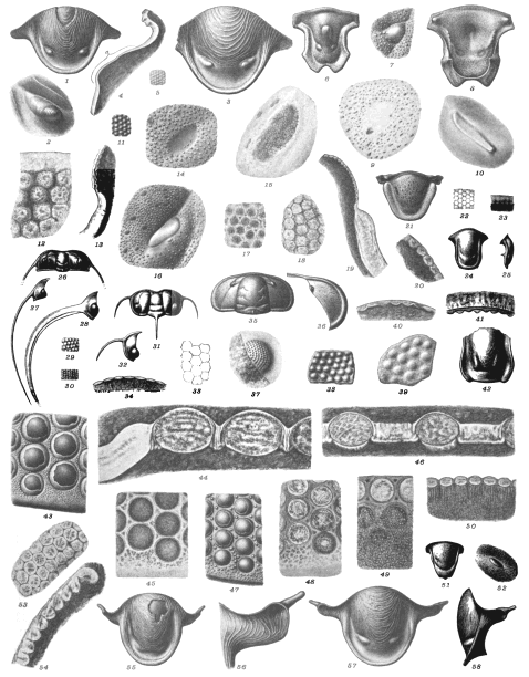
Bumastus n. sp. p. 47.
Fig. 1. Hypostoma 4/1. Specimen AY. From Wisby, Norderstrand. » 2. Its left macula 12/1.
Bumastus n. sp. (different from the preceding) p. 47.
Fig. 3. Hypostoma 4/1. Spec. AT from Wisby, Norderstrand. » 4. Vertical section of the macula a in its longest axis. » 5. A portion of the cephalic eye, the facets very low.
Calymmene intermedia Lindstr. p. 48.
Fig. 6. Hypostoma 4/1. Spec. BG from Gotland. » 7. The left macula 12/1.
Calymmene tuberculata Brünnich p. 48
Fig. 8. The inside of the hypostoma 3/1. Spec. BA from Gotland. » 9. Horizontal section near the surface, seen from the inside 20/1.
Chasmops macroura (?) p. 49.
Fig. 10. The left macula, cast of the inside 12/1. Rostock.
Chirurus glaber Ang. p. 50.
Fig. 11. A portion of the surface of the cephalic eye 12/1. From Dalecarlia.
Chirurus ornatus Dalm. p. 50.
Fig. 12. Horizontal section of a Cephalic eye near the surface 60/1. » 13. Vertical section of the same, 20/1, at the inferior margin. A line added to show the supposed size of the fragmentary lenses. » 14. The left macula of the hypostoma 12/1. From Östergötland, Westanå.
Chirurus spinulosus Nieczkowski p. 51.
Fig. 15. The right macula of the hypostoma from the inside 12/1. Belongs to Dr. G. Holm, from Esthonia, Kuckers C^2.
Chirurus sp. p. 51.
Fig. 16. The left hypostomic macula 12/1. Öland, Brunsby kanal.
Cyrtometopus clavifrons Dalm. p. 49.
Fig. 17 & 18. Horizontal sections of cephalic eyes 60/1. » 19. Vertical sections of cephalic eyes and a part of the cheek 20/1. » 20. A highly magnified portion of the eye in vertical section 60/1. » 21. Hypostoma, 3/1 the anterior wings completed after another specimen. From Östergötland.
Cyphaspis elegantula Angelin p. 52.
Fig. 22 & 23. Horizontal and Vertical sections of cephalic eyes 60/1. Diameter of facets 0,03 mm. » 24 & 25. Hypostomas from the front side and in side view 8/1. Upper Silurian, Gotland.
Ctenopyge pecten Salter p. 29.
Fig. 26. The head shield, the free cheeks added from other specimens 4/1. » 27. A free cheek, viewed without foreshortening, as in its natural position 4/1. Alumschists of Andrarum, Scania
Ctenopyge n. sp. (Linnarsson, Geol. För. Förhandl. 1880, pl. VI, figs. 14, 15) p. 29.
Fig. 28. A free cheek 6/1. » 29. Surface of the eyes, near the suture 30/1. » 30. The same next the free cheek 30/1. Andrarum.
Sphærophthalmus alatus Ang. p. 29.
Fig. 31. The head shield; the free cheeks have been added 8/1. » 32. A free cheek without foreshortening 8/1. » 33 & 34. Horizontal and Vertical sections of cephalic eyes 100/1. The cornea nearly 0,01 mm. in thickness. Andrarum.
Peltura scarabæoides Wahlenb. p. 29.
Fig. 35. The head shield; the free cheeks added 2/1. » 36. A free cheek, not foreshortened 4/1. » 37. A cast of the inside of the cephalic eye 20/1. » 38. A portion of the cephalic eye from the inside 60/1. » 39. A portion of the same in transmitted light 100/1. » 40. Vertical section of an eye 100/1. The rock on the inside. » 41. A similar one passing through two rows of lenses, the one behind the other. The rock on the outside 100/1. » 42. A hypostoma 8/1. Andrarum.
Dalmanites imbricatulus Angelin p. 53.
Fig. 43. The cephalic eye near the inferior margin 20/1. » 44. Vertical section of the same and a portion of the free cheek 60/1. The lenses are almost contiguous and the interspace between them is formed by the common test of the body, which is perforated by longitudinal tubes.
Dalmanites obtusus Lindstr. p. 53.
Fig. 45. Horizontal section of cephalic eye near the inferior margin 30/1. » 46. Vertical section, the interspaces being in this wider than in the preceding 60/1. From Gotland.
Dalmanites vulgaris Salter P. 54.
Fig. 47. Part of the cephalic eye near the inferior margin 20/1. » 48. Horizontal section near the surface at the inferior margin 30/1. » 49. A tangential section, so that the inferior part is below the lenses where the peculiar radiating structure is seen in horizontal section 30/1. » 50. Vertical section of a small Specimen, fibrous appendices below the lenses 30/1. » 51. Hypostoma 2/1. » 52. The right macula 12/1. English specimens from Dudley, Shropshire.
Dysplanus centrotus Dalman p. 55.
Fig. 53 & 54. Horizontal and vertical sections of an eye 60/1. » 55. Hypostoma 3/1. » 56. The anterior wing of the hypostoma seen in foreshortening 6/1. Specimen AX from Westanå, Östergötland.
Dysplanus ladogensis Holm p. 55.
Fig. 57. Hypostoma 3/1. » 58. The same, seen from the side 3/1. Specimen AI from Östergötland.
K. Sv. Vet. Akad. Handl., Bd 34, No 8. Lindström: Trilobites. Pl. III.
Click on image to view larger sized.
G. Liljevall del. Ljustryck af Chr. Westphal Stockholm.