Pl. II.
Reading edition
Asaphus (Isotelus) gigas J. Hall p. 42.
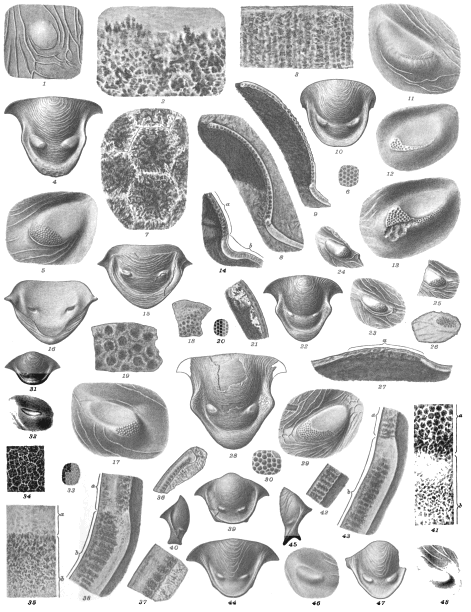
Fig. 1. The right macula surrounded by terrace lines 3/1. » 2. Horizontal section of a macula with a part of the test 60/1. » 3. Vertical section of the same, the surface of the outside upwards 60/1. All specimens from Cincinnati, Ohio, Hudson River group. Spec. BU.
Bronteus irradians Lindström. p. 43.
Fig. 4. The hypostoma 4/1. » 5. The left macula 12/1. Specimen from Gotland, Löjsta.
Bronteus laticauda Ang. p. 44.
Fig. 6. Horizontal section of the cephalic eye 20/1. » 7. The same highly magnified 250/1. » 8 & 9. Vertical sections of the cephalic eyes 20/1. In fig. 8 with integument, in fig. 9 decorticated. » 10. Hypostoma. Specim. N. 2/1. » 11. The right macula, from the outside. Spec. O. 12/1. » 12 & 13. Left maculæ, casts of the interior surface 12/1. Specimens from Dalecarlia.
Bronteus platyactin Ang. p. 43.
Fig. 14. Vertical section of cephalic eye, a the eye. b, cheek 20/1. » 15. Hypostoma 2/1. » 16. The same from the interior surface. Sockets of the maculæ sunk in the shell 2/1. » 17. The right macula 12/1. » 18. Portion of the eye on the macula in transmitted light 20/1. » 19. The same more magnified 60/1. Specimens from the stratum b. Wisby, Norderstrand.
Bronteus polyactin Ang. p. 8.
Fig. 20. Horizontal section of cephalic eye 20/1. » 21. Vertical section of the same 60/1. » 22. Hypostoma 4/1. » 23-25. Samples of right maculæ 20/1. » 26. Horizontal section of macula, the portion bearing the lenses 20/1. » 27. Vertical section of the same 60/1, a the lenses. Specimen from Stora Carlsö. » 28. Hypostoma 4/1. » 29. The right macula 12/1. » 30. A portion of the lenses on the macula in reflected light 30/1. The lenses are only faintly convex and encased in a white frame. The diameter of the largest lenses 0,06 millim. Specimen from Lansa, Fårö.
Bumastus barriensis Murch. p. 46.
Fig. 31. Hypostoma 4/1. » 32. The right macula 12/1. Specim. CU from Wisby.
Bumastus sulcatus Lindst. p. 46.
Fig. 33. Horizontal Section of a cephalic eye 20/1. » 34. The same magnified 60/1. » 35. A similar, near the inferior margin and close to the surface, a the eye, b cheek 60/1. Compare fig. 38. » 36. Vertical section of the cephalic eye near the suture 20/1. » 37. The same, more magnified 60/1. » 38. A similar, near the inferior margin, a eye, b cheek 60/1. » 39. Cast of the interior surface of a hypostoma 2/1. » 40. The same seen from the side. Originals to figs. 33-38 from Samsugu, Othem, Gotland, figs. 39 & 40 from Stora Vede, Follingbo, Gotland.
Bumastus sp. p. 47.
Fig. 41. Horizontal section of cephalic eye, near the inferior margin, a eye, b cheek, 60/1. » 42. Vertical section of the same 60/1. » 43. A similar near the inferior margin, a eye, b cheek, 60/1. » 44. Hypostoma 4/1. » 45. Side view of the same. » 46. The right macula 12/1. Specimens from Korpklint near Wisby.
Bumastus sp. p. 46.
Fig. 47. Hypostoma 4/1. » 48. The right macula 12/1. Specimen from Klints, Othem, Gotland.
K. Sv. Vet. Akad. Handl., Bd 34, No 8. Lindström: Trilobites. Pl. II.
Click on image to view larger sized.
G. Liljevall del. Ljustryck af Chr. Westphal Stockholm.