Chapter III · MORPHOLOGY
Reading edition
GENERAL ACCOUNT OF LICHEN STRUCTURE
I. ORIGIN OF LICHEN STRUCTURES
The two organisms, fungus and alga, that enter into the composition of the lichen plant are each characterized by the simplicity of their original structure in which there is little or no differentiation into tissues. The gonidia-forming algae are many of them unicellular, and increase mainly by division or by sporulation into daughter-cells which become rounded off and repeat the life of the mother-cell; others, belonging to different genera, are filaments, mostly of single cell-rows, with apical growth. The hyphal elements of the lichen are derived from fungi in which the vegetative body is composed of branching filaments, a character which persists in the lichen thallus.
The union of the two symbionts has stimulated both, but more especially the fungus, to new developments of vegetative form, in which the fungus, as the predominant partner, provides the framework of the lichen plant-body. Varied structures have been evolved in order to secure life conditions favourable to both constituents, though more especially to the alga; and as the close association of the assimilating and growing tissues is maintained, the thallus thus formed is capable of indefinite increase.
A. Forms of Cell-Structure
There is no true parenchyma or cellular structure in the lichen thallus such as forms the ground tissue of the higher plants. The fungal hyphae are persistently filamentous and either simple or branched. By frequent and regular cell-division—always at right angles to the long axis—and by coherent growth, a pseudoparenchyma may however be built up which functions either as a protective or strengthening tissue (Fig. 36).
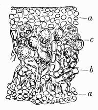
Fig. 36. Vertical section of young stage of stratose thallus (Xanthoria parietina Th. Fr.). a, plectenchyma of cortex; b, medullary hyphae; c, gonidial zone. × 500 (after Schwendener).
Lindau[311] proposed the name “plectenchyma” for the tangled weft of hyphae that is the principal tissue system in fungi as well as lichens. The more elaborated pseudoparenchyma he designates as “paraplectenchyma,” while the term “prosoplectenchyma” he reserved for the fibrous or chondroid strands of compact filaments that occur frequently in the thallus of the larger fruticose lichens, and are of service in strengthening the fronds. The term plectenchyma is now generally used for pseudoparenchyma.
B. Types of Thallus
Three factors, according to Reinke[312], have been of influence in determining the thalline development. The first, and most important, is the necessity to provide for the work of photosynthesis on the part of the alga. There is also the building up of a tissue that should serve as a storage of reserve material, essential in a plant the existence of which is prolonged far beyond the natural duration of either of the component organisms; and, finally, there is the need of protecting the long-lived plant as a whole though more particularly the alga.
Wallroth was the first to make a comparative study of the different lichen thalli. He distinguished those lichens in which the green cells and the colourless filaments are interspersed equally through the entire thallus as “homoiomerous” (Fig. 2), and those in which there are distinct layers of cortex, gonidia, and medulla, as “heteromerous” (Fig. 1), terms which, though now considered of less importance in classification, still persist and are of service in describing the position of the alga with regard to the general structure. A less evident definition of the different types of thallus has been proposed by Zukal[313] who divides them into “endogenous” and “exogenous.”
a. Endogenous Thallus. The term has been applied to a comparatively small number of homoiomerous lichens in which the alga predominates in the development, and determines the form of the thallus. These algae, members of the Myxophyceae, are extremely gelatinous, and the hyphae grow alongside or within the gelatinous sheath. In the simpler forms the vegetative structure is of the most primitive type: the alga retains its original character almost unchanged, and the ascomycetous fungus grows along with and beside it (Fig. 4). Such are the minutely tufted thalli of Thermutis and Spilonema and the longer strands of Ephebe, in which the associated Scytonema or Stigonema, filamentous blue-green algae, though excited to excessive growth, scarcely lose their normal appearance, making it difficult at times to recognize the lichenoid character unless the fruits also are present.
Equally primitive in most cases is the structure of the thallus associated with Gloeocapsa. The resulting lichens, Pyrenopsis, Psorotichia, etc. are simply gelatinous crusts of the alga with a more or less scanty intermingling of fungal hyphae.
In the Collemaceae, the gonidial cells of which are species of Nostoc (Fig. 2), there appears a more developed thallus; but in general, symbiosis in Collema has wrought the minimum of change in the habit of the alga, hence the indecision of the earlier botanists as to the identification and classification of Nostoc and Collema. Though in many of the species of the genus Collema no definite tissue is formed, yet, under the influence of symbiosis, the plants become moulded into variously shaped lobes which are specifically constant. In some species there is an advance towards more elaboration of form in the protective tissues of the apothecia, a layer of thin-walled plectenchyma being occasionally formed beneath or around the fruit as in Collema granuliferum.
In all these lichens, it is only the thallus that can be considered as primitive: the fruit is a more or less open apothecium—more rarely a perithecium—with a fully developed hymenium. Frequently it is provided with a protective thalline margin.
b. Exogenous Thallus. In this group, composed almost exclusively of heteromerous lichens, Zukal includes all those in which the fungus takes the lead in thalline development. He counts as such Leptogium, a genus closely allied to Collema but with more membranous lobes, in which the short terminal cells of the hyphae have united to form a continuous cortex. A higher development, therefore, becomes at once apparent, though in some genera, as in Coenogonium, the alga still predominates, while the simplest forms may be merely a scanty weft of filaments associated with groups of algal cells. Such a thallus is characteristic of the Ectolechiaceae, and some Gyalectaceae, etc., which have, indeed, been described by Zahlbruckner[314] as homoiomerous though their gonidia belong to the non-gelatinous Chlorophyceae.
Heteromerous lichens have been arranged by Hue[315] according to their general structure in three great series:
1. Stratosae. Crustaceous, squamulose and foliose lichens with a dorsiventral thallus.
2. Radiatae. Fruticose, shrubby or filamentous lichens with a strap-shaped or cylindrical thallus of radiate structure.
3. Stratosae-Radiatae. Primary dorsiventral thallus, either crustaceous or squamulose, with a secondary upright thallus of radiate structure called the podetium (Cladoniaceae).
II. STRATOSE THALLUS
1. CRUSTACEOUS LICHENS
A. General Structure
In the series “Stratosae,” the plant is dorsiventral, the tissues forming the thallus being arranged more or less regularly in strata one above the other (Fig. 37). On the upper surface there is a hyphal layer constituting a cortex, either rudimentary or highly elaborated; beneath the cortex is situated the gonidial zone composed of algae and hyphae in close association; and deeper down the medulla, generally a loose tissue of branching hyphae. The lower cortex which abuts on the medulla may be as fully developed as the upper or it may be absent.
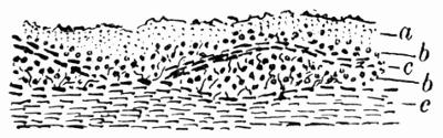
Fig. 37. Vertical section of crustaceous lichen (Lecanora subfusca var. chlarona Hue) on bark. a, lichen cortex; b, gonidia; c, cells of the periderm. × 100.
The growing tissue is chiefly marginal; the hyphae on the outer edge remain “meristematic”[316] and provide for horizontal as well as vertical extension; and there is also continual increase of the algal cells. There is in addition a certain amount of intercalary growth due to the activity of the gonidial tissue, both algal and fungal, providing for the renewal of the cortex, and even interposing new tissue.
B. Saxicolous Lichens
a. Epilithic Lichens. The crustaceous lichens forming this group spread over the rock surfaces. The support must be stable to allow the necessary time for the slowly developing organism, and therefore rocks that are friable or subject to continual weathering are bare of lichens.
aa. Hypothallus or Prothallus. The first stage of growth in the lichen thallus can be most easily traced in epilithic crustaceous species, especially in those that inhabit a smooth rock surface. The spore, on germination, produces a delicate branching septate mycelium which radiates on all sides, as was so well observed and recorded by Tulasne[317] in Verrucaria muralis (Fig. 14). Zukal[318] has called this first beginning the prothallus. In time the cell-walls of the filaments become much thicker and though, in some species, they remain colourless, in others they become dark-coloured, all except the extreme tips, owing to the presence of lichen pigments—a provision, Zukal[319] considers, to protect them against the ravages of insects, etc. The prothallic filaments adhere closely to the substratum and the branching becomes gradually more dendroid in form, though sometimes hyphae are united into strands, or even form a kind of plectenchymatous tissue. This purely hyphal stage may persist for long periods without much change. In time there may be a fortuitous encounter with the algae (Fig. 38 A) which become the gonidia of the plant. Either these have been already established on the substratum as free-growing organisms, or, as accidentally conveyed, they alight on the prothallus. The contact between alga and hypha excites both to active growth and to cell-division; and the rapidly multiplying gonidia are as speedily surrounded by the vigorously growing hyphal filaments.
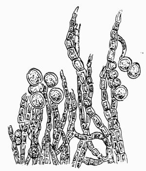
Fig. 38 A. Hypothallus of Rhizocarpon confervoides DC., from the extreme edge, with loose gonidia × 600.
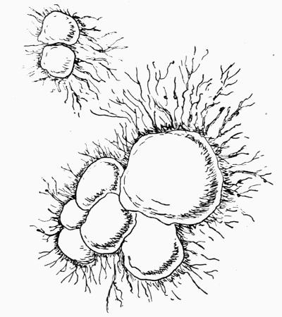
Fig. 38 B. Young thallus of Rhizocarpon confervoides DC., with various centres of gonidial growth on the hypothallus × 30.
Schwendener[320] has thus described the origin and further development of prothallus and gonidia: on the dark-coloured proto- or prothallus, he noted small nestling groups of green cells which he, at that time, regarded as direct outgrowths from the lichen hyphae. These gonidial cells, increasing by division, multiplied gradually and gathered into a connected zone. He also observed that the hyphae in contact with the gonidia became more thin-walled and produced many new branches. Some of these newly formed branches grow upwards and form the cortex, others grow downwards and build up the medulla or pith; the filaments at the circumference continue to advance and may start new centres of gonidial activity (Fig. 38 B). In many species, however, this prothallus or, as it is usually termed at this stage, the hypothallus, becomes very soon overgrown and obscured by the vigorous increase of the first formed symbiotic tissue and can barely be seen as a white or dark line bordering the thallus (Fig. 39). Schwendener[321] has stated that probably only lichens that develop from the spore are distinguished by a protothallus, and that those arising from soredia do not form these first creeping filaments.
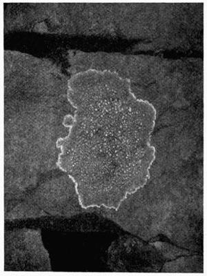
Fig. 39. Lecanora parella Ach. Determinate thallus with white bordering hypothallus, reduced (M. P., Photo.).
bb. Formation of crustaceous tissues. Some crustaceous lichens have a persistently scanty furfuraceous crust, the vegetative development never advancing much beyond the first rather loose association of gonidia and hyphae; but in those in which a distinct crust or granules are formed, three different strata of tissue are discernible:
1st. An upper cortical tissue of interlaced hyphae with frequent septation and with swollen gelatinous walls, closely compacted and with the lumen of the cells almost obliterated, not unfrequently a layer of mucilage serving as an outer cuticle. This type of cortex has been called by Hue[322] “decomposed.” It is subject to constant surface weathering, thin layers being continually peeled off, but it is as continually being renewed endogenously by the upward growth of hyphae from the active gonidial zone. Exceptions to this type of cortex in crustaceous lichens are found in some Pertusariae where a secondary plectenchymatous cortex is formed, and in Dirina where it is fastigiate[323] as in Roccella.
2nd. The gonidial zone—a somewhat irregular layer of algae and hyphae below the cortex—which varies in thickness according to the species.
3rd. The medullary tissue of somewhat loosely intermingled branching hyphae, with generally rather swollen walls and narrow lumen. It rests directly on the substratum and follows every inequality and crack so closely, even where it does not penetrate, that the thallus cannot be detached without breaking it away.
In Verrucaria mucosa, a smooth brown maritime lichen found on rocks between tide-levels, the thallus is composed of tightly packed vertical rows of hyphae, slender, rather thin-walled, and divided into short cells. The gonidia are chiefly massed towards the upper surface, but they also occur in vertical rows in the medulla. One or two of the upper cells are brown and form an even cortex. The same formation occurs in some other sea-washed species; the arrangement of the tissue elements recalls that of crustaceous Florideae such as Hildenbrandtia, Cruoria, etc.
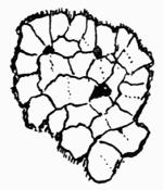
Fig. 40. Young thallus of Rhizocarpon geographicum DC., with primary and subsequent (dotted lines) areolation × 5.
cc. Formation of areolae. An “areolate” thallus is seamed and scored by cracks of varying width and depth which divide it into minute compartments. These cracks or fissures or chinks originate in two ways depending on the presence or absence of hypothallic hyphae. Where the hypothallus is active, new areolae arise when the filaments encounter new groups of algae. More vigorous growth starts at once and proceeds on all sides from these algal centres, until similarly formed areolae are met, a more or less pronounced fissure marking the limits of each. This primary areolation, termed rimose or rimulose, is well seen in the thin smooth thallus of Rhizocarpon geographicum (Fig. 40); but the first-formed areolae are also very frequently slightly marked by subsequent cracks due to unequal growth. The areolation caused by primary growth conditions tends to become gradually less obvious or to disappear altogether.
Secondary areolation is due to unequal intercalary growth of the otherwise continuous thallus[324]. A more active increase of any minute portions provokes a tension or straining of the cortex between the swollen areas and the surrounding more sluggish tissues; the surface layers give way and chinks arise, a condition described by older lichenologists as “rimose-diffract” or sometimes as “rhagadiose.” The thallus is generally thicker, more broken and granular in the older central parts of the lichen. Towards the circumference, where the tissue is thinner and growth more equal, the chinks are less evident. Sometimes the more vigorously growing areolae may extend over those immediately adjoining, in which case the covered portions become brown and their gonidia gradually disappear.
Strongly marked intersecting lines, similar to those round the margin of the thallus, are formed when hypothalli that have themselves started from different centres touch each other. A large continuous patch of crustaceous thallus may thus be composed of many individuals (Fig. 41).
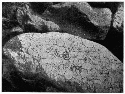
Fig. 41. Rhizocarpon geographicum DC. on boulder, reduced (M. P., Photo.).
b. Endolithic Lichens. In many species, only the lower hyphae penetrate the substratum either of rock or soil. In a few, more especially those growing on limestone, the greater part or even the whole of the vegetative thallus and sometimes also the fruits are, to some extent, immersed in the rock. It has now been demonstrated that a number of lichens, formerly described as athalline, possess a considerable vegetative body which cannot be examined until the limestone in which they are embedded is dissolved by acids. One such species, Petractis (Gyalecta) exanthematica, studied by Steiner[325] and later by Fünfstück[326], is associated with the blue-green filamentous alga, Scytonema, and is homoiomerous in structure, the alga growing through and permeating the whole of the embedded thallus. A partly homoiomerous thallus, associated with Trentepohlia, has been described by Bachmann[327]. He found the bright-yellow filaments of the alga covering the surface of a calcareous rock. By reason of their apical growth, they pierced the rock and dissolved a way for themselves, not only among the loose particles, but right through a clear calcium crystal reaching generally to a depth of about 200µ, though isolated threads had gone 350µ below the surface. Near the outside the tendency was for the algae to become stouter and to increase by intercalary growth and by budded yeast-like outgrowths; lower down they were somewhat smaller. The hyphae that became united with the algae were unusually slender and were characterized by frequent anastomoses. They closely surrounded the gonidia and also filled the loose spaces of the limestone with their fine thread-like strands. Though oil was undoubtedly present in the lower hyphae there were no swollen nor sphaeroid cells[328]. Some interesting experiments with moisture proved that the part of the rock permeated with the lichen absorbed much more water and retained it longer than the part that was lichen-free.
Generally the embedded tissues follow the same order as in other crustaceous lichens: an upper layer of cortical hyphae, next a gonidial zone, and beneath that an interlaced tissue of medullary or rhizoidal hyphae which often form fat-cells[328]. Friedrich[329] has given measurements of the immersed thallus of Lecanora (Biatorella) simplex: under a cortical layer of hyphae there was a gonidial zone 600-700µ thick, while the lower hyphae reached a depth of 12 mm.; he has also recorded an instance of a thallus reaching a depth of 30 mm.
On siliceous rocks such as granite, rhizoidal hyphae penetrate the rock chiefly between the thin separable flakes of mica. Bachmann[330] has recognized in these conditions three distinct series of cell-formations: (1) slender long-celled sparsely branched hyphae which form a network by frequent anastomoses; (2) further down, though only occasionally, hyphae with short thick-walled bead-like cells; and (3) beneath these, but only in or near mica crystals, spherical cells containing oil or some albuminous substance.
c. Chemical Nature of the Substratum. Lichens growing on calcareous rocks or soils are more or less endolithic, those on siliceous rocks are largely epilithic, but Bachmann[331] found that the mica crystals in granite were penetrated, much in the same way as limestone, by the lichen hyphae. These travel through the mica in all directions, though they tend to follow the line of cleavage, thus taking the direction of least cohesion. He found that oil-hyphae were formed, and also certain peculiar bristle-like terminal branches; in other cases there were thin layers of plectenchyma, and gonidia were also present. If however felspar or quartz crystals, no matter how thin, blocked the way, further growth was arrested, the hyphae being unable to pierce through or even to leave any trace on the quartz[332]. On granite containing no mica constituents the hyphae can only follow the cracks between the different impenetrable crystals.
Stahlecker[333] has confirmed Bachmann’s observations, but he considers that the difference in habit and structure between the endolithic and epilithic series of lichens is due rather to the chemical than to the physical nature of the substratum. Thus in a rock of mixed composition such as granite, the more basic constituents are preferred by the hyphae, and are the first to be surrounded: mica, when present, is at once penetrated; particles of hornblende, which contain 40 to 50 per cent. only of silicic acid, are laid hold of by the filaments of the lichen before the felspar, of which the acid content is about 60 per cent.; quartz grains which are pure silica are attacked last of all, though in the course of time they also become corroded.
The character of the substratum also affects to a great extent the comparative development of the different thalline layers: the hyphal tissues in silicicolous lichens are much thinner than in lichens on limestone, and the gonidial zone is correspondingly wider. In a species of Staurothele on granite, Stahlecker[333] estimated the gonidial zone to be about 600 µ thick, while the lower medullary hyphae, partly burrowing into the rock, measured about 6 mm. Other measurements at different parts of the thallus gave a rhizoidal depth of 3 mm., while on a more finely granular substratum, with a gonidial zone of 350 µ, the rhizoidal hyphae measured only 1-1/2 mm. On calcareous rocks, on the contrary, with a gonidial zone that is certainly no larger, the hyphal elements penetrate the rock to varying depths down to 15 mm. or even more.
Lang[334] has recorded equally interesting measurements for Sarcogyne (Biatorella) latericola: on slaty rock which contained no mixture of lime, the gonidial zone had a thickness of 80 µ, a considerable proportion of the very thin thallus. Fünfstück[335] has indeed suggested that this lichen on acid rocks is only a starved condition of Sarcogyne (Biatorella) simplex, which on calcareous rocks, though with a broader gonidial zone, has, as noted above, a correspondingly much larger hyphal tissue.
Stahlecker’s theory is that the hyphae require more energy to grow in the acid conditions that prevail in siliceous rocks, and therefore they make larger demands on the algal symbionts. It follows that the latter must be stimulated to more abundant growth than in circumstances favourable to the fungus, such as are found in basic (calcareous) rocks; he concludes that on the acid (siliceous) rocks, the epilithic or superficial condition is not only a physical but a biological necessity, to enable the algae to grow and multiply in a zone well exposed to light with full opportunity for active photosynthesis and healthy increase.
C. Corticolous Lichens
The crustaceous lichens occurring on bark or on dead wood, like those on rocks, are either partly or wholly immersed in the substratum (hypophloeodal), or they grow on the surface (epiphloeodal); but even those with a superficial crust are anchored by the lower hyphae which enter any crack or crevice of wood or bark and so securely attach the thallus, that it can only be removed by cutting away the underlying substance.
a. Epiphloeodal Lichens. These lichens originate in the same way as the corresponding epilithic series from soredia or from germinating spores, and follow the same stages of growth; first a hypothallus with subsequent colonization of gonidia, the formation of granules, areolae, etc. The small compartments are formed as primary or secondary areolae; the larger spaces are marked out by the encounter of hypothalli starting from different centres.
The thickness of the thallus varies considerably according to the species. In some Pertusariae with a stoutish irregular crust there is a narrow amorphous cortical layer of almost obliterated cells, a thin gonidial zone about 35 µ in width and a massive rather dense medulla of colourless hyphae. Darbishire[336] has described and figured in Varicellaria microsticta, one of the Pertusariaceae, single hyphae that extend like beams across the wide medulla and connect the two cortices. In some Lecanorae and Lecideae there is, on the contrary, an extremely thin thallus consisting of groups of algae and loose fungal filaments, which grow over and between the dead cork cells of the outer bark. On palings, there is often a fairly substantial granular crust present, with a gonidial zone up to about 80 µ thick, while the underlying or medullary hyphae burrow among the dead wood fibres.
b. Hypophloeodal Lichens. These immersed lichens are comparable with the endolithic species of the rock formations, as their thallus is almost entirely developed under the outer bark of the tree. They are recognizable, even in the absence of any fructification, by the somewhat shining brownish, white or olive-green patches that indicate the underlying lichen. This type of thallus occurs in widely separated families and genera, Lecidea, Lecanora, etc., but it is most constant in Graphideae and in those Pyrenolichens of which the algal symbiont belongs to the genus Trentepohlia. The development of these lichens is of peculiar interest as it has been proved that though both symbionts are embedded in the corky tissues, the hyphae arrive there first, and, at some later stage, are followed by the gonidia. There is therefore no question of the alga being a “captured slave” or “unwilling mate.”
Frank[337] made a thorough study of several subcortical forms. He found that in Arthonia radiata, the first outwardly visible indication of the presence of the lichen on ash bark was a greenish spot quite distinct from the normal dull-grey colour of the periderm. Usually the spots are round in outline, but they tend to become ellipsoid in a horizontal direction, being influenced by the growth in thickness of the tree. At this early stage only hyphae are present; Bornet[338] as well as Frank described the outer periderm cells as penetrated and crammed with the colourless slender filaments. Lindau[339], in a more recent work, disputes that statement: he found that the hyphae invariably grew between the dead cork cells, splitting them up and disintegrating the bark, but never piercing the membranes. The purely prothallic condition, as a weft of closely entangled hyphae, may last, Frank considers, for a long period in an almost quiescent condition—possibly for several years—before the gonidia arrive.
It is always difficult to observe the entrance of the gonidia but they seem to spread first under the second or third layers of the periderm. With care it is possible to trace a filament of Trentepohlia from the surface downwards, and to see that the foremost cell is really the growing and advancing apex of the creeping alga. Both symbionts show increased vigour when they encounter each other: the thallus at once develops in extent and in depth, and, ultimately, reproductive bodies are formed. In some species the apothecia or perithecia alone emerge above the bark, in others the outer peridermal cells are thrown off, and the thallus thus becomes superficial to some extent as a white scurfy or furfuraceous crust.
The change from a hypophloeodal to a partly epiphloeodal condition depends largely on the nature of the bark. Frank[337] found that Lecanora pallida remained for a long time immersed when growing on the thick rugged bark of oak trunks. When well lighted, or on trees with a thin periderm, such as the ash, the lichen emerges much earlier and becomes superficial.
Black (or occasionally white) lines intersect the thallus and mark, as in saxicolous lichens (Fig. 41), the boundary lines between different individuals or different species. The pioneer hyphae of certain lichens very frequently become dark-coloured, and Bitter[340] has suggested as the reason for this that in damp weather the hypothallic growth is exceptionally vigorous. When dry weather supervenes, with high winds or strong sunshine, the outlying hyphae, unprotected by the thallus, become dark-coloured. On the return of more normal conditions the blackened tips are thrown off. Bitter further states that species of Graphideae do not form a permanent black limiting line when they grow in an isolated position: it is only when their advance is checked by some other thallus that the dark persistent edge appears, a characteristic also to be seen in the crust of other lichens. The dark boundary is always more marked in sunny exposed situations: in the shade, the line is reduced to a mere thread.
Bitter’s restriction of black boundary lines to cases of encountering thalli only, would exclude the comparison one is tempted to make between the advancing hyphae of lichens and those of many woody fungi where the extreme edge of the white invaded woody tissue is marked by a dark line. In the latter case however it is the cells of the host that are stained black by the fungus pigment.
2. SQUAMULOSE LICHENS
A. Development of the Squamule
The crustaceous thallus is more or less firmly adherent to, or confused with, the substratum. Further advance to a new type of thallus is made when certain hyphal cells of soredium or granule take the lead in an ascending direction both upwards and outwards. As growth becomes definitely apical or one-sided, the structure rises free from the substratum, and small lobules or leaflet-like squamules are formed. Each squamule in this type of thallus is distinct in origin and not merely the branch of a larger whole.
In a few lichens the advance from the crustaceous to the squamulose structure is very slight. The granules seem but to have been flattened out at one side, and raised into minute rounded projections such as those that compose the thallus of Lecanora badia generally described as “subsquamulose.” The squamulose formation is more pronounced in Lecidea ostreata, and in some species of Pannaria; and the whole thallus may finally consist of small separate lobes as in Lecidea lurida, Lecanora crassa, L. saxicola, species of Dermatocarpon and the primary thallus of the Cladoniae. Most of these squamules are of a firm texture and more or less round in outline; in some species of Cladonia, etc., they are variously crenate, or cut into pinnate-like leaflets. Squamulose lichens grow mostly on rocks or soil, occasionally on dead wood, and are generally attached by single rhizoidal hyphae, either produced at all points of the under surface, or from the base only, growth in the latter case being one-sided. In a few instances, as in Heppia Guepini, there is a central hold-fast.
A frequent type of squamulose thallus is that termed “placodioid,” or “effigurate,” in which the squamulose character is chiefly apparent at the circumference. The thallus is more or less orbicular in outline; the centre may be squamulose or granular and cracked into areolae; the outer edge is composed of radiating lobules closely appressed to the substratum (Fig. 42).
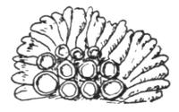
Fig. 42. Placodium murorum DC. Part of placodioid thallus with apothecia × 2.
All lichens with this type of thallus were at one time included in the genus Placodium, now restricted by some lichenologists to squamulose or crustaceous species with polarilocular spores. Many of them rival Xanthoria parietina in their brilliant yellow colouring.
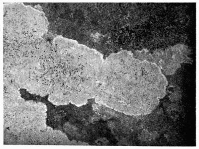
Fig. 43. Lecania candicans A. Zahlbr., with placodioid thallus, reduced (S. H., Photo.).
There are also greyish-white effigurate lichens such as Lecanora saxicola, Lecania candicans (Fig. 43) and Buellia canescens, well-known British species.
B. Tissues of Squamulose Thallus
The anatomical structure of the squamules is in general somewhat similar to that of the crustaceous thallus: an upper cortex, a gonidial zone, and below that a medullary layer of loose hyphae with sometimes a lower cortex.
1. The upper cortex, as in crustaceous lichens, is generally of the “decomposed”[341] or amorphous type: interlaced hyphae with thick gelatinous walls. A more highly developed form is apparent in Parmeliella and Pannaria where the upper cortex is formed of plectenchyma, while in the squamules of Heppia the whole structure is built up of plectenchyma, with the exception of a narrow band of loose hyphae in the central pith.
2. The gonidia are Myxophyceae or Chlorophyceae; the squamules in some instances may be homoiomerous as in Lepidocollema, but generally they belong to the heteromerous series, with the gonidia in a circumscribed zone, and either continuous or in groups. Friedrich[342] held that, as in crustaceous lichens the development of the gonidial as compared with the other tissues depended on the substratum. The squamules of Pannaria microphylla on sandstone were 100 µ thick, and the gonidial layer occupied 80 or 90 µ of the whole[343]. With that may be compared Placodium Garovagli on lime-containing rock: the gonidial layer measured only 50 µ across, the pith hyphae 280 µ and the rhizoidal hyphae that penetrated the rock 500 µ.
3. The medullary layer, as a rule, is of closely compacted hyphae which give solidity to the squamules; in those of Heppia it is almost entirely formed of plectenchyma.
4. The lower cortex is frequently little developed or absent, especially when the squamules are closely applied to the support as in some species of Dermatocarpon. In some of the squamulose Lecanorae (L. crassa and L. saxicola) the lowest hyphae are somewhat more closely interwoven; they become brown in colour, and the lichen is attached to the substratum by rhizoid-like branches. In Lecanora lentigera there is a layer of parallel hyphae along the under surface. Further development is reached when a plectenchyma of thick-walled cells is formed both above and below, as in Psoroma hypnorum, though on the under surface the continuity is often broken. The squamules of Cladoniae are described under the radiate-stratose series.
3. FOLIOSE LICHENS
A. Development of foliose Thallus
The larger leafy lichens are occasionally monophyllous and attached at a central point as in Umbilicaria, but mostly they are broken up into lobes which are either imbricate and crowded, or represent the dividing and branching of the expanding thallus at the circumference. They are horizontal spreading structures, with marginal and apical growth. The several tissues of the squamule are repeated in the foliose thallus, but further provision is made to meet the requirements of the larger organism. There is the greater development of cortical tissue, especially on the lower surface, and the more abundant formation of rhizoidal organs to attach the large flat fronds to the support. There are also various adaptations to secure the aeration of the internal tissues[344].
B. Cortical Tissues
Schwendener[345] was the first who, with the improved microscope, made a systematic study of the minute structure of lichens. He examined typical species in genera of widely different groups and described their anatomy in detail. The most variable and perhaps the most important of the tissues of lichens is the cortex, which is most fully developed in the larger thalli, and as the same type of cortical structures recurs in lichens widely different in affinity as well as in form, it seems well to group together here the ascertained facts about these covering layers.
a. Types of Cortical Structure. Zukal[346], and more recently Hue[347], have made independent studies in the comparative morphology of the thallus and have given particular attention to the different varieties of cortex. They each find that the variations come under a definite series of types. Zukal recognized five of these:
1. Pseudoparenchymatous (plectenchyma): by frequent septation of regularly arranged hyphae and by coalescence a kind of continuous cell-structure is formed.
2. Palisade cells: the outer elongate ends of the hyphae lie close together in a direction at right angles to the surface of the thallus and form a coherent row of parallel cells.
3. Fibrous: the cortical hyphae lie in strands of fine filaments parallel with the surface of the thallus.
4. Intricate: hyphae confusedly interwoven and becoming dark in colour form the lower cortex of some foliose lichens.
These four types, Zukal finds, are practically without interstices in the tissue and form a perfect protection against excessive transpiration. He adds yet another form:
5. A cortex formed of hyphae with dark-coloured swollen cells, which is not a protection against transpiration. It occurs among lower crustaceous forms.
Hue has summed up the different varieties under four types, but as he has omitted the “fibrous” cortex, we arrive again at five different kinds of cortical formation, though they do not exactly correspond to those of Zukal. A definite name is given to each type:
1. Intricate: an intricate dense layer of gelatinous-walled hyphae, branching in all directions, but not coalescent (Fig. 44). This rather unusual type of cortex occurs in Sphaerophorus and Stereocaulon, both of which have an upright rigid thallus (fruticose).
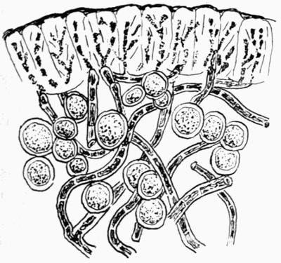
Fig. 44. Sphaerophorus coralloides Pers. Transverse section of cortex and gonidial layer near the growing point of a frond × 600.
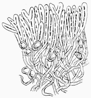
Fig. 45. Roccella fuciformis DC. Transverse section of cortex near the growing point of a frond × 600.
2. Fastigiate: the hyphae bend outwards or upwards to form the cortex. A primary filament can be distinguished with abundant branches, all tending in the same direction; anastomosis may take place between the hyphae. The end branches are densely packed, though there are occasional interstices (Fig. 45). Such a cortex occurs in Thamnolia; in several genera of Roccellaceae—Roccellographa, Roccellina, Reinkella, Pentagenella, Combea, Schizopelte and Roccella—and also in the crustaceous genus Dirina. The fastigiate cortex corresponds with Zukal’s palisade cells.
3. Decomposed: in this, the most frequent type of cortex, the hyphae that travel up from the gonidial layer become irregularly branched and frequently septate. The cell-walls of the terminal branches become swollen into a gelatinous mass, the transformation being brought about by a change in the molecular constituents of the cell-walls which permits the imbibition and storage of water. The tissue, owing to the enormous increase of the wall, is so closely pressed together that the individual hyphae become indistinct; the cell-lumen finally disappears altogether, or, at most, is only to be detected in section as a narrow disconnected dark streak. The decomposed cortex is characteristic of many lichens, crustaceous (Fig. 46) and squamulose, as well as of such highly developed genera as Usnea, Letharia, Ramalina, Cetraria, Evernia and certain Parmeliae.
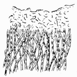
Fig. 46. Lecanora glaucoma var. corrugata Nyl. Vertical section of cortex × 500 (after Hue).
Zukal took no note of the decomposed cortex but the omission is intentional and is due to his regarding the structure of the youngest stages of the thallus near the growing point as the most typical and as giving the best indication as to the true arrangement of hyphae in the cortex. He thus describes palisade tissue as the characteristic cortex of Evernia, since the formation near the growing point of the fronds is somewhat palisade-like and he finds fibrous cortex at the tips of Usnea filaments. In both these instances Hue has described the cortex as decomposed because he takes account only of the fully formed thallus in which the tissues have reached a permanent condition.
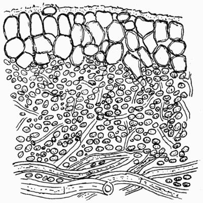
Fig. 47. Peltigera canina DC. Vertical section of cortex and gonidial zone × 600.
4. Plectenchymatous: the last of Hue’s types corresponds with the first described by Zukal. It is the result of the lateral coherence and frequent septation of the hyphae into short almost square or rounded cells (Fig. 47). The simplest type of such a cortex can be studied in Leptogium, a genus of gelatinous lichens in which the tips of the hyphae are cut off at the surface by one or more septa. The resulting cells are wider than the hyphae and they cohere together to form, in some species, disconnected patches of cells; in others, a continuous cortical covering one or more cells thick, while in the margin of the apothecium they form a deep cellular layer. The cellular type of cortex is found also, as already stated, in some crustaceous Pertusariae, and in a few squamulose genera or species. It forms the uppermost layer of the Peltigera thallus and both cortices of many of the larger foliose lichens such as Sticta, Parmelia, etc.
5. The “fibrous” cortex must be added to this series, as was pointed out by Heber Howe[348] who gave the less appropriate designation of “simple” to the type. It consists of long rather sparingly branched slender hyphae that grow in a direction parallel with the surface of the thallus (Fig. 48). It is characteristic of several fruticose and foliose lichens with more or less upright growth, such as we find in several of the Physciae, and in the allied genus Teloschistes, in Alectoria, several genera of Roccellaceae, in Usnea longissima and in Parmelia pubescens, etc. Zukal would have included all the Usneae as the tips are fibrous.
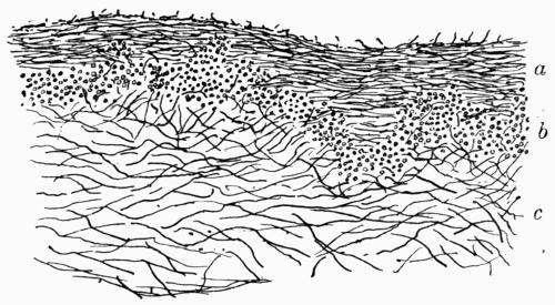
Fig. 48. Physcia ciliaris DC. Vertical section of thallus. a, cortex; b, gonidial zone; c, medulla. × 100.
More than one type of cortex, as already stated, may appear in a genus: a striking instance of variability occurs in Solorina where, as Hue[349] has pointed out, the cortex of S. octospora is fastigiate, that of all the other species being plectenchymatous. Cortical development is a specific rather than a generic characteristic.
b. Origin of Variation in Cortical Structure. The immediate causes making for differentiation in cortical development are: the prevailing direction of growth of the hyphae as they rise from the gonidial zone; the amount of branching and the crowding of the filaments; the frequency of septation; and the thickening or degeneration of the cell-walls which may become almost or entirely mucilaginous. In the plectenchymatous cortex, the walls may remain quite thin and the cells small as in Xanthoria parietina, or the walls may be much thickened as in both cortices of Sticta. As a result of stretching the cell may increase enormously in size: in some instances where the internal hyphae are about 3 µ to 4 µ in width, the cortical cells formed from these hyphae may have a cell cavity 15 µ to 16 µ in diameter.
c. Loss and Renewal of Cortex. Very frequently the cortex is covered over by a layer of homogeneous mucilage which forms an outer cuticle. It arises from the continual degeneration of the outer cell-walls and it is liable to friction and removal by atmospheric agency as was first described by Schwendener[350] in the weather-beaten cortex of Umbilicaria pustulata. He had noted the irregular jagged outline of the cross section of the thallus, and he then suggested, as the probable reason, the decay of the outer rind with the constant renewal of it by the hyphae from the underlying gonidial zone, though he was unable definitely to prove his theory. The peeling of the dead outer layer (with its replacement by new tissue) has however been observed many times since his day. It has been described by Darbishire[351] in Pertusaria: in that genus there is at first a primary cortex formed of hyphae that grow in a radial direction, parallel to the surface of the thallus. The walls of these hyphae become gradually more and more mucilaginous till the cells are obliterated. Meanwhile short-celled filaments grow up in serried ranks from the gonidial layer and finally push off the dead “fibrous” cortex. The new tissue takes on a plectenchymatous character, and the outer cells in time become decomposed and provide a mucilaginous cuticle which in turn is also subject to wasting.
The same process of peeling was noted by Rosendahl[352] in some species of brown Parmeliae, where the dead tissues were thrown off in shreds, though only in isolated patches. But whether in patches or as a continuous sheath, there is constant degeneration, with continual renewal of the dead material from the internal tissues.
The cortex is the most highly developed of all the lichen structures and is of immense importance to the plant as may be judged from the various adaptations to different needs[353]. The cortical cell-walls are frequently impregnated with some dark-coloured substance which, in exposed situations, must counteract the influence of too direct sunlight and be of service in sheltering the gonidia. Lichen acids—sometimes very brightly coloured—and oxalic acid are deposited in the cortical tissues in great abundance and aid in retaining moisture; but the two chief functions to which the cortex is specially adapted are the checking of transpiration and the strengthening of the thallus against external strains.
d. Cortical Hairs or Trichomes. Though somewhat rare, cortical hairs are present on the upper surface of several foliose lichens. They take rise, in all the instances noted, as a prolongation of one of the cell-rows forming a plectenchymatous cortex.
In Peltidea (Peltigera) aphthosa they are especially evident near the growing edges of the thallus; and they take part in the development of the superficial cephalodia[354] which are a constant feature of the lichen. They tend to disappear with age and leave the central older parts of the thallus smooth and shining. In several other species of Peltigera (P. canina, etc.) they are present and persist during the life of the cortex. In these lichens the cells of the cortical tissue are thin-walled, all except the outer layer, the membranes of which are much thicker. The hairs rising from them are also thick-walled and septate. Generally they branch in all directions and anastomose with neighbouring hairs so that a confused felted tangle is formed; they vary in size but are, as a rule, about double the width of the medullary hyphae as are the cortical cells from which they rise. They disappear from the thallus, frequently in patches, probably by weathering, but over large surfaces, and especially where any inequality affords a shelter, they persist as a soft down.
Hairs are also present on the upper surface of some Parmeliae. Rosendahl[355] has described and figured them in P. glabra and P. verruculifera—short pointed unbranched hyphae, two or more septate and with thickened walls. They are most easily seen near the edge of the thallus, though they persist more or less over the surface; they also grow on the margins of the apothecia. In P. verruculifera they arise from the soredia; in P. glabra a few isolated hairs are present on the under surface.
In Nephromium tomentosum there is a scanty formation of hairs on the upper surface. They are abundant on the lower surface, and function as attaching organs. A thick tomentum of hairs is similarly present on the lower surface of many of the Stictaceae either as an almost unbroken covering or in scattered patches. In several species of Leptogium they grow out from the lower cortical cells and attach the thin horizontal fronds; and very occasionally they are present in Collema.
C. Gonidial Tissues
With the exception of some species of Collema and Leptogium lichens included under the term foliose, are heteromerous in structure, and the algae that form the gonidial zone are situated below the upper cortex and, therefore, in the most favourable position for photosynthesis. Whether belonging to the Myxophyceae or the Chlorophyceae, they form a green band, straight and continuous in some forms, in others somewhat broken up into groups. In certain species they push up at intervals among the cortical cells, as in Gyrophora and in Parmelia tristis. In Solorina crocea a regular series of gonidial pyramids rises towards the upper surface. The green cells are frequently more dense at some points than at others, and they may penetrate in groups well into the medulla.
The fungal tissue of the gonidial zone is composed of hyphae which have thinner walls, and are generally somewhat loosely interlacing. In Peltigera[356] the gonidial hyphae are so connected by frequent branching and by anastomosis that a net-like structure is formed, in the meshes of which the algae—a species of Nostoc—are massed more or less in groups. In lichens with a plectenchymatous cortex, the cellular tissue may extend downwards into the gonidial zone and the gonidia thus become enmeshed among the cells, a type of formation well seen in the squamulose species, Dermatocarpon lachneum and Heppia Guepini, where the massive plectenchyma of both the upper and lower cortices encroaches on the pith. In Endocarpon and in Psoroma the gonidia are also surrounded by short cells.
A similar type of structure occurs in Cora Pavonia, one of the Hymenolichenes: the gonidial hyphae in that species form a cellular tissue in which are embedded the blue-green Chroococcus cells[357].
D. Medulla and Lower Cortex
a. Medulla. The hyphal tissue of the dorsiventral thallus that lies between the gonidial zone and the lower cortex or base of the plant is always referred to as the medulla or pith. It is, as a rule, by far the most considerable portion of the thallus. In Parmelia caperata (Fig. 49), for instance, the lobes of which are about 300 µ thick, over 200 µ of the space is occupied by this layer. It varies however very largely in extent in different lichens according to species, and also according to the substratum. In another Parmelia with a very thin thallus, P. alpicola growing on quartzite, the medulla measures scarcely twice the width of the gonidial zone. It forms a fairly massive tissue in some of the crustaceous lichens—in some Pertusariae and Lecanorae—attaining a width of about 600 µ.
Nylander[358] distinguished three types of medullary tissue in lichens:
(1) felted, which includes all those of a purely filamentous structure;
(2) cretaceous or tartareous, more compact than the felted, and containing granular or crystalline substances as in some Pertusariae; and lastly
(3) the cellular medulla in which the closely packed hyphae are divided into short cells and a kind of plectenchyma is formed, as in Lecanora (Psoroma) hypnorum, in Endocarpon, etc.
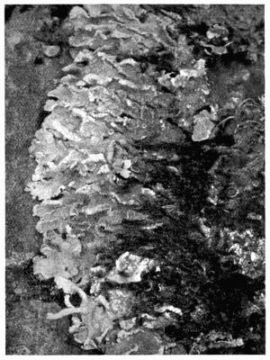
Fig. 49. Parmelia caperata Ach. (S. H., Photo.).
The felted medulla is characteristic of most lichens and is formed of loose slender branching septate hyphae with thickish walls. This interwoven hyphal texture provides abundant air-spaces.
Hue[359] has noted that the walls of the medullary hyphae in Parmeliae are smooth, unless they have been exposed to great extremes of heat or cold, when they become wrinkled or scaly. They are very thick-walled in Peltigera (Fig. 50).
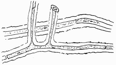
Fig. 50. Hyphae from lower medulla of Peltigera canina DC. × 600.
b. Lower Cortex. In some foliose lichens such as Peltigera there is no special tissue developed on the under surface. In Lobaria pulmonaria large patches of the under surface are bare, and the medulla is exposed to the outer atmosphere, sheltered only by its position. In some other lichens the lowermost hyphae lie closer together and a kind of felt of almost parallel filaments is formed, generally darker in colour, as in Lecanora lentigera, and in some species of Physcia.
Most frequently however the tissues of the upper cortex are repeated on the lower surface, though differing somewhat in detail. In all of the brown Parmeliae, according to Rosendahl[360], the structure is identical for both cortices, though the upper develops now hairs, now isidia, breathing pores, etc., while the lower produces rhizinae. The amorphous mucilaginous cuticle so often present on the upper surface is absent from the lower, the walls of the latter being often charged instead with dark-brown pigments.
c. Hypothallic Structures. An unusual development of hyphae from the lower cortex occurs in the genera Anzia and Pannoparmelia—both closely related to Parmelia—whereby a loose sponge-like hypothallus of anastomosing reticulate strands is formed. In one of the simpler types, Anzia colpodes, a North American species, the hyphae passing out from the lower medulla become abruptly dark-brown in colour, and are divided into short thick-walled cells. Frequent branching and anastomosis of these hyphae result in the formation of a cushion-like structure about twice the bulk of the thallus. In another species from Australia (A. Japonica) there is a lower cortex, distinct from the medulla, consisting of septate colourless hyphae with thick walls. From these branch out free filaments, similar in structure but dark in colour, which branch and anastomose as in the previous species.
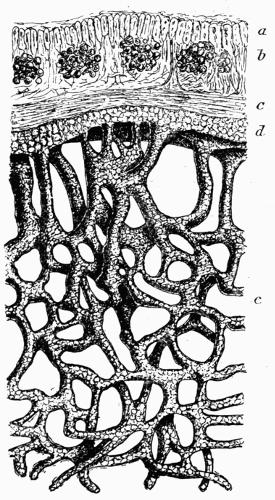
Fig. 51. Pannoparmelia anzioides Darb. Vertical section of thallus and hypothallus. a, cortex; b, gonidial zone; c, medulla; d, lower cortex; e, hypothallus. × ca. 450 (after Darbishire).
In Pannoparmelia the lower cortex and the outgrowths from it are several cells thick; they may be thick-walled as in Anzia, or they may be thin-walled as described and figured by Darbishire[361] in Pannoparmelia anzioides, a species from Tierra del Fuego (Fig. 51). A somewhat dense interwoven felt of hyphae occurs also in certain parts of the under surface of Parmelia physodes[362].
This peculiar structure, regarded as a hypothallus, is probably of service in the retention of moisture. The thick cell-walls in most of the forms suggest some such function.
E. Structures for Protection and Attachment
Such structures are almost wholly confined to the larger foliose and fruticose lichens and are all of the same simple type; they are fungal in origin and very rarely are gonidia associated with them.
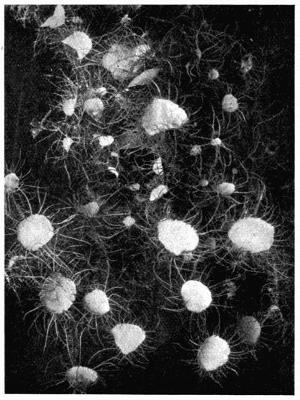
Fig. 52. Usnea florida Web. Ciliate apothecia (S. H., Photo.).
a. Cilia. In a few widely separated lichens stoutish cilia are borne, mostly on the margins of the thallus lobes, or on the margins of the apothecia (Fig. 52). They arise from the cortical cells or hyphae, several of which grow out in a compact strand which tapers gradually to a point. Cilia vary in length up to about 1 cm. or even longer. In some lichens they retain the colour of the cortex and are greyish or whitish-grey, as in Physcia ciliaris or in Physcia hispida (Fig. 110). They provide a yellow fringe to the apothecia of Physcia chrysophthalma and a green fringe to those of Usnea florida. They are dark-brown or almost black in Parmelia perlata var. ciliata and in P. cetrata, etc. as also in Gyrophora cylindrica. The fronds of Cetraria islandica and other species of the genus are bordered with short spinulose brown hairs whose main function seems to be the bearing of “pycnidia” though in many cases they are barren (Fig. 128).
Superficial cilia are more rarely formed than marginal ones, but they are characteristic of one not uncommon British species, Parmelia proboscidea (P. pilosella Hue). Scattered over the surface of that lichen are numerous crowded groups of isidia which, frequently, are prolonged upwards as dark-brown or blackish cilia. Nearly every isidium bears a small brown spot on the apex at an early stage of growth. Similar cilia are sparsely scattered over the thallus, but their base is always a rather stouter grey structure, which suggests an isidial origin. Cilia also occur on the margin of the lobes.
As lichens are a favourite food of snails, insects, etc., it is considered that these structures are protective in function, and that they impede, if they do not entirely prevent, the larger marauders in their work of destruction.
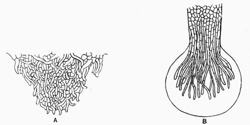
Fig. 53. Rhizoid of Parmelia exasperata Carroll (P. aspidota Rosend.). A, hyphae growing out from lower cortex × 450. B, tip of rhizoid with gelatinous sheath × 335 (after Rosendahl).
b. Rhizinae. Lichen rootlets are mainly for the purpose of attachment and have little significance as organs of absorption. They have been noted in only one crustaceous lichen, Varicellaria microsticta[363], an alpine species that spreads over bark or soil, and which is further distinguished by being provided with a lower cortex of plectenchyma. In foliose lichens they are frequently abundant, though by no means universal, and attach the spreading fronds to the support. They originate, as Schwendener[364] pointed out, from the outer cortical cells, exactly as do the cilia, and are scattered over the under surface or are confined to special areas. Rosendahl[365] has described their development in the brown species of Parmeliae: the under cortex in these lichens is formed of a cellular plectenchyma with thickish walls; the rootlets arise by the outgrowth of several neighbouring cells from some slight elevation near the edge of the thallus. Branching and interlacing of these growing rhizinal hyphae follow, the outermost frequently spreading outwards at right angles to the axis, and forming a cellular cortex. The apex of the rhizoid is generally an enlarged tuft of loose hyphae involved in mucilage (Fig. 53), a provision for securing firmer cohesion to the support; or the tips spread out as a kind of sucker. Not unfrequently neighbouring “rootlets” are connected by mucilage at the tips, or by outgrowths of their hyphae, and a rather large hold-fast sheath is formed.
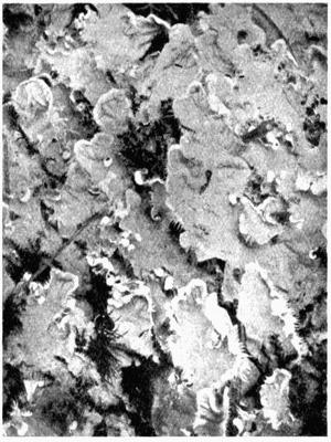
Fig. 54. Peltigera canina DC. (S. H., Photo.).
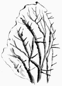
Fig. 55. Peltigera canina DC. Under surface with veins and rhizoids (after Reinke).
In species of Peltigera (Fig. 54) the rhizinae are confined to the veins or ridges (Fig. 55); they are thickish at the base, and are generally rather long and straggling. Meyer[366] states that the central hyphae are stoutish and much entangled owing to the branching and frequent anastomosis of one hypha with another; the peripheral terminal branches are thinner-walled and free. These rhizinae vary in colour from white in Peltigera canina to brown or black in other species. Most species of Peltigera spread over grass or mosses, to which they cling by these long loose “rootlets.”
Lichen rhizinae, distinguished by Reinke[367] as “aerial rhizinae,” are more or less characteristic of all the species of Parmelia with the exception of those belonging to the subgenus Hypogymnia in which they are of very rare occurrence, arising, according to Bitter[368], only in response to some external friction. They are invariably dark-coloured, rather short, about one to a few millimetres in length, and are simple or branched. The branches may go off at any angle and are sometimes curved back at the ends in anchor-like fashion. The Parmeliae grow on firm substances, trees, rocks, etc., and the irregularities of their attaching structures are conditioned by the obstacles encountered on the substratum. Not unfrequently the lobes are attached by the rhizinae to underlying portions of the thallus.
In the genus Gyrophora, the rhizinae are simple strands of hyphae (G. polyrhiza) or they are corticate structures (G. murina, G. spodochroa and G. vellea). They are also present in species of Solorina, Ricasolia, Sticta and Physcia and very sparingly in Cetraria (Platysma).
c. Haptera. Sernander[369] has grouped all the more distinctively aerial organs of attachment, apart from rhizinae, under the term “hapteron” and he has described a number of instances in which cilia and even the growing points of the thallus may become transformed to haptera or sucker-like sheaths.
The long cilia of Physcia ciliaris occasionally form haptera at their tips where the hyphae are loose and in active growing condition. Contact with some substance induces branching by which a spreading sheath arises; a plug-like process may also be developed which pierces the substance encountered—not unfrequently another lobe of its own thallus. The long flaccid fronds of Evernia furfuracea are frequently connected together by bridge-like haptera which rise at any angle of the thallus or from any part of the surface.
The spinous hairs that border the thalline margins in Cetraria may also, in contact with some body—often another frond of the lichen—form a hapteron, either while the spermogonium, which occupies the tip of the spine, is still in a rudimentary stage, or after it has discharged its spermatia. The small sucker sheath may in that case arise either from the apex of the cilium, from the wall of the spermogonium or from its base. By means of these haptera, not only different individuals become united together, but instances are given by Sernander in which Cetraria islandica, normally a ground lichen, had become epiphytic by attaching itself in this way to the trunk of a tree (Pinus sylvestris).
In Alectoria, haptera are formed at the tip of the thallus filament as an apical cone-like growth from which hyphae may branch out and penetrate any convenient object. A species of this genus was thus found clinging to stems of Betula nana. Apical haptera are very frequent in Cladonia rangiferina and Cl. sylvatica, induced here also by contact. These two plants, as well as several species of Cetraria, tend, indeed, to become entirely epiphytic on the heaths of the Calluna formations. Haptera similar to those of Alectoria occur in Usnea, Evernia, Ramalina and Cornicularia (Cetraria). In Evernia prunastri var. stictoceros, a heath form, the fronds become attached to the stems and branches of Erica tetralix by hapteroid strands of slender glutinous hyphae which persist on the frond of the lichen after it is detached as small very dark tubercles surmounted, as Parfitt[370] pointed out, by a dark-brown grumous mass of cells. Plug-like haptera may be formed at the base of Cladoniae which attach them to each other and to the substratum. The brightly coloured fronds of Letharia vulpina are attached to each other in somewhat tangled fashion by lateral bridges or by fascicles of hyphae dark-brown at the base but colourless at the apices, exactly like aerial adventitious rhizinae. They grow out from the fronds generally at or near the tips and lay hold of a neighbouring frond by means of mucilage. These haptera are evidently formed in response to friction. Haptera along with other lichen attachments have received considerable attention from Galløe[371]. He finds them arising on various positions of the lichen fronds and has classified them accordingly.
After the haptera have become attached, they increase in size and strength and supply a strong anchorage for the plant; the point of contact frequently forms a basis for renewed growth while the part beneath the hapteron may gradually die off. Haptera are more especially characteristic of fruticose lichens, but Sernander considers that the rhizinae of foliose species may function as haptera. They are important organs of tundra and heath formations as they enable the lichens to get a foothold in well-lighted positions, and by their aid the fronds are more able to resist the extreme tearing strains to which they are subjected in high and unsheltered moorlands.
F. Strengthening Tissues of Stratose Lichens
Squamulose and foliose lichens grow mostly in close relation with the support, and the flat expanding thallus, as in the Parmeliae, is attached at many points to the substance—tree, rock, etc.—over which the plants spread. Special provision for support is therefore not required, and the lobes remain thin and flaccid. Yet, in a number of widely different genera the attachment to the substratum is very slight, and in these we find an adaptation of existing tissues fitted to resist tearing strains, resistance being almost invariably secured by the strengthening of the cortical layers.
a. By development of the Cortex. Such a transformation of tissue is well illustrated in Heppia Guepini. The thallus consists of rigid squamules which are attached at one point only; the cortex of both surfaces is plectenchymatous and very thick and even the medulla is largely cellular.
The much larger but equally rigid coriaceous thallus of Dermatocarpon miniatum (Fig. 56) has also a single central attachment or umbilicus, and both cortices consist of a compact many-layered plectenchyma. The same structure occurs in Umbilicaria pustulata and in some species of Gyrophora, which, having only a single central hold-fast, gain the necessary stiffening through the increase of the cortical layers.
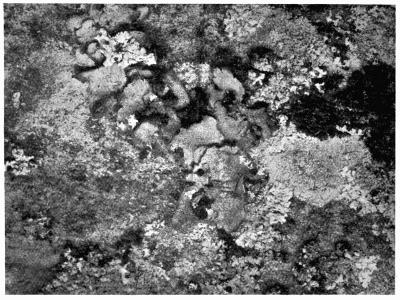
Fig. 56. Dermatocarpon miniatum Th. Fr. (S. H., Photo.).
In the Stictaceae there are a large number of widely-expanded forms, and as the attachment depends mostly on a somewhat short tomentum, strength is obtained here also by the thick plectenchymatous cortex of both surfaces. When areas denuded of tomentum and cortex occur, as in Lobaria pulmonaria, the under surface is not sensibly weakened, since the cortical tissue remains connected in a stout and firm reticulation.
b. By development of Veins or Nerves. Certain ground lichens belonging to the Peltigeraceae have a wide spreading thallus often with very large lobes. The upper cortex is a many-layered plectenchyma, but the under surface is covered only by a loose felt of hyphae which branch out into a more or less dense tomentum. As the firm upper cortex continues to increase by intercalary growth from the branching upwards of hyphae from the meristematic gonidial zone, there occurs an extension of the upper thallus with which the lower cannot keep pace[372]. A little way back from the edge, the result of the stretching is seen in the splitting asunder of the felted hyphae of the under surface, and in the consequent formation of a reticulate series of ridges known as the veins or nerves; they represent the original tomentose covering, and are white, black or brown, according to the colour of the tomentum itself. The naked ellipsoid interstices show the white medulla, and, if the veins are wide, the colourless areas are correspondingly small. Rhizinae are formed on the nerves in several of the species, and anchor the thallus to the support. In Peltigera canina, the under surface is almost wholly colourless, the veins are very prominent (Fig. 55), and are further strengthened by the growth and branching of the parallel hyphae of which they are composed. They serve to strengthen the large and flabby thallus and form a rigid base for the long rhizinae by which the lichen clings to the grass or moss over which it grows.
The most perfect development of strengthening nerves is to be found in Hydrothyria venosa[373], a rather rare water lichen that occurs in the streams of North America. It consists of fan-like lobes of thin structure, the cortex being only about one cell thick. The fronds are about 3 cm. wide and they are contracted below into a stalk which serves to attach the plant to the substratum. Several fronds may grow together in a dense tuft, the expanded upper portion floating freely in the water. Frequently the plants form a dense growth over the rocky beds of the stream.
At the point where the stalk expands into the free erect frond, there arise a series of stout veins which spread upwards and outwards. They are definitely formed structures and not adaptations of pre-existing tissues: certain hyphae arise from the medulla at the contracted base of the frond, take a radial direction and, by increase, become developed into firm strands. The individual hyphae also increase in size, and the swelling of the nerve gives rise to a ridge prominent on both surfaces. They seldom anastomose at first but towards the tips they become smaller and spread out in delicate ramifications which unite at various points. There is no doubt, as Bitter[372] points out, that the nerves function as strengthening tissues and preserve the frond from the strain of the water currents which would, otherwise, tear apart the delicate texture.
III. RADIATE THALLUS
1. CHARACTERS OF RADIATE THALLUS
In the stratose dorsiventral thallus, there is a widely extended growing area situated round the free margins of the thallus. In the radiate thallus of the fruticose or filamentous lichens, growth is confined to an apical region. Attachment to the substratum is at one point only—the base of the plant—thus securing the exposure of all sides equally to light. The cortex surrounds the fronds, and the gonidia (mostly Protococcaceae) lie in a zone or in groups between the cortex and the medulla. It is the highest type of vegetative development in the lichen kingdom, since it secures the widest room for the gonidial layer, and the largest opportunity for photosynthesis.
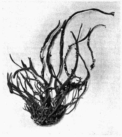
Fig. 57. Roccella fuciformis DC.
Shrubby upright lichens consist mostly of strap-shaped fronds, either simple or branched, which may be broadened to thin bands (Fig. 57) or may be narrowed and thickened till they are almost cylindrical. The fronds vary in length according to the species from a few millimetres upwards: those of Roccella have been found measuring 30 cm. in length; those of Ramalina reticulata, the largest of all the American lichens, extend to considerably more.
Lichens of filamentous growth are more or less cylindrical (Fig. 58). They are in some species upright and of moderate length, but in a few pendulous forms they grow to a great length: specimens of Usnea longissima have been recorded that measured 6 to 8 metres from base to tip.
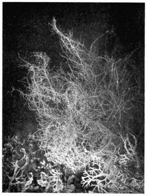
Fig. 58. Usnea barbata Web. (S. H., Photo.).
The radiate type of thallus occurs in most of the lichen groups but most frequently in the Gymnocarpeae. In gelatinous Discolichens it is represented in the Lichinaceae. It is rare among Pyrenocarpeae: there is one very minute British lichen in that series, Pyrenidium actinellum, and one from N. America, Pyrenothamnia, that are of fruticose habit.
2. INTERMEDIATE TYPES OF THALLUS
Between the foliose and the fruticose types, there are intermediate forms that might be, and often are, classified now in one group and now in the other. These are chiefly: Physcia (Anaptychia) ciliaris, Ph. leucomelas and the species of Evernia.
In the two former the habit is more or less fruticose as the plants are affixed to the substratum at a basal point, but the fronds are decumbent and the internal structure is of the dorsiventral type: there is an upper “fibrous” cortex of closely compacted parallel hyphae, a gonidial zone—the gonidia lying partly in the cortex and partly among the loose hyphae of the medulla—and a lower cortex formed of a weft of hyphae which also run somewhat parallel to the surface. Both species are distinguished by the numerous marginal cilia, either pale or dark in colour. These two lichens are greyish-coloured on the upper surface and greyish or whitish below.
Evernia furfuracea with a basal attachment[374], and with a partly horizontal and partly upright growth, has a dorsiventral thallus, dark greyish-green above and black beneath, with occasional rhizinae towards the base. The cortex of both surfaces belongs to the “decomposed” type; the gonidial zone lies below the upper surface, and the medullary tissue is of loose hyphae. In certain forms of the species isidia are abundant on the upper surface, a character of foliose rather than of fruticose lichens. E. furfuracea grows on trees and very frequently on palings.
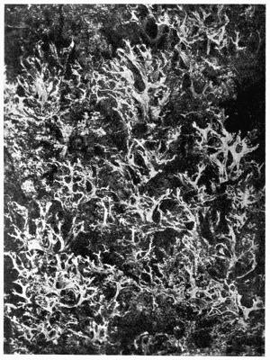
Fig. 59. Evernia prunastri Ach. (M. P., Photo.).
E. prunastri, the second species of the genus, is more distinctly upright in habit, with a penetrating basal hold-fast and upright strap-shaped branching fronds, light-greyish green on the “upper” surface and white on the other (Fig. 59). The internal structure is sub-radiate; both cortices are “decomposed”; the gonidial zone consists of somewhat loose groups of algae, very constant below the “upper” surface, with an occasional group in the pith near to the lower cortex in positions that are more exposed to light. There is also a tendency for the gonidial zone to pass round the margin and spread some way along the under side. The medulla is of loose arachnoid texture and the whole plant is very limp when moist. It grows on trees, often in dense clusters.
3. FRUTICOSE AND FILAMENTOUS
A. General Structure of Thallus
The conditions of strain and tension in the upright plant are entirely different from those in the decumbent thallus, and to meet the new requirements, new adaptations of structure are provided either in the cortex or in the medulla.
Cortical Structures. With the exception of the distinctly plectenchymatous cortex, all the other types already described recur in fruticose lichens; in various ways they have been modified to provide not only covering but support to the fronds.
a. The fastigiate cortex. This reaches its highest development in Roccella in which the branched hyphal tips, slightly clavate and thick-walled, lie closely packed in palisade formation at right angles to the main axis (Fig. 45). They afford not only bending power, but give great consistency to the fronds. The cortex is further strengthened in R. fuciformis[375] by the compact arrangement of the medullary hyphae that run parallel with the surface, and among which occur single thick-walled filaments. The plant grows on maritime rocks in very exposed situations; and the narrow strap-shaped fronds, as stated above, may attain a length of 30 cm., though usually they are from 10 to 18 cm. in height. The same type of cortex, but less highly differentiated, affords a certain amount of stiffness to the cylindrical much weaker fronds of Thamnolia.
b. The fibrous cortex. This type is found in a number of lichens with long filamentous hanging fronds. It consists of parallel hyphae, rarely septate and rarely branched, but frequently anastomosing and with strongly thickened “sclerotic” walls. Such a cortex is the only strengthening element in Alectoria, and it affords great toughness and flexibility to the thong-like thallus. It is also present in Ramalina (Alectoria) thrausta, a species with slender fronds (Fig. 60).
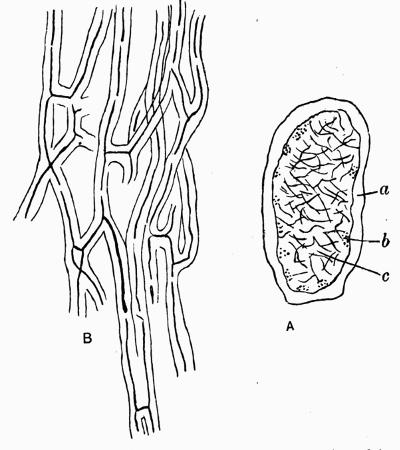
Fig. 60. Alectoria thrausta Ach. A, transverse section of frond; a, cortex; b, gonidia; c, arachnoid medulla × 37. B, fibrous hyphae from longitudinal section of cortex. × 430 (after Brandt).
In Usnea longissima the cortex both of the fibrillose branchlets and of the main axis is fibrous, and is composed of narrow thick-walled hyphae which grow in a long spiral round the central strand. The hyphae become more frequently septate further back from the apex (Fig. 61). Such a type of cortex provides an exceedingly elastic and efficient protection for the long slender thallus.
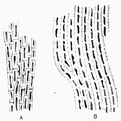
Fig. 61. Usnea longissima Ach. Longitudinal sections of outer cortex. A, near the apex; B, the middle portion of a fibril. × 525 (after Schulte).
The same type of cortex forms the strengthening element in the fruticose or partly fruticose members of the family Physciaceae. One of these, Teloschistes flavicans, is a bright yellow filamentous lichen with a somewhat straggling habit. The fronds are very slender and are either cylindrical or slightly flattened. The hyphae of the outer cortex are compactly fibrous; added toughness is given by the presence of some longitudinal strands of hyphae in the central pith.
Another still more familiar grey lichen, Physcia ciliaris, has long flat branching fronds which, though dorsiventral in structure, are partly upright in habit. Strength is secured as in Teloschistes by the fibrous upper cortex. Other species of Physciae are somewhat similar in habit and in structure.
In Dendrographa leucophaea, a slender strap-shaped rock lichen, Darbishire[376] has described the outer cortex as composed of closely compacted parallel hyphae resembling the strengthening cortex of Alectoria and very different from the fastigiate cortex of the Roccellae with which it is usually classified.
B. Special strengthening Structures
a. Sclerotic strands. This form of strengthening tissue is characteristic of Ramalina. With the exception of R. thrausta (more truly an Alectoria) all the species have a rather weak cortical layer of branching intricate thick-walled hyphae, regarded by Brandt[377] as plectenchymatous, but more correctly by Hue[378] as “decomposed” on account of the gelatinous walls and diminishing lumen of the irregularly arranged cells.
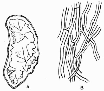
Fig. 62. Ramalina minuscula Nyl. A, transverse section of frond × 37; B, longitudinal strengthening hyphae of inner cortex × 430 (after Brandt).
In R. evernioides, a plant with very wide flat almost decumbent fronds of soft texture, in R. ceruchis and in R. homalea there is a somewhat compact medulla which gives a slight stiffness to the thallus. The other species of the genus are provided with strengthening mechanical tissue within the cortex formed of closely united sclerotic hyphae that run parallel to the surface (Fig. 62). In a transverse section of the thallus, this tissue appears sometimes as a continuous ring which may project irregularly into the pith (R. calicaris); more frequently it is in the form of strands or bundles which alternate with the groups of gonidia (R. siliquosa, R. Curnowii, etc.). In R. fraxinea these strands may be scarcely discernible in young fronds, though sometimes already well developed near the tips. Occasionally isolated strands of fibres appear in the pith (R. Curnowii), or the sclerotic projections may even stretch across the pith to the other side (R. strepsilis) (Fig. 75 B).
In the Cladoniae support along with flexibility is secured to the upright podetium by the parallel closely packed hyphae that form round the hollow cylinder a band called the “chondroid” layer from its cartilage-like consistency.
b. Chondroid axis. The central medullary tissue in Ramalina is, with few exceptions, a loose arachnoid structure; often the fronds are almost hollow. In one species of Usnea, U. Taylori, found in polar regions, there is a similar loose though very circumscribed medullary and gonidial tissue in the centre of the somewhat cylindrical thallus, and a wide band of sclerotic fibres towards the cortex.
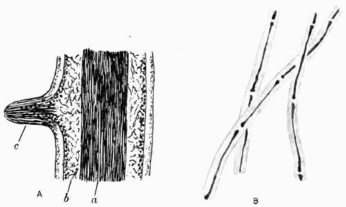
Fig. 63 A. A, Usnea barbata Web. Longitudinal section of filament with young adventitious branch. a, chondroid axis; b, gonidial tissue; c, cortex. × 100 (after Schwendener). B, U. longissima Ach. Hyphae from central axis × 525 (after Schulte).
In all other species of Usnea the medulla itself is transformed into a strong central strand of long-celled thick-walled hyphae closely knit together by frequent anastomoses (Fig. 63 A). This central strand of the Usneas is known as the “chondroid axis.” A narrow band of loose air-containing hyphae and a gonidial zone lie round the central axis between it and the outer cortex (Fig. 63 A, b). At the extreme apex, the external cortical hyphae grow in a direction parallel with the long axis of the plant, but further back, they branch out at right angles and become swollen and mostly “decomposed” as in the cortex of Ramalina.
In Letharia (L. vulpina, etc.) the structure is midway between Ramalina and Usnea: the central axis is either a solid strand of chondroid hyphae or several separate strands.
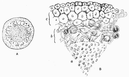
Fig. 63 B. Usnea longissima Ach. A, transverse section of fibril × 85. B, a, chondroid axis; b, gonidial tissue; c, cortex × 525 (after Schulte).
In three other genera with upright fruticose thalli, Sphaerophorus, Argopsis and Stereocaulon, rigidity is maintained by a medulla approaching the chondroid type. In Sphaerophorus the species may have either flattened or cylindrical branching stalks, but in all of them, the centre is occupied by longitudinal strands of hyphae. Argopsis, a monotypic genus from Kerguelen, has a cylindrical branching thallus with a strong solid axis; it is closely allied to Stereocaulon, a genus of familiar moorland lichens. The central tissue of the stalks in Stereocaulon is also composed of elongate, thick-walled conglutinate hyphae, formed into a strand which is, however, not entirely solid.
C. Survey of Mechanical Tissues
Mechanical tissues scarcely appear among fungi, except perhaps as stoutish cartilaginous hyphae in the stalks of some Agarics (Collybiae, etc.), or as a ring of more compact consistency round the central hyphae of rhizomorphic strands. It is practically a new adaptation of hyphal structure confined to lichens of the fruticose group, where there is the same requirement as in the higher plants for rigidity, flexure and tenacity.
Rigidity is attained as in other plants by groups or strands of mechanical tissue situated close to the periphery, as they are so arranged in Ramalina and Cladonia; or the same end is achieved by a strongly developed fastigiate cortex as in Roccella. Bending strains to which the same lichens are subjected, are equally well met by the peripheral disposition of the mechanical elements.
Tenacity and elasticity are provided for in the pendulous forms either by a fibrous cortex as in Alectoria, or by the chondroid axis in Usnea. Haberlandt[379] has recorded some interesting results of tests made by him as to the stretching capacity of a freshly gathered pendulous species in which the central strand was from ·5 to 1 mm. thick. He found he could draw it out 100 to 110 per cent. of its normal length before it gave way. In an upright species the frond broke when stretched 60 to 70 per cent. In both of the plants tested, the central strand retained its elasticity up to 20 per cent. of stretching. The outer cortical tissue was cracked and broken in the experiments. Schulte[380] calculated somewhat roughly the tenacity of Usnea longissima and found that a piece of the main axis 8 cm. long carried up to 300 grms. without breaking.
D. Reticulate Fronds
In the upright radiate thallus, more especially among the Ramalinae, though also among Cladoniae[381], there has appeared a reticulate thallus resulting from the elongate splitting of the tissues, and due to unequal growth tension and straining of the gelatinous cortex when swollen with moisture. In several species of Ramalina, the strap-shaped frond is hollow in the centre; and strands of strengthening fibres give rise to a series of cortical ridges. The thinner tissue between is frequently torn apart and ellipsoid openings appear which do not however pierce beyond the central hollow. Such breaks are irregular and accidental though occurring constantly in Ramalina fraxinea, R. dilacerata, etc.
A more complete type of reticulation is always present in a Californian lichen, Ramalina reticulata, in which the large flat frond is a delicate open network from tip to base (Fig. 64). It grows on the branches of deciduous trees and hangs in crowded tufts up to 30 cm. or more in length. Usually it is so torn, that the real size attainable can only be guessed at. It is attached at the base by a spreading discoid hold-fast, and, in mature plants, consists of a stoutish main axis from which side branches are irregularly given off. These latter are firm at the base like the parent stalk, but soon they broaden out into very wide fronds. Splitting begins at the tips of the branches while still young; they are then spathulate in form with a slightly narrower recurved tip, below which the first perforations are visible, small at first, but gradually enlarging with the growth of the frond.
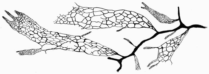
Fig. 64. Ramalina reticulata Krempelh. Portion of frond (after Cramer).
Ramalina reticulata is an extremely gelatinous lichen and the formation of the network was supposed by Lutz[382] to be entirely due to the swelling of the tissues, or the imbibition of water, causing tension and splitting. A more exact explanation of the phenomenon is given by Peirce[383]: he found that it was due to the thickened incurved tip, which, on the addition of moisture, swells in length, breadth and thickness, causing it to bend slightly upwards and then curve backwards over the thallus, thus straining the part immediately behind. These various movements result in the splitting of the frond while it is young and the cortices are thin and weak.
Peirce made a series of experiments to test the capacity of the tissues to support tensile strains. In a dry state, a piece of the lichen held a weight up to 150 grms.; when wet it broke with a weight of 30 grms. It was also observed that the thickness of the frond doubled on wetting.
E. Rooting Base in Fruticose Lichens
Fruticose and filamentous lichens are distinguished by their mode of attachment to the substratum: instead of a system of rhizinae or of hairs spread over a large area, there is usually one definite rooting base by which the plant maintains its hold on the support.
Intermediate between the foliose and fruticose types of thallus are several species which are decumbent in habit, but which are attached at one (or sometimes more) definite points, with but little penetration of the underlying substance. One such lichen, Evernia furfuracea, has been classified now as foliose, and again as fruticose. The earliest stage of the thallus is in the form of a rosette-like sheath which bears rhizinae on the under surface, very numerous at the centre of the sheath, but entirely wanting towards the periphery. A secondary thallus of strap-shaped rather narrow fronds rises from the sheath and increases by irregular dichotomous branching. These branches, which are considered by Zopf[384] as adventitious, may also come into contact with the substratum and produce a few rhizinae at that point; or if the frond is more closely applied, the irritation thus produced causes a still greater outgrowth of rhizinae and the formation of a new base from which other fronds originate. These renewed centres of growth are not of very frequent occurrence; they were first observed and described by Lindau[385] in another species, Evernia prunastri, and were aptly compared by him to the creeping stolons of flowering plants.
Evernia furfuracea grows frequently on dead wood, palings, etc., as well as on trees. E. prunastri grows invariably on trees, and has a more constantly upright fruticose habit; in this species also, a basal sheath is present, and the attachment is secured by means of rhizoidal hyphae which penetrate deeply into the periderm of the tree, taking advantage of the openings afforded by the lenticels. The sheath hyphae are continuous with the medullary hyphae of the frond, and gonidia are frequently enclosed in the tissues; the sheath spreads to some extent over the surface of the bark, and round the base of the fronds, thus rendering the attachment of the lichen to the tree doubly secure.
Among Ramalinae, the development of the base was followed by Brandt[386] in one species, R. Landroensis, an arboreal lichen from S. Tyrol. A rosette-like sheath was formed consisting solely of strands of thick-walled hyphae which spread over the bark. There were no gonidia included in the tissue.
A different type of attachment was found by Lilian Porter[387] in corticolous Ramalinae—R. fraxinea, R. fastigiata, and R. pollinaria. The lichens were anchored to the tree by strands of closely compacted hyphae longitudinally arranged and continuous with the cortical hyphae. These enter the periderm of the tree by cracks or lenticels, and by wedge action cause extensive splitting. The strands may also spread horizontally and give rise to new plants. The living tissues of the tree were thus penetrated and injured, and there was evidence that hypertrophied tissue was formed and caused erosion of the wood.
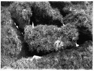
Fig. 65. Ramalina siliquosa A. L. Sm., on rocks, reduced (M. P., Photo.).
Several Ramalinae—R. siliquosa, R. Curnowii, etc.—grow on rocks, often in extremely exposed situations, in isolated tufts or in crowded swards (Fig. 65). The separate tufts are not unfrequently connected at the base by a crustaceous thallus. It is possible also to see on the rock, here and there, small areas of compact thalline granules that have scarcely begun to put out the upright fronds. These granules are corticate on the upper surface and contain gonidia; from the lower surface, slender branching hyphae in rhizoid-like strands penetrate down between the inequalities and separable particles of the rock, if the formation is granitic. They frequently have groups of gonidia associated with them, and they continue to ramify and spread, the pure white filaments often enough enclosing morsels of the rock. The upright fronds are continuous with the base and are thus securely anchored to the substratum.
On a smooth rock surface such as quartzite a continuous sward of Ramalina growth is impossible. The basal hyphae being unable to penetrate the even surface of the rock, the attachment is slight and the plants are easily dislodged. They do however succeed, sometimes, in taking hold, and small groups of fronds arise from a crustaceous base which varies in depth from ·5 to 1 mm. The tissues of this base are very irregularly arranged: towards the upper surface loose hyphae with scattered groups of algae are traversed by strands of gelatinized sclerotic hyphae similar to the strengthening tissues of the upright fronds, while down below there are to be found not only slender hyphae, but a layer of gonidia visible as a white and green film on the rock when the overlying particles are scaled off.
Darbishire[388] found that attachment to the substratum by means of a basal sheath was characteristic of all the genera of Roccellaceae. He looks on this sheath, which is the first stage in the development of the plant, as a primary or proto-thallus, analogous to the primary squamules of the Cladoniae, and he carries the analogy still further by treating the upright fronds as podetia. The sheath of the Roccellaceae varies in size but it is always of very limited extent; it is mainly composed of medullary hyphae, and gonidia may or may not be present. The whole structure is permanent and important, and is generally protected by a well-developed upper cortex similar in structure to that of the upright thallus, i.e. of a fastigiate type. There is no lower cortex.
The two British species of Roccella—R. fuciformis and R. phycopsis—grow on maritime rocks, the latter also occasionally on trees. In R. fuciformis, the attaching sheath is a flat structure which slopes up a little round the base of the upright frond. It is about 2 mm. thick, the cortex occupying about 40 µ of that space; a few scattered gonidia are present immediately below. The remaining tissue of the sheath is composed of firmly wefted slender filaments. Towards the lower surface, there is a more closely compacted dark brown layer from which pass out the hyphae that penetrate the rock.
The sheath of R. phycopsis is a small structure about 3 to 4 mm. in width and 1·5 mm. thick. A few gonidia may be found below the dense cortical layer, but they tend to disappear as the upright fronds become larger and the shade, in consequence, more dense. Lower down the hyphae take an intensely yellow hue; mixed with them are also some brown filaments. A somewhat larger sheath 7 to 8 mm. wide forms the base of R. tinctoria. In structure it corresponds—as do those of the other species—with the ones already described.
In purely filamentous species such as Usnea there is also primary sheath formation: the medullary hyphae spread out in radiating strands which force their way wherever possible into the underlying substance; on trees they enter into any chink or crevice of the outer bark like wedges; or they ramify between the cork cells which are split up by the mere growth pressure. By the vertical increase of the base, the fronds may be hoisted up and an intercalary basal portion may arise lacking both gonidia and cortical layer. Very frequently several bases are united and the lichen appears to be of tufted habit.
A basal sheath provides a similar firm attachment for Alectoria jubata and allied species: these are slender mostly dark brown lichens which hang in tangled filaments from the branches of trees, rocks, etc.
These attaching sheaths differ in function as well as in structure from the horizontal thallus of the Cladoniaceae. They may be more truly compared with the primary thallus of the red algae Dumontia and Phyllophora which are similarly affixed to the substratum, while upright fronds of subsequent formation bear the fructifications.
IV. STRATOSE-RADIATE THALLUS
1. STRATOSE OR PRIMARY THALLUS
A. General Characteristics
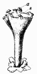
Fig. 66. Cladonia pyxidata Hoffm. Basal squamule and podetium. a, apothecia; s, spermogonia (after Krabbe).
This series includes the lichens of one family only, the Cladoniaceae, the genera of which are characterized by the twofold thallus, one portion being primary, horizontal and stratose, the other secondary and radiate, the latter an upright simple or branching structure termed a “podetium” which narrows above, or widens to form a trumpet-shaped cup or “scyphus” (Fig. 66). The apothecia are terminal on the podetium or on the margins of the scyphi; in a few species they are developed on the primary thallus. Some degree of primary thallus-formation has been demonstrated in all the genera, if not in all the species of the family. The genus Cladina was established to include those species of Cladonia in which, it was believed, only a secondary podetial thallus was present, but Wainio[389] found in Cladonia sylvatica a granular basal crust and, in Cladonia uncialis, minute round scales with crenate margins measuring from ·5 to 1 mm. in width. In some species (subgenus Cladina) the primary thallus is quickly evanescent, in others it is granular or squamulose and persistent. Where the basal thallus is so much reduced as to be practically non-existent, apothecia are rarely developed and soredia are absent. Renewal of growth in these lichens is secured by the dispersal of fragments of the podetial thallus; they are torn off and scattered by the wind or by animals, and, if suitable conditions are met, a new plant arises.
Cladonia squamules vary in size from very small scales as in Cl. uncialis to the fairly large foliose fronds of Cl. foliacea which extend to 5 cm. in length and about 1 cm. or more in width. It is interesting to note that when the primary thallus is well developed, the podetia are relatively unimportant and frequently are not formed. As a rule the squamules are rounded or somewhat elongate in form with entire or variously cut and crenate margins. They may be very insignificant and sparsely scattered over the substratum, or massed in crowded swards of leaflets which are frequently almost upright. In colour they are bluish-grey, yellowish or brownish above, and white beneath (red in Cl. miniata), frequently becoming very dark-coloured towards the rooting base. These several characteristics are specific and are often of considerable value in diagnosis. In certain conditions of shade or moisture, squamules are formed on the podetium; they repeat the characters of the basal squamules of the species.
B. Tissues of the Primary Thallus
The stratose layers of tissue in the squamules of Cladonia are arranged as in other horizontal thalli.
a. Cortical tissue. In nearly all these squamules the cortex is of the “decomposed” type. In a few species there is a plectenchymatous formation—in Cl. nana, a Brazilian ground species, and in two New Zealand species, Cl. enantia f. dilatata and Cl. Neo-Zelandica. The principal growing area is situated all round the margins though generally there is more activity at the apex. Frequently there is a gradual perishing of the squamule at the base which counterbalances the forward increase.
The upper surface in some species is cracked into minute areolae; the cracks, seen in section, penetrate almost to the base of the decomposed gelatinous cortex. They are largely due to alternate swelling and contraction of the gelatinous surface, or to extension caused, though rarely, by intercalary growth from the hyphae below. The surface is subject to weathering and peeling as in other lichens; but the loss is constantly repaired by the upward growth of the meristematic hyphae from the gonidial zone; they push up between the older cortical filaments and so provide for the expansion as well as for the renewal of the cortical tissue.
b. Gonidial tissue. The gonidia consisting of Protococcaceous algae form a layer immediately below the cortex. Isolated green cells are not unfrequently carried up by the growing hyphae into the cortical region, but they do not long survive in this compact non-aerated tissue. Their empty membranes can however be picked out by the blue stain they take with iodine and sulphuric acid.
Krabbe[390] has described the phases of development in the growing region: he finds that differentiation into pith, gonidial zone and cortex takes place some little way back from the edge. At the extreme apex the hyphae lie fairly parallel to each other; further back, they branch upwards to form the cortex, and to separate the masses of multiplying gonidia, by pushing between them and so spreading them through the whole apical tissue. The gonidia immediately below the upper cortex, where they are well-lighted, continue to increase and gradually form into the gonidial zone; those that lie deeper among the medullary hyphae remain quiescent, and before long disappear altogether.
Where the squamules assume the upright position (as in Cladonia cervicornis), there is a tendency for the gonidia to pass round to the lower surface, and soredia are occasionally formed.
c. Medullary tissue. The hyphae of the medulla are described by Wainio as having long cells with narrow lumen, and as being encrusted with granulations that may coalesce into more or less detachable granules; in colour they are mostly white, but pale-yellow in Cl. foliacea and blood-red in Cl. miniata, a subtropical species. They are connected at the base of the squamules with a filamentous hypothallus which penetrates the substratum and attaches the plant. In a few species rhizinae are formed, while in others the hyphae of the podetium grow downwards, towards and into the substratum as a short stout rhizoid.
d. Soredia. Though frequent on the podetia, soredia are rare on the squamules, and, according to Wainio[391], always originate at the growing region, from which they spread over the under surface—rather sparsely in Cl. cariosa, Cl. squamosa, etc., but abundantly in Cl. digitata and a few others. In some instances, they develop further into small corticate areolae on the under surface (Cl. coccifera, Cl. pyxidata and Cl. squamosa).
2. RADIATE OR SECONDARY THALLUS
A. Origin of the Podetium
The upright podetium, as described by Wainio[392] and by Krabbe[393], is a secondary product of the basal granule or squamule. It is developed from the hyphae of the gonidial zone, generally where a crack has occurred in the cortex and rather close to the base or more rarely on or near the edge of the squamule (Cl. verticillata, etc.). At these areas, certain meristematic gonidial hyphae increase and unite to form a strand of filaments below the upper cortex but above the gonidial layer, the latter remaining for a time undisturbed as to the arrangement of the algal cells.
This initial tissue—the primordium of the podetium—continues to grow not only in width but in length: the basal portion grows downwards and at length displaces the gonidial zone, while the upper part as a compact cylinder forces its way through the cortex above, the cortical tissue, however, taking no part in its formation; as it advances, the edges of the gonidial and cortical zones bend upwards and form a sheath distinguishable for some time round the base of the emerging podetium.
Even when the primary horizontal thallus is merely crustaceous, the podetia take origin similarly from a subcortical weft of hyphae in an areola or granule.
B. Structure of the Podetium
a. General structure. In the early stages of development the podetium is solid throughout, two layers of tissue being discernible—the hyphae forming the centre of the cylinder being thick-walled and closely compacted, and the hyphae on the exterior loosely branching with numerous air-spaces between the filaments.
In all species, with the exception of Cl. solida, which remains solid during the life of the plant, a central cavity arises while the podetium is still quite short (about 1 to 1·5 mm. in Cl. pyxidata and Cl. degenerans). The first indication of the opening is a narrow split in the internal cylinder, due to the difference in growth tension between the more free and rapid increase of the external medullary layer and the slower elongation of the chondroid tissue at the centre. The cavity gradually widens and becomes more completely tubular with the upward growth of the podetium; it is lined by the chondroid sclerotic band which supports the whole structure (Fig. 67).